Review

Duplication and Segregation of Centrosomes during Cell Division

1
Centre de Recherche de Biologie Cellulaire de Montpellier, Université de Montpellier, Centre Nationale de la Recherche Scientific, 34293 Montpellier, France
2
Faculté de Médecine, Université de Tours, 37032 Tours, France
3
Faculty of Bioengineering and Bioinformatics, Moscow State University, 119992 Moscow, Russia
*
Author to whom correspondence should be addressed.
Cells 2022, 11(15), 2445; https://doi.org/10.3390/cells11152445
Submission received: 15 July 2022 / Revised: 1 August 2022 / Accepted: 5 August 2022 / Published: 7 August 2022
(This article belongs to the Special Issue The Regulation of the Cell Cycle)

Abstract

:
During its division the cell must ensure the equal distribution of its genetic material in the two newly created cells, but it must also distribute organelles such as the Golgi apparatus, the mitochondria and the centrosome. DNA, the carrier of heredity, located in the nucleus of the cell, has made it possible to define the main principles that regulate the progression of the cell cycle. The cell cycle, which includes interphase and mitosis, is essentially a nuclear cycle, or a DNA cycle, since the interphase stages names (G1, S, G2) phases are based on processes that occur exclusively with DNA. However, centrosome duplication and segregation are two equally important events for the two new cells that must inherit a single centrosome. The centrosome, long considered the center of the cell, is made up of two small cylinders, the centrioles, made up of microtubules modified to acquire a very high stability. It is the main nucleation center of microtubules in the cell. Apart from a few exceptions, each cell in G1 phase has only one centrosome, consisting in of two centrioles and pericentriolar materials (PCM), which must be duplicated before the cell divides so that the two new cells formed inherit a single centrosome. The centriole is also the origin of the primary cilia, motile cilia and flagella of some cells.

1. What Is the Centrosome?

In animal cells, the centrosome is a membrane-less organelle consisting of two interconnected centrioles, PCM and some additional structures [1]. The centriole is a cylinder shape structure with a diameter of about 200 nm and a length of about 500 nm made of nine triplets of microtubules (Figure 1).
The triplets of microtubules are always twisted counterclockwise when viewed from the proximal end of the centriole [2]. Each triplet is composed of microtubules A, B and C, with microtubule C being shorter than the other two and ending in a characteristic hook at the distal end of the centriolar cylinder (Figure 2 middle). Three types of centrioles can simultaneously exist in a cell (Figure 2). The older and most mature centriole (at least 2 cell cycles old) is called the mother centriole in opposition to the younger one, called the daughter centriole (which appeared in the previous cell cycle), and possesses two kinds of appendages named distal and subdistal appendages. Each triplet of microtubules possesses one distal appendage which makes nine distal appendages per centriole [3]. During the formation of the primary cilium distal appendages are essential to attach the centriole to the plasma membrane [4]. The daughter centriole has electron-dense ribs at the site of the future formation of distal appendages (Figure 2, middle).
Regarding subdistal appendages their number varies from 0 to 13 depending on the cell types [5]. The number of subdistal appendages depends on the microtubule nucleating activity of the centrosome, in some types of cells subdistal appendages can be arranged in two or even three rows. The base of each subdistal appendage rests on two or three centriole triplets [3].
Proteins responsible for the nucleation and anchoring of microtubules localize on the heads of the subdistal appendages [6]. These appendages are absent in mitotic centrosomes; they disassemble during G2 phase and reappear on the surface of the mother centriole at the beginning of G1 phase [7].
In the proximal part inner lumen of the centriolar cylinder of the mother and daughter of the centrioles, a system of ligaments is located, which connects the triplets from the inside to each other. In the youngest centriole—procentriole, which arose in the current cell cycle, in the inner lumen there is a “cartwheel structure”, which determines the nine-beam symmetry of centrioles during their formation due to the lateral interaction of SAS6 protein dimers, the angle between which is 40 degrees [8,9].
The centrosome includes not only centrioles, but also several additional structures: satellites, striated rootlets, less morphologically pronounced pericentriolar material and a system of ligaments between the proximal ends of the mother and daughter centrioles. The centrosome is the main nucleation center of microtubules in the cell [10]. Connected to the centrosome are the minus ends of microtubules that attach to the heads of the subdistal appendages, the surface of the predominantly mother centriole and the pericentriolar material (Figure 3).
Finally, the centrosomes nucleate microtubules that participate to four main structures in the cell: the radial MT network in interphase, the bipolar spindle in mitosis, the procentriole and the cilia and flagella [5].
The two centrioles act as a platform to localize and assemble functional protein complexes. For example, the pericentriolar material surrounding the centrosomes consists of proteins that will ensure microtubule nucleation, the main function of centrosomes. Two dynamics of nucleation of microtubules follow each other at the centrosomes during the cell cycle progression. In interphase the nucleated microtubules are not very dynamic and of great length, they will constitute the cytoskeleton of microtubule of the interphase cells and will take part in giving them their shape. In mitosis, the network of interphase microtubules disappears to make room for a much more dynamic network with more numerous and very short microtubules that will only ensure one function: the assembly of the bipolar spindle essential to the segregation of chromosomes. In mitosis, the centrosomes recruit more proteins dedicated to intense nucleation of microtubules, the pericentriolar material of centrosomes becomes more extensive and denser: this mechanism is called the maturation of centrosomes. The mitotic spindle is bipolar, so the cell in mitosis must possess two centrosomes, one for each pole of the spindle. The duplication of centrosomes in interphase must therefore be strictly controlled so that it produces only two centrosomes. A higher number of centrosomes prevents the assembly of a bipolar spindle, disrupts chromosome segregation and induces aneuploidy, a situation frequently observed in cancer cells.

2. Common Features between the DNA and the Centrosome Cycle

DNA replication and centrosome duplication during a cell cycle must share common regulations because each of the two new cells formed must inherit only one copy of the genome and one copy of the centrosome from the initial cell [11,12].
The genome of eukaryotic cells is composed of DNA, structured in a double helix that carries the genes. The genome is present in two copies in each cell; each gene is therefore present in the form of two alleles. The cells are therefore diploid. This genome must be replicated (copied) at each cell cycle so that the two daughter cells inherit the same genome as the mother cell. To copy the genome, the double helix is opened and each strand is copied so that each double helix generates two new double helices, each consisting of a mother and a daughter strand [13,14].
The centrosome of higher eukaryotic cells (multicellular organisms) after mitosis consists of two centrioles. The centrosome must be duplicated (copied) during each cell cycle so that the two daughter cells inherit the same centrosome (two centrioles) as the mother cell. During the cell cycle, a new centriole is constructed on the surface of each centriole. Each daughter cell then inherits a centrosome composed of an old centriole (that of the mother cell) and a new one [15].
The temptation was therefore great to consider that, as with DNA, each centriole serves as a model for recreating a new one. However, unlike DNA, which must be copied to preserve genetic sequences and therefore cannot be synthesized de novo, the centriole is not copied. The old centriole only serves as a platform to recruit proteins that will build a new centriole. As evidence, the mechanism can occur de novo in the absence of any centriole [16,17].
The initiation of DNA replication is under the control of the restriction point (R), a checkpoint in G1 phase which when satisfied authorizes the cell to enter the cell cycle which will lead to cell division. As for the DNA, the duplication of the centrosome also depends on this restriction point.
The authorization to enter the cycle results in the production of the E2F family of transcription factors that will transactivate the genes whose products are required either for DNA replication or for centrosome duplication. The start of centrosome duplication precedes DNA replication [18] and the two events are independent of each other. Indeed, the addition of arabinosyl cytosine to cells inhibits DNA replication without affecting centriole duplication [19]. Similarly, centrioles continue to duplicate in sea urchin egg extracts in the presence of aphidicolin, an inhibitor of DNA replication, or even in previously enucleated sea urchin egg extracts [20]. Treatment of CHO cells with hydroxyurea or aphidicolin for a time equivalent to the duration of several cell cycles blocks DNA replication but does not prevent centrioles from continuing to replicate [21].
Another commonality between the DNA and the centrosome is the rule that allows DNA replication and centrosome duplication to occur only once and only once per cell cycle. As replication proceeds, the newly synthesized DNA is no longer allowed to be replicated again, only the passage through a phase of mitosis removes this blockage. The same rule applies to centrosomes which once duplicated are only allowed to replicate once they have passed through mitosis. The modalities of these authorizations are different for the DNA and the centrosome; we will come back to these mechanisms in more detail.

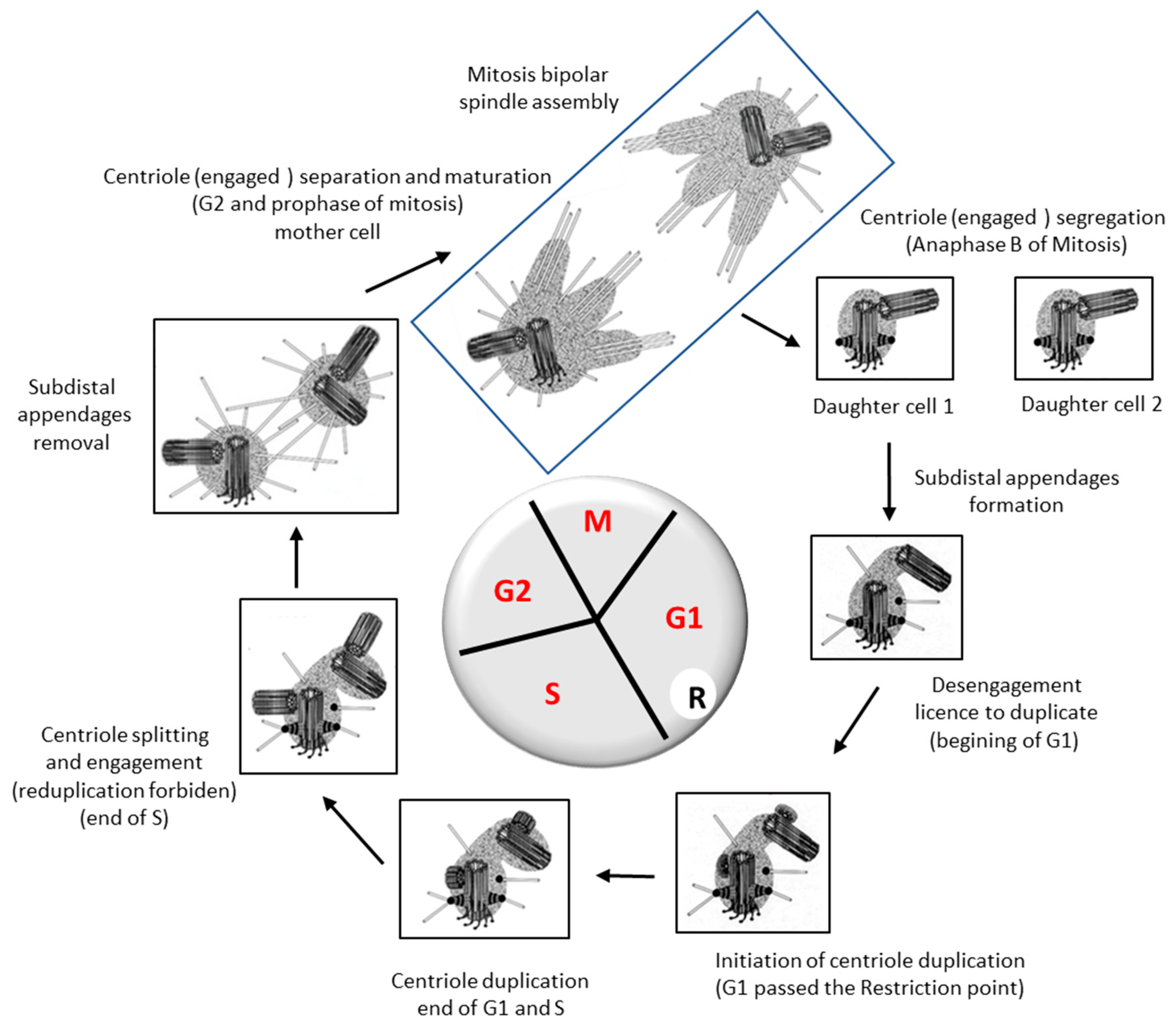
3. Behavior of the Centrosome during a Cell Cycle

In G1 phase of the cell cycle the mother centriole with its appendages is connected to the daughter centriole, both centrioles are “disengaged” i.e., allowed to duplicate. The passage of the restriction point will trigger the initiation of their duplication.
This duplication is under the control of the protein kinase PLK4 [22,23,24]. PLK4 comes to localize to each centriole by interacting with the two proteins Cep192 and Cep152 on the centriole wall, initially as a ring around the centriole [25,26,27]. The STIL protein then localizes as a dot on the ring of PLK4 on the centriole wall, STIL recruits Plk4 which relocalizes to the STIL dot, and activates it, in turn Plk4 phosphorylates STIL and this phosphorylated form recruits the SAS6 protein to initiate cartwheel assembly [28,29,30,31,32,33].
The cartwheel, first observed in 1960 in EM images of the base of flagella, is the structural unit of symmetry nine of centrioles [34]. This structure forms even before the nucleation of the microtubules that will form the procentriole. It is from this cartwheel that the growth of the procentriole will start (Figure 4). Once the first cartwheel is formed, others will be made to form a stack of cartwheels that will serve as a scaffold around which the nine triplets of microtubules will be organized.
Our current knowledge of the molecular mechanism of cartwheel establishment is still fragmented and incomplete. The SAS6 protein provides the cartwheel structure [39]. SAS6 forms a homodimer with the N-terminal domains at the center of the cartwheel and the long C-terminal coiled-coil portions of the dimer organize the spokes, 9 dimers organized in a disk form the cartwheel of symmetry 9 (Figure 5).
At the end of each spoke, the STIL protein makes contact with CPAP protein that cooperates with CEP120 to regulate centriolar microtubule growth [40,41,42]. CPAP is essential for procentriole construction [43,44,45] it binds the cartwheel to the nine triplets of microtubules via STIL that might replace by CEP135 during procentriole assembly [46].
Interestingly, CPAP interacts with STIL and/or CEP135, but the function of this interaction is unknown [47]. Both STIL and CEP135 also interact with SAS6. Why and in what cases does SAS6 interact with STIL or CEP135 remains to be determined.

4. One and Only One Procentriole per Centriole per Cell Cycle

Each centriole must be duplicated once and only once per cell cycle [48]. What is the mechanism that governs this restriction?
Nucleation of the new procentriole begins on the wall of the old centriole. As discussed above, while PLK4 is initially localized as a ring around the old centriole, a signal breaks this symmetry and induces relocalization of PLK4 to a single site (Figure 5). How is this site defined on the wall of the centriole?
The symmetry breaking of PLK4 localization would be induced by the STIL protein, but this only shifts the problem, how does STIL choose its localization site on the PLK4 ring [49]. Furthermore, it seems that PLK4 is able to self-activate locally and form a focus even in the absence of STIL [50].
In Drosophila syncytial embryos, it was recently observed that the assembly of the new procentriole always starts on the side of the wall that faces the nuclear envelope [51]. The centrosome is indeed always located close to the nucleus and is even physically bound to the nuclear envelope since the centrosome co-purifies with the nuclei during cell fractionations [52].
Could it be that a molecule associated with the nuclear envelope could designate the site of STIL anchoring and/or local PLK4 activation? This remains to be determined.
Obviously, the designation of a single site of construction of the new procentriole on the wall of the old centriole restricts de facto the number of procentrioles made to one per centriole per cycle.
When a new procentriole is built during the S phase of the cycle on the wall of an old centriole, re-duplication is no longer allowed on this centriole. The resulting centrosome is thus formed of a new centriole and old one; both centrioles are considered as “engaged” positioned orthogonally to each other.

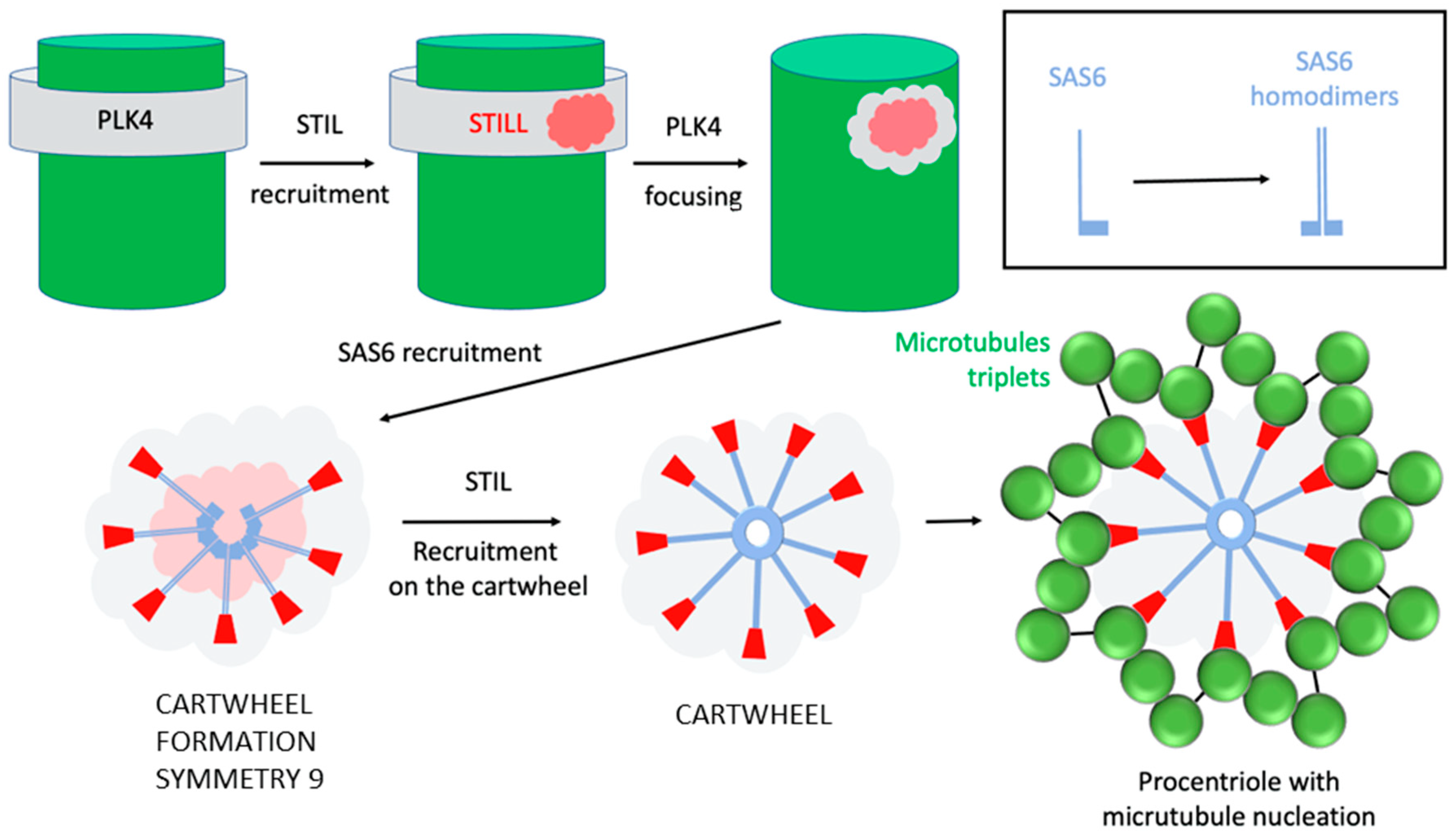
What Are the Proteins Involved in This Engagement?

The first protein identified as essential for this event is CEP215 (or CDK5RAP2 for Cdk5 regulatory subunit associated protein 2), a centriolar protein of the centrosomin family that localizes to the centrosomes during duplication. A deletion of this protein causes an amplification of centrosomes (continuous duplication) due to an engagement defect [53].
It is only when crossing mitosis following duplication that the two centrioles disengage [54]. This event is entirely dependent on the activity of a protease that is activated only when the Spindle Assembly Checkpoint is satisfied, and the cell makes the metaphase to anaphase transition [55,56,57]. This protease degrades cohesin, which binds sister chromatids together allowing their separation, from which it derives its name separase, after having been originally called “sister-separating” factor [58]. The degradation of cohesin thus leads to the segregation of chromosomes. The protease activity of separase is also required to disengage centrioles [57], yet it does not degrade the CEP215 protein [53].
Therefore, there is another protein that ensures centriole engagement and must be degraded by separase to trigger disengagement. This protein is the pericentrin. Indeed, the expression of a pericentrin that cannot be cleaved by separase blocks centriole disengagement and thus duplication [59]. It has been proposed that degradation of pericentrin by separase leads to removal of the CEP215 protein thus eliminating the two proteins ensuring centriole engagement [60].

5. Centrosome Splitting, Separation and Segregation

At the end of the duplication process, the two old centrioles are now each engaged with a new centriole. These two old centrioles are still linked by a network of fibrils that must disappear to allow their dissociation and thus form the two new independent centrosomes.
This dissociation of the two old centrioles is under the control of the protein kinase Nek2 (for NIMA-related kinase 2) which phosphorylates the Cep250 protein (CNAP1) to delocalize it [61,62]. This CNAP-1 protein plays a central role, but other proteins also participate in centriole cohesion such as rootletin [63,64], CEP68 and CEP215 [65], conductin [66], LRRC45 [67] and centlein [68].
This process of dissociation of the two centrosomes is essential to allow the separation of the centrosomes, i.e., their migration around the nuclear envelope to a position opposite to each other [69].
The protein that plays a central role in this process is the kinesin-like protein KIF11 (or Kinesin 5). Inhibition of KIF11 by microinjection of antibodies inhibits centrosome separation and induces the formation of monopolar spindle in mitosis [70]. These data were reproduced with the drug monastrol a small molecule that specifically inhibits KIF11 [71]. KIF11 is a motor protein with a motor head domain and a long coiled-coil C terminal domain, forms head-to-head dimers that subsequently dimerized again to form tetramers with two head motors at each end [72]. This tetramer possesses a MT-Sliding activity allowing binding and sliding of two adjacent microtubules [73]. The two centrosomes that are going to separate nucleate microtubules, the microtubules between the two centrosomes will be cross-linked by the KIF11 protein. The MT-sliding activity of KIF11 will result in a push on the centrosomes inducing their separation (Figure 6).
During their separation, the two centrosomes remain connected to the nuclear envelope by binding to dynein, which associates with BICD2 (dynein adaptor) at the nuclear pores [74] or/and the Nup133 protein (a nuclear pore protein) [75].
When the centrosomes split, both centrioles are different; there is a mother centriole and a daughter one. The presence of appendages on a centriole identifies the mother centriole [77]. Information regarding the nature and function of these appendages is still fragmentary and remains to be investigated [78,79]. Unlike distal appendages, subdistal appendages are not present on the mother centriole throughout the cell cycle [80].
Ninein and the dynactin subunit p150 (Glued), for example, are among the proteins involved in the construction of subdistal appendages on the wall of the mother centriole, and p150 (Glued) is required to localize ninein [81]. The p150 (Glued) protein is not present on the mother centriole during mitosis, is released at the end of G2 phase, and reappears in G1 phase. The subdistal appendage disappears from the mother centriole in mitosis during the process of centrosome separation and mitotic spindle assembly. The difference between the two centrioles is then limited to the presence of the distal appendages on the mother centriole [7].
The function of the subdistal appendage is to anchor microtubules during interphase [6], the reason why it is no longer present in mitosis remains to be determined. This could be related to the change in dynamics of microtubule nucleation by centrosomes and thus to their maturation.

6. Conclusions

Eventually, it is not so surprising that the centrosome cycle is a complex and strictly regulated process. The cell goes through various stages to duplicate its centrosome and segregate each of the new one in the two newly generated cells that must inherit one and only one centrosome. The different stages of the cell cycle G1, S, G2 and M have been defined in reference to the cycle of the DNA, consequently, it is not always an easy task to describe the centrosome cycle using the phases of the DNA cycle (Figure 7).
However, the comparison with DNA (the genome) is not without interest because as each somatic cell in G1 should contain one and only one copy of its diploid genome, it must also contain one and only one centrosome. This proportion is maintained after each cell division. Chromosome segregation during mitosis is performed by a dynamic spindle of microtubules and in order for the mother cell to separate two sets of chromosomes for the two newly generated cells, the spindle must be bipolar.
This bipolarity is controlled by the number centrosomes, one at each pole. Any changes in the centrosome number affects spindle assembly and consequently cell ploidy. There are few exceptions such the megakaryocyte that is polyploid and contains multiple centrosomes, in this cell the proportion is conserved two copies of the diploid genome with two centrosomes, 4 with 4, 8 with 8, 16 with 16 and so on. This is probably because abortive mitoses are at the origin of this polyploidy and centrosome amplification [82]. However, the more surprising behavior of megakaryocytes is that they still assemble multiple bipolar spindle using a mechanism that is not fully understood [83].
In agreement with Boveri’s theory many cancer cells are aneuploid and possess more than two centrosomes [84,85,86]. However, one of the properties of cancer cells is their ability to adapt, which often poses problems for treatment. For instance, cancer cells have found a way to deal with multiple centrosomes and still assemble a bipolar spindle even with more than two centrosomes. The mechanism named centrosome clustering consists by being able to form a bipolar spindle with one centrosome at one pole and three clustered centrosomes at the other pole [87]. Even if the mechanism is not fully understood, an inhibition of the clustering mechanism has been shown to be effective in eliminating cancer cells that have developed this strategy to proliferate in the presence of supernumerary centrosomes [88].
It is then established that some cells can assemble a bipolar spindle with different number of centrosomes at each pole, but what would happen in the absence of centrosome? Are the centrosomes essential to assemble a bipolar spindle? The answer is clearly “No” for three main reasons; (1) meiotic bipolar spindle in mouse oocytes is assembled in the absence of centrosomes [17], (2) bipolar spindle can assemble after laser ablation of centrosomes [89] and (3) Drosophila PLK4 mutants assemble bipolar mitotic spindle without centrosome [90]. So, cells can assemble bipolar spindles without centrosomes and cells can generate a centriole de novo. Then why do somatic cells keep conserving a centrosome? Moreover, why does the canonical mechanism imply that the new centriole is assembled on the surface of another one?
The presence of the centrosome might be necessary to limit duplication: each centriole giving birth to only one centriole. Indeed, de novo centriole amplification can be obtained by overexpressing PLK4 only after having removed the existing centrosome implying that its presence is limiting duplication [91].
Although the centrosome was discovered almost 150 years ago [92,93,94], the mechanism of duplication as well as its regulation during cell cycle progression are still not fully understood. Its conservation during evolution and its involvement in so many functions in the cell should attract more young researchers. To borrow a phrase from Michel Bornens, who has passed on his passion for the centrosome to many of us: “there are many important questions raised by this organelle” [95].

Author Contributions

C.P. and R.U. wrote and edited the paper. All authors have read and agreed to the published version of the manuscript

Funding

This research received no external funding.

Acknowledgments

We thank Tang K Tang and Pierre Gönczy for the helpful discussion. We would like to dedicate this review to Michel Bornens, a pioneer in centrosome biology, who passed away in March of this year. We all miss him greatly.

Conflicts of Interest

The authors declare no conflict of interest.

Abbreviations

BICD2 (dynein adaptor)Bicaudal-D2 (BICD2) is a dynein activating adaptor protein that plays a critical role in microtubule-based minus-end-directed transport
CHO cellsChinese hamster ovary cells
G1 phase (from Gap 1)phase after mitosis before DNA replication
G2 phase (from Gap 2)phase of cell cycle after S pahse and before mitosis
CEP68Centrosomal Protein 68 kDa
CEP120Centrosomal Protein 120 kDa
CEP135Centrosomal Protein 135 kDa
Cep152Centrosomal Protein 152 kDa
Cep192Centrosomal Protein 192 kDa
CEP215Centrosomal Protein 215 kDa (or CDK5RAP2 for Cdk5 regulatory subunit associated protein 2)
CNAP1 (Centrosomal Protein 250 kDa–Cep250)centrosomal Nek2-associated protein 1
CPAPcentriolar protein (centrosomal P4.1-associated protein)
EMelectron microscopy
LRRC45Leucine Rich Repeat Containing 45 kDa
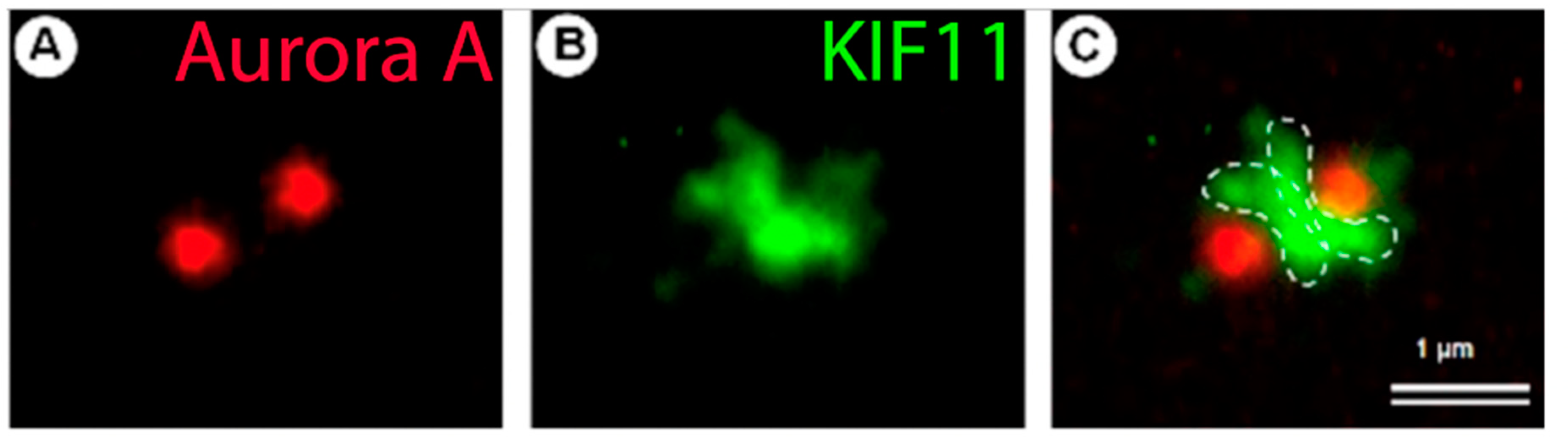
Kinesin-like protein KIF11 (or Kinesin 5 or XlEg5)plus end directed microtubule associated motor.
M phasemitosis
MTmicrotubule
Nek2for NIMA-related kinase 2
Nup133 proteinnuclear pore protein 133kDa
p150 (Glued) proteinDynactin subunit, potent anti-catastrophe factor for microtubules
PCMpericentriolar material
PLK4Polo-like kinase 4 (Serine/threonine-protein kinase)
S phase (from synthesis)phase of cell cycle during which DNA replicated
SAS6Centriolar Assembly Protein (Spindle Assembly 6 Homolog from Caenorabditis Elegans)
STIL proteinCentriolar Assembly Protein, the SCL/TAL interrupting locus is a human oncogene that was originally identified from human T-cell lymphoblastic leukemia, where it functions as a hematopoietic transcription factor.

References

  1. Bornens, M. The centrosome in cells and organisms. Science 2012, 335, 422–426. [Google Scholar] [CrossRef] [PubMed]
  2. Uzbekov, R.; Prigent, C. Clockwise or anticlockwise, turning the centriole triplets in the right direction! FEBS Lett. 2007, 581, 1251–1254. [Google Scholar] [CrossRef] [PubMed] [Green Version]
  3. Uzbekov, R.; Alieva, I. Who are you, subdistal appendages of centriole? Open Biol. 2018, 8, 180062. [Google Scholar] [CrossRef] [PubMed] [Green Version]
  4. Tanos, B.E.; Yang, H.J.; Soni, R.; Wang, W.J.; Macaluso, F.P.; Asara, J.M.; Tsou, M.F.B. Centriole distal appendages promote membrane docking, leading to cilia initiation. Genes Dev. 2013, 27, 163–168. [Google Scholar] [CrossRef] [Green Version]
  5. Uzbekov, R.E.; Avidor-Reiss, T. Principal postulates of centrosomal biology. Cells 2020, 9, 2156. [Google Scholar] [CrossRef]
  6. Delgehyr, N.; Sillibourne, J.; Bornens, M. Microtubule nucleation and anchoring at the centrosome are independent processes linked by ninein function. J. Cell Sci. 2005, 118 Pt 8, 1565–1575. [Google Scholar] [CrossRef] [Green Version]
  7. Vorobjev, I.A.; Chentsov, Y.S. Centrioles in the Cell Cycle. I Epithelial Cells. J. Cell Biol. 1982, 98, 938–949. [Google Scholar] [CrossRef]
  8. Kitagawa, D.; Vakonakis, I.; Olieric, N.; Hilbert, M.; Keller, D.; Olieric, V.; Bortfeld, M.; Erat, M.C.; Fluckiger, I.; Gonczy, P.; et al. Structural basis of the 9-fold symmetry of centrioles. Cell 2011, 144, 364–375. [Google Scholar] [CrossRef] [Green Version]
  9. van Breugel, M.; Hirono, M.; Andreeva, A.; Yanagisawa, H.A.; Yamaguchi, S.; Nakazawa, Y.; Morgner, N.; Petrovich, M.; Ebong, I.O.; Robinson, C.V.; et al. Structures of sas-6 suggest its organization in centrioles. Science 2011, 331, 1196–1199. [Google Scholar] [CrossRef] [Green Version]
  10. Osborn, M.; Weber, K. Cytoplasmic microtubules in tissue culture cells appear to grow from an organizing structure towards the plasma membrane. Proc. Natl. Acad. Sci. USA 1976, 73, 867–871. [Google Scholar] [CrossRef] [Green Version]
  11. Diffley, J.F. Once and only once upon a time: Specifying and regulating origins of DNA replication in eukaryotic cells. Genes Dev. 1996, 10, 2819–2830. [Google Scholar] [CrossRef] [PubMed] [Green Version]
  12. Zitouni, S.; Francia, M.E.; Leal, F.; Montenegro Gouveia, S.; Nabais, C.; Duarte, P.; Gilberto, S.; Brito, D.; Moyer, T.; Kandels-Lewis, S.; et al. CDK1 Prevents Unscheduled PLK4-STIL Complex Assembly in Centriole Biogenesis. Curr. Biol. 2016, 26, 1127–1137. [Google Scholar] [CrossRef] [PubMed] [Green Version]
  13. Watson, J.D.; Crick, F.H. Genetical implications of the structure of deoxyribonucleic acid. Nature 1953, 171, 964–967. [Google Scholar] [CrossRef] [PubMed]
  14. Meselson, M.; Stahl, F.W. The replication of DNA in Escherichia coli. Proc. Natl. Acad. Sci. USA 1958, 44, 671–682. [Google Scholar] [CrossRef] [Green Version]
  15. Gönczy, P.; Hatzopoulos, G.N. Centriole assembly at a glance. J. Cell Sci. 2019, 132, jcs228833. [Google Scholar] [CrossRef] [Green Version]
  16. Dirksen, E.R. The presence of centrioles in artificially activated sea urchin eggs. J. Biophys. Biochem. Cytol. 1961, 11, 244–247. [Google Scholar] [CrossRef] [Green Version]
  17. Szöllösi, D.; Calarco, P.; Donahue, R.P. Absence of centrioles in the first and second meiotic spindles of mouse oocytes. J. Cell Sci. 1972, 11, 521–541. [Google Scholar] [CrossRef]
  18. Uzbekov, R.E. Centriole duplication in PE (SPEV) cells starts before beginning of DNA replication. Biochem. (Mosc.) Suppl. Ser. A Membr. Cell Biol. 2007, 1, 206–211. [Google Scholar] [CrossRef]
  19. Rattner, J.B.; Phillips, S.G. Independence of centriole formation and DNA synthesis. J. Cell Biol. 1973, 57, 359–372. [Google Scholar] [CrossRef] [Green Version]
  20. Sluder, G. Centrosomes and the cell cycle. J. Cell Sci. Suppl. 1989, 12, 253–275. [Google Scholar] [CrossRef] [Green Version]
  21. Balczon, R.; Bao, L.; Zimmer, W.E.; Brown, K.; Zinkowski, R.P.; Brinkley, B.R. Dissociation of centrosome replication events from cycles of DNA synthesis and mitotic division in hydroxyurea-arrested Chinese hamster ovary cells. J. Cell Biol. 1995, 130, 105–115. [Google Scholar] [CrossRef] [PubMed]
  22. O’Connell, K.F.; Caron, C.; Kopish, K.R.; Hurd, D.D.; Kemphues, K.J.; Li, Y.; White, J.G. The C. elegans zyg-1 gene encodes a regulator of centrosome duplication with distinct maternal and paternal roles in the embryo. Cell 2001, 105, 547–558. [Google Scholar] [CrossRef] [Green Version]
  23. Habedanck, R.; Stierhof, Y.D.; Wilkinson, C.J.; Nigg, E.A. The Polo kinase Plk4 functions in centriole duplication. Nat. Cell Biol. 2005, 7, 1140–1146. [Google Scholar] [CrossRef] [PubMed]
  24. Bettencourt-Dias, M.; Rodrigues-Martins, A.; Carpenter, L.; Riparbelli, M.; Lehmann, L.; Gatt, M.K.; Carmo, N.; Balloux, F.; Callaini, G.; Glover, D.M. SAK/PLK4 is required for centriole duplication and flagella development. Curr. Biol. 2005, 15, 2199–2207. [Google Scholar] [CrossRef] [Green Version]
  25. Kim, T.S.; Park, J.E.; Shukla, A.; Choi, S.; Murugan, R.N.; Lee, J.H.; Ahn, M.; Rhee, K.; Bang, J.K.; Kim, B.Y.; et al. Hierarchical recruitment of Plk4 and regulation of centriole biogenesis by two centrosomal scaffolds, Cep192 and Cep152. Proc. Natl. Acad. Sci. USA 2013, 110, E4849–E4857. [Google Scholar] [CrossRef] [Green Version]
  26. Kemp, C.A.; Kopish, K.R.; Zipperlen, P.; Ahringer, J.; O'Connell, K.F. Centrosome maturation and duplication in C. elegans require the coiled-coil protein SPD-2. Dev. Cell 2004, 6, 511–523. [Google Scholar] [CrossRef] [Green Version]
  27. Pelletier, L.; Ozlü, N.; Hannak, E.; Cowan, C.; Habermann, B.; Ruer, M.; Müller-Reichert, T.; Hyman, A.A. The Caenorhabditis elegans centrosomal protein SPD-2 is required for both pericentriolar material recruitment and centriole duplication. Curr. Biol. 2004, 14, 863–873. [Google Scholar] [CrossRef] [Green Version]
  28. Delattre, M.; Leidel, S.; Wani, K.; Baumer, K.; Bamat, J.; Schnabel, H.; Feichtinger, R.; Schnabel, R.; Gönczy, P. Centriolar SAS-5 is required for centrosome duplication in C. elegans. Nat. Cell Biol. 2004, 6, 656–664. [Google Scholar] [CrossRef]
  29. Lettman, M.M.; Wong, Y.L.; Viscardi, V.; Niessen, S.; Chen, S.H.; Shiau, A.K.; Zhou, H.; Desai, A.; Oegema, K. Direct binding of SAS-6 to ZYG-1 recruits SAS-6 to the mother centriole for cartwheel assembly. Dev. Cell 2013, 25, 284–298. [Google Scholar] [CrossRef] [Green Version]
  30. Ohta, M.; Ashikawa, T.; Nozaki, Y.; Kozuka-Hata, H.; Goto, H.; Inagaki, M.; Oyama, M.; Kitagawa, D. Direct interaction of Plk4 with STIL ensures formation of a single procentriole per parental centriole. Nat. Commun. 2014, 5, 5267. [Google Scholar] [CrossRef] [Green Version]
  31. Dzhindzhev, N.S.; Tzolovsky, G.; Lipinszki, Z.; Schneider, S.; Lattao, R.; Fu, J.; Debski, J.; Dadlez, M.; Glover, D.M. Plk4 phosphorylates Ana2 to trigger Sas6 recruitment and procentriole formation. Curr. Biol. 2014, 24, 2526–2532. [Google Scholar] [CrossRef] [Green Version]
  32. Moyer, T.C.; Clutario, K.M.; Lambrus, B.G.; Daggubati, V.; Holland, A.J. Binding of STIL to Plk4 activates kinase activity to promote centriole assembly. J. Cell Biol. 2015, 209, 863–878. [Google Scholar] [CrossRef] [PubMed] [Green Version]
  33. Kratz, A.-S.; Bärenz, F.; Richter, K.T.; Hoffmann, I. Plk4-dependent phosphorylation of STIL is required for centriole duplication. Biol. Open 2015, 4, 370–377. [Google Scholar] [CrossRef] [PubMed] [Green Version]
  34. Uzbekov, R.E.; Alieva, I.B. Centriole duplication or DNA replication—what starts earlier. In Cytoskeleton: Cell Movement, Cytokinesis and Organelles Organization (Editors S. Lansing and and T. Rousseau); Chapter 5; Nosva Science Publishers, Inc.: New York, NY, USA, 2010. [Google Scholar]
  35. Uzbekov, R.E.; Alieva, I.B. Centrosome–history of investigations and new discoveries. In From Cytoplasmic Granule to the Centre of Intracellular Regulation; Nadezhdina, E.S., Ed.; Moscow University Press: Moscow, Russia, 2013; p. 320. [Google Scholar]
  36. Kim, M.; O’Rourke, B.P.; Soni, R.K.; Jallepalli, P.V.; Hendrickson, R.C.; Tsou, M.-F.B. Promotion and Suppression of Centriole Duplication Are Catalytically Coupled through PLK4 to Ensure Centriole Homeostasis. Cell Rep. 2016, 16, 1195–1203. [Google Scholar] [CrossRef] [PubMed] [Green Version]
  37. Sakharov, V.N.; Voronkova, L.N. The kinetics of the transition to DNA synthesis in the cell cycle of sister cells of the ESK line in culture. Tsitologiia 1993, 35, 79–85. [Google Scholar]
  38. Gibbons, I.R.; Grimstone, A.V. On flagellar structure in certain flagellates. J. Biophys. Biochem. Cytol. 1960, 7, 697–716. [Google Scholar] [CrossRef] [Green Version]
  39. Leidel, S.; Delattre, M.; Cerutti, L.; Baumer, K.; Gönczy, P. SAS-6 defines a protein family required for centrosome duplication in C. elegans and in human cells. Nat. Cell Biol. 2005, 7, 115–125. [Google Scholar] [CrossRef]
  40. Mahjoub, M.R.; Xie, Z.; Stearns, T. Cep120 is asymmetrically localized to the daughter centriole and is essential for centriole assembly. J. Cell Biol. 2010, 191, 331–346. [Google Scholar] [CrossRef] [Green Version]
  41. Lin, Y.N.; Wu, C.T.; Lin, Y.C.; Hsu, W.B.; Tang, C.J.; Chang, C.W.; Tang, T.K. CEP120 interacts with CPAP and positively regulates centriole elongation. J. Cell Biol. 2013, 202, 211–219. [Google Scholar] [CrossRef] [Green Version]
  42. Comartin, D.; Gupta, G.D.; Fussner, E.; Coyaud, É.; Hasegan, M.; Archinti, M.; Cheung, S.W.; Pinchev, D.; Lawo, S.; Raught, B.; et al. CEP120 and SPICE1 cooperate with CPAP in centriole elongation. Curr. Biol. 2013, 23, 1360–1366. [Google Scholar] [CrossRef] [Green Version]
  43. Leidel, S.; Gönczy, P. SAS-4 is essential for centrosome duplication in C elegans and is recruited to daughter centrioles once per cell cycle. Dev. Cell 2003, 4, 431–439. [Google Scholar] [CrossRef] [Green Version]
  44. Dammermann, A.; Müller-Reichert, T.; Pelletier, L.; Habermann, B.; Desai, A.; Oegema, K. Centriole assembly requires both centriolar and pericentriolar material proteins. Dev. Cell 2004, 7, 815–829. [Google Scholar] [CrossRef] [Green Version]
  45. Kirkham, M.; Müller-Reichert, T.; Oegema, K.; Grill, S.; Hyman, A.A. SAS-4 is a C. elegans centriolar protein that controls centrosome size. Cell 2003, 112, 575–587. [Google Scholar] [CrossRef] [Green Version]
  46. Lin, Y.C.; Chang, C.W.; Hsu, W.B.; Tang, C.J.; Lin, Y.N.; Chou, E.J.; Wu, C.T.; Tang, T.K. Human microcephaly protein CEP135 binds to hSAS-6 and CPAP, and is required for centriole assembly. EMBO J. 2013, 32, 1141–1154. [Google Scholar] [CrossRef] [PubMed] [Green Version]
  47. Tang, C.J.; Lin, S.Y.; Hsu, W.B.; Lin, Y.N.; Wu, C.T.; Lin, Y.C.; Chang, C.W.; Wu, K.S.; Tang, T.K. The human microcephaly protein STIL interacts with CPAP and is required for procentriole formation. EMBO J. 2011, 30, 4790–4804. [Google Scholar] [CrossRef] [PubMed] [Green Version]
  48. Nigg, E.A.; Holland, A.J. Once and only once: Mechanisms of centriole duplication and their deregulation in disease. Nat. Rev. Mol. Cell Biol. 2018, 19, 297–312. [Google Scholar] [CrossRef]
  49. Leda, M.; Holland, A.J.; Goryachev, A.B. Autoamplification and Competition Drive Symmetry Breaking: Initiation of Centriole Duplication by the PLK4-STIL Network. iScience 2018, 8, 222–235. [Google Scholar] [CrossRef] [Green Version]
  50. Yamamoto, S.; Kitagawa, D. Self-organization of Plk4 regulates symmetry breaking in centriole duplication. Nat. Commun. 2019, 10, 1810. [Google Scholar] [CrossRef] [Green Version]
  51. Cunningham, N.H.J.; Bouhlel, I.B.; Conduit, P.T. Daughter centrioles assemble preferentially towards the nuclear envelope in Drosophila syncytial embryos. Open Biol. 2022, 12, 210343. [Google Scholar] [CrossRef]
  52. Nadezhdina, E.S.; Fais, D.; Chentsov, Y.S. Partial purification of centrioles from spleen cells. Cell Biol. Int. Rep. 1978, 2, 601–606. [Google Scholar] [CrossRef]
  53. Barrera, J.A.; Kao, L.R.; Hammer, R.E.; Seemann, J.; Fuchs, J.L.; Megraw, T.L. CDK5RAP2 regulates centriole engagement and cohesion in mice. Dev. Cell 2010, 18, 913–926. [Google Scholar] [CrossRef] [PubMed] [Green Version]
  54. Vidwans, S.J.; Wong, M.L.; O'Farrell, P.H. Mitotic regulators govern progress through steps in the centrosome duplication cycle. J. Cell Biol. 1999, 147, 1371–1378. [Google Scholar] [CrossRef] [PubMed] [Green Version]
  55. Uhlmann, F.; Lottspeich, F.; Nasmyth, K. Sister-chromatid separation at anaphase onset is promoted by cleavage of the cohesin subunit Scc1. Nature 1999, 400, 37–42. [Google Scholar] [CrossRef] [PubMed]
  56. Uhlmann, F.; Wernic, D.; Poupart, M.A.; Koonin, E.V.; Nasmyth, K. Cleavage of cohesin by the CD clan protease separin triggers anaphase in yeast. Cell 2000, 103, 375–386. [Google Scholar] [CrossRef] [Green Version]
  57. Tsou, M.F.; Stearns, T. Mechanism limiting centrosome duplication to once per cell cycle. Nature 2006, 442, 947–951. [Google Scholar] [CrossRef] [PubMed]
  58. Ciosk, R.; Zachariae, W.; Michaelis, C.; Shevchenko, A.; Mann, M.; Nasmyth, K. An ESP1/PDS1 complex regulates loss of sister chromatid cohesion at the metaphase to anaphase transition in yeast. Cell 1998, 93, 1067–1076. [Google Scholar] [CrossRef] [Green Version]
  59. Matsuo, K.; Ohsumi, K.; Iwabuchi, M.; Kawamata, T.; Ono, Y.; Takahashi, M. Kendrin is a novel substrate for separase involved in the licensing of centriole duplication. Curr. Biol. 2012, 22, 915–921. [Google Scholar] [CrossRef] [Green Version]
  60. Pagan, J.K.; Marzio, A.; Jones, M.J.; Saraf, A.; Jallepalli, P.V.; Florens, L.; Washburn, M.P.; Pagano, M. Degradation of Cep68 and PCNT cleavage mediate Cep215 removal from the PCM to allow centriole separation, disengagement and licensing. Nat. Cell Biol. 2015, 17, 31–43. [Google Scholar] [CrossRef] [Green Version]
  61. Fry, A.M.; Mayor, T.; Meraldi, P.; Stierhof, Y.D.; Tanaka, K.; Nigg, E.A. C-Nap1, a novel centrosomal coiled-coil protein and candidate substrate of the cell cycle-regulated protein kinase Nek2. J. Cell Biol. 1998, 141, 1563–1574. [Google Scholar] [CrossRef] [Green Version]
  62. Mayor, T.; Hacker, U.; Stierhof, Y.D.; Nigg, E.A. The mechanism regulating the dissociation of the centrosomal protein C-Nap1 from mitotic spindle poles. J. Cell Sci. 2002, 115 Pt 16, 3275–3284. [Google Scholar] [CrossRef]
  63. Bahe, S.; Stierhof, Y.D.; Wilkinson, C.J.; Leiss, F.; Nigg, E.A. Rootletin forms centriole-associated filaments and functions in centrosome cohesion. J. Cell Biol. 2005, 171, 27–33. [Google Scholar] [CrossRef] [PubMed] [Green Version]
  64. Yang, J.; Adamian, M.; Li, T. Rootletin interacts with C-Nap1 and may function as a physical linker between the pair of centrioles/basal bodies in cells. Mol. Biol. Cell 2006, 17, 1033–1040. [Google Scholar] [CrossRef] [PubMed]
  65. Graser, S.; Stierhof, Y.D.; Nigg, E.A. Cep68 and Cep215 (Cdk5rap2) are required for centrosome cohesion. J. Cell Sci. 2007, 120 Pt 24, 4321–4331. [Google Scholar] [CrossRef] [PubMed] [Green Version]
  66. Hadjihannas, M.V.; Brückner, M.; Behrens, J. Conductin/axin2 and Wnt signalling regulates centrosome cohesion. EMBO Rep. 2010, 11, 317–324. [Google Scholar] [CrossRef] [PubMed] [Green Version]
  67. He, R.; Huang, N.; Bao, Y.; Zhou, H.; Teng, J.; Chen, J. LRRC45 is a centrosome linker component required for centrosome cohesion. Cell Rep. 2013, 4, 1100–1107. [Google Scholar] [CrossRef] [Green Version]
  68. Fang, G.; Zhang, D.; Yin, H.; Zheng, L.; Bi, X.; Yuan, L. Centlein mediates an interaction between C-Nap1 and Cep68 to maintain centrosome cohesion. J. Cell Sci. 2014, 127 Pt 8, 1631–1639. [Google Scholar] [CrossRef] [Green Version]
  69. Uzbekov, R.; Kireyev, I.; Prigent, C. Centrosome separation: Respective role of microtubules and actin filaments. Biol. Cell 2002, 94, 275–288. [Google Scholar] [CrossRef]
  70. Blangy, A.; Lane, H.A.; d’Hérin, P.; Harper, M.; Kress, M.; Nigg, E.A. Phosphorylation by p34cdc2 regulates spindle association of human Eg5, a kinesin-related motor essential for bipolar spindle formation in vivo. Cell 1995, 83, 1159–1169. [Google Scholar] [CrossRef] [Green Version]
  71. Mayer, T.U.; Kapoor, T.M.; Haggarty, S.J.; King, R.W.; Schreiber, S.L.; Mitchison, T.J. Small molecule inhibitor of mitotic spindle bipolarity identified in a phenotype-based screen. Science 1999, 286, 971–974. [Google Scholar] [CrossRef] [Green Version]
  72. Kashina, A.S.; Baskin, R.J.; Cole, D.G.; Wedaman, K.P.; Saxton, W.M.; Scholey, J.M. A bipolar kinesin. Nature 1996, 379, 270–272. [Google Scholar] [CrossRef]
  73. Kapitein, L.C.; Peterman, E.J.; Kwok, B.H.; Kim, J.H.; Kapoor, T.M.; Schmidt, C.F. The bipolar mitotic kinesin Eg5 moves on both microtubules that it crosslinks. Nature 2005, 435, 114–118. [Google Scholar] [CrossRef] [PubMed]
  74. Guo, Y.; Zheng, Y. Lamins position the nuclear pores and centrosomes by modulating dynein. Mol. Biol. Cell 2015, 26, 3379–3389. [Google Scholar] [CrossRef] [PubMed]
  75. Bolhy, S.; Bouhlel, I.; Dultz, E.; Nayak, T.; Zuccolo, M.; Gatti, X.; Vallee, R.; Ellenberg, J.; Doye, V. A Nup133-dependent NPC-anchored network tethers centrosomes to the nuclear envelope in prophase. J. Cell Biol. 2011, 192, 855–871. [Google Scholar] [CrossRef] [PubMed] [Green Version]
  76. Giet, R.; Uzbekov, R.; Cubizolles, F.; Le Guellec, K.; Prigent, C. The Xenopus laevis aurora-related protein kinase pEg2 associates with and phosphorylates the kinesin-related protein XlEg5. J. Biol. Chem. 1999, 274, 15005–15013. [Google Scholar] [CrossRef] [Green Version]
  77. Paintrand, M.; Moudjou, M.; Delacroix, H.; Bornens, M. Centrosome organization and centriole architecture: Their sensitivity to divalent cations. J. Struct. Biol. 1992, 108, 107–128. [Google Scholar] [CrossRef]
  78. Ibrahim, R.; Messaoudi, C.; Chichon, F.J.; Celati, C.; Marco, S. Electron tomography study of isolated human centrioles. Microsc. Res. Tech. 2009, 72, 42–48. [Google Scholar] [CrossRef]
  79. Chong, W.M.; Wang, W.J.; Lo, C.H.; Chiu, T.Y.; Chang, T.J.; Liu, Y.P.; Tanos, B.; Mazo, G.; Tsou, M.B.; Jane, W.N.; et al. Super-resolution microscopy reveals coupling between mammalian centriole subdistal appendages and distal appendages. Elife 2020, 9, e53580. [Google Scholar] [CrossRef] [Green Version]
  80. Chen, T.Y.; Syu, J.S.; Han, T.Y.; Cheng, H.L.; Lu, F.I.; Wang, C.Y. Cell Cycle-Dependent Localization of Dynactin Subunit p150 glued at Centrosome. J. Cell Biochem. 2015, 116, 2049–2060. [Google Scholar] [CrossRef]
  81. Kodani, A.; Salomé Sirerol-Piquer, M.; Seol, A.; Garcia-Verdugo, J.M.; Reiter, J.F. Kif3a interacts with Dynactin subunit p150 Glued to organize centriole subdistal appendages. EMBO J. 2013, 32, 597–607. [Google Scholar] [CrossRef] [Green Version]
  82. Vitrat, N.; Cohen-Solal, K.; Pique, C.; Le Couedic, J.P.; Norol, F.; Larsen, A.K.; Katz, A.; Vainchenker, W.; Debili, N. Endomitosis of human megakaryocytes are due to abortive mitosis. Blood 1998, 91, 3711–3723. [Google Scholar] [CrossRef]
  83. Anastasi, J. Some observations on the geometry of megakaryocyte mitotic figures: Buckyballs in the bone marrow. Blood 2011, 118, 6473–6474. [Google Scholar] [CrossRef] [PubMed] [Green Version]
  84. Nigg, E.A. Centrosome aberrations: Cause or consequence of cancer progression? Nat. Rev. Cancer 2002, 2, 815–825. [Google Scholar] [CrossRef] [PubMed]
  85. Boveri, T. Concerning the origin of malignant tumours by Theodor Boveri. Translated and annotated by Henry Harris. J. Cell Sci. 2008, 121 (Suppl. S1), 1–84. [Google Scholar] [CrossRef] [PubMed]
  86. Hardy, P.A.; Zacharias, H. Reappraisal of the Hansemann-Boveri hypothesis on the origin of tumors. Cell Biol. Int. 2005, 29, 983–992. [Google Scholar] [CrossRef]
  87. Kalkan, B.M.; Ozcan, S.C.; Quintyne, N.J.; Reed, S.L.; Acilan, C. Keep Calm and Carry on with Extra Centrosomes. Cancers 2022, 14, 442. [Google Scholar] [CrossRef] [PubMed]
  88. Fan, G.; Sun, L.; Meng, L.; Hu, C.; Wang, X.; Shi, Z.; Hu, C.; Han, Y.; Yang, Q.; Cao, L.; et al. The ATM and ATR kinases regulate centrosome clustering and tumor recurrence by targeting KIFC1 phosphorylation. Nat. Commun. 2021, 12, 20. [Google Scholar] [CrossRef]
  89. Khodjakov, A.; Cole, R.W.; Oakley, B.R.; Rieder, C.L. Centrosome-independent mitotic spindle formation in vertebrates. Curr. Biol. 2000, 10, 59–67. [Google Scholar] [CrossRef] [Green Version]
  90. Basto, R.; Lau, J.; Vinogradova, T.; Gardiol, A.; Woods, C.G.; Khodjakov, A.; Raff, J.W. Flies without centrioles. Cell 2006, 125, 1375–1386. [Google Scholar] [CrossRef] [Green Version]
  91. Takumi, K.; Kitagawa, D. Experimental and Natural Induction of de novo Centriole Formation. Front. Cell Dev. Biol. 2022, 10, 861864. [Google Scholar] [CrossRef]
  92. Flemming, W. Studien in der Entwicklungsgeschichte der Najaden. Sitz. Akad. Wissensch Wien. 1875, 71, 81–147. (In German) [Google Scholar]
  93. Hertwig, O. Beitrage zur Kenntniss der Bildung, Befruchtung und Theilung des thierischen Eies. Morphol. Jb. 1875, 1, 347–434. (In German) [Google Scholar] [CrossRef]
  94. Van Beneden, E. Recherches sur les Dicyémides, survivants actuels d’un embranchement des mésozoaires. Bull. Acad. Roy. Méd. Belg. 1876, 41, 1160–1205. (In French) [Google Scholar]
  95. Bornens, M. Centrosome organization and functions. Curr. Opin. Struct. Biol. 2021, 66, 199–206. [Google Scholar] [CrossRef] [PubMed]
Figure 1. 9 triplets of microtubules to form a centriole.
Cells 11 02445 g001
Figure 2. Procentriole, daughter and mother centrioles. Three morphological types of centriolar cylinders that can simultaneously exist in a cell. The mother centriole has an “age” at least 2 cell cycles, the daughter centriole originated in the previous cell cycle, the procentriole appeared in the current cell cycle.
Cells 11 02445 g002
Figure 3. Centrosome in the late G1 phase of the cell cycle. The pericentriolar material completely surrounds the mother centriole and the proximal end of the daughter centriole, the satellites are located around the centrosome, in contact with the pericentriolar material, the striated rootlet is not an obligatory component of the centrosome in non-ciliary cells, its functions are not known.
Cells 11 02445 g003
Figure 4. Ultrastructure of the early stages of centriole formation on the mother centriole in the G1 phase of the cell cycle ((A,B)—from [18,35,36] with modifications) and localization of the proteins involved in this process (E,H) after [32,37]). The photographs show cells of the porcine embryonic kidney line (SPEV) fixed after mitosis at a time significantly less than the average (B) or even minimum (A,C,D) duration of the G1 phase of the cell cycle for this cell line [38]. (A,E)—at the stage of a dense disc, a complex of PLK4 kinase and STIL protein is formed on the centriole surface. (B,F)—at the stage of the removed disk, 9 dimers of the SAS6 protein are added to this complex, which, due to the interaction of their lateral domains, form the “cartwheel structure”. (C,G)—nucleation of centriole microtubules occurs at the ends of the “cartwheel structure” spokes; the figure shows MT singlets. (D,H)—MT singlets are completed to MT doublets and further to MT triplets. The length of procentrioles at the end of the G1 phase of the cell cycle is about 100 nm; further elongation of triplets is activated when the cell enters the S phase of the cell cycle. On the surface of the mother centriole, the schemes show 9 distal appendages and two subdistal appendages (such a number of subdistal appendages is typical for this cell line). Microtubules extending from the heads of subdistal appendages and pericentriolar material are not shown in the diagrams. Arrows show procentrioles. The photo and diagrams show only one (mother) centriole from a pair. It should be noted that the mother centriole in the diagrams is shown when viewed from the proximal end and the triplets are twisted counterclockwise at this point of view, the procentriole is shown when viewed from the distal end, so the triplets in the diagram are twisted clockwise [2]. Scale bar 200 nm.
Cells 11 02445 g004
Figure 5. Cartwheel assembly: PLK4 (grey), located as a ring around the centriole, relocates by focusing on the STIL protein (pink). The assembly of the cartwheel begins with 9 SAS6 doublets stabilized by STIL, at the extremity of the coiled-coil regions of each doublet. STIL will ensure the link with each triplet of microtubules of the centrioles.
Cells 11 02445 g005
Figure 6. Kinesin-like motor KIF11 participate in centrosome separation in G2 phase of cell cycle. (A) Centrosomes labeled with monoclonal antibodies against Aurora A are shown in red, (B) localization of KIF11 between two centrosomes at the beginning its separation was detected with labelling by polyclonal antibodies against KIF11 in green; (C) double labelling show superposition. The dotted lines show the probable binding regions of the KIF11 protein to microtubules extending from two centrosomes. Scale bar 1 µm. Photos made by R. Uzbekov from [76] with modifications.
Cells 11 02445 g006
Figure 7. Stages of the centriolar cycle and their relationship with the stage of the cellular (nuclear) cycle.
Cells 11 02445 g007
Publisher’s Note: MDPI stays neutral with regard to jurisdictional claims in published maps and institutional affiliations.

