Review

The Potential Role of Cytokines and Growth Factors in the Pathogenesis of Alzheimer’s Disease

by
Gilbert Ogunmokun
1,
Saikat Dewanjee
2,
Pratik Chakraborty
2,
Chandrasekhar Valupadas
3,4,
Anupama Chaudhary
5,
Viswakalyan Kolli
6,
Uttpal Anand
7,
Jayalakshmi Vallamkondu
8,
Parul Goel
9,
Hari Prasad Reddy Paluru
10,
Kiran Dip Gill
11,
P. Hemachandra Reddy
12,13,14,15,16,
Vincenzo De Feo
17,* and
Ramesh Kandimalla
18,19,*
1
School of Public Health, The University of Texas Health Science Center (UTHealth), Houston, TX 77030, USA
2
Advanced Pharmacognosy Research Laboratory, Department of Pharmaceutical Technology, Jadavpur University, Kolkata 700032, India
3
Department of Medicine, Mahatma Gandhi Memorial Hospital, Warangal 506007, India
4
Department of Medicine, Kakatiya Medical College Superspeciality Hospital, Warangal 506007, India
5
Orinin-BioSystems, LE-52, Lotus Road 4, CHD City, Karnal 132001, India
6
Department of Biochemistry, GITAM Institute of Medical Sciences and Research, Visakhapatnam 530045, India
7
Department of Life Sciences, Ben-Gurion University of the Negev, Beer-Sheva 84105, Israel
8
Department of Physics, National Institute of Technology, Warangal 506004, India
9
Department of Biochemistry, Maharishi Markandeshwar Institute of Medical Sciences & Research, Mullana, Ambala 133207, India
10
Department of Biotechnology, Sri Krsihnadevaraya University, Anantapur 515003, India
11
Department of Biochemistry, Research Block-A, Posgraduate Institute of Medical Education & Research (PGIMER), Chandigarh 160012, India
12
Department of Internal Medicine, Texas Tech University Health Sciences Center, Lubbock, TX 79430, USA
13
Department of Neuroscience and Pharmacology, Texas Tech University Health Sciences Center, Lubbock, TX 79430, USA
14
Departments of Neurology, School of Medicine, Texas Tech University Health Sciences Center, Lubbock, TX 79430, USA
15
Public Health Department of Graduate School of Biomedical Sciences, Texas Tech University Health Sciences Center, Lubbock, TX 79430, USA
16
Department of Speech, Language and Hearing Sciences, School Health Professions, Texas Tech University Health Sciences Center, Lubbock, TX 79430, USA
17
Department of Pharmacy, University of Salerno, 84084 Fisciano, Italy
18
Applied Biology, CSIR-Indian Institute of Technology, Uppal Road, Tarnaka, Hyderabad 500007, India
19
Department of Biochemistry, Kakatiya Medical College, Warangal 506007, India
*
Authors to whom correspondence should be addressed.
Cells 2021, 10(10), 2790; https://doi.org/10.3390/cells10102790
Submission received: 9 August 2021 / Revised: 6 October 2021 / Accepted: 10 October 2021 / Published: 18 October 2021
(This article belongs to the Collection Advances in Neurodegenerative Disease)

Abstract

:
Alzheimer’s disease (AD) is one of the most prominent neurodegenerative diseases, which impairs cognitive function in afflicted individuals. AD results in gradual decay of neuronal function as a consequence of diverse degenerating events. Several neuroimmune players (such as cytokines and growth factors that are key players in maintaining CNS homeostasis) turn aberrant during crosstalk between the innate and adaptive immunities. This aberrance underlies neuroinflammation and drives neuronal cells toward apoptotic decline. Neuroinflammation involves microglial activation and has been shown to exacerbate AD. This review attempted to elucidate the role of cytokines, growth factors, and associated mechanisms implicated in the course of AD, especially with neuroinflammation. We also evaluated the propensities and specific mechanism(s) of cytokines and growth factors impacting neuron upon apoptotic decline and further shed light on the availability and accessibility of cytokines across the blood-brain barrier and choroid plexus in AD pathophysiology. The pathogenic and the protective roles of macrophage migration and inhibitory factors, neurotrophic factors, hematopoietic-related growth factors, TAU phosphorylation, advanced glycation end products, complement system, and glial cells in AD and neuropsychiatric pathology were also discussed. Taken together, the emerging roles of these factors in AD pathology emphasize the importance of building novel strategies for an effective therapeutic/neuropsychiatric management of AD in clinics.

Graphical Abstract

1. Introduction

Neurodegeneration has been a puzzle gradually elucidated by the progress of ample research and the investigation of dementia and progressive cognitive decline. Dementia which is marked by the affliction of Alzheimer’s disease (AD), is understood as the decline in memory and other fundamental cognitive functions. AD is the most commonly occurring neurodegenerative disease in the world. AD has been extensively characterized by the gradual decline of neuronal health. Neurotoxins, TAU protein neurofibrillary tangles, amyloid-beta (Aβ) plaque accumulation in mature neuron phenotypes [1,2,3,4,5], mitochondria dysfunction (fusion-fission imbalance) [6,7], and neuroinflammation collectively involves in neurodegeneration in AD [8,9,10,11]. Mitochondrial dysfunction results in the accumulation of harmful reactive oxygen species (ROS), which subsequently trigger CNS apoptotic decline [7]. Neuroinflammation is mainly governed by the actions of cytokines, chemokines, and growth factors, which play key roles in neurodegeneration [8,9,10]. These aberrancies have been widely reported as fundamental hallmarks of AD and its pathological quantification [12,13].
Cytokines are non-structural proteins within the molecular weight range of 8000–40,000 Da. They can be described as inflammatory peptides aiding the immune defense response. The majority of nucleated cells are capable of synthesizing cytokines but they are predominantly produced by macrophages/microglia and lymphocytes [14]. These cells can in turn also respond to and interact with cytokines. Cytokines can be grouped into certain classes based on their biological activities which could be pro-inflammatory or anti-inflammatory. The biological activities of cytokines are vast and range from cell proliferation to apoptosis and from cell differentiation to inflammatory responses. Cytokines are also termed lymphokines since they are primarily involved in the differentiation of different types of T lymphocytes viz. T helper cells, and T regulatory cells from undifferentiated cells [15]. Many of these proteins, for example, interleukins (ILs), interferons (INFs), tumor necrosis factors (TNFs), and certain growth factors are produced by neurons and glial cells of the brain in the event of neuroinflammation. Levels of IL-1α, IL-1β, IL-6, TNF-α, IFN-α, macrophage colony-stimulating factors (MCSFs), IFN-α and IL-8 receptor type B are enhanced in blood and cerebrospinal fluid (CSF) in AD patients. Nerve growth factors (NGF), growth-promoting properties of APP, vascular endothelial growth factor (VEGF) also play vital roles in the pathophysiology of AD. Growth factors are proteins by nature and support the survival of cells within the nervous system. Moreover, they are vital players for the proper development of the brain. In the CNS and PNS, they stimulate axonal growth and regulate the growth of different kinds of cells.
AD is named after German psychiatrist and neurologist Alois Alzheimer [16]. In 1906, the doctor noted some peculiar findings in the brain of a patient who passed away after suffering from memory loss, disorientation, paranoia, and unpredictable behaviors. AD causes a gradual decline in cognitive processes starting with mild cognitive impairment (MCI) reaching a stage of severe irreversible loss of cognition and functionality (Table 1). AD, by nature, is an insidious, progressive, and degenerative disorder. Given the fact that the improvements in medical science considerably improve the quality of life and increase life expectancy in afflicted individuals, a longitudinal study that began with a cohort of normal subjects revealed a higher incidence of AD in women compared to men with the largest incidence in age group ≥ 85 (95% CI 5.01 to 8.38) [17], and epidemiological studies of the prevalence of AD show a positive correlation with increasing age [18]. AD invariably starts from the hippocampus (responsible for new memory generation) making anterograde amnesia a primary symptom of the disease. As neurofibrillary tangles start to spread outward towards the frontal lobe, dementia is followed by progressive speech problems, mood imbalance, and inability in decisions making [19]. Several genes including the senilins, SORL1, APP, and ApoE4 were found to play crucial roles in the onset and progression of AD [19]. Early AD onset is generally familial, while late AD onset is largely related to SORL1. From the viewpoint of pathophysiology, AD is characterized by intracellular neurofibrillary tangles and extracellular senile plaques. Assessment of Instrumental activities of daily living in a geropsychiatry clinic revealed that impairment and memory loss was higher in patients with mild cognitive impairment (MCI) (n = 66) compared to control subjects (n = 61) [20]. During the course of AD progression, individuals begin to experience cognitive decline prior to clinically diagnosed MCI. In a longitudinal study by Cloutier et al., compared to healthy controls who did not progress to an MCI diagnosis, individuals who were previously healthy and later expressed cognitive impairment showed different patterns of impairment years prior to an MCI diagnosis and escalating severity of decline was observed over time [21]. The incidence of neuropsychological decline constituting memory loss, episodic cognitive decline, and executive function decline 12 years before MCI diagnosis indicate that neuroinflammation is present in neurodegeneration that leads to AD prior to diagnosable MCI [22]. Brain hypometabolism map PET scan analysis corroborated that the activation of microglial regional clusters in the brains of individuals is predominantly involved in the transition from healthy status to dementia [23], which divulges the involvement of inflammation in neurodegeneration leading to AD.
Identification and elucidation of the roles of cytokines and their co-associating factors, such as growth factors, in the immune system and in response to the pathogenesis of AD, is a key step to explore their potentials for therapeutic interventions. This review aims to analyze research data, prior AD-related research, and affiliations between connected fates of inflammatory and immune responses of AD, to help identify the role of cytokines and key growth factors implicated in AD.

2. Immune Response in AD: Role of Cytokines

Cytokines mediate cell functioning, cell signaling behaviors, and neuro-immune activity and are classified by the actions that they solicit. During AD immune response, such cytokines include pro-inflammatory cytokines, anti-inflammatory cytokines, and cytokines that are known to inhibit virus replication. These cytokines can activate macrophages, B-cells, T-cells, and mast-cells and constitute a cytokine network in the brain. In AD, certain cytokines are involved in the immune responses that precede and stimulate the actions of other cytokines in the innate neuroimmune inflammatory reactions. It was observed in AD consequent of aberrant pathologies in the brain and concomitant to CNS insults that include neurotoxicity, accumulation of Aβ senile plaque, and TAU pathologies (Table 2). IL-1α containing plasmids were analyzed in IL-1 cDNA clones by the hybrid selection of biologically active mRNA that resulted in abundant IL-1 expression in LPS-stimulated macrophages [24].
Of the classes of cytokines that are implicated in AD, specialized groups of cytokines are differentiated by the availability of their receptors expressed on the cell surface of implicated cell types and the condition of the genes that regulate these receptors. Cytokines play a major role in routine neurological activities of the CNS in the transfer and reception of chemical cues that confer instructions on cell actions and reactions. Chemotactic cytokines that function as chemoattractant cytokines, such as IL-8 and IP-10/CXCL10 may experience N-terminal proteolytic alteration after being secreted.

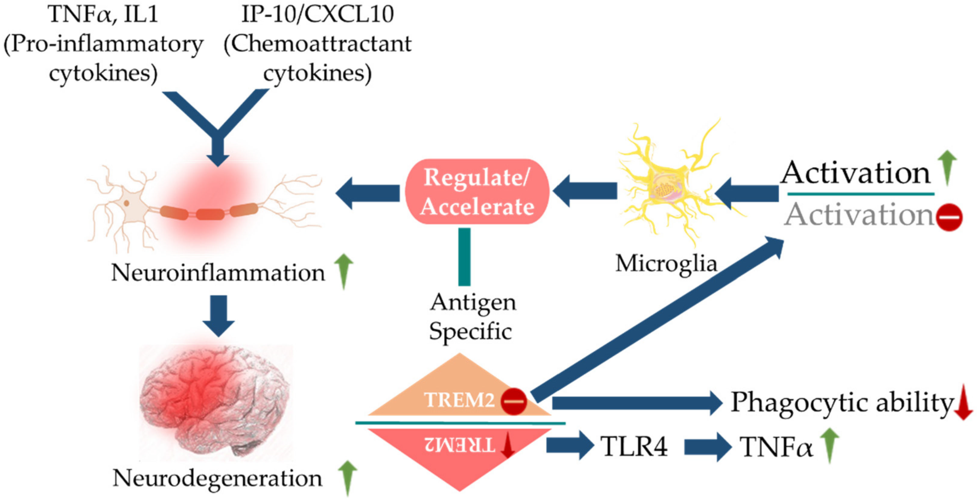
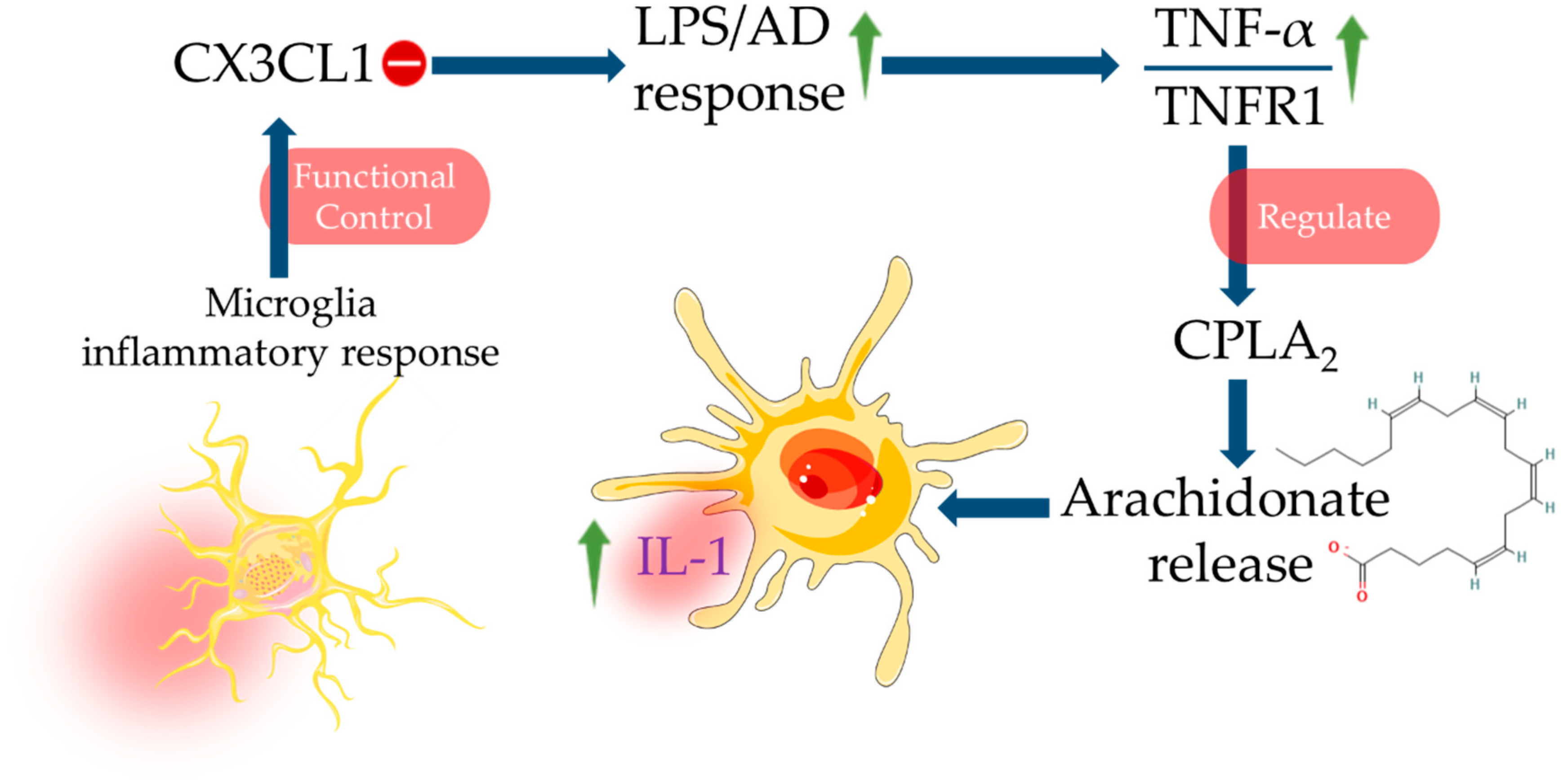
2.1. Immune System in AD and Cytokines

At the beginning of neurodegeneration, the immune reactions trigger macrophage activation (predominantly M2 and sometimes M1) [25]. These macrophages secrete chemical messengers in interneuronal communications and develop autoimmune neurotoxicity including those reactions that lead to neuroinflammation and escalation of AD. The immune system employs cytokines, which play a major role in immune responses following the activation of microglia in the pathology of AD. Cytokines determine the mechanisms and reactions that take place in the immune system in response to abnormal changes in the neurons. These trigger the recruitment of other defensive cells including neutrophils and macrophage progenitor cells.
In the case of AD, Aβ originating from APP trigger the rest of the pathologies. Aβ outside the neurons and neurofibrillary tangles inside the neurons make up for the development of AD [77,78]. Aβ further produces immune response activating complement systems. In CNS, the immune system is programmed to functionally respond to pathological changes such as those presented by the progression of AD [25]. The immune system activation observed in AD is labelled as neuroinflammation [79]. Herein, misfolded and aggregated proteins i.e., Aβ act through danger-associated molecular pathways (DAMP) to bind pathogen recognition receptors such as CD14, CD36, α6β1, integrin, and toll-like receptors (TLRs) [80]. These, in turn, control functions of ROS, NO, IL-1β and TNF-α. It has been experimentally shown that, contrary to antiquated conclusions about neuroinflammation, observed in MCI, early, and late AD onset are initiating events predominantly driven by the CNS resident immune cells, such as microglia and perivascular myeloid cells [79]. An up-regulation of TNF-α with concomitant suppression in TGF-β synergize Aβ42 deposition in MCI, which further trigger neuroinflammation via recruiting IL-1β (Figure 1). Genetic variants and transcription factors also determine the expression of activated microglia in the pathological environment. Damaging or degenerating neurons give off signals acting as a form of microglial control switch that stimulates microglia which could become cytotoxic from the reactive intermediates solicited such as pro-inflammatory cytokines [81]. In response to a change in homeostasis, microglia must first be activated, changing it from a static to a primed state. Changes in infiltrating monocytes that support CNS immune response in the parenchyma and neuronal progenitor granule crossing the BBB might be a hallmark for early detection of AD and propensity of inflammatory response and neurodegeneration [82]. Asymmetrical changes in serum and plasma levels of cytokines may indicate changes in early cytokine levels widely reported in macrophage precursor cells that may confer a greater risk of developing neurodegeneration and abnormal macrophage morphology.

2.2. Roles of Cytokines in Autophagy

Aβ burden has been revealed to be positively correlated with age [51] and exacerbated by oxidative stress, such as GAPs that promote the generation of ROS [54] that perturb brain health [83,84,85]. Glycation end products that confer oxidative stress in AD, which was found to be heavily associated with ApoE in its dimeric form greater than its monomeric form at Aβ accumulation site [55]. An increase of ApoE can lower the Aβ40–42 turnover rate on greater cognitive decline in AD [57]. The same has also been found to negatively influence or disturb autophagy by disrupting autophagosome formation [59]. This, in turn, leads to greater deterioration of neuronal health in AD pathology. Autophagy is critical for Aβ clearance and important in the maintenance of homeostasis in the CNS. In concert with dysfunction of autophagy, mitophagy was observed to express excessive fragmentation, decline in synaptic integrity [60], and an imbalance of mitochondrial dynamics [61,62]. Dysfunction of autophagy/mitophagy indicates a notable neuroinflammatory pathology and involvement of cytokines. IL-1β and IFN-γ (which are known to be expressed in AD pathogenesis) exposure to primary rat β-islet cells hindered autophagy resulting in cell apoptosis [64] and additionally, IL-1β was reported to modulate microglia autophagy in LPS cultures in the presence and absence of Aβ42 [67,86]. This evidence suggests that IL-1β and IFN-γ maintain control of inflammation in AD via lysosomal pathway and initiation of phagophore assembly.

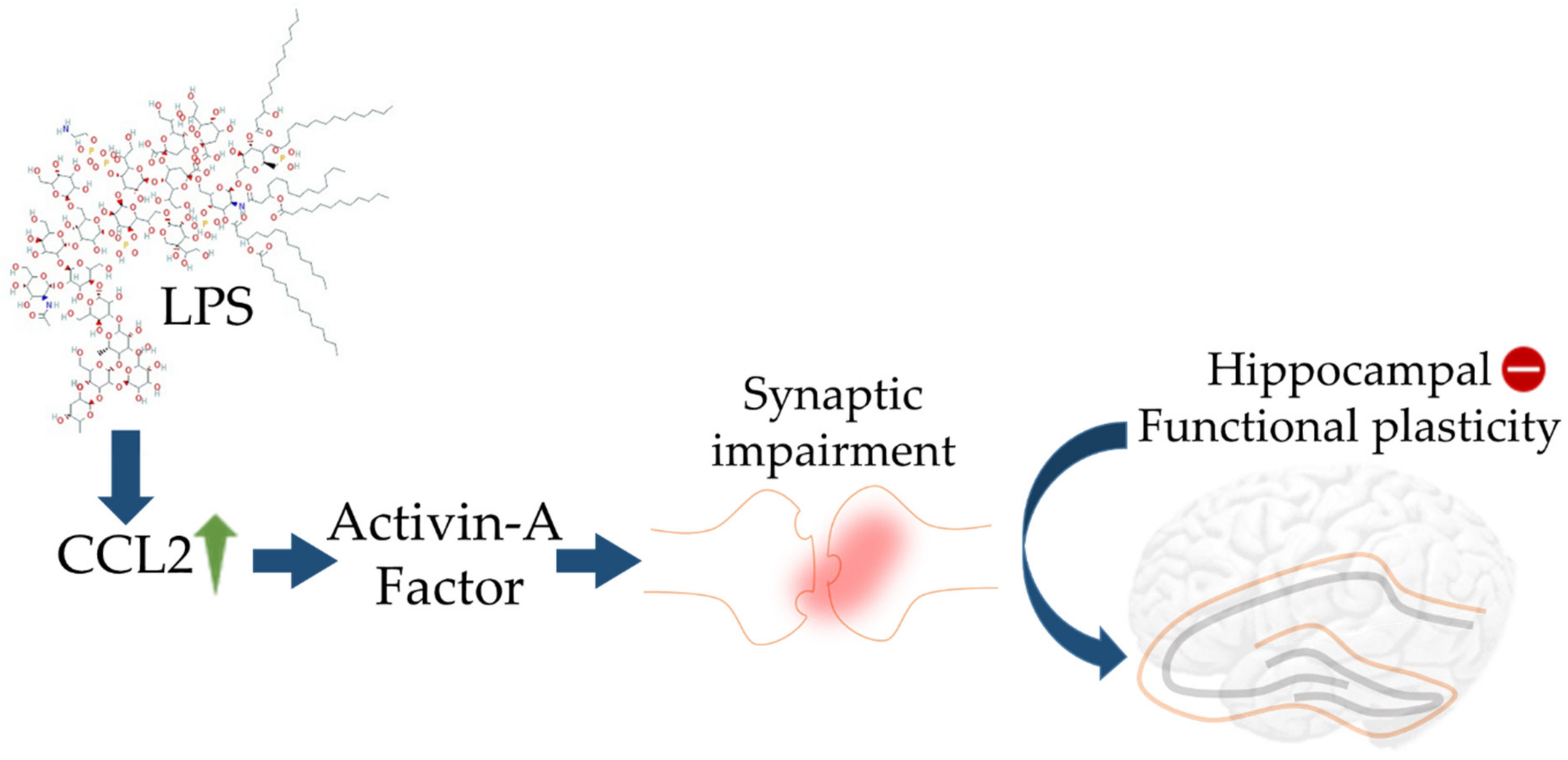
2.3. Cytokines and BBB

There exists a definite correlation between brain cytokine levels and neuropsychiatric disorders. Right at this point, selectivity, and integrity of BBB to cytokines become important. Cytokines are pleiotropic, hence their release, unlike hormones has more complicated effects on the regulation of neurotransmission. Cytokines can cross BBB, activate free calcium, and by disrupting the compartmental model of brain calcium homeostasis, compromise the integrity of BBB [87]. Many cytokines can pass through BBB directly [88]. Interestingly, glial cell-derived neurotrophic factors bypass the BBB by simple diffusion through circumventricular organs. Whereas passage of IL-1α, IL-6, and TNF-α involves saturable influx transport through retrograde axonal transport system [87,89]. TNF-α, a downstream cytokine of chemokine IP10, decreases tight junction proteins leading to the destruction of endothelial tight junctions of BBB to affect its permeability [90]. On the other side, inhibition of mTOR hyperactivity has been reported to protect the integrity of BBB in AD [91]. Therefore, BBB dysfunction brings about early aging in the brain paving the way for AD and other neurodegenerative disorders.

3. Role of Cytokines and Chemokines in Neuropsychiatry

The study of cytokines to understand the pathophysiology of neuropsychiatric disorders such as dementia, anxiety, and delirium has been pioneered by Dr. M. Maes who first linked vegetative symptoms with enhanced presence of IL-1, IL-6, and haptoglobin [87,92]. Chemokines regulate the migration of microglia and the recruitment of astrocytes to the sites of inflammation. Cytokines may act in an autocrine, paracrine, or endocrine fashion and generally are upregulated at sites of Aβ plaques. Aβ peptides mediate cell mediators, such as monocytes are also responsible for the generation of IL-8, monocyte chemoattractant protein 1 (MCP1), MIP1α, and MIP1β. LPS stimulates astrocytes to secrete cytokines including IL-6 and TNF-α, activates astrocytoma cells to secrete IL-6 and IL-8 and monocytes to secrete IL-8 under the influence of Aβ peptides [93]. Synergistic activity of cytokines has also been reported along with Aβ peptides e.g., TNF-γ synergizes with Aβ to enhance secretion of TNF-α and reactive nitrogen species [39]. IL-1β displays pro-inflammatory actions via MEK 1/2, JNK-activated α-secretase cleavage and upregulated a disintegrin and metalloprotease (ADAM)-17/TNF-α converting enzyme (TACE) pathway to increase sAPPα secretion [94]. On the contrary, IL-1β can also serve as an anti-amyloidogenic factor by decreasing sAPPβ and amyloidogenic Aβ fragment levels by reducing β-secretase cleavage [95]. It was also suggested that increased Aβ clearance by microglia in models of sustained IL-1β neuroinflammation could involve Th2 cytokines, such as IL-4 [30]. Moreover, a feedback signalling loop between Aβ and IL-1β was also proposed in which Aβ can induce the production of IL-1β [96]. The migration of astrocytes to Aβ plaques is promoted by chemokines CCL2 and CCL3, which are generally released by activated microglial cells. Upregulation of CCL2 by LPS was found to promote synaptic impairment through recruiting activin A leading to loss of hippocampal plasticity (Figure 2).
Important pathways involved in the pathogenesis of AD include the amyloid cascade hypothesis, TAU hypothesis, cholinergic hypothesis, and excitotoxicity hypothesis. In the case of AD, CSF dysfunction is noticed even before cognitive decline. Activities of mTOR cause vascular irregularities in the brain decreasing cerebral blood flow which in turn sets up cognitive decline. The amyloid cascade hypothesis identifies the accumulation of Aβ plaques at different areas of CNS and related changes as the principal factor behind the development of AD [97]. TAU hypothesis proposed that hyperphosphorylation of TAU leads to form neurofibrillary tangles preventing its regular role of supporting axonal microtubules and subsequently plays a critical role in neurodegeneration [98]. Cholinergic hypothesis focuses on symptoms of cognitive decline and presents malfunctioning of cholinergic neurons as a pathophysiological factor towards initiation of AD [99]. Excitotoxicity refers to the unprecedented death of nerve cells due to the overstimulation of certain amino acid receptors [100]. A high concentration of glutamates activates N-methyl-d-aspartate and α-amino-3-hydroxy-5-methylisoxazole propionic acid receptors. As a result, voltage-gated calcium allows the entry of extracellular calcium into cells and thus a hindrance in neuronal energy metabolism leads to cell death.

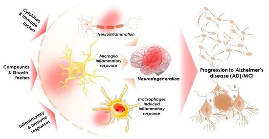
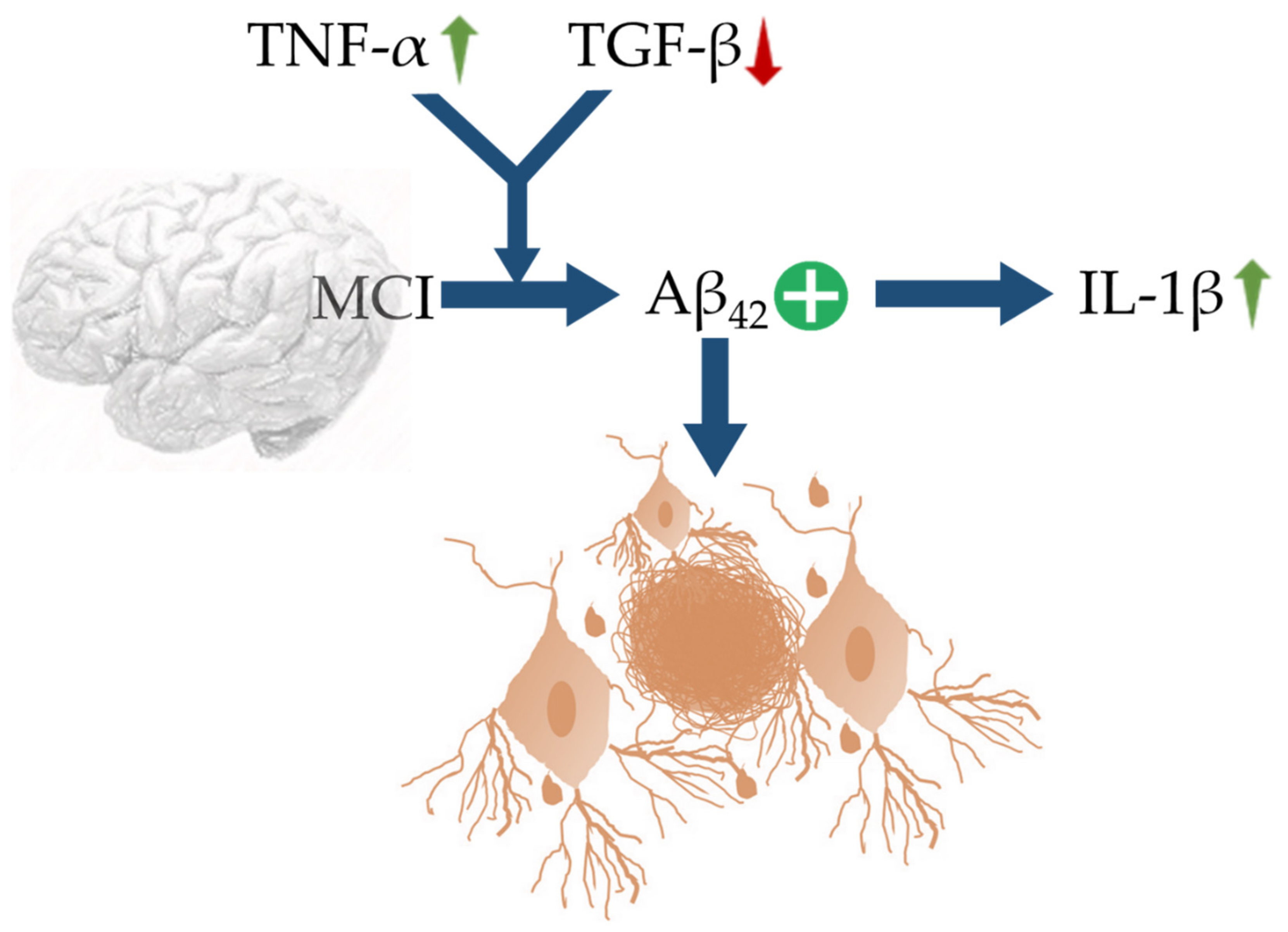
4. Neuroinflammation

Inflammation is the response of our body system to eliminate both sources of cell injury along with the cell and tissue debris originating from the insult. The immune system activation observed in AD is labelled as neuroinflammation. Though classical signs of inflammation such as swelling, heat, and pain are absent in brain inflammation, it characteristically involves increased monocytes and glial macrophage cells [31]. During the initial phase of neurodegeneration, immune reactions are triggered through the activation of macrophages (mainly M2 and sometimes M1) [101]. These activated macrophages secrete chemical messengers in interneuronal communications and develop autoimmune neurotoxicity including those reactions that lead to neuroinflammation and the escalation of AD. Activated cells strongly produce inflammatory mediators such as pro-inflammatory cytokines, chemokines, macrophage inflammatory proteins, monocyte chemo-attractant proteins, prostaglandins, leukotrienes, thromboxanes, coagulation factors, ROS (and other radicals), nitric oxide, complement factors, proteases, protease inhibitors, pentraxins, and C-reactive protein. Upregulated immunoinflammatory events play important roles in the pathogenesis of AD.
Chronic neuroinflammation (immune response to the formation of Aβ peptides and neurofibrillary tangles) is characterized by persistent activation of microglia and release of inflammatory mediators. Hence, an inflammatory cycle is perpetuated since microglia and astrocytes are constantly activated, leading to a further increase in the levels of cytokines and chemokines. These mediators, in turn, alter APP processing encourage the formation of Aβ plaques. These alterations also result in reduced production of neuroprotective sAPPα. Senile plaques activate the complement system resulting in inflammation within CNS. Thus, neuroinflammation-mediated tissue damage initiates the degeneration process. During the early stages of AD, neuroinflammation leads to the entry of PNS cells with chemokine receptors into the brain crossing BBB [102]. As a result of Aβ deposition, chemokines e.g., CCL2, IL-8, CXCL10, CCL5 are released from PNS.
Aβ plaques containing dystrophic neuritis, activated microglia, and reactive astrocytes that along with released inflammatory mediators contribute to neuronal dystrophy. Inflammatory mediators and activated glial cells together kill neighboring neurons and encourage amyloidogenic processing of APP. Nuclear receptor binding factor 2 (NRBF2) is a key factor for maintaining autophagic degradation of APP and production of Aβ by controlling maturation of APP-containing vesicles through the interaction of APP with CCZ1-MON1A-RAB7 module [103,104]. The inability of CNS phagocytes to clear Aβ plaques and upregulated formation of plaques as a result of chronic neuroinflammation play instrumental roles in AD [105]. In agreement with this, in a cohort study, Taipa and colleagues reported elevated levels of eotaxin, IL-1 receptor antagonist (IL-1ra), IL-4, IL-7, IL-8, IL-9, IL-10, IL-15, TNF-α, granulocyte colony-stimulating factor (GCSF), MCP1, and platelet-derived growth factor in CSF of AD patients in comparison with non-demented controls [40]. The same study also reported inverse relations between CSF levels of IL-1β, IL-4, IL-6, IL-9, IL-17A, IFN-γ, basic FGF/FGF2, GCSF, GMCSF, and MIP1β with AD progression [40]. In this section, we reviewed the roles of several neuroinflammatory factors including pro- and anti-inflammatory cytokines, APP and TAU proteins, glial cells, advanced glycation end products, and complement systems in the pathogenesis and development of AD.

4.1. Pro-Inflammatory Cytokines

Cytokines are secreted by glial cells around Aβ plaques. Disturbances in inflammatory and immune pathways in AD have been strongly associated with altered levels of some acute-phase proteins and pro-inflammatory cytokines in the blood, CSF, and brains. Aβ peptides can directly trigger the expression of several pro-inflammatory cytokines such as IL-1β, IL-6, TNF-α, and IFN-γ by glial cells. Pro-inflammatory cytokines like MMIF, YKL40, TNFs, and their receptors, sTREM2 are clearly engaged in TAU pathology and in the aging process [32]. Additionally, IL-15, MCP-1, VEGFR-1, sICAM1, sVCAM-1, and VEGF-D are found to be associated with TAU pathology and correlate with CSF TAU level [106]. Pro-inflammatory cytokines were found to induce indoleamine 2,3 dioxygenase to increase blood levels of quinolinic acid, a neurotoxic factor [107]. Pro-inflammatory cytokines, in conjugation with chemoattractants endorse neurodegeneration via promoting neuroinflammation, which can be triggered by the activation of defective microglia. TREM2 deficiency strongly triggers neuroinflammation via potentiating microglial activation and reducing microglia-mediated Aβ phagocytosis. TREM2 deficiency is also associated with activation of inflammatory markers, such as TNF-α through a TLR-dependent pathway (Figure 3).
High levels of pro-inflammatory cytokines, such as IL-1β, IL-6, and TNF-α, have been detected in the brain of AD subjects [108]. Pro-inflammatory molecules produced by the reactive astrocytes can elevate the expression of secretases in neurons, enhancing the production of Aβ and activating microglia to produce inflammatory factors [109]. In transgenic mice model, pro-inflammatory cytokines viz. IL-1 β, TNF-α, IL-6, IL-12, and IL-23 have also been found to correlate with Aβ load [110].
IL-1α and IL-1β are known to initiate cell activation upon binding with cell membrane receptors. Physiologically, an elevated level of IL-1β is a characteristic feature of brain parenchymal cells immediately after injury [111], while IL-1 hastens neuronal degeneration by increasing the production of IL-6 and the activity of iNOS. In addition to that, IL-1 is also responsible for enhanced acetylcholinesterase activity, activation of astrocytes and microglial cells, expression of S100β, production of macrophage colony-stimulating factor (MCSF), and further additional production of IL-1. IL-6 is a major player in host inflammatory response. IL-6 displays neurotrophic effects by activating microglia, promoting astrogliosis, and stimulating the production of acute-phase proteins. IFN-γ endorses TNFs and NO activities. TNF-α centrally regulates cytokine activities during inflammatory response through regulating an autocrine cascade of production of IL-1 and TNF-α from glial cells. In the AD brain, IL-1 regulates APP processing. In an experiment, rat cortical glial cells presented themselves with increased IL-6 mRNA on being exposed to the first 105 carboxy-terminal amino acids of APP [112]. Dose-dependent increments were also observed in levels of IL-1, IL-6, TNF-α, MIP-1α, and MCP-1 in glial cells on exposure to Aβ peptides [74].
In contrast, IL-1ra, IL-4, IL-10, IL-11, IL-13, TGF-β act as anti-inflammatory cytokines, specific receptors for IL-1, TNF-α, and IL-18 act as inhibitors of pro-inflammatory cytokines. Anti-inflammatory cytokines belonging to Th2 and Th3 cell subsets exert a protective effect against AD by counteracting the effects of pro-inflammatory cytokines [80]. Of note, TGF- β, produced by Th3 cells is capable of ameliorating Aβ-induced cytotoxicity both in vivo and in vitro; while, deficiency of TGF-β1 promotes accumulation of Aβ peptides and formation of neurofibrillary tangles [113]. Dysregulation of the balance between pro-inflammatory and anti-inflammatory cytokines in the favor of pro-inflammatory cytokines leads to a cycle of further cytokine production, cytokine synergism, and cellular activation. It has been shown that an absence of chemokine (CX3CL1) can increase TNF-α and TNFR1 expression by intensifying LPS action, which simultaneously triggers the release of other pro-inflammatory cytokines like IL-1 by macrophages mediated through enhanced arachidonate release. Microglial hyperactivation can lead to CX3CL1 impairment in the brain, which ultimately impacts by amplifying and worsening the neuroinflammatory conditions (Figure 4).

4.2. Anti-Inflammatory Cytokines

Interestingly IL-4, IL-10, and IL-13 can suppress pro-inflammatory cytokine genes e.g., IL-1, TNFs, and chemokines. IL-1ra directly antagonizes the activities of IL-1α and IL-1β by competitive inhibition. Experimental results suggest that IL-1ra suppresses IL-1β-induced TNF-α production and iNOS expression in astrocytes by preferentially binding with IL-1R1 [29]. In addition to protecting against IL-1β-induced neurotoxicity, IL-1ra also attenuates neuronal damage caused by ischaemic excitations. IL-4 can suppress pro-inflammatory cytokines such as IL-1, TNF-α, IL-6, IL-8, and MIP-1α by inhibiting their expressions. Further IL-4 is associated with increased production of IL-1ra and inhibition of IFN-γ leading to a decrease in TNF-α and NO. IL-10, acting through specific cell surface receptors reduces the synthesis of IL-1 and TNF-α. IL-10 also inhibits TNF-α, IL-1, IL-6, IL-8, IL-12, GMSF, MIP-1α, and MIP-2α. Secretion of pro-inflammatory cytokines by glial cells is halted on pre-exposure to IL-10. IL-10 has been hypothesized to exert the actions by suppressing cytokine receptor expression, inhibiting receptor activation, while TGF-β has been shown to impede the production of IL-2, IFN-γ, and TNFs. Of note, three mammalian isoforms of TGF-β i.e., TGF-β1, TGF-β2, and TGF-β3 are prevalent within the CNS. As a result of this, TGF-β is associated with a plethora of activities including microglial activation to inflammatory response, astrocytes, and regulation of COX-2 and APP. Interestingly, elevated levels of TGF-β1 and TGF-β2 have been observed in CSF and blood of AD patients [114,115].

4.3. APP Protein

APP is a transmembrane protein present in the cell membrane of all neurons. Under normal conditions, α-secretase and γ-secretase cleave APP into three fragments which in turn get digested via proteosomes (non-amyloidogenic pathway). During the initial phases of AD, the amyloidogenic pathway takes over and β-secretase becomes involved in the process in place of α-secretase [116]. The α-secretase activity is exerted by three members of the ADAM family viz. ADAM9, ADAM10, and ADAM17/TACE. The β-secretase activity has been mainly attributed to the β-site APP cleaving enzyme. The γ-secretase complex comprises presenilin (PSEN), nicastrin, anterior pharynx defective-1 (APH-1), and presenilin enhancer-2 (Pen-2). The amyloidogenic pathway predominantly gives rise to fragments like sAPPβ, APP intracellular domain (AICD), and Aβ peptide spanning from 1-40 amino acid residues. It further exacerbates AD symptoms as these abnormal fragments are not naturally digested resulting in extracellular accumulation of aggregates or plaques of those fragments. Eventually, these senile plaques are termed Aβ peptides or Aβ lipoproteins. These senile plaques, in general, lead to neurotoxicity, apoptosis, oxidative stress, and neuroinflammation. In addition to generating inflammatory responses, Aβ also causes mechanical disruption in synaptic transmission [117].

4.4. TAU

TAU protein stabilizes microtubules which are very important for the cytoskeletal integrity of a cell. They reside throughout the axon to aid transport proteins to move nutrients and neurotransmitters. Microtubules lose their structure in absence of TAU and break apart. When β-secretase becomes more active than α-secretase, thus a high amount of Aβ is produced that in turn, causes hyperpolarisation of TAU protein through excessive phosphorylation of TAU [118]. On hyperpolarisation, TAU protein starts aggregating with each other. Unlike senile plaques, TAU clumps stay inside neuronal cells. As a consequence of this, the cytoskeleton starts to fall apart that hampers axonal transport. Neurotransmitter transport from soma to synaptic bud becomes affected and neuronal function decreases. Not only neurotransmitters, but the flow of nutrients inside the longest cell of the body also suffers, and gradually axons and dendrons start to degenerate. As a result of this, the cluster of such neurons forms neurofibrillary tangles. Cytokines with kinase activity on TAU include cyclin-dependent kinase 5 (CDK5), glycogen synthase kinase-3β (GSK-3β), and p38 mitogen-activated protein kinases (p38-MAPK) [119].
In AD, these TAU-led neurofibrillary tangles have been observed to be further propagated through the toxicity presented by Aβ plaque accumulation and loss of cholinergic neurons in rat basal forebrain primary septal culture [120]. Additionally, Aβ was found to prevent microtubule binding in primary cultures of fetal rat hippocampal neurons. While in the human cortical neurons induced hyperphosphorylation of TAU at Ser-202 and Ser-396 was found to be accumulated in the somatodendritic compartment of Aβ-treated neurons [121].
The constituents of axonal projections in the mammalian brain are neurofilaments that form side projections of carboxy-terminals from the core filament, believed to be heavily phosphorylated; while TAU-embellished microtubules are also known to be differentially phosphorylated. The α- and β-globulin subunits that constitute axonal microtubules are formed by the energy-consuming nucleation process. An energy-expensive neuro-process would require optimal active mitochondria to properly conduct impulse. Hyperphosphorylation of TAU has been credited to play an active role in the impairment of axonal support functioning that optimizes interneuronal communications amongst associated organelles. The oxidative stress in AD brains also may lead to hyperphosphorylation of TAU. Of note, where the absence of superoxide dismutase (SOD) was observed to increase oxidation damage from ROS, an escalation of Ser-36 phospho-TAU was revealed in treatments of SOD-null mice. Untreated mice did not survive past one week, reflecting SOD deficiency was, therefore, deleterious [122].

4.5. Glial Cells

Progress in AD-related research has revealed the important roles of glial cells including the astrocytes, microglia, NG2 glia, and oligodendrocytes that contribute to the pathogenesis of the disease [123]. Astrocytes and microglia participate by functioning as effector cells to release cytokines by somehow failing to live up to their homeostatic functions. NG2 glia, a novel and distinct class of glial cells in CNS are responsible for myelination and remyelination of axons thus playing a vital role in high-speed nerve impulse transport and cognition [124]. It is interesting to note that amyloid peptides and their precursor APP protein act as glial activators. Disruption of the APP gene and its proteolytic products delay and decrease amyloid-dependent microglial activation.
Astrocytes are star-shaped glial cells in CNS involved in energy reserves, regulation of extracellular ions, as well as the clearance, metabolism of neurotransmitters, and modulation of oxidative stress. Among the notable neurotransmitters, glutamate is released during neuroinflammatory conditions mainly which in the long-term is proved to be toxic to neurons via the excitotoxicity pathway. Of note, astrocytes can take up glutamate and recycle it to neurons after transforming into glutamine, an amino acid [125]. In the AD, Aβ peptides decrease uptake of glutamate, resulting in increasing redox insult. Interestingly, alongside the neuroprotective activities of astrocytes through Aβ clearance and degradation, they could also be a source of Aβ owing to their overexpression of beta-secretase 1 (BACE1) in response to chronic stress [126].
The migration of astrocytes to Aβ plaques is promoted by chemokines CCL2 and CCL3, which are released by activated microglial cells. In an experimental model, mouse astrocytes plated on amyloid-rich brain sections from APP transgenic mice have been found to reduce amyloids [45]. Of note, astrocytes respond to CNS insults through a process named reactive astrogliosis, an early pathological feature of AD, and can represent a response to the accumulation of Aβ and/or to the increasing number of degenerating neurons [127]. Astrocytes can be stimulated by oxidative stress, free saturated fatty acids, pathogens, and LPSs. Additionally, contrary to quiescent astrocytes, reactive astrocytes can produce cytokines, such as TNF-α, IFN-γ, and ILs [41]. IFN-β, TNF-α, and IL-1β induce the generation of Aβ in primary human astrocytes and astrocytoma cells. Astrogliosis is also characterized by a high level of the astrocyte marker glial fibrillary acidic protein (GFAP). The latter occurs around Aβ deposits both in the brain parenchyma and in the cerebral microvasculature. Senile plaques are associated with GFAP-positive activated astrocytes. In various neuropathological states, the increased expression of GFAP corresponds to the severity of astroglial activation [128]. Microglial cells and astrocytes express pathogen recognition receptors e.g., TLRs, integrin α6β1, A1, CD36, CD47, CD14 to act as class A scavenger receptors through DAMP [80].
Oligodendrocytes, under the influence of NG2 cells, are responsible for myelin sheath generation around axons. A study concluded that Aβ peptides induce local translation of myelin basic protein 18.5 kDa isoform in distal cell processes [129]. It is interesting to note that Aβ oligomers modulate the expression of myelin basic protein with the help of the integrin β1 receptor, Src-family kinase Fyn, and Ca2+/CaMKII. The pharmacological inhibition of Fyn kinase was found to attenuate oligodendrocyte differentiation and survival induced by Aβ. Interestingly, in ex vivo organotypic cerebellar slices, Aβ caused upregulation of myelin basic protein through Fyn kinase and modulated oligodendrocyte population dynamics by inducing cell proliferation and differentiation [129]. Application of Aβ oligomers to cerebellar organotypic slices, enhance remyelination and oligodendrocyte lineage recovery was suggested in the case of lysolecithin-induced demyelination.

4.6. Advanced Glycation End Products

Advanced glycation end products mediate crosslinking of certain proteins resulting in age-related decline in cognition and other cellular functions [130]. RAGE (receptor for advanced glycation end-products), a ligand for both Aβ and S100B is also associated with the activity [131]. In hyperglycaemic patients, unusual glucose metabolism and oxidative stress aggravate the activities of advanced glycation end-products [132]. This may be correlated with the notion that excess dietary carbohydrates and deficient cholesterol may lead to AD development. Intracellular neurofibrillary tangles and extracellular senile plaques serve as substrates for glycation. Advanced glycation end products induce the production of ROS and cytokines through activation of microglial RAGE leading to engagement of nuclear factor kappa B (NF-κB) [133]. It has been clinically observed that low dietary intake of advanced glycation end products is directly related to reduced oxidative stress and inflammation that can further exacerbate AD symptoms [134,135].

4.7. Complement System

At an early stage of AD, Aβ peptides activate the complement system. The complement system works as a part of the immune system to remove unwanted bodies through antibody-mediated phagocytosis. In course of doing this, complementary proteins interact with cell surface receptors to promote an inflammatory response in the host system. Complement system attacks and destroys invaders in four steps viz. recognition, opsonization, inflammatory stimulation, and killing. In the human brain, astrocytes are the major center of complement activity. Astrocytes can synthesize complement proteins including C1-C9, regulatory factors B, D, H, I, and complement receptors namely C1qR, C3aR, and C5aR locally to defend through both classical and alternative pathways [74]. Microglia also supports phagocytosis by expressing C1q, C3 proteins, and C1qR, CR3, and C5aR receptors [136]. Apart from neuroglia, neurons also express regulatory factors H, S, and receptors C1qR, C3aR, and C5aR. Complement protein C1q affects the formation of Aβ plaques containing β-sheet structures [137]. In transgenic AD mice, inhibition of the complement system by C3-knockout resulted in the increased formation of Aβ plaques. These results have further supported a neuroprotective role of the complement system [137,138,139].

5. MMIFs in AD: Pathogenic or Protective?

MMIF, also termed as a glycosylation inhibiting factor, is classified as a pro-inflammatory cytokine is an important regulator of innate immunity. Expression of MMIF correlates with expression of VEGF in CNS [140,141]. Interestingly, glucocorticoids stimulate the secretion of MMIF, whereas glucocorticoids are known to suppress most of the other cytokines. Thus, MMIF acts against the general anti-inflammatory response of glucocorticoids. There exists a debate on whether endogenous MMIFs support or counter the pathogenesis of AD. Enhanced MMIFs have been reported in mouse models of neurodegenerative disorders [80,142]. Again, several studies reported that MMIF-knockdown in mutant mice has resulted in the acceleration of neurodegenerative disorders [143,144]. MMIFs have also been reported to regulate neuroinflammation and autophagy in the favor of neuroprotection [144,145,146].
MMIF has a notable function in controlling the synthesis and release of TNF-α, IL-1, and other cytokines. MMIF is also involved in macrophage functions such as phagocytosis and tumoricidal activities. On the other note, a brain insulin-resistant state arises due to prolonged exposure of cortical neurons to high concentrations of insulin. MMIF contributes to this insulin-resistant state through inhibition of Akt phosphorylation [147]. In some cases, a structural homolog of MMIF, D-dopachrome tautomerase (MIF-2) exhibits synergistic activities in combination with MMIF [148]. Moreover, MMIF and fragments of senile plaques display similar neurotoxicity patterns [149]. The study also reported enhanced MMIF levels in CSF of AD patients [149]. In silico studies further suggest that MMIF may be involved in neuronal apoptosis during AD [150]. However, it is interesting to note that Popp and colleagues earlier did not find any difference in MMIF levels of AD patients with mild, moderate, and severe dementia [151]. Conclusively, we can say that imbalance between oxidized and reduced isoforms of MMIF is the key to regulate the switch to either a diseased or normal state [151].

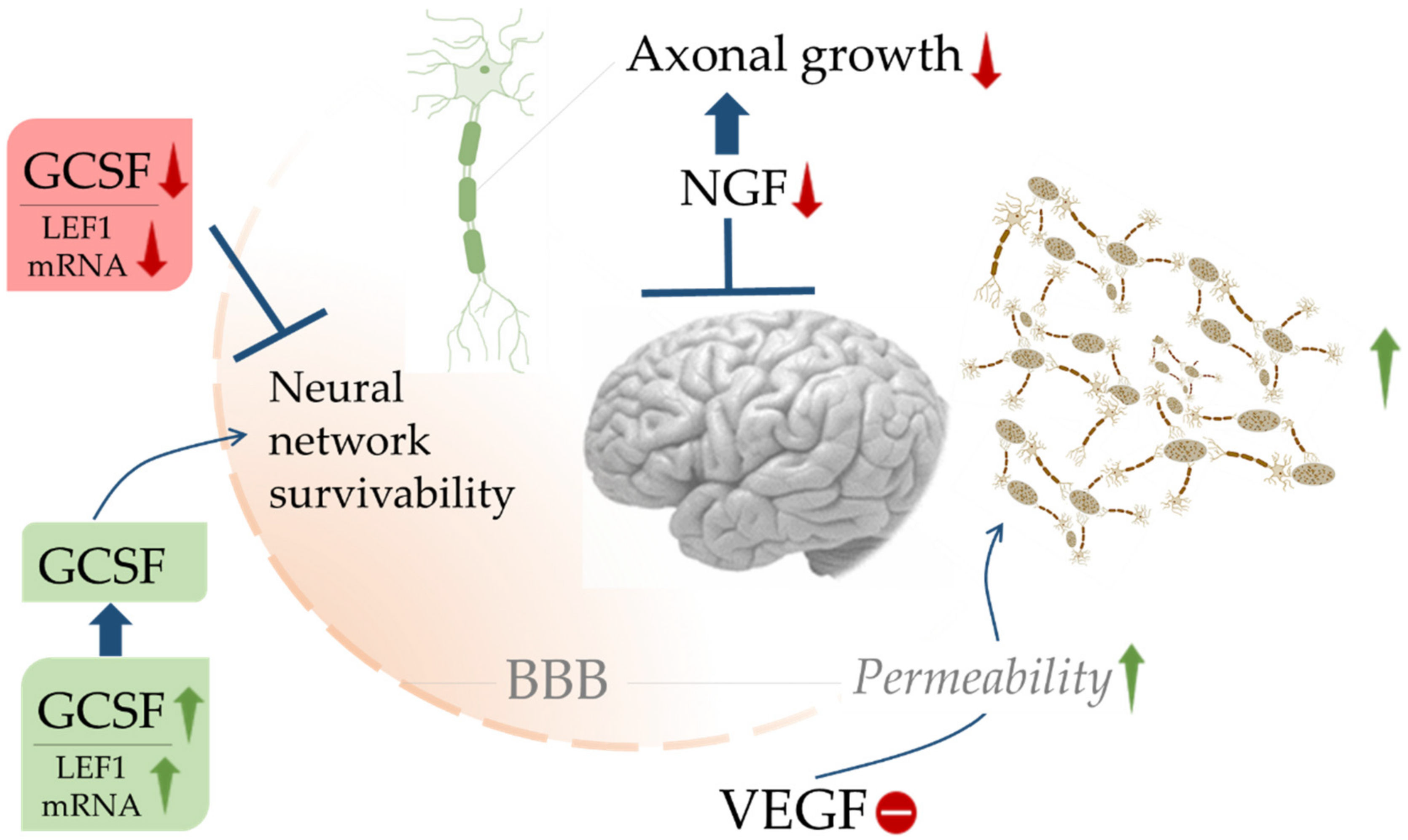
6. Choroid Plexus Growth Factors and AD

The growth-promoting properties of APP, along with other growth factors, play vital roles in the development of AD. The choroid plexus supports neuronal function by secreting CSF. VEGF and FGF can be found in epithelial cells of the choroid plexus. It is rich in various proteins and their receptors. Proteins include FGF-2, TGF-α, and TGF-β along with mRNA expressions for TGF-β, IGF-II, FGF-2, and NGF receptors. The choroid plexus also contains receptor binding sites for FGF-7, keratinocyte growth factor, IGF-1, and IGF-2. Blood-CSF barrier made up of epithelial cells and tight junctions at the choroid plexus allow selective passage of materials into the brain. FGF-2 has been reported to increase in brain parenchyma of AD patients. Moreover, infusion of FGF-2 in rats has resulted in hydrocephalus ex vacuo, which is a clinical feature of AD [152]. It is important to note that improper CSF circulation and impaired clearance of CSF may give rise to dementia and neurodegeneration due to lack of nutrition to CNS cells and enhanced toxic accumulations within CSF. In this section, we shed light on the specific roles of VEGF and FGF growth factors in the development of AD.

6.1. Vascular Endothelial Growth Factors (VEGFs)

VEGFs and their receptors have been reported to localize at the area with lesions and AD-related developments. Different isoforms of VEGF act as pro-inflammatory cytokines, which increase endothelial cell permeability, induce the expression of endothelial cell adhesion molecules and act as monocyte chemoattractants [153]. VEGF is involved in the regulation of GLUT1 and tissue thromboplastin, which in turn regulate vascular pathologies of AD. GLUT1, present in BBB mediates glucose transport into the brain and reduced expression of GLUT1 is relatable with aggravated AD conditions. Tissue thromboplastin and derived factors play a pro-inflammatory role leading to vascular dementia [154]. AD patients tend to present with enhanced VEGF activity within reactive astrocytes [155]. Rats subjected to cerebral ischemia displayed increased perivascular VEGF reactivity in the clusters of reactive astrocytes [156].

6.2. Fibroblast Growth Factors (FGF)

FGFs are circulatory proteins that play important roles in the activation of cell surface receptors. Around 23 FGF subtypes have been known to exert distinct functions to date [157]. Acidic FGF-1 and basic FGF-2, among eight other FGF family proteins, act through four families of FGF receptors. However, FGF-11-14 does not act through FGF receptors.
FGF-1 and FGF-2 are more potent angiogenic factors than VEGF [52]. Within CNS, FGFs play important roles in the proliferation and differentiation of neuronal stem cells including neurogenesis and axonal growth. FGFs also support the self-renewal of radial glial cells. FGF-8 is a vital player for the proper functioning of the cerebral cortex. Increased levels of FGF-2 have reportedly been associated with AD brain leading to enlargement of ventricles [158]. FGFs regulate not only neuronal stem cells but also adult neurogenesis. Additionally, the maintenance and survival of neurons throughout their life depend greatly on FGF-2. Synaptic plasticity, to some extent, is controlled by FGF-1 and FGF-2. Thus, the conduction of nerve impulses through axons and synapses for proper cognition is dependent upon FGFs. Belluardo and colleagues demonstrated that upregulation of FGF-2 can successfully prevent neuronal loss in cortical and hippocampal regions of the brain [159]. In the rat models, FGF-21 has been found to ameliorate senile plaques-mediated neurodegeneration [160]. The effects were achieved via minimizing oxidative stress through PP2A/MAPK/HIF-1α-mediated pathways [160].

7. Neurotrophic Factors

Neurotrophic growth factors produced by neural stem cells are involved in the differentiation of cells and cell survival. Neurotrophic growth factors consist of NGFs, GDNF, neurokines, and non-neuronal growth factors. NGF is probably the most discussed neurotrophic growth factor/neuropeptide that involves in growth regulation, maintenance, proliferation, and survival of certain target neurons. NGF was the first neurotrophin to be discovered followed by BDNF, neurotrophin-3, neurotrophin-4/5, and neurotrophin-6 [72]. Neurotrophins bind to cognate TrK receptors and p75NTR. The low-affinity p75NTR can bind with all neurotrophin family members. Neurokines and cytokines related to IL-6 bind to cell surface receptor complexes, which share a common structural organization. The four ligands interchangeably employ two distinct receptor subunits, leukemia inhibitory factor receptor b (LIFRb) and gp130; some employ a ligand-specific α subunit [76].
NGF exhibits protective action over cholinergic neurodegeneration. NGF can influence APP processing towards the non-amyloidogenic pathway via protein kinase C-coupled M1 and M3 receptors. Interestingly, NGFs are upregulated in AD brain and CSF, while NGF receptor TrKA is downregulated [74]. BDNFs alone and in chimeric combination with NGF have been found to protect cholinergic neurons in prosencephalon [58]. Interestingly, AD brains have been diagnosed with decreased levels of mRNAs for BDNFs but normal levels of mRNAs for NGF and neurotrophin-3 [161]. In the AD brain, astrogliosis may contribute to increasing NGF and reducing TrKA in the cortex and nucleus basalis. Vinculin-dependent adhesions are central to the functioning of NGF to promote axonal outgrowth. Vinculin-dependent coupling regulates the level of myosin needed for NGF stimulation. The role of NGF as a growth factor amongst a bouquet of proteins is paramount in cognitive processes that may be involved in the survival and phosphorylation of fibrils in axons, that are involved in AD and other chronic diseases closely related to AD [56].

8. Hematopoietic Growth Factors

Apart from controlling hematopoiesis in blood progenitor cells, hematopoietic growth factors such as IL-3, GCSF, GMCSF, MCSF, and erythropoietin play vital roles in the functional activation of all mature cells. In the biological and pathological role of the immune system, the immune system achieves its role by cells that encapsulate it as a whole. Such cells originate from hematopoietic stem cells in the bone marrow by a blood-forming process of hematopoiesis that gives rise to myeloid progenitor cells and lymphoid progenitor cells [162]. Myeloid progenitor cells constitute megakaryocytes, erythrocytes, mast cells, and myeloblast. The myeloblast cells differentiate into immune cells, such as basophil, neutrophil, eosinophil, and monocytes. Of the subset of the myoblast cells are the monocytes that later develop into macrophages, which play an initiating part in immune system responses that counter foreign material, pathogens, and compromised cells in the CNS.
Hematopoietic growth factors are important contributors to brain marrow for neuropoiesis. They can prevent neuronal death to some extent. Jin and colleagues have pointed out enhanced neurogenesis during AD progression [163], though many pose doubts on the marker doublecortin [164,165]. In a mouse model, GCSF has been observed to restore cognition by restoring acetylcholine levels [61]. The survivability of neural networks in the brain largely depends on GCSF and LEF1 availability, which enter through the BBB and promote their survivability. VEGF increases BBB permeability; however, a defective VEGF expression can trigger immunoreactivity, which is a characteristic feature in AD (Figure 5). Stem cell factors, in combination with receptor c-kit, stimulate neurogenesis [62]. The lower level of stem cell factor in blood and CSF were observed to accelerate cognitive decline during AD [63]. Increased levels of angiopoietins 1 and 2 indicate a cognitive decline in AD. In the mouse models, angiopoietin 1 accelerates AD via FOXA2/PEN2/APP-dependent pathway [166]. Increased neurogenesis, anti-apoptotic influences, and mobilization of microglia contribute to brain repair involving hematopoietic growth factors.

9. Potential Strategies Involving Cytokines for Management of AD

AD affects millions of individuals worldwide among the aging population, yet no therapeutic intervention is available to stop and eliminate this disorder. Neuropathological hallmarks of AD are extracellular deposits of Aβ peptides assembled in plaques, intraneuronal accumulation of hyperphosphorylated TAU protein forming neurofibrillary tangles, and chronic neuroinflammation. No absolute cure for AD is available so far [167].
Among the available therapeutic options against AD, cholinesterase inhibitors and NMDA antagonists display moderate relief in the case of AD. Donepezil, an inhibitor of acetylcholinesterase improved cognitive conditions in AD and increased BDNFs [168]. Pharmacotherapy against Aβ and TAU has yielded limited success only. Treatment with β-sheet breaker peptides results in reduced brain inflammation by disrupting amyloids [169]. RAGE/NF-κB axis could be a potential therapeutic target in AD [170]. Some dietary nutraceuticals display inhibitory effects on the formation of advanced glycation end-products [171]. Resveratrol has been found to modulate levels of Aβ and certain inflammatory markers in AD patients [172]. Luteolin can play a prophylactic role against AD [173]. Additionally, moderate activation of microglia is thought to have beneficial effects in removing neurotoxins, cellular debris, and dying cells or in promoting neuronal survival. Since MMIF is augmented in AD, measuring blood and CSF levels of MMIF may represent a diagnostic biomarker useful both for diagnosis and therapeutic monitoring of the disease [174]. Moderate activation of microglia by acute neuroinflammation is thought to have beneficial effects in removing neurotoxins, cellular debris, or dying cells and also in promoting neuronal survival [175]. IL-1ra, a glycosylated protein antagonizes the cell activating action of IL-1. Furthermore, TNF-α has been reported to possess neuroprotective effects [176]. TGF-β is capable of converting an active site of inflammation into one dominated by reparations [177]. Kitazawa et al. described that blocking IL-1 signaling in 3xtg AD mice with an IL-1 receptor blocking antibody was beneficial since it leads to a decrease in certain Aβ fibrillar forms and plaques [27].
It has been suggested that a blockade of the ongoing inflammatory processes may delay the progression of AD [178]. Studies suggest lesser incidents of developing AD in arthritis patients receiving NSAIDs, regularly [179,180]. The fact that COX-2 mRNA is upregulated in the AD brain further supports this claim. Therefore, receptors for hematopoietic growth factors expressed on neurons provide novel targets for drug discovery in the search for agents that can reverse the progression of AD.
It is interesting to observe that peripheral phagocytes can effectively clear plaques and therapeutic strategies aiming at favoring the recruitment of these cells into the CNS are actively being pursued [80]. In a mouse model, the BDNFs have improved AD conditions by delaying synaptic loss, improving cell signaling, and enhancing cognition and spatial learning [181]. GCSF and analogs have proven neuroprotective activity, which may possibly be used therapeutically. In vivo intraperitoneal VEGF administration reduced cognitive impairment in a mice model of AD [53]. As discussed earlier, NGFs are potential candidates for significant improvement of cognitive functions. Biogenetic exosome-mediated activation of microglia and deregulation of microRNA can be useful to fight against neuroinflammation [182]. Erythropoietin, together with NF-κB can prevent neuronal injury triggered by Aβ toxicity [183]. Inhibitors of TNF-α have exhibited potential promise to slow down the progress of AD-associated cognitive decline [183]. Experimentally delivered mature NGFs into the AD brain showed potential for improving AD condition [56]. ApoE4-centric treatment approaches are gaining interest in recent times since ApoE4 is involved in more than 50% of AD cases [184]. M2 microglia are generally engaged in the restoration of homeostatic balance after an inflammatory insult by releasing anti-inflammatory factors. Thus, the therapeutic promise is there to prevent and treat neuroinflammation with protective functions of microglia [185,186,187]. Another potential strategy might be to inhibit BACE1 to reduce the production of Aβ, however, clinical success is yet to be achieved [188]. Recently, multitarget-directed ligand-based treatment strategies have started to evolve centering on inhibition of GSK-3β, a crucial enzyme for TAU hyperphosphorylation, and some other CNS-specific signaling pathways [119]. Nowadays, in the war against AD and associated disorders, researchers are focusing more on regulating neurotransmitters, lipid metabolism, autophagy, circadian rhythm, gene therapy, etc. [189].

10. Conclusions

In this review, ample evidence reflects the potential roles of cytokines and growth factors in the pathogenesis of AD or pathologically related to AD-like neurodegenerative conditions. It helps us to understand the propensities and action of cytokines and growth factors regulating their effects on neurons upon neurodegeneration. Altogether, evidence evinced in previous research on the rather novel concentration on the topic of cytokines in neuroimmune system responses and their role in inflammation. These two factors possibly preceding neurotoxicity and intrathecal generation of immune molecules and cytokine-producing cells show that cytokines mediate and even activate innate neuroimmune agents. Cytokines regulate the response of pro-inflammatory and anti-inflammatory signals to maintain CNS machinery homeostasis [190]. Pro-inflammatory cytokines induce inflammation in AD and AD-like pathogenesis in response to the apoptotic scenarios. Some growth factors are implicated in the expression of cytokinetic reactions to activate microglia that cause inflammation in AD. Cytokines and growth factors such as NGF, VEGF, TNF-α, and IL-1 additionally impact intricate molecular processes necessary for balance and homeostasis in cognitive mechanisms. To conclude, there exists ample scope of improvement regarding clinically useful strategies to mitigate AD.

Author Contributions

S.D., V.D.F. and R.K. contributed to the conceptualization and designing the manuscript. G.O., P.C., C.V., V.K., A.C., U.A., J.V., P.G., H.P.R.P., K.D.G. and P.H.R. edited and corrected the manuscript. The final correction and editing were done by G.O., S.D., P.C., V.D.F. and R.K. All authors have read and agreed to the published version of the manuscript.

Funding

This research received no external funding. V.D.F. provided APC for publishing this manuscript and all the authors acknowledged the same.

Institutional Review Board Statement

Not applicable.

Informed Consent Statement

Not applicable.

Data Availability Statement

Not applicable.

Acknowledgments

The authors are thankful to the Council of Scientific and Industrial Research, New Delhi, India, for awarding research project (grant number 02(0275)/16/EMR-II) to Saikat Dewanjee. Authors sincerely acknowledge Jadavpur University, India, CSIR-Indian Institute of Chemical Technology, Hyderabad, India, for providing necessary facilities, and DBT-India for providing Ramalingaswami Re-entry Fellowship to Ramesh Kandimalla (RK) for the period of 2018-2023 (No. BT/RLF/Re-entry/22/2016 and SAN.No. 102/IFD/SAN/1117/2018-19). Finally, the authors are exceedingly grateful to the editor and reviewers for their serious comments to improve the quality of this review.

Conflicts of Interest

The authors declare no conflict of interest.

Abbreviations

ADAlzheimer’s disease
ADAMA disintegrin and metalloprotease
AICDAPP intracellular domain
APH-1Anterior pharynx defective-1
ApoEApolipoprotein E
APP Myloid precursor 69 protein
Amyloid-beta
basic FGF/FGF2Basic fibroblast growth factor
BBBBlood-brain barrier
BDNFBrain-derived neurotrophic factor
CDK5Cyclin-dependent kinase 5
CNSCentral nervous system
CPLA2Cytosolic phospholipase A2
CSFCerebrospinal fluid
DAMPDanger-associated molecular pathways
GCSFGranulocyte colony-stimulating factor
GDNFGlial cell line-derived neurotrophic factor
GFAPGlial fibrillary acidic protein
GMCSFGranulocyte macrophage colony-stimulating factor
GSK-3βGlycogen synthase kinase-3beta
IGFInsulin-like growth factor
IL-1raIL-1 receptor antagonist
ILInterleukin
INFInterferon
LIFRbLeukemia inhibitory factor receptor b
LPSLipopolysaccharide
MCIMild cognitive impairment
MCP1Monocyte chemoattractant protein 1
MCSFMacrophage colony-stimulating factor
MIIBMyosin IIB
MIPMacrophage inflammatory protein
MMIFMacrophage migration inhibitory factor
NGFNerve growth factors
p38-MAPKMitogen-activated protein kinases
p75NTRp75 neurotrophin receptor
Pen-2Presenilin enhancer-2
PNSPeripheral nervous system
PSENPresenilin
ROSReactive oxygen species
TACETNF-α converting enzyme
TGFβTransforming growth factor beta
TNFsTumor necrosis factors
TrKTropomyosin Receptor Kinases
VEGFVascular endothelial growth factor

References

  1. Fricker, M.; Tolkovsky, A.M.; Borutaite, V.; Coleman, M.; Brown, G.C. Neuronal Cell Death. Physiol. Rev. 2018, 98, 813–880. [Google Scholar] [CrossRef]
  2. Niikura, T.; Tajima, H.; Kita, Y. Neuronal cell death in Alzheimer’s disease and a neuroprotective factor, humanin. Curr. Neuropharmacol. 2006, 4, 139–147. [Google Scholar] [CrossRef]
  3. Caccamo, A.; Branca, C.; Piras, I.S.; Ferreira, E.; Huentelman, M.J.; Liang, W.S.; Readhead, B.; Dudley, J.T.; Spangenberg, E.E.; Green, K.N.; et al. Necroptosis activation in Alzheimer’s disease. Nat. Neurosci. 2017, 20, 1236–1246. [Google Scholar] [CrossRef]
  4. Menon, P.K.; Koistinen, N.A.; Iverfeldt, K.; Ström, A.L. Phosphorylation of the amyloid precursor protein (APP) at Ser-675 promotes APP processing involving meprin β. J. Biol. Chem. 2019, 294, 17768–17776. [Google Scholar] [CrossRef] [PubMed] [Green Version]
  5. Bergström, P.; Agholme, L.; Nazir, F.H.; Satir, T.M.; Toombs, J.; Wellington, H.; Strandberg, J.; Bontell, T.O.; Kvartsberg, H.; Holmström, M.; et al. Amyloid precursor protein expression and processing are differentially regulated during cortical neuron differentiation. Sci. Rep. 2016, 6, 29200. [Google Scholar] [CrossRef] [Green Version]
  6. Medala, V.K.; Gollapelli, B.; Dewanjee, S.; Ogunmokun, G.; Kandimalla, R.; Vallamkondu, J. Mitochondrial dysfunction, mitophagy, and role of dynamin-related protein 1 in Alzheimer’s disease. J. Neurosci. Res. 2021, 99, 1120–1135. [Google Scholar] [CrossRef] [PubMed]
  7. Tönnies, E.; Trushina, E. Oxidative Stress, Synaptic Dysfunction, and Alzheimer’s Disease. J. Alzheimers. Dis. 2017, 57, 1105–1121. [Google Scholar] [CrossRef] [Green Version]
  8. Roy, E.R.; Wang, B.; Wan, Y.W.; Chiu, G.; Cole, A.; Yin, Z.; Propson, N.E.; Xu, Y.; Jankowsky, J.L.; Liu, Z.; et al. Type I interferon response drives neuroinflammation and synapse loss in Alzheimer disease. J. Clin. Investig. 2020, 130, 1912–1930. [Google Scholar] [CrossRef] [PubMed]
  9. Hampel, H.; Caraci, F.; Cuello, A.C.; Caruso, G.; Nisticò, R.; Corbo, M.; Baldacci, F.; Toschi, N.; Garaci, F.; Chiesa, P.A.; et al. A Path Toward Precision Medicine for Neuroinflammatory Mechanisms in Alzheimer’s Disease. Front. Immunol. 2020, 11, 456. [Google Scholar] [CrossRef]
  10. Wang, M.M.; Miao, D.; Cao, X.P.; Tan, L.; Tan, L. Innate immune activation in Alzheimer’s disease. Ann. Transl. Med. 2018, 6, 177. [Google Scholar] [CrossRef] [PubMed]
  11. Suresh, J.; Khor, I.W.; Kaur, P.; Heng, H.L.; Torta, F.; Dawe, G.S.; Tai, E.S.; Tolwinski, N.S. Shared signaling pathways in Alzheimer’s and metabolic disease may point to new treatment approaches. FEBS J. 2021, 288, 3855–3873. [Google Scholar] [CrossRef] [PubMed]
  12. Murphy, M.P.; LeVine, H., 3rd. Alzheimer’s disease and the amyloid-beta peptide. J. Alzheimers Dis. 2010, 19, 311–323. [Google Scholar] [CrossRef] [PubMed] [Green Version]
  13. Cheignon, C.; Tomas, M.; Bonnefont-Rousselot, D.; Faller, P.; Hureau, C.; Collin, F. Oxidative stress and the amyloid beta peptide in Alzheimer’s disease. Redox Biol. 2018, 14, 450–464. [Google Scholar] [CrossRef] [PubMed]
  14. Arango Duque, G.; Descoteaux, A. Macrophage cytokines: Involvement in immunity and infectious diseases. Front. Immunol. 2014, 5, 491. [Google Scholar] [CrossRef] [PubMed] [Green Version]
  15. Finkelman, F.D.; Holmes, J.; Katona, I.M.; Urban, J.F., Jr.; Beckmann, M.P.; Park, L.S.; Schooley, K.A.; Coffman, R.L.; Mosmann, T.R.; Paul, W.E. Lymphokine control of in vivo immunoglobulin isotype selection. Annu. Rev. Immunol. 1990, 8, 303–333. [Google Scholar] [CrossRef] [PubMed]
  16. Alzheimer, A.; Forstl, H.; Levy, R. On certain peculiar diseases of old age. Hist. Psychiatry 1991, 2, 71–101. [Google Scholar] [CrossRef]
  17. Kawas, C.; Gray, S.; Brookmeyer, R.; Fozard, J.; Zonderman, A. Age-specific incidence rates of Alzheimer’s disease: The Baltimore Longitudinal Study of Aging. Neurology 2000, 54, 2072–2077. [Google Scholar] [CrossRef]
  18. Qiu, C.; Kivipelto, M.; Von Strauss, E. Epidemiology of Alzheimer’s disease: Occurrence, determinants, and strategies toward intervention. Dialogues Clin. Neurosci. 2009, 11, 111–128. [Google Scholar] [CrossRef]
  19. Kumar, A.; Singh, A.; Ekavali. A review on Alzheimer’s disease pathophysiology and its management: An update. Pharmacol. Rep. 2015, 67, 195–203. [Google Scholar] [CrossRef]
  20. Ahn, I.S.; Kim, J.H.; Kim, S.; Ahn, I.S.; Kim, J.H.; Kim, S.; Chung, J.W.; Kim, H.; Kang, H.S.; Kim, D.K. Impairment of instrumental activities of daily living in patients with mild cognitive impairment. Psychiatry Investig. 2009, 6, 180–184. [Google Scholar] [CrossRef] [Green Version]
  21. Cloutier, S.; Chertkow, H.; Kergoat, M.J.; Gauthier, S.; Belleville, S. Patterns of Cognitive Decline Prior to Dementia in Persons with Mild Cognitive Impairment. J. Alzheimers Dis. 2015, 47, 901–913. [Google Scholar] [CrossRef] [PubMed] [Green Version]
  22. Mistridis, P.; Krumm, S.; Monsch, A.U.; Berres, M.; Taylor, K.I. The 12 Years Preceding Mild Cognitive Impairment Due to Alzheimer’s Disease: The Temporal Emergence of Cognitive Decline. J. Alzheimers Dis. 2015, 48, 1095–1107. [Google Scholar] [CrossRef] [PubMed] [Green Version]
  23. Berti, V.; Pupi, A.; Mosconi, L. PET/CT in diagnosis of dementia. Ann. N. Y. Acad. Sci. 2011, 1228, 81–92. [Google Scholar] [CrossRef] [PubMed] [Green Version]
  24. Werman, A.; Werman-Venkert, R.; White, R.; Lee, J.K.; Werman, B.; Krelin, Y.; Voronov, E.; Dinarello, C.A.; Apte, R.N. The precursor form of IL-1alpha is an intracrine proinflammatory activator of transcription. Proc. Natl. Acad. Sci. USA 2004, 101, 2434–2439. [Google Scholar] [CrossRef] [Green Version]
  25. Sattler, S. The Role of the Immune System Beyond the Fight Against Infection. Adv. Exp. Med. Biol. 2017, 1003, 3–14. [Google Scholar] [CrossRef] [PubMed]
  26. Bandyopadhyay, S.; Hartley, D.M.; Cahill, C.M.; Lahiri, D.K.; Chattopadhyay, N.; Rogers, J.T. Interleukin-1alpha stimulates non-amyloidogenic pathway by alpha-secretase (ADAM-10 and ADAM-17) cleavage of APP in human astrocytic cells involving p38 MAP kinase. J. Neurosci. Res. 2006, 84, 106–118. [Google Scholar] [CrossRef] [PubMed]
  27. Kitazawa, M.; Cheng, D.; Tsukamoto, M.R.; Koike, M.A.; Wes, P.D.; Vasilevko, V.; Cribbs, D.H.; LaFerla, F.M. Blocking IL-1 signaling rescues cognition, attenuates tau pathology, and restores neuronal beta-catenin pathway function in an Alzheimer’s disease model. J. Immunol. 2011, 187, 6539–6549. [Google Scholar] [CrossRef] [PubMed] [Green Version]
  28. Tsai, S.J. Effects of interleukin-1beta polymorphisms on brain function and behavior in healthy and psychiatric disease conditions. Cytokine Growth Factor Rev. 2017, 37, 89–97. [Google Scholar] [CrossRef] [PubMed]
  29. Liu, J.; Zhao, M.L.; Brosnan, C.F.; Lee, S.C. Expression of type II nitric oxide synthase in primary human astrocytes and microglia: Role of IL-1beta and IL-1 receptor antagonist. J. Immunol. 1996, 157, 3569–3576. [Google Scholar]
  30. Rivera-Escalera, F.; Pinney, J.J.; Owlett, L.; Ahmed, H.; Thakar, J.; Olschowka, J.A.; Elliott, M.R.; O’Banion, M.K. IL-1beta-driven amyloid plaque clearance is associated with an expansion of transcriptionally reprogrammed microglia. J. Neuroinflamm. 2019, 16, 261. [Google Scholar] [CrossRef]
  31. Kinney, J.W.; Bemiller, S.M.; Murtishaw, A.S.; Leisgang, A.M.; Salazar, A.M.; Lamb, B.T. Inflammation as a central mechanism in Alzheimer’s disease. Alzheimers Dement. 2018, 4, 575–590. [Google Scholar] [CrossRef] [PubMed]
  32. Alasmari, F.; Alshammari, M.A.; Alasmari, A.F.; Alanazi, W.A.; Alhazzani, K. Neuroinflammatory Cytokines Induce Amyloid Beta Neurotoxicity through Modulating Amyloid Precursor Protein Levels/Metabolism. Biomed. Res. Int. 2018, 2018, 3087475. [Google Scholar] [CrossRef] [PubMed]
  33. Bakshi, P.; Margenthaler, E.; Reed, J.; Crawford, F.; Mullan, M. Depletion of CXCR2 inhibits gamma-secretase activity and amyloid-beta production in a murine model of Alzheimer’s disease. Cytokine 2011, 53, 163–169. [Google Scholar] [CrossRef]
  34. Koper, O.M.; Kaminska, J.; Sawicki, K.; Kemona, H. CXCL9, CXCL10, CXCL11, and their receptor (CXCR3) in neuroinflammation and neurodegeneration. Adv. Clin. Exp. Med. 2018, 27, 849–856. [Google Scholar] [CrossRef]
  35. Magalhaes, C.A.; Carvalho, M.D.G.; Sousa, L.P.; Caramelli, P.; Gomes, K.B. Alzheimer’s disease and cytokine IL-10 gene polymorphisms: Is there an association? Arq. Neuropsiquiatr. 2017, 75, 649–656. [Google Scholar] [CrossRef] [Green Version]
  36. Culjak, M.; Perkovic, M.N.; Uzun, S.; Strac, D.S.; Erjavec, G.N.; Leko, M.B.; Simic, G.; Tudor, L.; Konjevod, M.; Kozumplik, O.; et al. The Association between TNF-alpha, IL-1 alpha and IL-10 with Alzheimer’s Disease. Curr. Alzheimer Res. 2020, 17, 972–984. [Google Scholar] [CrossRef]
  37. Ojala, J.O.; Sutinen, E.M. The Role of Interleukin-18, Oxidative Stress and Metabolic Syndrome in Alzheimer’s Disease. J. Clin. Med. 2017, 6, 55. [Google Scholar] [CrossRef] [Green Version]
  38. Lindberg, C.; Chromek, M.; Ahrengart, L.; Brauner, A.; Schultzberg, M.; Garlind, A. Soluble interleukin-1 receptor type II, IL-18 and caspase-1 in mild cognitive impairment and severe Alzheimer’s disease. Neurochem. Int. 2005, 46, 551–557. [Google Scholar] [CrossRef] [PubMed]
  39. Saha, R.N.; Pahan, K. Signals for the induction of nitric oxide synthase in astrocytes. Neurochem. Int. 2006, 49, 154–163. [Google Scholar] [CrossRef] [Green Version]
  40. Taipa, R.; das Neves, S.P.; Sousa, A.L.; Fernandes, J.; Pinto, C.; Correia, A.P.; Santos, E.; Pinto, P.S.; Carneiro, P.; Costa, P.; et al. Proinflammatory and anti-inflammatory cytokines in the CSF of patients with Alzheimer’s disease and their correlation with cognitive decline. Neurobiol. Aging 2019, 76, 125–132. [Google Scholar] [CrossRef]
  41. Liu, L.; Martin, R.; Chan, C. Palmitate-activated astrocytes via serine palmitoyltransferase increase BACE1 in primary neurons by sphingomyelinases. Neurobiol. Aging 2013, 34, 540–550. [Google Scholar] [CrossRef] [PubMed] [Green Version]
  42. Estrada, L.D.; Oliveira-Cruz, L.; Cabrera, D. Transforming Growth Factor Beta Type I Role in Neurodegeneration: Implications for Alzheimer s Disease. Curr. Protein Pept. Sci. 2018, 19, 1180–1188. [Google Scholar] [CrossRef] [PubMed]
  43. Khedr, E.M.; Gomaa, A.; Ahmed, O.G.; Sayed, H.M.; Gamea, A. Cognitive Impairment, P300, and Transforming Growth Factor β1 in Different Forms of Dementia. J. Alzheimer Dis. 2020, 78, 837–845. [Google Scholar] [CrossRef]
  44. Joly-Amado, A.; Hunter, J.; Quadri, Z.; Zamudio, F.; Rocha-Rangel, P.V.; Chan, D.; Kesarwani, A.; Nash, K.; Lee, D.C.; Morgan, D.; et al. CCL2 Overexpression in the Brain Promotes Glial Activation and Accelerates Tau Pathology in a Mouse Model of Tauopathy. Front. Immunol. 2020, 11, 997. [Google Scholar] [CrossRef]
  45. Wyss-Coray, T.; Loike, J.D.; Brionne, T.C.; Lu, E.; Anankov, R.; Yan, F.; Silverstein, S.C.; Husemann, J. Adult mouse strocytes degrade amyloid-beta in vitro and in situ. Nat. Med. 2003, 9, 453–457. [Google Scholar] [CrossRef]
  46. Park, J.; Baik, S.H.; Mook-Jung, I.; Irimia, D.; Cho, H. Mimicry of Central-Peripheral Immunity in Alzheimer’s Disease and Discovery of Neurodegenerative Roles in Neutrophil. Front. Immunol. 2019, 10, 2231. [Google Scholar] [CrossRef]
  47. Slanzi, A.; Iannoto, G.; Rossi, B.; Zenaro, E.; Constantin, G. In vitro models of neurodegenerative diseases. Front. Cell Dev. Biol. 2020, 8, 328. [Google Scholar] [CrossRef]
  48. Naveed, M.; Zhou, Q.G.; Han, F. Cerebrovascular inflammation: A critical trigger for neurovascular injury? Neurochem. Int. 2019, 126, 165–177. [Google Scholar] [CrossRef]
  49. Lee, S.; Xu, G.; Jay, T.R.; Bhatta, S.; Kim, K.W.; Jung, S.; Landreth, G.E.; Ransohoff, R.M.; Lamb, B.T. Opposing effects of membrane-anchored CX3CL1 on amyloid and tau pathologies via the p38 MAPK pathway. J. Neurosci. 2014, 34, 12538–12546. [Google Scholar] [CrossRef] [PubMed] [Green Version]
  50. Sarlus, H.; Heneka, M.T. Microglia in Alzheimer’s disease. J. Clin. Investig. 2017, 127, 3240–3249. [Google Scholar] [CrossRef] [PubMed]
  51. Alvarez, X.A.; Alvarez, I.; Aleixandre, M.; Linares, C.; Muresanu, D.; Winter, S.; Moessler, H. Severity-Related Increase and Cognitive Correlates of Serum VEGF Levels in Alzheimer’s Disease ApoE4 Carriers. J. Alzheimers Dis. 2018, 63, 1003–1013. [Google Scholar] [CrossRef]
  52. Cao, R.; Eriksson, A.; Kubo, H.; Alitalo, K.; Cao, Y.; Thyberg, J. Comparative evaluation of FGF-2-, VEGF-A-, and VEGF-C-induced angiogenesis, lymphangiogenesis, vascular fenestrations, and permeability. Circ. Res. 2004, 94, 664–670. [Google Scholar] [CrossRef] [PubMed] [Green Version]
  53. Wang, P.; Xie, Z.H.; Guo, Y.J.; Zhao, C.P.; Jiang, H.; Song, Y.; Zhu, Z.Y.; Lai, C.; Xu, S.L.; Bi, J.Z. VEGF-induced angiogenesis ameliorates the memory impairment in APP transgenic mouse model of Alzheimer’s disease. Biochem. Biophys. Res. Commun. 2011, 411, 620–626. [Google Scholar] [CrossRef] [PubMed]
  54. Donnini, S.; Cantara, S.; Morbidelli, L.; Giachetti, A.; Ziche, M. FGF-2 overexpression opposes the beta amyloid toxic injuries to the vascular endothelium. Cell Death Differ. 2006, 13, 1088–1096. [Google Scholar] [CrossRef] [PubMed] [Green Version]
  55. Cuello, A.C. The Involvement of NGF in the Alzheimer’s Pathology. Front. Neurosci. 2019, 13, 872. [Google Scholar] [CrossRef] [PubMed]
  56. Mitra, S.; Behbahani, H.; Eriksdotter, M. Innovative Therapy for Alzheimer’s Disease-With Focus on Biodelivery of NGF. Front. Neurosci. 2019, 13, 38. [Google Scholar] [CrossRef] [Green Version]
  57. Shabani, S.F.; Sarkaki, A.; Marda, S.A.; Ahangarpour, A.; Khorsandi, L. The effect of triiodothyronine on the hippocampal long-term potentiation in an animal model of the Alzheimer’s disease: The role of BDNF and reelin. Neurol. Psychiatry Brain Res. 2019, 33, 82–88. [Google Scholar] [CrossRef]
  58. Friedman, W.J.; Black, I.B.; Persson, H.; Ibanez, C.F. Synergistic trophic actions on rat basal forebrain neurons revealed by a synthetic NGF/BDNF chimaeric molecule. Eur. J. Neurosci. 1995, 7, 656–662. [Google Scholar] [CrossRef]
  59. Sharif, M.; Noroozian, M.; Hashemian, F. Do serum GDNF levels correlate with severity of Alzheimer’s disease? Neurol. Sci. 2021, 42, 2865–2872. [Google Scholar] [CrossRef]
  60. Rahi, V.; Jamwal, S.; Kumar, P. Neuroprotection through G-CSF: Recent advances and future viewpoints. Pharmacol. Rep. 2021, 73, 372–385. [Google Scholar] [CrossRef]
  61. Laske, C.; Stellos, K.; Stransky, E.; Leyhe, T.; Gawaz, M. Decreased plasma levels of granulocyte-colony stimulating factor (G-CSF) in patients with early Alzheimer’s disease. J. Alzheimers Dis. 2009, 17, 115–123. [Google Scholar] [CrossRef]
  62. Laske, C.; Stellos, K.; Stransky, E.; Seizer, P.; Akcay, O.; Eschweiler, G.W.; Leyhe, T.; Gawaz, M. Decreased plasma and cerebrospinal fluid levels of stem cell factor in patients with early Alzheimer’s disease. J. Alzheimers Dis. 2008, 15, 451–460. [Google Scholar] [CrossRef]
  63. Boese, A.C.; Hamblin, M.H.; Lee, J.P. Neural stem cell therapy for neurovascular injury in Alzheimer’s disease. Exp. Neurol. 2020, 324, 113112. [Google Scholar] [CrossRef]
  64. Dalan, B.; Timirci-Kahraman, O.; Gulec-Yilmaz, S.; Altinkilic, E.M.; Duman, S.; Ayhan, H.; Isbir, T. Potential Protective Role of SDF-1 and CXCR4 Gene Variants in the Development of Dementia. Psychiatr. Danub. 2020, 32, 92–96. [Google Scholar] [CrossRef]
  65. Karaca, I.; Wagner, H.; Ramirez, A. Search for risk genes in Alzheimer’s disease. Der Nervenarzt 2017, 88, 744–750. [Google Scholar] [CrossRef]
  66. Karakus, S.; Bagci, B.; Bagci, G.; Sancakdar, E.; Yildiz, C.; Akkar, O.; Cetin, A. SDF-1/CXCL12 and CXCR4 gene variants, and elevated serum SDF-1 levels are associated with preeclampsia. Hypertens. Pregnancy 2017, 36, 124–130. [Google Scholar] [CrossRef] [PubMed]
  67. Hayashi, S.I.; Rakugi, H.; Morishita, R. Insight into the Role of Angiopoietins in Ageing-Associated Diseases. Cells 2020, 9, 2636. [Google Scholar] [CrossRef]
  68. Eklund, L.; Saharinen, P. Angiopoietin signaling in the vasculature. Exp. Cell Res. 2013, 319, 1271–1280. [Google Scholar] [CrossRef]
  69. Zhang, Z.G.; Zhang, L.; Croll, S.D.; Chopp, M. Angiopoietin-1 reduces cerebral blood vessel leakage and ischemic lesion volume after focal cerebral embolic ischemia in mice. Neuroscience 2002, 113, 683–687. [Google Scholar] [CrossRef]
  70. Schindowski, K.; Belarbi, K.; Buee, L. Neurotrophic factors in Alzheimer’s disease: Role of axonal transport. Genes Brain Behav. 2008, 7, 43–56. [Google Scholar] [CrossRef] [PubMed]
  71. Shen, L.L.; Li, W.W.; Xu, Y.L.; Gao, S.H.; Xu, M.Y.; Bu, X.L.; Liu, Y.H.; Wang, J.; Zhu, J.; Zeng, F.; et al. Neurotrophin receptor p75 mediates amyloid beta-induced tau pathology. Neurobiol. Dis. 2019, 132, 104567. [Google Scholar] [CrossRef]
  72. Levi-Montalcini, R.; Skaper, S.D.; Dal Toso, R.; Petrelli, L.; Leon, A. Nerve growth factor: From neurotrophin to neurokine. Trends Neurosci. 1996, 19, 514–520. [Google Scholar] [CrossRef]
  73. Savaskan, E.; Muller-Spahn, F.; Olivieri, G.; Bruttel, S.; Otten, U.; Rosenberg, C.; Hulette, C.; Hock, C. Alterations in trk A, trk B and trk C receptor immunoreactivities in parietal cortex and cerebellum in Alzheimer’s disease. Eur. Neurol. 2000, 44, 172–180. [Google Scholar] [CrossRef]
  74. Rubio-Perez, J.M.; Morillas-Ruiz, J.M. A review: Inflammatory process in Alzheimer’s disease, role of cytokines. Sci. World J. 2012, 2012, 756357. [Google Scholar] [CrossRef] [PubMed]
  75. Yamada, K.; Nabeshima, T. Brain-derived neurotrophic factor/TrkB signaling in memory processes. J. Pharmacol. Sci. 2003, 91, 267–270. [Google Scholar] [CrossRef] [PubMed] [Green Version]
  76. Siegel, G.J.; Chauhan, N.B. Neurotrophic factors in Alzheimer’s and Parkinson’s disease brain. Brain Res. Brain. Res. Rev. 2000, 33, 199–227. [Google Scholar] [CrossRef]
  77. Ohm, D.T.; Fought, A.J.; Martersteck, A.; Coventry, C.; Sridhar, J.; Gefen, T.; Weintraub, S.; Bigio, E.; Mesulam, M.M.; Rogalski, E.; et al. Accumulation of neurofibrillary tangles and activated microglia is associated with lower neuron densities in the aphasic variant of Alzheimer’s disease. Brain Pathol. 2021, 31, 189–204. [Google Scholar] [CrossRef] [PubMed]
  78. Allen, H.B.A.; Cusack, C.A.; Joshi, S.G. Alzheimer’s Disease: Intracellular Beta Amyloid Completes the Irreversible Pathway from Spirochetes to Biofilms to Beta Amyloid to Hyperphosphorylated Tau Protein. J. Neuroinfect. Dis. 2018, 9, 276. [Google Scholar] [CrossRef]
  79. Heppner, F.L.; Ransohoff, R.M.; Becher, B. Immune attack: The role of inflammation in Alzheimer disease. Nat. Rev. Neurosci. 2015, 16, 358–372. [Google Scholar] [CrossRef]
  80. Petralia, M.C.; Mazzon, E.; Fagone, P.; Basile, M.S.; Lenzo, V.; Quattropani, M.C.; Di Nuovo, S.; Bendtzen, K.; Nicoletti, F. The cytokine network in the pathogenesis of major depressive disorder. Close to translation? Autoimmun. Rev. 2020, 19, 102504. [Google Scholar] [CrossRef]
  81. Biber, K.; Neumann, H.; Inoue, K.; Boddeke, H.W. Neuronal ‘On’ and ‘Off’ signals control microglia. Trends Neurosci. 2007, 30, 596–602. [Google Scholar] [CrossRef]
  82. Sikora, E.; Bielak-Zmijewska, A.; Dudkowska, M.; Krzystyniak, A.; Mosieniak, G.; Wesierska, M.; Wlodarczyk, J. Cellular Senescence in Brain Aging. Front. Aging Neurosci. 2021, 13, 646924. [Google Scholar] [CrossRef]
  83. Uddin, M.S.; Stachowiak, A.; Mamun, A.A.; Tzvetkov, N.T.; Takeda, S.; Atanasov AGAtanasov, A.G. Autophagy and Alzheimer’s Disease: From Molecular Mechanisms to Therapeutic Implications. Front. Aging Neurosci. 2018, 10, 4. [Google Scholar] [CrossRef]
  84. Lambelet, M.; Terra, L.F.; Fukaya, M.; Meyerovich, K.; Labriola, L.; Cardozo, A.K.; Allagnat, F. Dysfunctional autophagy following exposure to pro-inflammatory cytokines contributes to pancreatic β-cell apoptosis. Cell Death Dis. 2018, 9, 96. [Google Scholar] [CrossRef]
  85. Manczak, M.; Kandimalla, R.; Yin, X.; Reddy, P.H. Hippocampal mutant APP and amyloid beta-induced cognitive decline, dendritic spine loss, defective autophagy, mitophagy and mitochondrial abnormalities in a mouse model of Alzheimer’s disease. Hum. Mol. Genet. 2018, 27, 1332–1342. [Google Scholar] [CrossRef] [Green Version]
  86. François, A.; Terro, F.; Janet, T.; RiouxBilan, A.; Paccalin, M.; Page, G. Involvement of interleukin-1β in the autophagic process of microglia: Relevance to Alzheimer’s disease. J. Neuroinflamm. 2013, 10, 151. [Google Scholar] [CrossRef] [Green Version]
  87. Yarlagadda, A.; Alfson, E.; Clayton, A.H. The blood brain barrier and the role of cytokines in neuropsychiatry. Psychiatry 2009, 6, 18–22. [Google Scholar]
  88. Dantzer, R.; Kelley, K.W. Twenty years of research on cytokine-induced sickness behavior. Brain Behav. Immun. 2007, 21, 153–160. [Google Scholar] [CrossRef] [PubMed] [Green Version]
  89. Gutierrez, E.G.; Banks, W.A.; Kastin, A.J. Murine tumor necrosis factor alpha is transported from blood to brain in the mouse. J. Neuroimmunol. 1993, 47, 169–176. [Google Scholar] [CrossRef]
  90. Wang, K.; Wang, H.; Lou, W.; Ma, L.; Li, Y.; Zhang, N.; Wang, C.; Li, F.; Awais, M.; Cao, S.; et al. IP-10 Promotes Blood-Brain Barrier Damage by Inducing Tumor Necrosis Factor Alpha Production in Japanese Encephalitis. Front. Immunol. 2018, 9, 1148. [Google Scholar] [CrossRef] [PubMed]
  91. Uddin, M.S.; Rahman, M.A.; Kabir, M.T.; Behl, T.; Mathew, B.; Perveen, A.; Barreto, G.E.; Bin-Jumah, M.N.; Abdel-Daim, M.M.; Ashraf, G.M. Multifarious roles of mTOR signaling in cognitive aging and cerebrovascular dysfunction of Alzheimer’s disease. IUBMB Life 2020, 72, 1843–1855. [Google Scholar] [CrossRef]
  92. Maes, M. A review on the acute phase response in major depression. Rev. Neurosci. 1993, 4, 407–416. [Google Scholar] [CrossRef]
  93. Bahniwal, M.; Little, J.P.; Klegeris, A. High Glucose Enhances Neurotoxicity and Inflammatory Cytokine Secretion by Stimulated Human Astrocytes. Curr. Alzheimer Res. 2017, 14, 731–741. [Google Scholar] [CrossRef]
  94. Griffin, W.S.; Liu, L.; Li, Y.; Mrak, R.E.; Barger, S.W. Interleukin-1 mediates Alzheimer and Lewy body pathologies. J. Neuroinflamm. 2006, 3, 5. [Google Scholar] [CrossRef] [PubMed] [Green Version]
  95. Tachida, Y.; Nakagawa, K.; Saito, T.; Saido, T.C.; Honda, T.; Saito, Y.; Murayama, S.; Endo, T.; Sakaguchi, G.; Kato, A.; et al. Interleukin-1 beta up-regulates TACE to enhance alpha-cleavage of APP in neurons: Resulting decrease in Abeta production. J. Neurochem. 2008, 104, 1387–1393. [Google Scholar] [CrossRef] [PubMed]
  96. Fang, F.; Lue, L.F.; Yan, S.; Xu, H.; Luddy, J.S.; Chen, D.; Walker, D.G.; Stern, D.M.; Yan, S.; Schmidt, A.M.; et al. RAGE-dependent signaling in microglia contributes to neuroinflammation, Abeta accumulation, and impaired learning/memory in a mouse model of Alzheimer’s disease. FASEB J. 2010, 24, 1043–1055. [Google Scholar] [CrossRef] [PubMed] [Green Version]
  97. Tolar, M.; Abushakra, S.; Sabbagh, M. The path forward in Alzheimer’s disease therapeutics: Reevaluating the amyloid cascade hypothesis. Alzheimers Dement. 2020, 16, 1553–1560. [Google Scholar] [CrossRef]
  98. Arnsten, A.F.T.; Datta, D.; Tredici, K.D.; Braak, H. Hypothesis: Tau pathology is an initiating factor in sporadic Alzheimer’s disease. Alzheimers Dement. 2021, 17, 115–124. [Google Scholar] [CrossRef] [PubMed]
  99. Hampel, H.; Mesulam, M.M.; Cuello, A.C.; Khachaturian, A.S.; Vergallo, A.; Farlow, M.R.; Snyder, P.J.; Giacobini, E.; Khachaturian, Z.S. Revisiting the Cholinergic Hypothesis in Alzheimer’s Disease: Emerging Evidence from Translational and Clinical Research. J. Prev. Alzheimers Dis. 2019, 6, 2–15. [Google Scholar] [CrossRef] [PubMed]
  100. Olloquequi, J.; Cornejo-Cordova, E.; Verdaguer, E.; Soriano, F.X.; Binvignat, O.; Auladell, C.; Camins, A. Excitotoxicity in the pathogenesis of neurological and psychiatric disorders: Therapeutic implications. J. Psychopharmacol. 2018, 32, 265–275. [Google Scholar] [CrossRef]
  101. Chen, S.; Yang, J.; Wei, Y.; Wei, X. Epigenetic regulation of macrophages: From homeostasis maintenance to host defense. Cell Mol. Immunol. 2020, 17, 36–49. [Google Scholar] [CrossRef] [Green Version]
  102. Ramesh, G.; MacLean, A.G.; Philipp, M.T. Cytokines and chemokines at the crossroads of neuroinflammation, neurodegeneration, and neuropathic pain. Mediat. Inflamm. 2013, 2013, 480739. [Google Scholar] [CrossRef] [Green Version]
  103. Cai, C.Z.; Yang, C.; Zhuang, X.X.; Yuan, N.N.; Wu, M.Y.; Tan, J.Q.; Song, J.X.; Cheung, K.H.; Su, H.; Wang, Y.T.; et al. NRBF2 is a RAB7 effector required for autophagosome maturation and mediates the association of APP-CTFs with active form of RAB7 for degradation. Autophagy 2021, 17, 1112–1130. [Google Scholar] [CrossRef]
  104. Yang, C.; Cai, C.Z.; Song, J.X.; Tan, J.Q.; Durairajan, S.S.K.; Iyaswamy, A.; Wu, M.Y.; Chen, L.L.; Yue, Z.; Li, M.; et al. NRBF2 is involved in the autophagic degradation process of APP-CTFs in Alzheimer disease models. Autophagy 2017, 13, 2028–2040. [Google Scholar] [CrossRef] [PubMed]
  105. Ennerfelt, H.E.; Lukens, J.R. The role of innate immunity in Alzheimer’s disease. Immunol. Rev. 2020, 297, 225–246. [Google Scholar] [CrossRef] [PubMed]
  106. Popp, J.; Oikonomidi, A.; Tautvydaite, D.; Dayon, L.; Bacher, M.; Migliavacca, E.; Henry, H.; Kirkland, R.; Severin, I.; Wojcik, J.; et al. Markers of neuroinflammation associated with Alzheimer’s disease pathology in older adults. Brain Behav. Immun. 2017, 62, 203–211. [Google Scholar] [CrossRef] [PubMed] [Green Version]
  107. Bansal, Y.; Singh, R.; Parhar, I.; Kuhad, A.; Soga, T. Quinolinic Acid and Nuclear Factor Erythroid 2-Related Factor 2 in Depression: Role in Neuroprogression. Front. Pharmacol. 2019, 10, 452. [Google Scholar] [CrossRef]
  108. Khemka, V.K.; Ganguly, A.; Bagchi, D.; Ghosh, A.; Bir, A.; Biswas, A.; Chattopadhyay, S.; Chakrabarti, S. Raised serum proinflammatory cytokines in Alzheimer’s disease with depression. Aging Dis. 2014, 5, 170–176. [Google Scholar] [CrossRef]
  109. Kaur, D.; Sharma, V.; Deshmukh, R. Activation of microglia and astrocytes: A roadway to neuroinflammation and Alzheimer’s disease. Inflammopharmacology 2019, 27, 663–677. [Google Scholar] [CrossRef]
  110. Patel, N.S.; Paris, D.; Mathura, V.; Quadros, A.N.; Crawford, F.C.; Mullan, M.J. Inflammatory cytokine levels correlate with amyloid load in transgenic mouse models of Alzheimer’s disease. J. Neuroinflamm. 2005, 2, 9. [Google Scholar] [CrossRef] [Green Version]
  111. Winter, C.D.; Iannotti, F.; Pringle, A.K.; Trikkas, C.; Clough, G.F.; Church, M.K. A microdialysis method for the recovery of IL-1beta, IL-6 and nerve growth factor from human brain in vivo. J. Neurosci. Methods 2002, 119, 45–50. [Google Scholar] [CrossRef]
  112. Chong, Y. Effect of a carboxy-terminal fragment of the Alzheimer’s amyloid precursor protein on expression of proinflammatory cytokines in rat glial cells. Life Sci. 1997, 61, 2323–2333. [Google Scholar] [CrossRef]
  113. Agbo, D.B.; Neff, F.; Seitz, F.; Binder, C.; Oertel, W.H.; Bacher, M.; Dodel, R. Immunization as treatment for Parkinson’s disease. J. Neural. Transm. Suppl. 2009, 73, 311–315. [Google Scholar] [CrossRef]
  114. Wyss-Coray, T.; Lin, C.; von Euw, D.; Masliah, E.; Mucke, L.; Lacombe, P. Alzheimer’s disease-like cerebrovascular pathology in transforming growth factor-beta 1 transgenic mice and functional metabolic correlates. Ann. N. Y. Acad. Sci. 2000, 903, 317–323. [Google Scholar] [CrossRef]
  115. Chao, C.C.; Hu, S.; Frey, W.H., 2nd; Ala, T.A.; Tourtellotte, W.W.; Peterson, P.K. Transforming growth factor beta in Alzheimer’s disease. Clin. Diagn. Lab. Immunol. 1994, 1, 109–110. [Google Scholar] [CrossRef]
  116. Schaduangrat, N.; Prachayasittikul, V.; Choomwattana, S.; Wongchitrat, P.; Phopin, K.; Suwanjang, W.; Malik, A.A.; Vincent, B.; Nantasenamat, C. Multidisciplinary approaches for targeting the secretase protein family as a therapeutic route for Alzheimer’s disease. Med. Res. Rev. 2019, 39, 1730–1778. [Google Scholar] [CrossRef]
  117. Mroczko, B.; Groblewska, M.; Litman-Zawadzka, A.; Kornhuber, J.; Lewczuk, P. Amyloid beta oligomers (AbetaOs) in Alzheimer’s disease. J. Neural. Transm. 2018, 125, 177–191. [Google Scholar] [CrossRef]
  118. Alonso, A.D.; Cohen, L.S.; Corbo, C.; Morozova, V.; ElIdrissi, A.; Phillips, G.; Kleiman, F.E. Hyperphosphorylation of Tau Associates with Changes in Its Function Beyond Microtubule Stability. Front. Cell. Neurosci. 2018, 12, 338. [Google Scholar] [CrossRef] [Green Version]
  119. De Simone, A.; Tumiatti, V.; Andrisano, V.; Milelli, A. Glycogen Synthase Kinase 3beta: A New Gold Rush in Anti-Alzheimer’s Disease Multitarget Drug Discovery? J. Med. Chem. 2021, 64, 26–41. [Google Scholar] [CrossRef]
  120. Gotz, J.; Chen, F.; van Dorpe, J.; Nitsch, R.M. Formation of neurofibrillary tangles in P301l tau transgenic mice induced by Abeta 42 fibrils. Science 2001, 293, 1491–1495. [Google Scholar] [CrossRef]
  121. Busciglio, J.; Lorenzo, A.; Yeh, J.; Yankner, B.A. beta-amyloid fibrils induce tau phosphorylation and loss of microtubule binding. Neuron 1995, 14, 879–888. [Google Scholar] [CrossRef] [Green Version]
  122. Melov, S.; Adlard, P.A.; Morten, K.; Johnson, F.; Golden, T.R.; Hinerfeld, D.; Schilling, B.; Mavros, C.; Masters, C.L.; Volitakis, I.; et al. Mitochondrial oxidative stress causes hyperphosphorylation of tau. PLoS ONE 2007, 2, e536. [Google Scholar] [CrossRef]
  123. Dzamba, D.; Harantova, L.; Butenko, O.; Anderova, M. Glial Cells—The Key Elements of Alzheimer’s Disease. Curr. Alzheimer Res. 2016, 13, 894–911. [Google Scholar] [CrossRef] [PubMed]
  124. Baaklini, C.S.; Rawji, K.S.; Duncan, G.J.; Ho, M.F.S.; Plemel, J.R. Central Nervous System Remyelination: Roles of Glia and Innate Immune Cells. Front. Mol. Neurosci. 2019, 12, 225. [Google Scholar] [CrossRef] [PubMed]
  125. Vesce, S.; Rossi, D.; Brambilla, L.; Volterra, A. Glutamate release from astrocytes in physiological conditions and in neurodegenerative disorders characterized by neuroinflammation. Int. Rev. Neurobiol. 2007, 82, 57–71. [Google Scholar] [CrossRef] [PubMed]
  126. Rossner, S.; Lange-Dohna, C.; Zeitschel, U.; Perez-Polo, J.R. Alzheimer’s disease beta-secretase BACE1 is not a neuron-specific enzyme. J. Neurochem. 2005, 92, 226–234. [Google Scholar] [CrossRef] [PubMed]
  127. Liu, C.Y.; Yang, Y.; Ju, W.N.; Wang, X.; Zhang, H.L. Emerging Roles of Astrocytes in Neuro-Vascular Unit and the Tripartite Synapse with Emphasis on Reactive Gliosis in the Context of Alzheimer’s Disease. Front. Cell Neurosci. 2018, 12, 193. [Google Scholar] [CrossRef] [Green Version]
  128. Chen, Y.; Qin, C.; Huang, J.; Tang, X.; Liu, C.; Huang, K.; Xu, J.; Guo, G.; Tong, A.; Zhou, L. The role of astrocytes in oxidative stress of central nervous system: A mixed blessing. Cell Prolif. 2020, 53, e12781. [Google Scholar] [CrossRef] [PubMed] [Green Version]
  129. Quintela-Lopez, T.; Ortiz-Sanz, C.; Serrano-Regal, M.P.; Gaminde-Blasco, A.; Valero, J.; Baleriola, J.; Sanchez-Gomez, M.V.; Matute, C.; Alberdi, E. Abeta oligomers promote oligodendrocyte differentiation and maturation via integrin beta1 and Fyn kinase signaling. Cell Death Dis. 2019, 10, 445. [Google Scholar] [CrossRef]
  130. Li, Y.M.; Dickson, D.W. Enhanced binding of advanced glycation endproducts (AGE) by the ApoE4 isoform links the mechanism of plaque deposition in Alzheimer’s disease. Neurosci Lett. 1997, 226, 155–158. [Google Scholar] [CrossRef]
  131. Prasad, K. AGE-RAGE stress: A changing landscape in pathology and treatment of Alzheimer’s disease. Mol. Cell Biochem. 2019, 459, 95–112. [Google Scholar] [CrossRef]
  132. Gella, A.; Durany, N. Oxidative stress in Alzheimer disease. Cell Adhes. Migr. 2009, 3, 88–93. [Google Scholar] [CrossRef] [Green Version]
  133. Perrone, A.; Giovino, A.; Benny, J.; Martinelli, F. Advanced Glycation End Products (AGEs): Biochemistry, Signaling, Analytical Methods, and Epigenetic Effects. Oxid. Med. Cell Longev. 2020, 2020, 3818196. [Google Scholar] [CrossRef] [PubMed] [Green Version]
  134. Palimeri, S.; Palioura, E.; Diamanti-Kandarakis, E. Current perspectives on the health risks associated with the consumption of advanced glycation end products: Recommendations for dietary management. Diabetes Metab. Syndr. Obes. 2015, 8, 415–426. [Google Scholar] [CrossRef] [PubMed] [Green Version]
  135. Van Puyvelde, K.; Mets, T.; Njemini, R.; Beyer, I.; Bautmans, I. Effect of advanced glycation end product intake on inflammation and aging: A systematic review. Nutr. Rev. 2014, 72, 638–650. [Google Scholar] [CrossRef] [PubMed]
  136. Fatoba, O.; Itokazu, T.; Yamashita, T. Complement cascade functions during brain development and neurodegeneration. FEBS J. 2021. [Google Scholar] [CrossRef] [PubMed]
  137. Tenner, A.J. Complement-Mediated Events in Alzheimer’s Disease: Mechanisms and Potential Therapeutic Targets. J. Immunol. 2020, 204, 306–315. [Google Scholar] [CrossRef] [PubMed]
  138. Tenner, A.J. Complement in Alzheimer’s disease: Opportunities for modulating protective and pathogenic events. Neurobiol. Aging 2001, 22, 849–861. [Google Scholar] [CrossRef]
  139. Wyss-Coray, T.; Yan, F.; Lin, A.H.; Lambris, J.D.; Alexander, J.J.; Quigg, R.J.; Masliah, E. Prominent neurodegeneration and increased plaque formation in complement-inhibited Alzheimer’s mice. Proc. Natl. Acad. Sci. USA 2002, 99, 10837–10842. [Google Scholar] [CrossRef] [PubMed] [Green Version]
  140. Luo, Y.; Hou, W.T.; Zeng, L.; Li, Z.P.; Ge, W.; Yi, C.; Kang, J.P.; Li, W.M.; Wang, F.; Wu, D.B.; et al. Progress in the study of markers related to glioma prognosis. Eur. Rev. Med. Pharmacol. Sci. 2020, 24, 7690–7697. [Google Scholar] [CrossRef]
  141. Munaut, C.; Boniver, J.; Foidart, J.M.; Deprez, M. Macrophage migration inhibitory factor (MIF) expression in human glioblastomas correlates with vascular endothelial growth factor (VEGF) expression. Neuropathol. Appl. Neurobiol. 2002, 28, 452–460. [Google Scholar] [CrossRef] [Green Version]
  142. Li, S.; Nie, K.; Zhang, Q.; Guo, M.; Qiu, Y.; Li, Y.; Gao, Y.; Wang, L. Macrophage Migration Inhibitory Factor Mediates Neuroprotective Effects by Regulating Inflammation, Apoptosis and Autophagy in Parkinson’s Disease. Neuroscience 2019, 416, 50–62. [Google Scholar] [CrossRef] [PubMed]
  143. Leyton-Jaimes, M.F.; Kahn, J.; Israelson, A. AAV2/9-mediated overexpression of MIF inhibits SOD1 misfolding, delays disease onset, and extends survival in mouse models of ALS. Proc. Natl. Acad. Sci. USA 2019, 116, 14755–14760. [Google Scholar] [CrossRef] [PubMed] [Green Version]
  144. Leyton-Jaimes, M.F.; Benaim, C.; Abu-Hamad, S.; Kahn, J.; Guetta, A.; Bucala, R.; Israelson, A. Endogenous macrophage migration inhibitory factor reduces the accumulation and toxicity of misfolded SOD1 in a mouse model of ALS. Proc. Natl. Acad. Sci. USA 2016, 113, 10198–10203. [Google Scholar] [CrossRef] [Green Version]
  145. Dewanjee, S.; Vallamkondu, J.; Kalra, R.S.; John, A.; Reddy, P.H.; Kandimalla, R. Autophagy in the diabetic heart: A potential pharmacotherapeutic target in diabetic cardiomyopathy. Ageing Res. Rev. 2021, 68, 101338. [Google Scholar] [CrossRef] [PubMed]
  146. Dewanjee, S.; Vallamkondu, J.; Kalra, R.S.; Chakraborty, P.; Gangopadhyay, M.; Sahu, R.; Medala, V.; John, A.; Reddy, P.H.; De Feo, V.; et al. The Emerging Role of HDACs: Pathology and Therapeutic Targets in Diabetes Mellitus. Cells 2021, 10, 1340. [Google Scholar] [CrossRef]
  147. Stosic-Grujicic, S.; Saksida, T.; Miljkovic, D.; Stojanovic, I. MIF and insulin: Lifetime companions from common genesis to common pathogenesis. Cytokine 2020, 125, 154792. [Google Scholar] [CrossRef]
  148. Liang, C.J.; Li, J.H.; Zhang, Z.; Zhang, J.Y.; Liu, S.Q.; Yang, J. Suppression of MIF-induced neuronal apoptosis may underlie the therapeutic effects of effective components of Fufang Danshen in the treatment of Alzheimer’s disease. Acta Pharmacol. Sin. 2018, 39, 1421–1438. [Google Scholar] [CrossRef]
  149. Bacher, M.; Deuster, O.; Aljabari, B.; Egensperger, R.; Neff, F.; Jessen, F.; Popp, J.; Noelker, C.; Reese, J.P.; Al-Abed, Y.; et al. The role of macrophage migration inhibitory factor in Alzheimer’s disease. Mol. Med. 2010, 16, 116–121. [Google Scholar] [CrossRef]
  150. Makhouri, F.R.; Ghasemi, J.B. In Silico Studies in Drug Research Against Neurodegenerative Diseases. Curr. Neuropharmacol. 2018, 16, 664–725. [Google Scholar] [CrossRef]
  151. Popp, J.; Bacher, M.; Kolsch, H.; Noelker, C.; Deuster, O.; Dodel, R.; Jessen, F. Macrophage migration inhibitory factor in mild cognitive impairment and Alzheimer’s disease. J. Psychiatr. Res. 2009, 43, 749–753. [Google Scholar] [CrossRef]
  152. Krishnamurthy, S.; Li, J.; Schultz, L.; McAllister, J.P., 2nd. Intraventricular infusion of hyperosmolar dextran induces hydrocephalus: A novel animal model of hydrocephalus. Cereb. Fluid Res. 2009, 6, 16. [Google Scholar] [CrossRef] [Green Version]
  153. Zachary, I. Signaling mechanisms mediating vascular protective actions of vascular endothelial growth factor. Am. J. Physiol. Cell Physiol. 2001, 280, C1375–C1386. [Google Scholar] [CrossRef] [PubMed]
  154. Sabbatinelli, J.; Ramini, D.; Giuliani, A.; Recchioni, R.; Spazzafumo, L.; Olivieri, F. Connecting vascular aging and frailty in Alzheimer’s disease. Mech. Ageing Dev. 2021, 195, 111444. [Google Scholar] [CrossRef]
  155. Price, B.R.; Johnson, L.A.; Norris, C.M. Reactive astrocytes: The nexus of pathological and clinical hallmarks of Alzheimer’s disease. Ageing Res. Rev. 2021, 68, 101335. [Google Scholar] [CrossRef] [PubMed]
  156. Kalaria, R.N.; Cohen, D.L.; Premkumar, D.R.; Nag, S.; LaManna, J.C.; Lust, W.D. Vascular endothelial growth factor in Alzheimer’s disease and experimental cerebral ischemia. Brain Res. Mol. Brain Res. 1998, 62, 101–105. [Google Scholar] [CrossRef]
  157. Edmonston, D.; Wolf, M. FGF23 at the crossroads of phosphate, iron economy and erythropoiesis. Nat. Rev. Nephrol. 2020, 16, 7–19. [Google Scholar] [CrossRef] [PubMed]
  158. Govindpani, K.; McNamara, L.G.; Smith, N.R.; Vinnakota, C.; Waldvogel, H.J.; Faull, R.L.; Kwakowsky, A. Vascular Dysfunction in Alzheimer’s Disease: A Prelude to the Pathological Process or a Consequence of It? J. Clin. Med. 2019, 8, 651. [Google Scholar] [CrossRef] [Green Version]
  159. Belluardo, N.; Mudo, G.; Blum, M.; Itoh, N.; Agnati, L.; Fuxe, K. Nicotine-induced FGF-2 mRNA in rat brain is preserved during aging. Neurobiol. Aging 2004, 25, 1333–1342. [Google Scholar] [CrossRef]
  160. Chen, S.; Chen, S.T.; Sun, Y.; Xu, Z.; Wang, Y.; Yao, S.Y.; Yao, W.B.; Gao, X.D. Fibroblast growth factor 21 ameliorates neurodegeneration in rat and cellular models of Alzheimer’s disease. Redox Biol. 2019, 22, 101133. [Google Scholar] [CrossRef]
  161. Du, Y.; Wu, H.T.; Qin, X.Y.; Cao, C.; Liu, Y.; Cao, Z.Z.; Cheng, Y. Postmortem Brain, Cerebrospinal Fluid, and Blood Neurotrophic Factor Levels in Alzheimer’s Disease: A Systematic Review and Meta-Analysis. J. Mol. Neurosci. 2018, 65, 289–300. [Google Scholar] [CrossRef]
  162. Shizuru, J.A.; Negrin, R.S.; Weissman, I.L. Hematopoietic stem and progenitor cells: Clinical and preclinical regeneration of the hematolymphoid system. Annu. Rev. Med. 2005, 56, 509–538. [Google Scholar] [CrossRef] [Green Version]
  163. Jin, K.; Peel, A.L.; Mao, X.O.; Xie, L.; Cottrell, B.A.; Henshall, D.C.; Greenberg, D.A. Increased hippocampal neurogenesis in Alzheimer’s disease. Proc. Natl. Acad. Sci. USA 2004, 101, 343–347. [Google Scholar] [CrossRef] [Green Version]
  164. Sopova, K.; Gatsiou, K.; Stellos, K.; Laske, C. Dysregulation of neurotrophic and haematopoietic growth factors in Alzheimer’s disease: From pathophysiology to novel treatment strategies. Curr. Alzheimer Res. 2014, 11, 27–39. [Google Scholar] [CrossRef] [PubMed]
  165. Boekhoorn, K.; Joels, M.; Lucassen, P.J. Increased proliferation reflects glial and vascular-associated changes, but not neurogenesis in the presenile Alzheimer hippocampus. Neurobiol. Dis. 2006, 24, 1–14. [Google Scholar] [CrossRef]
  166. Peng, Z.; Luo, Y.; Xiao, Z.Y. Angiopoietin-1 accelerates Alzheimer’s disease via FOXA2/PEN2/APP pathway in APP/PS1 mice. Life Sci. 2020, 246, 117430. [Google Scholar] [CrossRef]
  167. Wimo, A.; Handels, R.; Winblad, B.; Black, C.M.; Johansson, G.; Salomonsson, S.; Eriksdotter, M.; Khandker, R.K. Quantifying and Describing the Natural History and Costs of Alzheimer’s Disease and Effects of Hypothetical Interventions. J. Alzheimers Dis. 2020, 75, 891–902. [Google Scholar] [CrossRef]
  168. Numakawa, T.; Odaka, H.; Adachi, N. Actions of Brain-Derived Neurotrophic Factor and Glucocorticoid Stress in Neurogenesis. Int. J. Mol. Sci. 2017, 18, 2312. [Google Scholar] [CrossRef] [PubMed] [Green Version]
  169. Permanne, B.; Adessi, C.; Saborio, G.P.; Fraga, S.; Frossard, M.J.; Van Dorpe, J.; Dewachter, I.; Banks, W.A.; Van Leuven, F.; Soto, C. Reduction of amyloid load and cerebral damage in a transgenic mouse model of Alzheimer’s disease by treatment with a beta-sheet breaker peptide. FASEB J. 2002, 16, 860–862. [Google Scholar] [CrossRef]
  170. Bortolotto, V.; Grilli, M. Every Cloud Has a Silver Lining: Proneurogenic Effects of Abeta Oligomers and HMGB-1 via Activation of the RAGE-NF-kappaB Axis. CNS Neurol. Disord. Drug Targets 2017, 16, 1066–1079. [Google Scholar] [CrossRef] [PubMed]
  171. Konar, A.; Kalra, R.S.; Chaudhary, A.; Nayak, A.; Guruprasad, K.P.; Satyamoorthy, K.; Ishida, Y.; Terao, K.; Kaul, S.C.; Wadhwa, R. Identification of Caffeic Acid Phenethyl Ester (CAPE) as a Potent Neurodifferentiating Natural Compound That Improves Cognitive and Physiological Functions in Animal Models of Neurodegenerative Diseases. Front. Aging Neurosci. 2020, 12, 561925. [Google Scholar] [CrossRef]
  172. Martin, I. Resveratrol for Alzheimer’s disease? Sci. Transl. Med. 2017, 9, eaam6055. [Google Scholar] [CrossRef]
  173. Delgado, A.; Cholevas, C.; Theoharides, T.C. Neuroinflammation in Alzheimer’s disease and beneficial action of luteolin. Biofactors 2021, 47, 207–217. [Google Scholar] [CrossRef]
  174. Di Costanzo, A.; Paris, D.; Melck, D.; Angiolillo, A.; Corso, G.; Maniscalco, M.; Motta, A. Blood biomarkers indicate that the preclinical stages of Alzheimer’s disease present overlapping molecular features. Sci. Rep. 2020, 10, 15612. [Google Scholar] [CrossRef]
  175. Biringer, R.G. The Role of Eicosanoids in Alzheimer’s Disease. Int. J. Environ. Res. Public Health 2019, 16, 2560. [Google Scholar] [CrossRef] [Green Version]
  176. Akiyama, H.; Barger, S.; Barnum, S.; Bradt, B.; Bauer, J.; Cole, G.M.; Cooper, N.R.; Eikelenboom, P.; Emmerling, M.; Fiebich, B.L.; et al. Inflammation and Alzheimer’s disease. Neurobiol. Aging 2000, 21, 383–421. [Google Scholar] [CrossRef]
  177. Letterio, J.J.; Roberts, A.B. TGF-beta: A critical modulator of immune cell function. Clin. Immunol. Immunopathol. 1997, 84, 244–250. [Google Scholar] [CrossRef]
  178. Newcombe, E.A.; Camats-Perna, J.; Silva, M.L.; Valmas, N.; Huat, T.J.; Medeiros, R. Inflammation: The link between comorbidities, genetics, and Alzheimer’s disease. J. Neuroinflamm. 2018, 15, 276. [Google Scholar] [CrossRef]
  179. Zhou, M.; Xu, R.; Kaelber, D.C.; Gurney, M.E. Tumor Necrosis Factor (TNF) blocking agents are associated with lower risk for Alzheimer’s disease in patients with rheumatoid arthritis and psoriasis. PLoS ONE 2020, 15, e0229819. [Google Scholar] [CrossRef] [Green Version]
  180. McGeer, P.L.; Schulzer, M.; McGeer, E.G. Arthritis and anti-inflammatory agents as possible protective factors for Alzheimer’s disease: A review of 17 epidemiologic studies. Neurology 1996, 47, 425–432. [Google Scholar] [CrossRef]
  181. Nagahara, A.H.; Merrill, D.A.; Coppola, G.; Tsukada, S.; Schroeder, B.E.; Shaked, G.M.; Wang, L.; Blesch, A.; Kim, A.; Conner, J.M.; et al. Neuroprotective effects of brain-derived neurotrophic factor in rodent and primate models of Alzheimer’s disease. Nat. Med. 2009, 15, 331–337. [Google Scholar] [CrossRef] [Green Version]
  182. Brites, D.; Fernandes, A. Neuroinflammation and Depression: Microglia Activation, Extracellular Microvesicles and microRNA Dysregulation. Front. Cell Neurosci. 2015, 9, 476. [Google Scholar] [CrossRef] [Green Version]
  183. Chong, Z.Z.; Li, F.; Maiese, K. Erythropoietin requires NF-kappaB and its nuclear translocation to prevent early and late apoptotic neuronal injury during beta-amyloid toxicity. Curr. Neurovasc. Res. 2005, 2, 387–399. [Google Scholar] [CrossRef]
  184. Safieh, M.; Korczyn, A.D.; Michaelson, D.M. ApoE4: An emerging therapeutic target for Alzheimer’s disease. BMC Med. 2019, 17, 64. [Google Scholar] [CrossRef] [Green Version]
  185. Cisbani, G.; Rivest, S. Targeting innate immunity to protect and cure Alzheimer’s disease: Opportunities and pitfalls. Mol. Psychiatry 2021. [Google Scholar] [CrossRef]
  186. Du, L.; Zhang, Y.; Chen, Y.; Zhu, J.; Yang, Y.; Zhang, H.L. Role of Microglia in Neurological Disorders and Their Potentials as a Therapeutic Target. Mol. Neurobiol. 2017, 54, 7567–7584. [Google Scholar] [CrossRef]
  187. Chaudhary, A.; Kalra, R.S.; Huang, C.; Prakash, J.; Kaul, S.C.; Wadhwa, R. 2,3-Dihydro-3beta-methoxy Withaferin-A Protects Normal Cells against Stress: Molecular Evidence of Its Potent Cytoprotective Activity. J. Nat. Prod. 2017, 80, 2756–2760. [Google Scholar] [CrossRef]
  188. Das, B.; Yan, R. A Close Look at BACE1 Inhibitors for Alzheimer’s Disease Treatment. CNS Drugs 2019, 33, 251–263. [Google Scholar] [CrossRef]
  189. Zhang, F.; Zhong, R.J.; Cheng, C.; Li, S.; Le, W.D. New therapeutics beyond amyloid-beta and tau for the treatment of Alzheimer’s disease. Acta Pharmacol. Sin. 2021, 42, 1382–1389. [Google Scholar] [CrossRef]
  190. Casali, B.T.; Reed-Geaghan, E.G. Microglial Function and Regulation during Development, Homeostasis and Alzheimer’s Disease. Cells 2021, 10, 957. [Google Scholar] [CrossRef]
Figure 1. Schematic representation of MCI, linked with up-regulation of TNF-α and decrease in TGF-β characterized by upregulation of IL-1β and Aβ42 expressions. The blue arrows () indicate downstream cellular events, upward green arrows () indicate upregulation, downward red arrow () indicates down-regulation, and plus sign (+) indicates enhanced activity.
Cells 10 02790 g001
Figure 2. Schematic diagram showing impact of LPS on elicited CCL2 activity in turn leading to aberrant hippocampal plasticity. The blue arrows () indicate downstream cellular events, upward green arrow () indicates upregulation, and minus sign () indicates decreased activity.
Cells 10 02790 g002
Figure 3. Pro-inflammatory cytokines and chemoattractant cytokines are key characteristic of neuroinflammation that can be acquired by the activation of microglia and can escalate neurodegeneration. Abnormalities in the TREM2 variant lead to defective microglial activation and decrease its phagocytic ability. The blue arrows () indicate downstream cellular events, upward green arrows () indicate upregulation, downward red arrows () indicate down-regulation, and minus signs () indicate decreased activity.
Cells 10 02790 g003
Figure 4. Absence of CX3CL1 upregulates LPS response leading to increase in TNF-α expression. TNFR1 in turn regulates CPLA2 to stimulate arachidonate release. Arachidonate release can further lead to IL-1 release from macrophages. The blue arrows () indicate downstream cellular events, upward green arrows () indicate upregulation, and minus sign () indicates decreased activity.
Cells 10 02790 g004
Figure 5. Schematic representation of functional control across BBB by hematopoietic growth factors. The blue arrows () indicate downstream cellular events, blue lines (T) indicate restriction, upward green arrows () indicate upregulation, downward red arrows () indicate down-regulation, and minus sign () indicates decreased activity.
Cells 10 02790 g005
Table 1. Stepwise progression of AD.
Serial. No.StagesPathological Symptoms
1Early onset AD/MCIImpairment of non-memory features of cognition, difficulty in word finding, decline in reasoning/judgement.
2Mild ADLoss of spontaneity, memory loss, anxiety, aggression, restlessness, altered personality, misplacing items.
3Moderate ADConfusion, attention deficit, continuous cognition problems, impulsive behavior, delusion, paranoia, hallucination, recognition problem.
4Severe ADSevere dementia, continued cognitive decline, seizures, functional limitations, lack of bowel/bladder control, weight loss, skin infection, swallowing difficulty, enhanced sleep time, brain atrophy.
Table 2. Changes mediated by cytokines and growth factors within CNS.
Serial No.MediatorsFunctionsReferences
1IL-1αIncreases α-secretase, decreases amyloidogenic processing, increases sAPPα[24,26,27]
2IL-1βIncreases APP mRNA, increases α-secretase and γ-secretase, downregulates β-secretase, upregulates TAU mRNA[28,29,30]
3IL-4Upregulates Aβ production, increases p-TAU[30,31]
4IL-6Upregulates APP mRNA, increases p-TAU[10,32]
5IL-8/CXCL8Upregulates γ-secretase activity by increasing substrates C83 and C99[33,34]
6IL-10Favors Aβ deposition[10,35,36]
7IL-18Increases APP, upregulates both β-secretase and γ-secretase, increases Aβ formation[10,37,38]
8TNF-αUpregulates APP mRNA, upregulates both β-secretase and γ-secretase, increases sAPPβ[10,36,39]
9IFN-γUpregulates APP intracellular domains, upregulates both β-secretase and γ-secretase, increases Aβ deposition[40,41,42,43]
10TGF-β1Increases APP mRNA, increases Aβ deposition[10,42,43]
11CCL2Increases Aβ formation and deposition [44,45]
12CCL3Upregulates β-secretase, increases C99, increases Aβ deposition[45,46]
13CCL5Upregulates β-secretase, increase C99, increases Aβ deposition[46,47]
14CXCL10Decreases Aβ deposition[34,48]
15CX3CL1Decreased Aβ deposition, upregulated p-TAU[49,50]
16VEGFUpregulates expressions of monocytes and macrophages, increases proliferation of endothelial cells[51,52,53]
17FGFAttenuates Aβ related pathologies[52,54]
18NGFIncreases degeneration leads to loss of cholinergic nerve endings in cortex and hippocampus[55,56]
19BDNFUpregulates sAPPα, promotes non-amyloidogenic pathway, astrocyte activation, improved memory performance[57,58]
20GDNFNeuroprotection[55,59]
21GCSFInduces neurogenesis[60,61]
22Stem cell factorMaintains hematopoietic brain support, neurogenesis[62,63]
23SDFNeurogenesis, inflammatory disruption of BBB[64,65]
24CXCR4Ligand for SDF-1[64,66]
25AngiopoeitinsAngiopoeitin-1 prevents neuronal apoptosis, Angiopoeitin-2 promotes neurogenesis via migration of neural progenitor cells[67,68,69]
26Neurotrophin-3Upregulates neuronal apoptosis inhibitory protein 1, limits cleavage of caspases 3, 8 and 9[70,71]
27Neurotrophin-4Regulates TAU dephosphorylation[70,72]
28TrKAReceptor protein for β-NGF[73,74]
29TrKBReceptor protein for brain derived neurotrophic factor and neurotrophins[73,75]
30TrKCReceptor protein for neurotrophin-3[73,76]
31p75Neurotrophin receptor protein, regulates phosphorylation of TAU[71,72]
Publisher’s Note: MDPI stays neutral with regard to jurisdictional claims in published maps and institutional affiliations.

