Article

Over-Expression of Soybean GmSAUL1 Enhances Disease Resistance in Nicotiana tabacum

1
Institute of Plant Genetics and Developmental Biology, College of Chemistry and Life Sciences, Zhejiang Normal University, Jinhua 321004, China
2
Zhejiang Provincial Key Laboratory of Biotechnology on Specialty Economic Plants, Zhejiang Normal University, Jinhua 321004, China
*
Author to whom correspondence should be addressed.
Agronomy 2023, 13(6), 1533; https://doi.org/10.3390/agronomy13061533
Submission received: 22 February 2023 / Revised: 16 May 2023 / Accepted: 28 May 2023 / Published: 31 May 2023
(This article belongs to the Special Issue Advances in Molecular Technologies on Plant Disease Management)

Abstract

:
E3 ubiquitin ligases play essential roles in plant defense responses. However, their roles in other plant species have not been investigated extensively. Here, we used a gain-of-function approach to interrogate the function of GmSAUL1 (Senescence-Associated E3 Ubiquitin Ligase 1) homologs in soybeans. Ectopic over-expression of GmSAUL1a in Nicotiana tabacum resulted in autoimmune responses that could be suppressed by high temperature, which is a hallmark of NLR (nucleotide-binding leucine-rich repeat protein)-mediated resistance. Interestingly, an elevated activation of NtMPK6, but reduced activation of NtMPK4, responding to flg22 treatment, was observed in these GmSAUL1a-over-expressing tobacco lines, suggesting that over-expressing GmSAUL1a activates immune signaling through activating NtMPK6, but suppressing NtMPK4 kinase activity. Collectively, these results demonstrated that GmSAUL1 plays a positive and pivotal role in soybean immunity that is likely governed by NLR proteins.

1. Introduction

Plants fight against pathogens via two layers of defense: pathogen-associated molecular pattern (PAMP)-triggered immunity (PTI) and effector-triggered immunity (ETI) [1]. Upon sensing and detection of PAMPs by plasma-membrane-localized receptor-like kinases (RLKs) or receptor-like protein (RLPs), PTI is activated [2,3]. Specific recognition of effectors secreted from pathogens into plant cells by host NLRs (nucleotide-binding leucine-rich repeat proteins) activates a stronger and long-lasting ETI [1]). Recently, it has been shown that PTI and ETI share common components and activate a set of overlapping responses [4,5]. In addition, PTI and ETI mutually regulate each other to achieve stronger immunity [6,7].
Ubiquitination is a conserved protein modification in regulating protein stability in eukaryotes [8]. Ubiquitination plays a wide range diverse functions in plants. Protein ubiquitination reactions are sequentially catalyzed by three family of enzymes: an ubiquitin-activating enzyme (E1), an ubiquitin-conjugating enzyme (E2), and an ubiquitin ligase (E3) [8,9]. The substrate specificity is determined by the E3 ligases [10]. Over 1400 E3 ligase-encoding genes have been identified in the Arabidopsis genome [11], which can be classified into five families: HECT, SCF, RING-finger, U-box, and APC [12,13]. It has been shown that U-box E3 ligases play a critical role in plant immunity [12,13,14].
Senescence-associated E3 ubiquitin ligase 1 (SAUL1) in Arabidopsis was initially identified as a U-box E3 ubiquitin ligase, whose function was to suppress premature senescence or spontaneous cell death [15]. Loss-of-function of SAUL1 led to elevated disease resistance to different biotrophic pathogens [16,17,18,19,20]. This activated autoimmunity is SA-dependent [16,17,18], and SOC3 served as a guard for monitoring SAUL1 levels (suppressors of chs1-2, 3) [19,21]. Surprisingly, SOC3 was also required for the auto-immune responses induced by overexpression of SAUL1 [19]. Interestingly, Liang et al. [20] recently found that SOC3 pairs with two different truncated TIR-NB (TN) proteins, CHS1 or TN2, to monitor the loss and over-expression of SAUL1, respectively.
Mitogen-activated protein kinase (MAPK) signaling pathways are major components downstream of PTI and ETI [22,23,24,25]. It has been established that, whereas MPK3 and MPK6 function as positive regulators in immunity [25,26,27,28], MPK4 plays both positive and negative roles in Arabidopsis immunity [29,30,31,32]. We have previously proved that GmMPK4 plays a negative role, and GmMPK6 plays both positive and negative roles, respectively, in soybean immunity [33,34].
BPMV-mediated VIGS is the most widely used approach in the interrogation of gene functions in soybean [35,36]. Using this approach, we have successfully silenced multiple genes associated with defense responses [33,34,37,38,39,40,41]. Recently, we showed that silencing two homologous SAUl1 genes simultaneously in soybean (GmSAUL1a and GmSAUL1b) activated defense responses [42]. To further understand the role of GmSAUL1 in immunity, we generated transgenic plants ectopically expressing GmSAUL1a in Nicotiana tabacum. We found that ectopic over-expression of the GmSAUL1a in N. tabacum activated immune responses that could be partially suppressed by high temperature, indicating that the GmSAUL1a plays a positive role in immunity. Interestingly, over-expression of the GmSAUL1a in N. tabacum led to activation of NtMPK6, but it led to repression of NtMPK4. Taken together, our results indicated that GmSAUL1a/1b plays a positive role in regulating cell death and immunity.

2. Results

2.1. Transient Over-Expression of GmSAUL1a Induces HR Cell Death on the Leaves of Nicotiana benthamiana Plants

We previously showed that simultaneously silencing two homologous GmSAUL1 genes, GmSAUL1a and GmSAUL1b, resulted in an autoimmune response in soybean [42]. Over-expression of AtSAUL1 in Arabidopsis also activates autoimmunity [19]. To test whether over-expressing GmSAUL1a results in a similar phenotype, we transiently over-expressed the GmSAUL1a driven by a 35S promoter on the leaves of N. benthamiana via agro-infiltration. As anticipated, transient over-expression of GmSAUL1a induced cell death, similar to what is induced by over-expression of GmMEKK1 (Figure 1A; [38]). As a negative control, infiltration of an agro-bacterial strain, expressing free GFP, did not induce HR-like cell death. This result demonstrated that the function of the SAUL1 homologs from different plant species is conserved.

2.2. Stable Transgenic N. tabacum Plants Over-Expressing GmSAUL1a Displayed an Auto-Immune Phenotype

Soybeans are recalcitrant for transformation. To circumvent the obstacle and to confirm the result from transient over-expression of GmSAUL1a (Figure 1A), over 20 independent stable transgenic N. tabacum (Samsun, NN) plants that overexpress the GmSAUL1a were generated. Genomic PCR and RT-PCR analysis confirmed that the GmSAUL1a was successfully transformed and over-expressed in the majority of these transgenic lines (Figure S1). Consistent with the result from transient over-expression of GmSAUL1a, spontaneous cell death was displayed on the leaves of all the GmSAUL1a-overexpressing plants. Representative images of the whole plants and the leaves from line #9 and #12 are shown in Figure 1B–D. The cell death was observed on the older leaves first and, subsequently, progressed to the younger leaves (Figure 1B,C). HR-like cell death was accompanied by significantly induced expression of multiple PR genes (Figure 1E) and over-accumulation of H2O2 (Figure 1F), suggesting that cell death is reminiscent of what occurred in the GmSAUL1a/b-silenced leaves [42].
Autoimmunity, resulting from the activation of R genes, is usually suppressed by high temperature [43,44]. To further test if the autoimmunity of the GmSAUL1a/1b-silenced plants resulted from NLR activation, the GmSAUL1a-overexpressing N. tabacum plants were treated at 30 °C. As anticipated, HR-like cell death on the leaves of the GmSAUL1a-overexpressing N. tabacum plants could be suppressed by high temperature (Figure S2), indicating that cell death could be a result of a NLR activation.

2.3. GmSAUL1a-Overexpressing N. tabacum Plants Exhibit Enhanced Resistance against Different Biotrophic Pathogens

To examine whether cell death is associated with enhanced resistance, we firstly infected two independent GmSAUL1a-overexpressing tobacco plants with Tobacco mosaic virus (TMV). Because the tobacco cultivar we used for generating transgenic plants carries the N gene, TMV infection induces HR cell death in the leaves of these plants [45]. The size of the HR reflects the extent of TMV replication or movement [46]. As seen in Figure 2A, the number of Trypan blue-stained HR spots formed on the leaves of the GmSAUL1a-overexpressing plants upon TMV infection was significantly reduced compared to that on the WT plants (Figure 2A). In addition, the sizes of the HRs on the GmSAUL1a-overexpressing plants were significantly reduced compared with those formed on the WT leaves (Figure 2B). Furthermore, RT-qPCR analysis showed that the transcript level of the TMV capsid protein (CP)-encoding gene was significantly lower in the leaves of the GmSAUL1a-overexpressing plants than in the leaves of the WT plants (Figure 2C), demonstrating that overexpressing GmSAUL1a enhances resistance to TMV in tobacco.
Next, WT and two independent GmSAUL1a-overexpressing plants were inoculated with a bacterial pathogen Pseudomonas syrangae pv. tomato DC 3000 (Pst DC3000). As anticipated, the multiplication of the Pst DC3000 on the leaves of the GmSAUL1a-overexpressing plants was significantly lower than that on the leaves of the WT plants (Figure 2D), indicating that overexpression of GmSAUL1a in tobacco enhances resistance to Pst DC3000.
Lastly, we examined the effect of over-expression of GmSAUL1a on the resistance to an Agrobacterial strain carrying a 35::GUS construct. The agro-bacterial strain was infiltrated in the leaves of the WT and the GmSAUL1a-overexpressing tobacco plants. At 2 dpi, the leaf discs from the infiltrated area were stained for GUS activity, which reflects the infectivity of the Agrobacterial strain. As shown in Figure 2E,F, the intensity of the GUS staining on the leaf discs collected from the GmSAUL1a-overexpressing plants was significantly reduced relative to that of the WT plants, indicating that overexpressing GmSAUL1a in tobacco enhances its resistance to Agrobacterium. Collectively, these results indicated that overexpressing GmSAUL1a in tobacco enhances resistance to multiple biotrophic pathogens with different lifestyles and infection strategies, and GmSAUL1a is a positive regulator of immunity.

2.4. Enhanced Disease Resistance of GmSAUL1a-Overexpressing Tobacco Plants Is Correlated with Elevated Callose Deposition

To understand the molecular mechanism of the enhanced resistance of the GmSAUL1a-overexpressing tobacco plants, we performed the callose deposition assay on the leaves of WT and the GmSAUL1a-overexpressing plants. It was found that callose deposition of the GmSAUL1a-overexpressing plants was significantly increased relative to the WT plants (Figure 3A,B), suggesting that the enhanced callose deposition is one of the mechanisms explaining the elevated resistance in the GmSAUL1a-overexpressing plants.

2.5. Over-Expressing GmSAUL1a in Tobacco Has Opposite Effects on the Activation of NtMPK4 and NtMPK6 Responding to flg22 Elicitation

To test the effect of the GmSAUL1a overexpression on the activation of the MAPK signaling pathways, MAPK kinase assay was performed using a Phospho-p44/42 MAP Erk1/2 antibody. As shown in Figure 4, the activation of NtMPK6 in response to flg22 elicitation was dramatically elevated in the GmSAUL1a-overexpressing plants compared with the WT plants. Interestingly, contrary to NtMPK6, the activation of NtMPK4 was drastically reduced in the GmSAUL1a-overexpressing plants relative to the WT plants. The activation of NtMPK3 was barely detectable on the GmSAUL1a-overexpressing leaves, but it was only visible on the WT leaves at 60 min of flg22 elicitation. Together, these results indicated that over-expression of the GmSAUL1a exerts opposite effects on the activation of NtMAPK6 and NtMPK3/4.

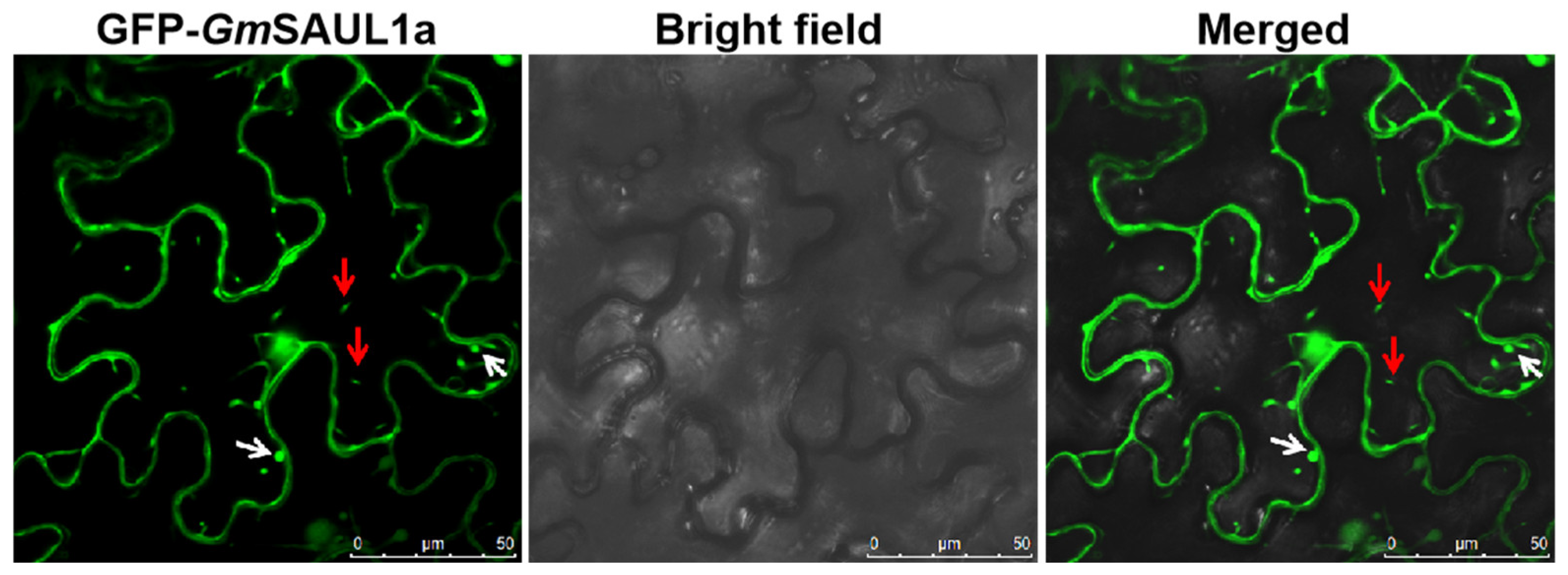
2.6. Subcellular Localization of GmSAUL1a

To visualize the subcellular localization of GmSAUL1a, the 35S::GFP-GmSAUL1a construct was transiently expressed in N. benthamiana leaves via agroinfiltration. Green fluorescence was observed, not only in the cytosol, but also in the nucleus (Figure 5). Interestingly, we frequently observed the fluorescent patches at the plasma membrane (PM) (Figure 5, see white arrows), These GFP patches may represent the sites for tethering the tonoplast and/or MVBs to the PM, as described in Arabidopsis [47,48,49]. In addition, mobile vesicles were also observed in the cytoplasm (Figure 5, see red arrows), suggesting that the function of SAUL1 homologues might be conserved in different plant species.

3. Discussion

3.1. The Function of SAUL1 Homologs Is Conserved across Plant Species

In Arabidopsis, either loss function of SAUL1 or ectopic over-expression of SAUL1 results in auto-immune phenotypes [16,17,19,20]. We showed that either ectopic over-expression of GmSAUL1a in N. tabacum (Figure 1, Figure 2 and Figure 3) or silencing GmSAUL1a/1b in soybean [42] led to similar autoimmunity. The autoimmunity of the Arabidopsis saul1-1 mutant is dependent on PAD4 and EDS1 [16,17], suggesting that the autoimmunity of saul1-1 mutant depends on the SA pathway. The elevated levels of both free and conjugated forms of SA in the GmSAUL1a/1b-silenced soybean plants suggest that the autoimmunity displayed in these plants is also dependent on SA [42]. Consistent with the auto-immune phenotypes, the tobacco GmSAUL1a-overexpressed plants exhibited an increased resistance to different biotrophic pathogens (Figure 2). These results indicated that the function of SAUL1 homologs is highly conserved in different plant species [47]. The previously published results, together with our results, strongly indicated that the homeostasis of SAUL1 must be properly maintained within a certain threshold range to prevent activation of immunity. A level of SAUL1 exceeding the higher end or below the lower end of the threshold range will result in the activation of immune responses.
In Arabidopsis, the Toll interleukin 1 receptor (TIR)-type NLR (TNL), as well as SOC3 (Suppressors of chs1-2, 3), which associates with SAUL1, serve as guards to monitor the homeostasis of SAUL1. Either loss or overexpression of SAUL1 triggers SOC3-mediated autoimmunity [19]. Later, the same group showed that SOC3 pairs with two atypical truncated TIR-NB (TN) proteins to guard the homeostasis of SAUL1 [20]. Over-accumulation of SAUL1 is guarded by the SOC3-TN2 pair, and knocking out or knocking down of SAUL1 is guarded by the SOC3-CHS1 (Chilling Sensitive 1) pair [20]. Autoimmunity, resulting from NLR gene activation, is usually suppressed at high temperature [43,44]. The fact that the auto-immune phenotypes of the GmSAUL1a-overexpressing tobacco plants were significantly alleviated by high temperature treatment (Figure S2) suggest that GmSAUL1a/1b is similarly guarded by a NLR(s), and the auto-immune phenotype might be a result of a NLR activation. It remains to be examined whether the SOC3-TN2 and SOC3-CHS1 homologues function the same way as in Arabidopsis to guard SAUL1 in soybean or tobacco.

3.2. The Roles of GmSAUL1s in PTI and ETI

An enhanced resistance to virulent pathogens was observed in the GmSAUL1a-overexpressing tobacco lines, which suggests that PTI is activated in these plants (Figure 2). If the homeostasis of GmSAUL1 is similarly guarded by SOC3-TN2 and SOC3-CHS1 homologous pairs in soybean [19,20], the activated immunity observed in the 35S::GmSAUL1a tobacco plants (Figure 1) actually results from activated NLRs. High temperature rescued the autoimmune phenotype of the GmSAUL1a-overexpressing plants (Figure S2), suggesting that certain NLR(s) likely act as guard(s) to monitor the level of GmSAUL1s in soybean or tobacco plants. Because PTI and ETI share overlapping components and are mutually required to maintain a prolonged and stronger immunity [6,7], it is not surprising that the enhanced disease resistance observed in the GmSAUL1a-overexpressing tobacco plants (Figure 2) could be a consequence of the activated ETI.
Interestingly, although both the saul1-1 mutant and the 35S::SAUL1 transgenic lines display autoimmunity [19,20], macroscopic lesions were only seen on the leaves of saul1-1 plants, but not on the leaves of the 35S::SAUL1 plants [20]. Different from the case in Arabidopsis, massive cell death was observed on the leaves of 35S::GmSAUL1a plants (Figure 1B–D). This disparity is probably caused by differences in the downstream signaling pathways between Arabidopsis and tobacco.

3.3. Silencing GmSAUL1a/1b in Soybean and Over-Expressing GmSAUL1a in N. tabacum Activate Immunity through Different Downstream MPKs

Silencing GmSAUL1a/1b caused a reduced activation of GmMPK6 in soybean [42], whereas over-expression of GmSAUL1a results in an elevated activation of NtMPK6 in N. tabacum (Figure 4), indicating that the positive effect of GmSAUL1 on MPK6 activation is consistent between soybean and tobacco. Silencing GmSAUL1a/1b has contrasting effects on the flg22-induced activation of GmMPK3 and GmMPK6 (Figure 4). In Arabidopsis, the enhanced resistance is usually correlated with the enhanced MPK3/MPK6 activity [25]. In Arabidopsis, MPK3 and MPK6 can compensate for each other’s function in mpk3 and mpk6 mutants, respectively [50].
Over-expressing GmSAUL1a in tobacco enhanced the flg22-induced activation of NtMPK6, but it reduced the activation of NtMPK4 (Figure 4). The elevated resistance is usually correlated with the enhanced MPK3/MPK6 activity and reduced MPK4 activity, respectively [22,23,24,25,26,27,28,29,30,31,32,51,52]. Consistent with this, silencing GmMPK4 results in cell death and activated immunity in soybeans [33]. Either silencing GmMPK6 in soybean or overexpressing GmMPK6 in Arabidopsis resulted in cell death and activation of immune responses, indicating that GmMPK6 plays both positive and negative roles in immune responses [34]. Based on these studies, it is reasonable to postulate that the cell death and the activated immune responses observed in the GmSAUL1a-overexpresssing tobacco plants are a result of combined effects of the reduced activation of NtMPK4 and enhanced activation of NtGMPK6. Collectively, our results demonstrated that silencing GmSAUL1a/1b and over-expressing GmSAUL1a activate immune responses through different downstream MAPK modules (Figure 6, [42]). Instead of through a simple linear MAPK pathway, the SAUL1 regulates cell death and immunity through a MAPK network consisting of different downstream MAPKs. It remains to be determined whether the differential activation and repression of downstream MAPK modules resulted from silencing GmSAUL1a/1b in soybean and overexpression of GmSAUL1a in tobacco are associated with the activation of the SOC3-TN2 and SOC3-CHS1, respectively.

4. Materials and Methods

4.1. Plant Materials

Nicotiana tabacum cv. Sumsun (NN) and Nicotiana benthamiana were used in this study. Tobacco plants were grown in the growth room or growth chamber at 22 °C with a 16-h light/8-h dark photoperiod. High temperature treatment was performed at 30 °C.

4.2. MAPK Activity Assay

After flg22 treatment, protein samples were prepared from tobacco leaf discs in extraction buffer containing 50 mM Tris-MES, pH 8, 0.5 M Suc, 1 mM MgCl2, 10 mM EDTA, and 5 mM DTT, as described [38,39]. Proteins were separated by 10% SDS-PAGE gel and transferred to PVDF membranes (Millipore, Burlington, MA, USA) by using semidry electrotransfer equipment (Bio-Rad, Hercules, CA, USA). Western blot analysis was performed using the anti-phospho-p44/p42 MAPK (anti-pTEpY) (Cell Signaling Technology, Danvers, MA, USA). The bands were visualized using chemiluminescent horseradish peroxidase (HRP) substrate (Millipore). Coomassie Blue stained gel (CBS) was used as loading control.

4.3. Histochemical Assays

Plant cell death on the infected leaves was visualized by staining with trypan blue in lactophenol and ethanol, as described [53].

4.4. SA Quantification

SA was measured, as described [54], using an Agilent 1260 HPLC system (Agilent Technologies, Santa Clara, CA, USA).

4.5. Generation of Transgenic N. tabacum Samsun (NN) Plants Over-Expressing GmSAUL1a

The full-length cDNA of GmSAUl1a was amplified using following pair of primers with KpnI and XmaI sites (in bold) attached to the 5′end and 3′ end, respectively:
  • GmSAUL1a-pE1776-F: aagGGTACCATGATGGCTGCGAGCTG
  • GmSAUL1a-pE1776-R: ttgCCCGGGTTCATCCCATGTTTGGAAAGATT
The amplified fragment was double-digested with KpnI and XmaI and subsequently cloned into the pE1776 vector [55] predigested with the same pair of restriction enzymes. The pE1776-GmSaul1a was transformed into the Agrobacterium strain GV3101 and then transformed into N. tabacum Samsun (NN), as described [56,57,58]. Briefly, tobacco leaves were sequentially sterilized with 70% ethanol and 0.5–2% NaClO3, respectively. Leaf discs from the sterilized leaves were pre-cultivated in MS medium for 3 days. After this, the leaf discs were co-incubated with Agrobacterium (GV3101) suspension solution for about a half hour, followed by cultivating in MS medium containing 1 mg/L Indole-3-acetic acid (IAA) and 1 mg/L 6-BA for 2 days. Afterward, these leaf discs were transferred to MS medium containing 1 mg/L IAA, 1 mg/L 6-BA, 50 mg/L Hygromycin, and 25 mg/L rifampicin. The shoots emerging from the callus were subsequently transferred to rooting medium containing IAA (1 mg/L), 50 mg/L Hygromycin, and 25 mg/L Timentin. After rooting, the seedlings were transferred to soil.

4.6. The Pst DC3000 Infection

The Pst DC3000 of OD600 = 0.00001 was infiltrated into the N. tabacum leaves, as described previously [59]. A colony-forming unit (cfu) at different days of infiltration (dpi) was counted and calculated, as described [59].

4.7. RNA Isolation and RT-qPCR

RNA isolation and RT-qPCR were performed, as described [34]. The RT-qPCRs were performed with three biological replicates and three technical replicates using an ABI550 Real-Time PCR machine (Applied Biosystems, Thermo Fisher Scientific, Auxtin, TX, USA) and the 2× SYBR Green qPCR Mix (Aidlab, Beijing, China). The annealing temperature for the RT-qPCRs was 60 °C. The comparative CT method was used for analysis.
The primers used for RT-qPCR are:
  • NtPR1-F: GCTGAGGGAAGTGGCGATTTC
  • NtPR1-R: CCTAGCACATCCAACACGAACC
  • NtPR3-F: GACCCATCCAATTGACAAACCAA
  • NtPR3-R: CCTGTGGTGTCATCCAGAACC
  • NtPR5-F: GGCGATTGTGGCTCAAACC
  • NtPR5-R: GAAATCTTGCTTCGTACCTGAGA
  • NtUBQ5-F: CCTAACGGGTAAAACAATCAC
  • NtUBQ5-R: AGCCATAAAAGTTCCAGCAC

4.8. Callose Deposition

Callose deposition in N. tabacum leaves was stained 24 h, as described by [59]. Callose deposits were visualized by ultraviolent epifluorescence using a fluorescence microscope (Olympus, Center Valley, PA, USA).

4.9. GUS Staining

GUS staining was performed, as described by [60]. A stereo microscope (Olympus SZH10, Center Valley, PA, USA) was used to take photographs of the GUS-stained leaves.

4.10. Sub-Cellular Localization of GFP-GmSAUL1a

The full-length cDNA of GmSAUL1a was amplified by RT-PCR by using the following pair of primers:
  • pGDG-F-Sal1: aaaGTC GAC ATGATGGCTGCGAGCTGG
  • pGDG-R-BamH1: ttgGGA TCC TCATCCCATGTTTGGAAAGATTC
The PCR product was double-digested with SalI and BamHI and cloned into pGDG vector [61] predigested with the same set of restriction enzymes to generate the GFP–GmSAUL1a fusion construct. This 35S:: GFP-GmSAUL1a construct was infiltrated onto N. benthamiana leaves, as described [62]. Images were captured with a confocal laser-scanning microscope (Carl Zeiss, LSM 880, Jena, Germany).

Supplementary Materials

The following supporting information can be downloaded at: https://www.mdpi.com/article/10.3390/agronomy13061533/s1, Figure S1: Screening of transgenic tobacco lines that ectopically expressing GmSAUL1. Figure S2: The cell death phenotype of the GmSAUL1a-overexpressing tobacco line is suppressed at 30 °C.

Author Contributions

J.-Z.L. designed the experiments and wrote the manuscript; J.-M.L., M.-Y.Y., X.-H.M. and N.-N.W. performed the experiments. J.-M.L. and J.-Z.L. prepared the figures. All authors have read and agreed to the published version of the manuscript.

Funding

This work was supported by grants from National Natural Science Foundation of China (32170761 and 31571423 to J.-Z.L.).

Data Availability Statement

Not applicable.

Acknowledgments

We thank Brian J. Staskawicz for kindly providing the pE1776 vector.

Conflicts of Interest

The authors declare that they have no known competing financial interests or personal relationships that could have appeared to influence the work reported in this paper.

Abbreviations

SAUL1: senescence-associated E3 ubiquitin ligase 1; PAMPs, pathogen-associated molecular patterns; MAPK, mitogen-activated protein kinase; HR, hypersensitive response; EDS1, enhanced disease susceptibility 1.

References

  1. Jones, J.D.G.; Dangl, J.L. The plant immune system. Nature 2006, 444, 323–329. [Google Scholar] [CrossRef] [PubMed]
  2. Chinchilla, D.; Zipfel, C.; Robatzek, S.; Kemmerling, B.; Nürnberger, T.; Jones, J.D.G.; Felix, G.; Boller, T. A flagellin-induced complex of the receptor FLS2 and BAK1 initiates plant defence. Nature 2007, 448, 497–500. [Google Scholar] [CrossRef] [PubMed]
  3. Boller, T.; Felix, G. A renaissance of elicitors: Perception of microbe-associated molecular patterns and danger signals by pattern-recognition receptors. Annu. Rev. Plant Biol. 2009, 60, 379–406. [Google Scholar] [CrossRef]
  4. Tena, G.; Boudsocq, M.; Sheen, J. Protein kinase signaling networks in plant innate immunity. Curr. Opin. Plant Biol. 2011, 14, 519–529. [Google Scholar] [CrossRef]
  5. Zhou, J.; Zhang, Y. Plant Immunity: Danger perception and signaling. Cell 2020, 181, 978–989. [Google Scholar] [CrossRef]
  6. Yuan, M.; Jiang, Z.; Bi, G. Pattern-recognition receptors are required for NLR-mediated plant immunity. Nature 2021, 592, 105–109. [Google Scholar] [CrossRef] [PubMed]
  7. Ngou, B.P.M.; Ahn, H.K.; Ding, P.; Jones, J.D. Mutual potentiation of plant immunity by cell-surface and intracellular receptors. Nature 2021, 592, 110–115. [Google Scholar] [CrossRef]
  8. Vierstra, R.D. The ubiquitin-26S proteasome system at the nexus of plant biology. Nat. Rev. Mol. Cell Biol. 2009, 10, 385–397. [Google Scholar] [CrossRef]
  9. Smalle, J.; Vierstra, R.D. The Ubiquitin 26S proteasome proteolytic pathway. Annu. Rev. Plant Biol. 2004, 55, 555–590. [Google Scholar] [CrossRef] [PubMed]
  10. Hershko, A.; Clechanover, A. The ubiquitin system. Annu. Rev. Biochem. 1998, 67, 425–479. [Google Scholar] [CrossRef]
  11. Cheng, Y.T.; Li, X. Ubiquitination in NB-LRR-mediated immunity. Curr. Opin. Plant Biol. 2012, 15, 392–399. [Google Scholar] [CrossRef] [PubMed]
  12. Zeng, L.; Vega-Sanchez, M.E.; Zhu, T.; Wang, G.L. Ubiquitination-mediated protein degradation and modification: An emerging theme in plant-microbe interactions. Cell Res. 2006, 16, 413–426. [Google Scholar] [CrossRef]
  13. Zhou, B.; Zeng, L. Conventional and unconventional ubiquitinnation in plant immunity. Mol. Plant Pathol. 2017, 18, 1313–1330. [Google Scholar] [CrossRef]
  14. Zeng, L.; Qu, S.; Bordeos, A.; Yang, C.; Baraoidan, M.; Yan, H.; Xie, Q.; Nahm, B.H.; Leung, H.; Wang, G.L. Soptted leaf 11, a negative regulator of plant cell death and defense, encodes a U-box/armadillo repeat protein endowed with E3 ubiquitin ligase activity. Plant Cell 2004, 16, 2795–2808. [Google Scholar] [CrossRef]
  15. Raab, S.; Drechsel, G.; Zarepour, M.; Hartung, W.; Koshiba, T.; Bittner, F.; Hoth, S. Identification of a novel E3 ubiquitin ligase that is required for suppression of premature senescence in Arabidopsis. Plant J. 2009, 59, 39–51. [Google Scholar] [CrossRef]
  16. Vogelmann, K.; Drechsel, G.; Bergler, J.; Subert, C.; Philippar, K.; Soll, J.; Engelmann, J.C.; Engelsdorf, T.; Voll, L.M.; Hoth, S. Early senescence and cell death in Arabidopsis saul1 mutants involves the PAD4-dependent salicylic acid pathway. Plant Physiol. 2012, 159, 1477–1487. [Google Scholar] [CrossRef] [PubMed]
  17. Disch, E.M.; Tong, M.; Kotur, T.; Koch, G.; Wolf, C.A.; Li, X.; Hoth, S. Membrane-associated ubiquitin ligase SAUL1 suppresses temperature-and humidity-dependent autoimmunity in Arabidopsis. Mol. Plant-Microbe Interact. 2016, 29, 69–80. [Google Scholar] [CrossRef] [PubMed]
  18. Lee, I.H.; Lee, I.C.; Kim, J.; Kim, J.H.; Chung, E.H.; Kim, H.J.; Park, S.J.; Kim, Y.M.; Kang, S.K.; Nam, H.G.; et al. NORE1/SAUL1 integrates temperature-dependent defense programs involving SGT1b and PAD4 pathways and leaf senescence in Arabidopsis. Physiol. Plant. 2016, 158, 180–199. [Google Scholar] [CrossRef]
  19. Tong, M.; Kotur, T.; Liang, W.; Vogelmann, K.; Kleine, T.; Leister, D.; Brieske, C.; Yang, S.; Ludke, D.; Wiermer, M.; et al. E3 ligase SAUL1 serves as a positive regulator of PAMP-triggered immunity and its homeostasis is monitored by immune receptor SOC3. New Phytol. 2017, 215, 1516–1532. [Google Scholar] [CrossRef]
  20. Liang, W.; van Wersch, S.; Tong, M.; Li, X. TIR-NB-LRR immune receptor SOC3 pairs with truncated TIR-NB protein CHS1 or TN2 to monitor the homeostasis of E3 ligase SAUL1. New Phytol. 2019, 221, 2054–2066. [Google Scholar] [CrossRef]
  21. Zhang, Y.; Wang, Y.; Liu, J.; Ding, Y.; Wang, S.; Zhang, X.; Liu, Y.; Yang, S. Temperature-dependent autoimmunity mediated by chs1 requires its neighboring TNL gene SOC3. New Phytol. 2016, 213, 1330–1345. [Google Scholar] [CrossRef]
  22. Colcombet, J.; Hirt, H. Arabidopsis MAPKs: A complex signal-ling network involved in multiple biological processes. Biochem. J. 2008, 413, 217–226. [Google Scholar] [CrossRef]
  23. Pitzschke, A.; Schikora, A.; Hirt, H. MAPK cascade signalling networks in plant defence. Curr. Opin. Plant. Biol. 2009, 12, 421–426. [Google Scholar] [CrossRef]
  24. Pitzschke, A.; Djamei, A.; Bitton, F.; Hirt, H. A major role of the MEKK1-MKK1/2-MPK4 pathway in ROS signalling. Mol. Plant 2009, 2, 120–137. [Google Scholar] [CrossRef]
  25. Meng, X.; Zhang, S. MAPK cascades in plant disease resistance signaling. Annu. Rev. Phytopathol. 2013, 51, 245–266. [Google Scholar] [CrossRef]
  26. Zhang, S.; Klessig, D.F. Resistance gene N-mediated de novo synthesis and activation of a tobacco mitogen-activated protein kinase by Tobacco mosaic virus infection. Proc. Natl. Acad. Sci. USA 1998, 95, 7433–7438. [Google Scholar] [CrossRef] [PubMed]
  27. Ren, D.; Liu, Y.; Yang, K.Y.; Han, L.; Mao, G.; Glazebrook, J.; Zhang, S. A fungal-responsive MAPK cascade regulates phytoalexin biosynthesis in Arabidopsis. Proc. Natl. Acad. Sci. USA 2008, 105, 5638–5643. [Google Scholar] [CrossRef] [PubMed]
  28. Mao, G.; Meng, X.; Liu, Y.; Zheng, Z.; Chen, Z.; Zhang, S. Phosphorylation of a WRKY transcription factor by two pathogen-responsive MAPKs drives phytoalexin biosynthesis in Arabidopsis. Plant Cell 2011, 23, 1639–1653. [Google Scholar] [CrossRef]
  29. Petersen, M.; Brodersen, P.; Naested, H.; Andreasson, E.; Lindhart, U.; Johansen, B.; Nielsen, H.B.; Lacy, M.; Austin, M.J.; Parker, J.E.; et al. Arabidopsis MAP kinase 4 negatively regulates systemic acquired resistance. Cell 2000, 103, 1111–1120. [Google Scholar] [CrossRef] [PubMed]
  30. Qiu, J.L.; Zhou, L.; Yun, B.W.; Nielsen, H.B.; Fiil, B.K.; Petersen, K.; Mackinlay, J.; Loake, G.J.; Mundy, J.; Morris, P.C. Arabidopsis mitogen-activated protein kinase kinases MKK1 and MKK2 have overlapping functions in defense signaling mediated by MEKK1, MPK4, and MKS1. Plant Physiol. 2008, 148, 212–222. [Google Scholar] [CrossRef]
  31. Gao, M.; Liu, J.; Bi, D.; Zhang, Z.; Cheng, F.; Chen, S.; Zhang, Y. MEKK1, MKK1/MKK2 and MPK4 function together in a mitogen-activated protein kinase cascade to regulate innate immunity in plants. Cell Res. 2008, 18, 1190–1198. [Google Scholar] [CrossRef] [PubMed]
  32. Kong, Q.; Qu, N.; Gao, M.; Zhang, Z.; Ding, X.; Yang, F.; Li, Y.; Dong, O.X.; Chen, S.; Li, X.; et al. The MEKK1-MKK1/MKK2-MPK4 kinase cascade negatively regulates immunity mediated by a mitogen-activated protein kinase kinase kinase in Arabidopsis. Plant Cell 2012, 24, 2225–2236. [Google Scholar] [CrossRef] [PubMed]
  33. Liu, J.Z.; Horstman, H.D.; Braun, E.; Graham, M.A.; Zhang, C.; Navarre, D.; Qiu, W.L.; Lee, Y.; Nettleton, D.; Hill, J.H. Soybean homologs of MPK4 negatively regulate defense responses and positively regulate growth and development. Plant Physiol. 2011, 157, 1363–1378. [Google Scholar] [CrossRef]
  34. Liu, J.Z.; Braun, E.; Qiu, W.L.; Shi, Y.F.; Marcelino-Guimars, F.C.; Navarre, D.; Hill, J.H.; Whitham, S.A. Positive and negative roles for soybean MPK6 in regulating defense responses. Mol. Plant-Microbe Interact. 2014, 27, 824–834. [Google Scholar] [CrossRef] [PubMed]
  35. Liu, J.Z.; Graham, M.A.; Pedley, K.F.; Whitham, S.A. Gaining insight into soybean defense responses using functional genomics approaches. Brief. Funct. Genom. 2015, 14, 283–290. [Google Scholar] [CrossRef]
  36. Liu, J.Z.; Fang, Y.; Pang, H. The current status of the soybean-soybean mosaic virus (SMV) pathosystem. Front. Microbiol. 2016, 7, 1906. [Google Scholar] [CrossRef]
  37. Liu, J.Z.; Whitham, S.A. Over-expression of a nuclear-localized DnaJ domain-containing HSP40 from soybean reveals its roles in cell death and disease resistance. Plant J. 2013, 74, 110–121. [Google Scholar] [CrossRef]
  38. Xu, H.Y.; Zhang, C.; Li, Z.C.; Wang, Z.R.; Jiang, X.X.; Shi, Y.F.; Tian, S.N.; Braun, E.; Mei, Y.; Qiu, W.L.; et al. The MAPK kinase kinase GmMEKK1 regulates cell death and defense responses. Plant Physiol. 2018, 178, 907–922. [Google Scholar] [CrossRef]
  39. Tian, S.N.; Liu, D.D.; Zhong, C.L.; Xu, H.Y.; Yang, S.; Fang, Y.; Ran, J.; Liu, J.Z. Silencing GmFLS2 enhances the susceptibility of soybean to bacterial pathogen through attenuating the activation of GmMAPK signaling pathway. Plant Sci. 2020, 292, 110386. [Google Scholar] [CrossRef]
  40. Hashimi, S.M.; Wu, N.N.; Ran, J.; Liu, J.Z. Silencing Autophagy-Related Gene 2 (ATG2) Results in Accelerated Senescence and Enhanced Immunity in Soybean. Int. J. Mol. Sci. 2021, 22, 11749. [Google Scholar]
  41. Liu, D.D.; Lan, H.J.; Masoud, H.S.; Ye, M.Y.; Dai, X.Y.; Zhong, C.L.; Tian, S.N.; Liu, J.Z. Silencing GmBIR1 in Soybean Results in Activated Defense Responses. Int. J. Mol. Sci. 2022, 23, 7450. [Google Scholar]
  42. Li, J.M.; Ye, M.Y.; Wang, C.; Ma, X.H.; Wu, N.N.; Zhong, C.L.; Zhang, Y.; Cheng, N.; Nakata, P.A.; Zeng, L.; et al. Soybean GmSAUL1, a Bona Fide U-Box E3 Ligase, Negatively Regulates Immunity Likely through Repressing the Activation of GmMPK3. Int. J. Mol. Sci. 2023, 24, 6240. [Google Scholar]
  43. Whitham, S.; McCormick, S.; Baker, B. The N gene of tobacco confers resistance to tobacco mosaic virus in transgenic tomato. Proc. Natl. Acad. Sci. USA 1996, 93, 8776–8781. [Google Scholar]
  44. Zhu, Y.; Qian, W.; Hua, J. Temperature Modulates Plant Defense Responses through NB-LRR Proteins. PLoS Pathog. 2010, 6, e1000844. [Google Scholar] [CrossRef]
  45. Whitham, S.; Dinesh-Kumar, S.P.; Choi, D.; Hehl, R.; Corr, C.; Baker, B. The product of the tobacco mosaic virus resistance gene N: Similarity to toll and the interleukin-1 receptor. Cell 1994, 78, 1101–1115. [Google Scholar] [CrossRef]
  46. Meshi, T.; Takamatsu, N.; Ohno, T.; Okada, Y. Molecular cloning of the complementary DNA copies of the common and cowpea strains of tobacco mosaic virus RNA. Virology 1982, 118, 4–75. [Google Scholar]
  47. Vogelmann, K.; Subert, C.; Danzberger, N.; Drechsel, G.; Bergler, J.; Kotur, T.; Burmester, T.; Hoth, S. Plasma membrane-association of SAUL1-type plant U-box armadillo repeat proteins is conserved in land plants. Front. Plant Sci. 2014, 5, 37. [Google Scholar] [CrossRef]
  48. Tao, K.; Waletich, J.R.; Wise, H.; Arredondo, F.; Tyler, B.M. Tethering of multi-vesicular bodies and the tonoplast to the plasma membrane in plants. Front. Plant Sci. 2019, 10, 636. [Google Scholar] [CrossRef]
  49. Knop, J.; Lienemann, T.; El-Kilani, H.; Falke, S.; Krings, C.; Sindalovskaya, M.; Bergler, J.; Betzel, C.; Hoth, S. Structural Features of a Full-Length Ubiquitin Ligase Responsible for the Formation of Patches at the Plasma Membrane. Int. J. Mol. Sci. 2021, 22, 9455. [Google Scholar] [CrossRef] [PubMed]
  50. Wang, H.; Liu, Y.; Bruffett, K.; Lee, J.; Hause, G.; Walker, J.; Zhang, S. Haplo-insufficiency of MPK3 in MPK6 mutant background uncovers a novel function of these two MAPKs in Arabidopsis ovule development. Plant Cell 2008, 20, 602–613. [Google Scholar] [CrossRef] [PubMed]
  51. Menke, F.L.; van Pelt, J.A.; Pieterse, C.M.; Klessig, D.F. Silencing of the mitogen-activated protein kinase MPK6 compromises disease resistance in Arabidopsis. Plant Cell 2004, 16, 897–907. [Google Scholar] [CrossRef]
  52. Zhang, S.; Liu, Y. Activation of salicylic acid-induced protein kinase, a mitogen-activated protein kinase, induces multiple defense responses in tobacco. Plant Cell 2001, 13, 1877–1889. [Google Scholar]
  53. Ren, D.; Yang, H.; Zhang, S. Cell death mediated by MAPK is associated with hydrogen peroxide production in Arabidopsis. J. Biol. Chem. 2002, 277, 559–565. [Google Scholar] [CrossRef] [PubMed]
  54. Zhang, Y.; Zhao, L.; Zhao, J.; Li, Y.; Wang, J.; Guo, R.; Gan, S.; Liu, C.J.; Zhang, K. S5H/DMR6 Encodes a Salicylic Acid 5-Hydroxylase That Fine-Tunes Salicylic Acid Homeostasis. Plant Physiol. 2017, 175, 1082–1093. [Google Scholar] [CrossRef]
  55. Leister, R.T.; Dahlbeck, D.; Day, B.; Li, Y.; Chesnokova, O.; Staskawicz, B.J. Molecular genetic evidence for the role of SGT1 in the intramolecular complementation of Bs2 protein activity in Nicotiana benthamiana. Plant Cell 2005, 17, 1268–1278. [Google Scholar] [CrossRef]
  56. Horsch, R.B.; Fry, J.E.; Hofmann, N.L.; Eichholtz, D.; Rogers, S.G.; Fraley, R.T. A simple and general method for transferring genes into plants. Science 1985, 227, 1229–1231. [Google Scholar] [CrossRef]
  57. Li, Z.C.; Ren, Q.W.; Guo, Y.; Ran, J.; Ren, X.T.; Wu, N.N.; Liu, X.; Liu, J.Z. Dual roles of GSNOR1 in cell death and immunity in tetraploid Nicotiana tabacum. Front. Plant Sci. 2021, 12, 596234. [Google Scholar] [CrossRef] [PubMed]
  58. Hinge, V.R.; Chavhan, R.L.; Kale, S.P.; Suprasanna, P.; Kadam, U.S. Engineering Resistance against Viruses in Field Crops Using CRISPR-Cas9. Curr. Genom. 2021, 22, 214–231. [Google Scholar] [CrossRef] [PubMed]
  59. Heese, A.; Hann, D.R.; Gimenez-Ibanez, S.; Jones, A.M.; He, K.; Li, J.; Schroeder, J.I.; Peck, S.C.; Rathjen, J.P. The receptor-like kinase SERK3/BAK1 is a central regulator of innate immunity in plants. Proc. Natl. Acad. Sci. USA 2007, 104, 12217–12222. [Google Scholar] [CrossRef]
  60. Jefferson, R.A.; Kavanagh, T.A.; Bevan, M.W. GUS fusions: Beta-glucuronidase as a sensitive and versatile gene fusion marker in higher plants. EMBO J. 1987, 6, 3901–3907. [Google Scholar] [CrossRef]
  61. Goodin, M.; Dietzgen, R.G.; Schichnes, D.; Ruzin, S.; Jackson, A.O. pGD vectors: Versatile tools for the expression of green and red fluorescent protein fusions in Agroinfiltrated plant leaves. Plant J. 2022, 31, 375–383. [Google Scholar] [CrossRef] [PubMed]
  62. Liu, J.Z.; Blancaflor, E.B.; Nelson, R.S. The tobacco mosaic virus 126-kilodalton protein, a constituent of the virus replication complex, alone or within the complex aligns with and traffics along microfilaments. Plant Physiol. 2005, 138, 1853–1865. [Google Scholar] [CrossRef] [PubMed]
Figure 1. Either transient or stable over-expression of the GmSAUL1a in N. tabacum cv. Samsun (NN) induces cell death and activates immune responses. (A). Transient overexpression of GmSAUL1a via agro-infiltration resulted in HR-like cell death on the leaves of N. benthamiana. The 35S::GmMEKK1 was used as a positive control, and the 35S::GFP was used as a negative control. The leaf was photographed at three days post-infiltration; (BD). Transgenic N. tabacum plants over-expressing GmSAUL1a induce HR-like cell death on the leaves (see red arrows). Line 8, Line 9, and Line 12 represent three independent transgenic lines; (E). The expressions of three Pathogenesis-related (PR) genes, NtPR1, NtPR3, and NtPR5, were highly induced in the GmSAUL1a-overexpressing tobacco plants. The NtUBQ5 gene was used as a reference gene. ** and *** represent significant differences at 0.01 and 0.001 levels, respectively, by Student’s t-test; (F). H2O2 was over-accumulated on the leaves of the GmSAUL1a-overexpressing tobacco plants. The oxidized DAB spots are indicated by the red arrows.
Agronomy 13 01533 g001
Figure 2. Transgenic tobacco plants over-expressing GmSAUL1a exhibit enhanced resistance to different types of pathogens. (A). Trypan blue-stained HR lesions on the leaves of the WT and the GmSAUL1a-overexpressing N. tabacum Samsun (NN) plants upon TMV infection. The photos were taken at 3 dpi; (B). Comparison of the diameters of the HR lesions on the TMV-infected WT and GmSAUL1a-overexpressing N. tabacum Samsun (NN) plants, shown in (A). The diameters of the HR lesions were measured using Image J; (C). RT-qPCR analysis showed that the transcript of the TMV capsid protein encoding gene (CP) was significantly reduced in three independent GmSAUL1a-overexpressing N. tabacum Samsun (NN) plants, relative to the WT plants. The NtActin gene was used as an internal reference gene; (D). Comparison of the colony forming units on the leaves of the control N. tabacum Samsun (NN) plants and the GmSAUL1a-overexppressing lines at different days post inoculation (dpi) of Pst DC3000 infection. The leaves of indicated 45-day-old tobacco lines were injected with Pst DC3000 to determine the colony forming units at different days post infiltration; (E). Overexpression of GmSAUL1a in N. tabacum leads to increased resistance to an agrobacterium strain (GV3101), carrying a construct of the pBI121-GUS; (F). The relative intensity of GUS staining was quantitated by Image J. ** and *** represent significant differences at 0.01 and 0.001 levels, respectively, by Student’s t-test.
Agronomy 13 01533 g002
Figure 3. Over-expressing GmSAUL1a in N. tabacum cv. Samsun (NN) enhances the flg22-induced callose deposition. (A). The callose deposition of two-week-old WT and GmSAUL1a-overexpressing tobacco plants was visualized by staining with Analine blue. The representative callose spots were pointed out by white arrows; (B). Comparison of the number of callose foci per mm2 of leaf surface area visualized by Aniline blue staining between WT and different GmSAUL1-overexpressing lines. The significant differences were determined by an ANOVA with a post hoc Duncan’s test, as indicated by different letters (p < 0.05, n > 10).
Agronomy 13 01533 g003
Figure 4. Over-expressing GmSAUL1a in N. tabacum cv. Samsun (NN) enhances the activation of NtMPK6, but it reduces the activation of NtMPK4. Protein samples were prepared, as described previously [38]. Western blotting was performed using a Phosph-p44/p42 MAP Erk1/2 antibody. An Arabidopsis sample treated with 10 μM flg22 for 10 min was used as a positive control. Coomassie blue-stained gel (CBS) was used as a loading control.
Agronomy 13 01533 g004
Figure 5. Subcellular localization of GFP-GmSAUL1a. GFP-GmSAUL1a localizes in the cytoplasm, nucleus, as well as at plasma membrane (PM) (see white arrows) and cytoplasmic vesicles (see red arrows) in the N. benthamiana epidermal cells. Bar = 50 μm.
Agronomy 13 01533 g005
Figure 6. A proposed model for how NtMPK6 and NtMPK4 are involved in the GmSAUL1a-triggered immunity in tobacco. Overexpression of GmSAUL1a in N. tabacum triggers cell death and activated immune responses through combined effects of the reduced activation of NtMPK4 and enhanced activation of NtGMPK6.
Agronomy 13 01533 g006
Disclaimer/Publisher’s Note: The statements, opinions and data contained in all publications are solely those of the individual author(s) and contributor(s) and not of MDPI and/or the editor(s). MDPI and/or the editor(s) disclaim responsibility for any injury to people or property resulting from any ideas, methods, instructions or products referred to in the content.

