Article

Suspension Cell Culture of Dioscorea deltoidea—A Renewable Source of Biomass and Furostanol Glycosides for Food and Pharmaceutical Industry

by
Maria V. Titova
1,*,
Elena V. Popova
1,
Svetlana V. Konstantinova
2,
Dmitry V. Kochkin
1,2,
Igor M. Ivanov
1,
Andrey G. Klyushin
1,
Elena G. Titova
3,
Elena A. Nebera
3,
Ekaterina R. Vasilevskaya
4,
Galina S. Tolmacheva
4,
Elena A. Kotenkova
4,
Alexandr M. Nosov
1,2 and
Kee-Yoeup Paek
5
1
K.A. Timiryazev Institute of Plant Physiology, Russian Academy of Sciences, 127276 Moscow, Russia
2
Biology Faculty, M.V. Lomonosov Moscow State University, 119234 Moscow, Russia
3
All-Russian State Research Institute for Control Standardization and Certification of Veterinary Preparations, 123022 Moscow, Russia
4
V.M. Gorbatov Federal Research Center for Food Systems of Russian Academy of Sciences, 109316 Moscow, Russia
5
Research Center for the Development of Advanced Horticultural Technology, Chungbuk National University, Cheongju 361-763, Korea
*
Author to whom correspondence should be addressed.
Agronomy 2021, 11(2), 394; https://doi.org/10.3390/agronomy11020394
Submission received: 19 January 2021 / Revised: 13 February 2021 / Accepted: 18 February 2021 / Published: 23 February 2021
(This article belongs to the Special Issue Role of Plant Tissue Culture in Agricultural Research and Production)

Abstract

:
Dioscorea deltoidea is a medicinal plant valued for its high content of steroidal glycosides (SG)—bioactive compounds with cardioprotective and immunomodulation actions, also used to treat reproductive system disorders. To overcome the limitations of natural resources of this species, a suspension cell culture of D. deltoidea was developed as a renewable and ecologically sustainable source of raw biomass and SG. Cell culture demonstrated stable and intensive growth in the laboratory (20 L) and industrial (630 L) bioreactors operated under a semi-continuous regime (specific growth rate 0.11–1.12 day−1, growth index 3.5–3.7). Maximum dry weight accumulation (8.5–8.8 g/L) and SG content (47–57 mg/g DW) were recorded during the stationary phase. Bioreactor-produced cell biomass contained inorganic macro (K, Ca, Mg, Na) and micro (Zn, Mn, Fe, B, Al, Cu, Cr, Se, Co, Ni) elements in concentrations within the safe range of dietary recommendations. Acute toxicity test showed no or insignificant changes in organ weight, hematological panel and blood biochemistry of laboratory animals fed with 2000 and 5000 mg/kg dry biomass. The results suggest that cell culture of D. deltoidea grown in bioreactors has great potential to be used as functional foods and a component of specialized dietary supplements in complex therapy of reproductive system disorders and mineral deficiency.

1. Introduction

Steroidal glycosides (SG), the products of isoprenoid metabolism in plants, are of particular interest for pharmacology, veterinary science, and food and agricultural industries as they have been demonstrated to be efficient and safe in treating or mitigating a variety of adverse disease symptoms [1,2]. SG from Dioscorea spp. have been shown to successfully stabilize the endocrine profile in both men and women [1,2,3,4]. They also have restorative and mild anabolic effects, improve stress resistance and endurance during intensive physical activity, and participate in the regulation of blood pressure and lipid content in blood [1,4,5,6,7]. SG from Dioscorea spp. were also shown to be efficient in cardiology to treat acute myocardial infarction and enhance heart regeneration processes [8,9,10,11,12]. In addition, SG affect human and animal reproductive systems [13]. They were found to have a pronounced immunomodulating effect influencing different lymphocyte subpopulations. Experiments on isolated leukocyte cell cultures revealed that the effect of SG was concentration-dependent: different SG levels either stimulated or suppressed immune response [13,14,15,16]. The ability of Dioscorea spp. to stimulate protein synthesis and to activate succinic dehydrogenase, lactate dehydrogenase, and cytochrome oxidase (enzymes involved in energy metabolism) was also reported [15].
Other potentially useful compounds found in Dioscorea spp. plants include phenolcarboxylic acids, monosaccharides (mannose, glucose, galactose), dopamine, starch, riboflavin, vitamin C, nicotinic acid, choline, thiamine, carotene, and microelements (barium, cobalt, chrome, iron, magnesium, phosphorus, potassium, copper), as well as dietary fiber, mucus, and allantoin. Polysaccharides from D. bulbifera were reported to have antitumor, anti-inflammatory, and antioxidant effects [7,15,17]. Plant rhizomes may be composed of up to 6% of proteins. Medicinal preparations from Dioscorea plants are usually well-tolerable by patients: side-effects are extremely rare and are generally limited to individual allergic reactions to certain ingredients.
Plants of different Dioscorea species were reported to have similar biochemical composition, which defines their similar biological actions [18]. Dioscorea deltoidea Wall., a perennial medicinal plant growing mainly in China and India, is a valuable natural source of diosgenin, an aglycone of SG that for many years was an irreplaceable substrate for pharmacological synthesis of steroid hormones such as cortisone, pregnenolone and progesterone [19]. However, despite well-documented benefits for human health, the use of D. deltoidea plants as a natural source of SG is hampered by several strong limitations. The concentration of SG in D. deltoidea plants rarely exceeds 1–2% of dry biomass. Natural resources of D. deltoidea are limited, and the content of individual SG in plants varies significantly depending on geographical region, environmental conditions and the plant age. Moreover, plants of this genus contain both furostanol (FG) and spirostanol types glycosides, which complicates the purification of individual compounds from plant material. Spirostanol glycosides act contradictorily to FG often impairing or mitigating their positive effects [13,20,21,22].
Earlier we reported the development of several strains of suspension cell culture of D. deltoidea as an alternative source of FG protodioscin and deltoside (protodeltonin) specific for this species [23]. The content of protodioscin and deltoside in the cell cultures varied in the range of 3–12% of the dry cell weight depending on culture conditions, which is higher than the average FG content in plants [23,24,25]. One of the strains, DM-05-03, was shown to contain mainly protodioscin and only trace amounts of deltoside which suggests a more specific biological activity [23]. Preparations from cell biomass of D. deltoidea were proved to be non-toxic in the in vitro studies [26]. In the experiments with human peripheral blood lymphocytes, cell preparations also demonstrated immunomodulating activities [26].
In this study, we explored the feasibility of cultivation of D. deltoidea cell suspension in bioreactors of different types and volumes, up to 630 L. The content of steroidal glycosides, macro and microelements in cell biomass and its acute toxicity on laboratory rats were analyzed to explore the possibility of using D. deltoidea cell biomass as a novel supplement and a source of FG in human and animal diet.

2. Materials and Methods

2.1. Cell Culture

Suspension cell culture of Dioscorea deltoidea Wall strain DM-05–03 with enhanced production of furostanol glycosides was received from the All-Russian Collection of Cell Cultures of Higher Plants at the Institute of Plant Physiology of Russian Academy of Sciences, Moscow. Stock cell culture was maintained in 250 mL Erlenmeyer flasks containing 35 mL liquid medium composed of MS mineral salts [27], Staba vitamins [28], 30 g/L sucrose, 1 mg/L 2,4-dichlorophenoxyacetic acid (2,4-D) and 0.1 mg/L kinetin [23]. Cultures were grown on an orbital shaker (90 rpm) at 26 °C and 75% relative air humidity in darkness with 14 days subculture intervals.

2.2. Large-Scale Cultivation of D. deltoidea Cell Suspension in Bioreactors

Two types of bioreactors were tested for cell culture: bubble-type 20 L glass bioreactors of our own design (Institute of Plant Physiology of RAS, Russia) with 15 L working volume, air supply through a sparger, semi-continuous cultivation mode; and 630 L bioreactor (1T series, CUC “EBEE”, Yoshkar-Ola, Russia) with 550 L working volume, air supply through a gas distributor ∅ 750 mm (Figure 1). In both types of bioreactors cultivation was performed using a semi-continuous cultivation regime at 26 ± 0.5 °C in darkness. To maintain the semi-continuous cultivation regime, fresh nutrient medium was fed into bioreactors at the beginning of the stationary growth phase (measured as cell concentration reaching 8–10 g dry weight per liter of medium) until dilution of the cell suspension to 1.5–2.0 g of dry weight per liter was achieved. Air supply varied from 0.1 to 1.0 v/vpm depending on the growth phase of cell culture. Concentration of dissolved oxygen (pO2) was maintained at 10–40% of saturation volume [25].

2.3. Assessment of Growth and Physiological Characteristics of Cell Suspension Culture

To assess the growth and physiological state of cell cultures in flasks and bioreactors, fresh (FW) and dry (DW) weights of cell biomass, level of cell aggregation and cell viability were recorded periodically during the cultivation cycle.
To estimate the fresh weight, 10–15 mL aliquots of cell suspension were pipetted on paper filters and the culture medium was removed under vacuum; cell biomass was washed three times by distilled water under vacuum and weighted. To estimate the dry weight, cell biomass was dried to a constant weight at 60 °C. The biomass samples for chemical analysis and for experiments on acute toxicity (see below) were prepared using the same method.
Cell viability was determined by staining with 0.025% Evans blue (modified from [29]) as the percentage of cell aggregates composed of colourless (living) cells. For each experimental conditions, a minimum of 250 cell aggregates were examined in each of three replicates.
In addition, the following growth parameters were calculated for D. deltoidea cell culture according to [30]:
  • growth index: I = (Xmax − Xo)/Xo,
  • specific growth rate at exponential growth phase: μ = (ΔlnXmax/Xo)/Δt, [day−1]
  • productivity on dry biomass: P = (Xmax − Xo)/Δtmax, [g/(L day)]
where:
Xo and Xmax are, respectively, initial and maximum dry cell biomass weights (g);
Δt is exponential growth phase duration (days);
Δtmax – time from the beginning of the subculture cycle to achieving maximum biomass accumulation (days).

2.4. Content of Macro and Microelements in Cell Biomass

The elemental composition of D. deltoidea cells was analyzed using dried cell biomass harvested from a 630 L bioreactor at the end of the subcultivation cycle (18 days after feeding fresh medium). The content of essential macro (K, Ca, Mg, Na) and micro (Zn, Mn, Fe, B, Al, Cu, Cr, Se, Co, Ni) elements were analyzed using mass spectrometry with the inductively coupled argon plasma (ICP-MS) [31].
Sample preparation and analysis were done in duplicate following [32]. Measurements were performed using an inductively coupled plasma mass spectrometer Agilent 7900 MS model ICP-MS system (Agilent, Santa Clara, CA, USA) equipped with an autosampler, a Babington nebulizer, nickel cones, and a peristaltic sample delivery pump. High purity argon gas was used to form the plasma in the ICP-MS. The pulse to analog factor was determined on the day of analysis. Agilent ICP-MS tuning solution of 10 μg/L (Ce, Co, Li, Tl, and Y) was used for the tuning of the instrument.
A microwave system Ethos up (Milestone, Milan, Italy) equipped with a microwave acid digestion bomb made of Teflon was used for microwave digestion. All solutions were prepared using ultra-high purity water (18.2 MΩ cm−1, Ultrapure Water System, Sartorius arium mini plus). All the reagents used were of analytical reagent grade. High purity ICP-MS multi-element standard solution obtained from Merck (Darmstadt, Germany) was used for the preparation of calibration curves in the quantitative analysis of the elements. This solution is a mixture of 10 mg/L of elements [31].

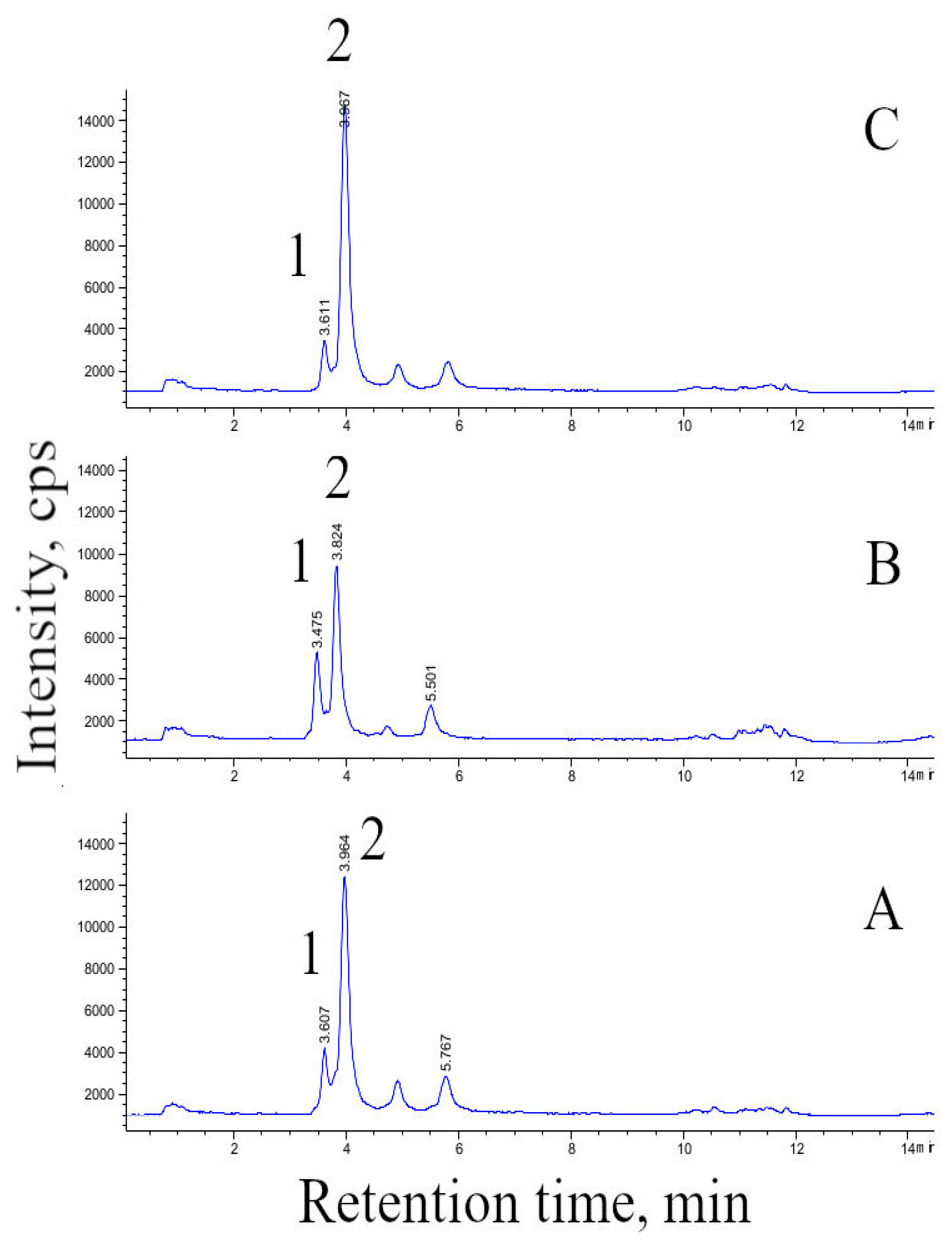
2.5. HPLC and HPLC–MS Analysis of Furostanol Glycosides (FG) in Cell Biomass

Sample preparation and quantitative analysis were performed according to [33] with modifications. Samples of dried cell biomass (10 mg each) were extracted three times, 30 min each, with distilled water (biomass:distilled water of 1:40) using ultrasound. The combined aqueous extract was applied on a Supelco (St. Louis, MO, USA) Supelclean ENVI-18 extraction cartridge for solid-phase extraction. Next, the cartridge was washed with distilled water followed by 70% ethanol. The ethanolic extract was evaporated until dryness in a rotary vacuum evaporator at 55 °C, then dissolved in an acetonitrile : water mixture (24:76 v/v) and filtered through a 0.2 μm pore diameter nylon membrane (Acrodisc, Pall Corporation, New York, NY, USA). Extracts were analyzed using the HPLC system Shimadzu Nexera LC30 (Tokyo, Japan) equipped with Shim-pack XR-ODS (75 × 2.0 mm, 2.2 μm) column. The injection volume was 0.4 μL. The mobile phase flow rate was set at 0.4 mL/min. Acetonitrile and water were used as eluents. The elution was performed at isocratic mode at acetonitrile : water volume ratio 23:77. HPLC-UV detection was performed at 203 nm. The quantitative content of individual furostanol glycosides was determined using a protodioscin standard (purity >98%, Sigma-Aldrich, St. Louis, MO, USA) [33].
The HPLC–MS was carried out in an Agilent 1260 Infinity system (Agilent Technologies, Santa Clara, CA, USA) equipped with a Single Quadrupole mass-selective detector (6120B, Agilent Technologies) with a Poroshell 120 EC-C18 column (100 × 3 mm, 2.7 μm, Agilent). The column temperature was 37 °C, the flow rate of the mobile phase was 0.5 mL/min, and the injection volume was 0.5 μL. The mobile phase consisted of a solution of formic acid (0.05% vol.) in water (solvent A) and acetonitrile (solvent B) with a gradient elution. In the course of analysis, the composition of the mobile phase changed as follows (solvent B, volume %): 0–7 min, 27%; 7–9 min 27→95%; 9–11 min, 95%; 11–11.5 min 95→27%; 11.5–14.5 min, 27%. The analysis was carried out in the positive-ion mode in the m/z range 100–1300 and fragmenter of 70. The parameters of the ionization source were as follows: quadrupole temperature 100 °C, carrier gas (nitrogen) temperature 305 °C, nitrogen supply rate (spraying gas) 13 L/min; nitrogen pressure 1294 Torr, and capillary voltage 3.0 kV. The chromatograms were recorded in the selected ion mode (m/z 1047.5 for protodeltonin and 1031.5 for protodioscin, both corresponding to [M + H − H2O]+).

2.6. Acute Toxicity Test of Dioscorea deltoidea Cell Biomass on Rats

Acute toxicity tests were performed on clinically healthy sexually naive laboratory rats of Wistar stock, specific pathogen-free category, age 10 weeks, average weight 225 ± 13 g. The animals were provided by the Department of Experimental Animal Genetic Resources of the Federal Research Center Institute of Cytology and Genetics of the Siberian Branch of the Russian Academy of Sciences (Novosibirsk, Russia).
The lyophilized powder of the D. deltoidea cell biomass was used. Five days prior to the experiment, the animals were habituated to the laboratory conditions. The rats were randomly selected and individually marked. Throughout the experiment the rats were kept in a system of individually ventilated cages (IVC) as part of VENT II ventilation unit and type III Bio.A.S. (EHRET, Mahlberg, Germany) cage shelving stand. This system does not only act as a barrier protecting laboratory animals and staff from contamination, dust, and allergens but also helps to establish the optimal microclimate in each particular cage. The housing conditions for each IVC were similar in terms of temperature (22 ± 3) °C, humidity (50–60%), and light/dark cycle 12/12 (daylight time from 6:00 am to 6:00 pm). Throughout the experiment the rats were provided with complete feed (‘Assortiment-Agro’, Sergiev Posad, Russia) and water ad libitum. Water was prepared using an EMD Millipore RiOs™ 50 (Merck Millipore, Darmstadt, Germany) water purification system. Drinking water temperature ranged from 18 to 20 °C.
Animal husbandry and management, feeding, experimental manipulations, and sacrifice were performed in accordance with the requirements of the Russian Ministry of Health Directive no. 267 «On approval of regulations of laboratory routine» (19 June 2003), no. 742 «On approval of regulations of laboratory routine using experimental animals» (13 November 1984) and no. 48 «On control of laboratory routine using experimental animals» (23 January 1985), and conforming to the International animal welfare act (EU Directive 2010/63/EU).
Acute toxicity test was performed in accordance with the All-union State Standard 32644. Two threshold doses of the lyophilized and powdered cell biomass, 2000 mg/kg and 5000 mg/kg, were considered in the experiment. The research was conducted as follows: a dose of 2000 mg/kg was used first and was administered to three animals. If at least one animal stayed alive, a dose of 5000 mg/kg was given to the other three animals. Each dose was administered intragastrically using a specialized tube.
Powdered D. deltoidea cell biomass was given to animals after 12 h of food deprivation. The feeding of animals was resumed 3–4 h after the administration of the cell biomass powder. A group of intact (control) animals (n = 3) were given equivalent doses of purified water.
The state of each animal was evaluated individually every 30 min during the first 4 h after the administration of the compound under study. Particular attention was paid to the manifestation, severity, and duration of the intoxication symptoms, recovery time, and death of the animal. During the two weeks that followed the administration of D. deltoidea biomass, the animals were examined daily in order to keep a record of clinical signs of intoxication, and dates of death or recovery.
The animals were weighted using the electronic laboratory scales (Ohaus, Parsippany, NJ, USA) before the experiment and every third day after the administration of D. deltoidea biomass. At the end of the experiment, the animals were euthanized by carbon dioxide in a special chamber (VetTech, Russia) and then autopsied.
General blood analysis was performed using an Abacus junior vet 2.7 (Diatron Messtechnik GmbH, Wien, Austria) automated veterinary blood analyzer. Eighteen parameters were analyzed with Diatron assay kits. The biochemical parameters of blood were examined at using a BioChem FC-360 (North Attleboro, MA, USA) automated clinical chemistry analyzer with High Technology (North Attleboro, MA, USA) assay kits.
The autopsy of every animal included a thorough examination of the body surface, all orifices, intracranial, chest, and abdominal cavities, and their contents. Liver, kidneys, adrenal glands, testicles, epididymis, prostate and seminal glands with coagulation gland, thymus, spleen, brain and heart were routinely detached from the neighboring tissues in all animals and weighted using electronic laboratory scales (Ohaus, Adventurer Pro, USA). These organs were weighted fresh immediately after the autopsy to prevent desiccation.

2.7. Statistical Analysis

Data are presented as the mean values and standard deviations recorded for the triplicates (3 flasks or 3 fixed-size samples of cell suspension collected from bioreactors) for each data point. Standard deviations that constitute less than 10% of mean values are not displayed in the graphs. STATISTICA10 software (StatSoft©, Moscow, Russia) was used for processing the data. Statistical significance of differences was estimated using the one-way ANOVA test followed by the Dunnett’s test at p < 0.05 for growth parameters and FG content and at p < 0.1 for acute toxicity tests on rats (comparison with intact animals’ parameters).

3. Results

3.1. Growth and Furostanol Glycoside Content of Dioscorea deltoidea Cell Suspension Culture in Bioreactors

Cell suspension of D. deltoidea was successfully cultured in laboratory (20 L) and industrial (630 L) bioreactors using the semi-continuous regime. The main growth characteristics of D. deltoidea cell suspension cultured in bioreactors were comparable to those recorded during cultivation in flasks (Table 1). The transfer from flask culture to bioreactors resulted in the reduction of all growth parameters such as cell viability, specific growth rate and accumulation of cell dry weight which, in turn, led to lower productivity (Table 1), however, the recorded differences were statistically insignificant.
Active synthesis of FG in cell culture was recorded during all phases of the cultivation cycle in all culture systems (Table 1, Figure 2).
The highest FG content in both flasks and bioreactors was observed during the stationary phase and corresponded to maximum dry weight accumulation (data not shown). Accumulation of FG in cells cultured in 20 L and 630 L bioreactors (47 and 57 mg/g DW respectively) was lower compared to cells cultured in flasks (76 mg/g DW); however, this difference was insignificant at p < 0.05 due to variation in FG content between individual subculture cycles in each type of bioreactor. In both flasks and bioreactors, FG were represented mainly by 25 R and 25 S protodioscin isomers while traces of 25 R and 25 S deltoside isomers were also detected (data not shown). The ratio of 25R:25S protodioscin isomers in the cell biomass was 55:45 and remained stable during the cultivation cycle.

3.2. Content of Macro and Microelements in Cell Biomass

The content of inorganic macro- and microelements in D. deltoidea cell biomass was measured at the end of the subculture cycle corresponding to a maximum accumulation of dry cell biomass in 630 L bioreactor. The cell culture was found to contain all elements assayed. Concentrations of individual elements are summarised in Table 2.

3.3. Acute Toxicity Test of Dioscorea deltoidea Cell Biomass

The acute toxicity test was performed using two doses of dry cell biomass, 2000 mg/kg and 5000 mg/kg. The preliminary dose (2000 mg/kg) for a single intragastric administration did not induce any toxicity signs in animals: motor, respiratory, and cardiac activity as well as responses to external stimuli remained unchanged after the administration and throughout the entire observation period. Similarly, rats exposed to the administration of the higher biomass dose of 5000 mg/kg showed no sign of intoxication. Animals consistently gained weight throughout the entire observation period, and a similar stable weight gain was recorded for intact animals. No difference in appearance and relative weight of heart, liver, kidney, spleen, or thymus were observed between control animals and animals that received the high dose (Table 3).
The hematology test panel showed that a single intragastric administration of D. deltoidea cell biomass at a dose of 2000 mg/kg caused only minor changes in blood biochemical composition (Table 4 and Table 5) [34]. There were no significant changes in leukocytes and their subpopulations, however, some insignificant rise was observed in monocytes, eosinophils, basophils and blast cells mixture, granulocytes (up to 20%), granulocyte percentage (to 20%), and monocytes (to 63%) compared to intact animals. The indicators of erythrocytes’ functional status (erythrocytes and hemoglobin concentration, hematocrit, mean hemoglobin contents in erythrocytes) did not differ significantly from the same parameters of the control group, with a moderate statistically significant rise in mean erythrocyte volume (by 5.1%), mean haemoglobin contents in erythrocytes (by 4.4%), and its distribution width (no more than 7%). The indicators of thrombocytes’ functional status (thrombocyte concentration, plateletcrit, mean thrombocyte volume, and platelet distribution) did not change significantly; however, an insignificant rise in mean thrombocyte volume by 2.4% was noted.
The increase in the dose of Dioscorea cell biomass up to 5000 mg/kg resulted in more prominent shifts in blood cell composition. A significant increase in leukocyte and lymphocyte content (2.2–2.3-fold, p < 0.1); monocytes, eosinophils, basophils and blast cells mixture (3.3-fold, p < 0.1), granulocytes (to 86%, p. < 0.1) and monocyte percentage (to 48%) compared to the same parameters of intact animals has been noted. A rise in hemoglobin and hematocrit concentration (to 5.5%, p < 0.1), and mean hemoglobin content in erythrocyte (no more than 3.3%, p <0.1) compared to the same parameters of intact animals was observed. Thrombocyte concentration did not change significantly. An insignificant increase in mean thrombocyte volume by no more than 1.7% and platelet distribution by 3.3% has been noted.
The results of comparing the blood chemistry values for blood serum of animals that once received threshold doses of D. deltoidea cell biomass (Table 5), and intact rats did not reveal any significant changes (within one group on average). However, in those animals that received the biomass study at a dose of 2000 mg/kg, an insignificant decrease in serum protein level (by no more than 4.2%) was noted due to a decrease in albumin concentration (by 3.5%) and creatinine concentration (by no more than 2.5%); however, no changes in urea and potassium concentration were observed. It is interesting to note an increase in aspartate aminotransferase (AspAT) activity by 31% (non-significant) and a decrease in alanine aminotransferase (ALAT) activity by 13.6% (non-significant), while alkaline phosphatase activity remained unchanged (Table 5).
In animals that were administered the biomass at a dose reaching 5000 mg/kg, no significant changes in blood serum biochemistry were observed, although an insignificant decrease in creatinine concentration (no more than 7%), ALAT (up to 12%), and gamma-glutamyl transferase (GGT, up to 20%) activity (Table 5) was recorded. The analysis of blood chemistry values revealed that changes in enzymatic activity may be indicative of the intensification of myocardium work (a drop in activity of liver enzymes ALAT and GGT when a dose is increased to 5000 mg/kg against the background of an increase in AspAT activity); the intensification of dietary proteins’ catabolism (reduction of serum creatinine concentration in animals), and carbohydrate and lipid metabolism (rise of cholesterol and glucose concentration after the administration at a dose of 2000 mg/kg).

4. Discussion

Plant cell culture technology and its wide-scale commercial application, which has flourished between the 1960s and 1980s is now experiencing its renaissance. Undifferentiated plant cells cultured in shake flasks and bioreactors and their extracts are frequently used as ingredients in the cosmetic industry [35] and for the production of secondary metabolites with proven biological activities [29,36]. In addition, it is believed that plant cells exhibit high nutritional value and should be tested for wider applications in the human diet [35]. Not surprisingly, the concept of “cellular agriculture” is getting increasing attention from both research and commercial perspectives [34,37]. One of good examples is the bioreactor-produced cell culture of a medicinal plant Polyscias filicifolia, which is used as the main component of the food supplement Vitagmal available in Russian and European markets [38]. Nevertheless, the application of cell culture-based products as food ingredients requires a more strict, hence longer and more expensive, approval process compared to cosmetic industry, often involving toxicity assays [39]. Another bottleneck is the efficient transfer of the cultivation process from the laboratory to the industrial facility, which requires the bioreactor type and culture conditions to be optimized. In this study, we explored the possibility of transferring cell culture technology from flasks to laboratory and industrial bioreactors for cell suspension of D. deltoidea, strain DM-0.5–03. We also assayed FG content, elemental composition and acute toxicity of the produced cell biomass as the first steps for its approval as a food supplement.

4.1. Bioreactor Culture of Dioscorea deltoidea Suspension Cells

Both the literature and the results of our previous studies evidenced that bubble-type bioreactors where the medium is stirred by a flow of pressurized sterile air are most efficient for the cultivation of undifferentiated plant cells [25,29]. In the present study, the growth characteristic of D. deltoidea cell culture decreased when the cultivation process was transferred from flasks to bioreactors, but this effect was statistically insignificant. The decline in cell viability and growth observed in this study may be attributed to lower oxygen concentration and a higher level of mechanical stress in bioreactors compared to flask culture. Earlier, it was demonstrated that cell culture of D. deltoidea was extremely sensitive to a lack of aeration [25]. Nevertheless, growth index and maximum biomass accumulation, productivity, and specific growth rate of D. deltoidea cell culture in 20 L and 630 L bioreactors were in line with those recorded for cell cultures of other medicinal plants. For example, the specific growth rate of Polyscias filicifolia suspension cell culture in 20 L and 630 L bioreactors were respectively 0.15 and 0.13 day−1, while accumulation of dry weight reached 12 g/L [40]. Maximum accumulation of dry biomass of 13 g/L and specific growth rate at exponential phase of 0.24 day−1 were reported for cell suspension of Tribulus terrestris cultured in 7 L bioreactors [41].
The content of FG in D. deltoidea in the present study was maximized at the stationary phase and remained comparable or higher than glycoside production usually reported for plant cell cultures. For example, the content of triterpene glycosides in suspension cell culture of Polyscias fruticosa was ca. 15 mg/g DW [42]. Production of ginsenosides in suspension cell cultures of various Panax species varied from 3 to 1130 mg/L depending on the culture conditions and elicitation strategy [43]. In the earlier studies, Tal and Goldberg [44] reported that cell culture of D. deltoidea grown in batch (periodic) regime accumulated up to 1.8% (of cell dry wt.) diosgenin while very little amounts of this substance were produced by growing cells in the chemostat. In contrast, Drapeau et al. [45] demonstrated that diosgenin biosynthesis by D. deltoidea cell culture in 14 L bioreactor was independent of culture growth rate. Accumulation of diosgenin in cell suspension cultures of Dioscorea zingiberensis reached its maximum of 5.25 mg/L after the elicitation treatment using fungal oligosaccharide [46]. Lower FG content determined in the present study for bioreactor-cultured cells compared to shake flask culture was, most likely, associated with mechanical damage and reduced oxygen concentration in a large volume of nutrient medium, which affected cells’ respiratory metabolism [24].
It has been reported that cell cultures of Dioscorea spp. accumulate predominantly SG of the furostanol type [47]. The content and ratio of individual glycosides in cell culture vary significantly depending on both physical (gas exchange, temperature, bioreactor type) and chemical (medium composition, growth regulators) factors [24,43]. In the present study, FG in Dioscorea cell culture was mainly represented by protodioscin with only a trace amount of deltoside, which may be specific to cell cultures of this species.
Overall, the results of this study suggest that suspension cells of D. deltoidea can be efficiently cultured in bioreactors of industrial volume, up to 630 L, and retain high growth and biosynthetic abilities. Both growth and FG content in the cell culture remained stable over course of six months of cultivation using the semi-continuous regime (data not shown).

4.2. Inorganic Elements in Cell Culture of Dioscorea deldoidea

The indisputable advantage of cell cultures over plants collected from the wild is a controllable, sterile production environment that supports standardization and implementation of quality control measures [30,35]. This cell-based biotechnology opens the door for the production of biomaterials with predetermined and controllable composition of macro- and micronutrients and, potentially, their manipulation towards increasing the content of the deficient elements. However, information about the elemental composition and nutritive value of plant cell cultures is scarce. Recently, Nordlung et al. [37] analyzed the elemental composition and nutritive value of cell cultures of cloudberry (Rubus chamaemorus), stoneberry (Rubus saxatilis) and lingonberry (Vaccinium vitisidaea) grown in flasks and found that they contained more proteins and dietary fiber than fruits of the same species. Composition and amounts of free sugars varied among cell cultures. Not surprisingly, the major element in all cultures was carbon (452.8–460.6 mg/g DW) followed by hydrogen (up to 61.6 mg/g DW) and nitrogen (38-72 mg/g DW). Sulfur was found in concentrations from 1.2 to 8.5 mg/g DW [37]. In the present study, the weight content of macro- and micro-elements in D. deltoidea cell biomass was determined using the ICP-MS method. This method has already been tested and used for analyzing macro- and micro-elements content in intact plants and cell cultures [47,48,49,50]. Cultured cells of D. deltoidea contained inorganic marco- and micro-elements that are essential for the human diet [50]. It is expected that D. deltoidea cell culture also contained carbon, hydrogen and nitrogen as major structural elements. However, in the present study we focused on inorganic elements, hence C, H, and N contents were not analyzed. The elemental composition of D. deltoidea cell biomass indicates that it is an environmentally safe plant product. In addition, the concentrations of individual elements do not exceed their threshold limits recommended for the human diet [51].
For the intensive and stable growth of plant cells and organs cultured in vitro, various natural or synthetic growth regulators are usually supplied to the culture medium at the beginning of the cultivation process. Some of these substances, for example, 2,4-D or indole-3-butiric acid (IBA), are considered harmful for human health; hence, the concern is often raised regarding their potential occurrence in food ingredients derived from cultured plant cells, tissues and organs [39]. In view of these concerns, several studies have confirmed that growth regulators are normally fully consumed during cell culture growth and were not detected or existed in trace amounts in the final biomass [52,53]. Our previous analysis revealed that 2,4-D added to culture medium at a concentration of 1 mg/L (similar to the present study) was found in the biomass of Arabidopsis thaliana cells cultured in flasks for 14 days in a very low amount of 16 × 10−12 g/g fresh cell weight which was far below the safety threshold limits (unpublished data). Based on these data and the data of toxicity assessment presented here, it is expected that biomass of D. deltoidea cells contain zero or insignificant amount of growth regulators, but the quantitative analysis may be further performed to obtain the safety assessment certification from a national authority.

4.3. Acute Toxicity Test of Dioscorea deltoidea Cell Biomass in Rats

The results of the present study demonstrate that oral administration of D. deltoidea cell biomass at threshold doses (2000 mg/kg and 5000 mg/kg) does not result in death or intoxication of laboratory animals. Moreover, the high doses tested had no or little effect on the hematological parameters and blood biochemistry of the animals. In other experiments with laboratory rats, the cell biomass of D. deltoidea administrated at a dose of 1 mg/kg prevented the development of arterial hypertension and showed pronounced endothelio- and atheroprotective effects [16]. Earlier reports suggested high biological activity of “Deltostim”, a preparation from D. deltoidea cell biomass containing a mixture of protodioscin and deltoside [13]. Deltostim stimulated ovulation and spermatogenesis in rats and rabbits and resulted in a 2.0–2.5-fold increase in the fertilization rate in cows with ovarial hypofunction [13]. In the experiments with isolated cultured lymphocytes, Deltostim displayed high immunomodulatory activity [13]. According to [51], Dioscorea deltoidea cell biomass can be referred to as a low-hazardous substance; therefore, its potential to be used in the production of functional foods and other specialized products such as food supplements and animal feed supplements may be further explored.

5. Conclusions

The results of the present study demonstrated that suspension cell culture of the medicinal plant D. deltoidea can be successfully grown in laboratory (20 L) and industrial (630 L) bioreactors under semi-continuous regime. Cell culture fully retained its growth and biosynthetic abilities and accumulated FG at concentrations of 47–57 mg/g DW which is higher or comparable to steroid glycoside accumulation in cell cultures of other Dioscorea species. Cell biomass is confirmed to contain safe concentrations of the essential inorganic macro- and microelements and has no toxic effects on laboratory rats. These results open the door for further investigations and approval of D. deltoidea cell culture as a natural safe source of furostanol type glycosides and its potential use as dietary supplement with high biological activity. Due to its high content of FG and non-toxic concentrations of inorganic elements, the cell strain DM-05–03 has great potential application for treating patients with disorders of the reproductive system and as a natural source of microelements to treat mineral deficiency.

Author Contributions

M.V.T., I.M.I. and A.G.K. performed bioreactor cultivation of cell suspension; S.V.K. and D.V.K. performed analysis of steroidal glycosides; E.G.T. and E.A.N. performed analysis of the elemental composition of cell biomass; E.R.V., G.S.T., and E.A.K. performed toxicity tests on laboratory rats; M.V.T., E.V.P., A.M.N., and K.-Y.P. prepared the manuscript. All authors contributed to data analysis and manuscript preparation. All authors have read and agreed to the published version of the manuscript.

Funding

Cultivation of cell suspensions and the analysis of steroidal glycoside content were funded by the Ministry of Science and Higher Education of Russian Federation through Megagrant project no. 075-15-2019-1882 and performed using the equipment of the large-scale research facilities “Experimental biotechnological facility” and “All-Russian Collection of cell cultures of higher plants” of the IPPRAS (EBF IPPRAS and ARCCC HP IPPRAS). Toxicology analysis and determination of elemental composition of the cell biomass were performed with the financial support of the Russian Science Foundation project no. № 19-14-00387.

Institutional Review Board Statement

Ethical review and approval were waived for this study as it was conducted in strict accordance to Russian and international directives on the experiments using laboratory animals: Russian Ministry of Health Directives no. 267 «On approval of regulations of laboratory routine» (19 June 2003), no. 742 «On approval of regulations of laboratory routine using experimental animals» (13 November 1984), no. 48 «On control of laboratory routine using experimental animals» (23 January 1985), the International animal welfare act (EU Directive 2010/63/EU).

Informed Consent Statement

Not applicable.

Data Availability Statement

No new data sets were generated during this study. Data sharing is not applicable to this article.

Conflicts of Interest

The authors declare no conflict of interest.

References

  1. Obidiegwu, J.E.; Lyons, J.B.; Chilaka, C.A. The Dioscorea genus (yam)—An appraisal of nutritional and therapeutic potentials. Foods 2020, 9, 1304. [Google Scholar] [CrossRef]
  2. Hooker, E. Final report of the amended safety assessment of Dioscorea villosa (wild yam) root extract. Int. J. Toxicol. 2004, 23 (Suppl. 2), 49–54. [Google Scholar] [CrossRef]
  3. Zeng, M.; Zhang, L.; Li, M.; Zhang, B.; Zhou, N.; Ke, Y.; Weisheng, F.; Zheng, X. Estrogenic effects of the extracts from the Chinese Yam (Dioscorea opposita Thunb.) and its effective compounds in vitro and in vivo. Molecules 2018, 23, 11. [Google Scholar] [CrossRef] [Green Version]
  4. Araghiniknam, M.; Chung, S.; Nelson-White, T.; Eskelson, C.; Watson, R. Antioxidant activity of dioscorea and dehydroepiandrosterone (DHEA) in older humans. Life Sci. 1996, 59, PL147–PL157. [Google Scholar] [CrossRef]
  5. Horii, N.; Hasegawa, N.; Fujie, S.; Iemitsu, K.; Uchida, M.; Hamaoka, T.; Iemitsu, M. Effects of Dioscorea esculenta intake with resistance training on muscle hypertrophy and strength in sprint athletes. J. Clin. Biochem. Nutr. 2020, 67, 338–343. [Google Scholar] [CrossRef]
  6. Son, I.S.; Kim, J.H.; Sohn, H.Y.; Son, K.H.; Kim, J.-S.; Kwon, C.-S. Antioxidative and hypolipidemic effects of diosgenin, a steroidal saponin of yam (Dioscorea spp.), on high-cholesterol fed rats. Biosci. Biotechnol. Biochem. 2007, 71, 3063–3071. [Google Scholar] [CrossRef] [PubMed] [Green Version]
  7. Dubinskaya, V.A.; Strelkova, L.B.; Vasil’eva, I.S.; Nikolaeva, S.S.; Rebrov, L.B.; Paseshnichenko, V.A. Anabolic properties of Dioscorea deltoidea Wall furostanol glycosides. Bull. Exp. Biol. Med. 1998, 126, 800–802. [Google Scholar] [CrossRef]
  8. Liang, X.-J.; Guo, Y.-C.; Sun, T.-Y.; Song, H.-R.; Gao, Y.-X. Anti-angiogenic effect of total saponins of Rhizoma Dioscorea nipponica on collagen induced-arthritis in rats. Exp. Ther. Med. 2016, 12, 2155–2160. [Google Scholar] [CrossRef] [Green Version]
  9. Qin, J.; Kang, Y.; Xu, Z.; Zang, C.; Fang, B.; Liu, X. Dioscin prevents the mitochondrial apoptosis and attenuates oxidative stress in cardiac H9c2 cells. Drug Res. 2013, 64, 47–52. [Google Scholar] [CrossRef] [PubMed]
  10. Tao, X.; Sun, X.; Yin, L.; Han, X.; Xu, L.; Qi, Y.; Xua, Y.; Lia, H.; Lina, Y.; Liu, K.; et al. Dioscin ameliorates cerebral ischemia/reperfusion injury through the downregulation of TLR4 signaling via HMGB-1 inhibition. Free Radic. Biol. Med. 2015, 84, 103–115. [Google Scholar] [CrossRef]
  11. Jayachandran, S.K.; Vasanthi, R.H.; Rajamanickam, V.G. Flavonoid rich fraction of Dioscorea bulbifera Linn. (yam) enhances mitochondrial enzymes and antioxidant status, thereby protects heart from isoproterenol induced myocardial infarction. Curr. Pharm. Biotechnol. 2010, 11, 887–894. [Google Scholar] [CrossRef] [PubMed]
  12. Tang, Y.-N.; He, X.-C.; Ye, M.; Huang, H.; Chen, H.-L.; Peng, W.-L.; Zhao, Z.-Z.; Tao, Y.; Chen, H.-B. Cardioprotective effect of total saponins from three medicinal species of Dioscorea against isoprenaline-induced myocardial ischemia. J. Ethnopharmacol. 2015, 175, 451–455. [Google Scholar] [CrossRef] [PubMed] [Green Version]
  13. Vasil’eva, I.S.; Paseshnichenko, V.A. Steroid glycosides from suspension cultures of Dioscorea deltoidea cells and their biological activity. In Saponins Used in Traditional and Modern Medicine. Advances in Experimental Medicine and Biology; Waller, G.R., Yamasaki, K., Eds.; Springer: Boston, MA, USA, 1996; Volume 404, pp. 15–22. [Google Scholar] [CrossRef]
  14. Galani, V.J.; Patel, D.M. A comprehensive phytopharmacological review of Dioscorea bulbifera Linn. Int. J. Environ. Sci. Nat. Res. 2017, 4, 555650. [Google Scholar] [CrossRef]
  15. Galakatu Sameer, S.; Surve Pankaj, P.; Ghotankar Aparna, M.; Kharat Ravindra, S. Review of Varahikanda (Dioscorea bulbifera) for its pharmacological properties. World J. Pharm. Res. 2016, 5, 1738–1746. [Google Scholar] [CrossRef]
  16. Korokin, M.; Gudyrev, O.; Gureev, V.; Korokina, L.; Peresypkina, A.; Pokrovskaia, T.; Pokrovskii, M. Studies to elucidate the effects of eurostanol glycosides from Dioscorea deltoidea cell culture in a rat model of endothelial dysfunction. Molecules 2019, 25, 169. [Google Scholar] [CrossRef] [Green Version]
  17. Cui, H.; Li, T.; Wang, L.; Su, Y.; Xian, C.J. Dioscorea bulbifera polysaccharide and cyclophosphamide combination enhances anti-cervical cancer effect and attenuates immunosuppression and oxidative stress in mice. Sci. Rep. 2016, 6. [Google Scholar] [CrossRef] [PubMed] [Green Version]
  18. Salehi, B.; Sener, B.; Kilic, M.; Sharifi-Rad, J.; Naz, R.; Yousaf, Z.; Mudau, F.N.; Fokou, P.V.T.; Ezzat, S.M.; El Bishbishy, M.H.; et al. Dioscorea plants: A genus rich in vital nutra-pharmaceuticals-a review. Iran. J. Pharm. Res. 2019, 18, 68–89. [Google Scholar] [CrossRef] [PubMed]
  19. Tahir, N.; Bibi, Y.; Iqbal, M.; Hussain, M.; Laraib, S.; Safdar, I.; Bibi, G. Overview of Dioscorea deltoidea Wall. Ex Griseb: An endangered medicinal plant from Himalaya region. J. Biodivers. Environ. Sci. 2016, 9, 13–24. [Google Scholar]
  20. Avula, B.; Wang, Y.-H.; Ali, Z.; Smillie, T.J.; Khan, I.A. Chemical fingerprint analysis and quantitative determination of steroidal compounds from Dioscorea villosa, Dioscorea species and dietary supplements using UHPLC-ELSD. Biomed. Chromatogr. 2013, 28, 281–294. [Google Scholar] [CrossRef] [PubMed] [Green Version]
  21. Sautour, M.; Mitaine-Offer, A.-C.; Lacaille-Dubois, M.-A. The Dioscorea genus: A review of bioactive steroid saponins. J. Nat. Med. 2007, 61, 91–101. [Google Scholar] [CrossRef]
  22. Shen, L.; Xu, J.; Luo, L.; Hu, H.; Meng, X.; Li, X.; Chen, S. Predicting the potential global distribution of diosgenin-contained Dioscorea species. Chin. Med. 2018, 13. [Google Scholar] [CrossRef]
  23. Khandy, M.T.; Titova, M.V.; Konstantinova, S.V.; Kochkin, D.V.; Ivanov, I.M.; Nosov, A.M. Formation of protodioscin and deltoside isomers in suspension cultures of Nepal yam (Dioscorea deltoidea Wall.) cells. Appl. Biochem. Microbiol. 2016, 52, 657–662. [Google Scholar] [CrossRef]
  24. Titova, M.V.; Khandy, M.T.; Konstantinova, S.V.; Kulichenko, I.E.; Sukhanova, E.S.; Kochkin, D.V.; Nosov, A.M. Effect of inhibitors of two isoprenoid biosynthetic pathways on physiological and biosynthetic characteristics of Dioscorea deltoidea cell suspension culture. Russ. J. Plant Physiol. 2016, 63, 894–900. [Google Scholar] [CrossRef]
  25. Titova, M.V.; Shumilo, N.A.; Kulichenko, I.E.; Ivanov, I.M.; Sukhanova, E.S.; Nosov, A.M. Features of respiration and formation of steroidal glycosides in Dioscorea deltoidea cell suspension culture grown in flasks and bioreactors. Russ. J. Plant Physiol. 2015, 62, 557–563. [Google Scholar] [CrossRef]
  26. Nosov, A.M. Aspects of Steroid Metabolism in Cultured Cells of Dioscorea deltoidea as the Base for Biotechnological Production of Furostanol Glycosides. Ph.D. Thesis, All-Russian Scientific Institute of Applied Biochemistry, Moscow, Russia, 1992. [Google Scholar]
  27. Murashige, T.; Skoog, F. A revised medium for rapid growth and bioassays with tobacco tissue cultures. Physiol. Plant 1962, 15, 473–479. [Google Scholar] [CrossRef]
  28. Kaul, B.; Staba, J. Biosynthesis and isolation of diosgenin from Dioscorea deltoidea callus and suspension cells. Lloydia 1968, 31, 171–179. [Google Scholar]
  29. Baker, J.C.; Mock, N.M. An improved method for monitoring cell death in cell suspension and leaf disc assays using evans blue. Plant Cell Tiss Organ Cult. 1994, 39, 7–12. [Google Scholar] [CrossRef]
  30. Nosov, A.M. Application of cell technologies for production of plant-derived bioactive substances of plant origin. Appl. Biochem. Microbiol. 2012, 48, 609. [Google Scholar] [CrossRef]
  31. Tokalıoğlu, Ş.; Dokan, F.K.; Köprü, S. ICP-MS multi-element analysis for determining the origin by multivariate analysis of red pepper flakes from three different regions of Turkey. LWT 2019. [Google Scholar] [CrossRef]
  32. Pinheiro, F.C.; Babos, D.V.; Barros, A.I.; Pereira-Filho, E.R.; Nóbrega, J.A. Microwave-assisted digestion using dilute nitric acid solution and investigation of calibration strategies for determination of As, Cd, Hg and Pb in dietary supplements using ICP-MS. J. Pharm. Biomed. Anal. 2019, 174, 471–478. [Google Scholar] [CrossRef]
  33. Kochkin, D.V.; Khandy, M.T.; Zaitsev, G.P.; Tolkacheva, N.V.; Shashkov, A.S.; Titova, M.V.; Chirva, V.Y.; Nosov, A.M. Protodioscin in Dioscorea deltoidea suspension cell culture. Chem. Nat. Compd. 2016, 52, 664–668. [Google Scholar] [CrossRef]
  34. Evans, G.O. (Ed.) Animal Clinical Chemistry: A Practical Handbook for Toxicologists and Biomedical Researchers, 2nd ed.; CRC Press: Boca Raton, FL, USA; Taylor & Francis: London, UK, 2009; 368p. [Google Scholar]
  35. Eibl, R.; Meier, P.; Stutz, I.; Schildberger, D.; Hühn, T.; Eibl, D. Plant cell culture technology in the cosmetics and food industries: Current state and future trends. Appl. Microbiol. Biotechnol. 2018, 102, 8661–8675. [Google Scholar] [CrossRef] [PubMed] [Green Version]
  36. Largest Global Supplier of Paclitaxel and Docetaxel via PCF®. Available online: https://phytonbiotech.com (accessed on 15 January 2021).
  37. Nordlund, E.; Lille, M.; Silventoinen, P.; Nygren, H.; Seppänen-Laakso, T.; Mikkelson, A.; Aura, A.M.; Heiniö, R.L.; Nohynek, L.; Puupponen-Pimiä, R.; et al. Plant cells as food—A concept taking shape. Food Res. Int. 2018, 107, 297–305. [Google Scholar] [CrossRef] [PubMed]
  38. Kotin, A.M.; Bichevaya, N.K. Anti-Teratogenic. Agent. Patent No. WO1996002266A1, 1 February 1996. [Google Scholar]
  39. Murthy, H.K.; Georgiev, M.I.; Park, S.-Y.; Dandin, V.S.; Paek, K.-Y. The safety assessment of food ingredients derived from plant cell, tissue and organ cultures: A review. Food Chem. 2015, 176, 426–432. [Google Scholar] [CrossRef] [PubMed]
  40. Sukhanova, E.S.; Chernyak, N.D.; Nosov, A.M. Obtaining and characteristics of callus and suspension cultures of Polyscias filicifolia and Polyscias fruticose. Biotekhnologiya 2010, 4, 44–50. [Google Scholar]
  41. Khandy, M.T.; Kochkin, D.V.; Tomilova, S.V.; Galishev, B.A.; Sukhanova, E.S.; Klyushin, A.G.; Ivanov, I.M.; Nosov, A.M. Obtaining and study of callus and suspension plant cell cultures of Tribulus terrestris L., a producer of steroidal glycosides. Appl. Biochem. Microbiol. 2017, 53, 800–806. [Google Scholar] [CrossRef]
  42. Kochkin, D.V.; Sukhanova, E.S.; Nosov, A.M. The accumulation of triterpene glycosides in the growing cycle cell suspension cultures Polyscias fruticosa. Vestn. Volga State Univ. Technol. Ser. Forest Ecol. Nat. Manag. 2014, 24, 67–73. [Google Scholar]
  43. Kim, Y.-J.; Zhang, D.; Yang, D.-C. Biosynthesis and biotechnological production of ginsenosides. Biotechnol. Adv. 2015, 33, 717–735. [Google Scholar] [CrossRef]
  44. Tal, B.; Goldberg, I. Growth and diosgenin production by Dioscorea deltoidea cells in batch and continuous cultures. Planta Med. 1982, 44, 107–110. [Google Scholar] [CrossRef]
  45. Drapeau, D.; Blanch, H.W.; Wilke, C.R. Growth kinetics of Dioscorea deltoidea and Catharanthus roseus in batch culture. Biotechnol. Bioeng. 1986, 28, 1555–1563. [Google Scholar] [CrossRef]
  46. Zhang, R.; Li, P.; Xu, L.; Chen, Y.; Sui, P.; Zhou, L.; Li, J. Enhancement of diosgenin production in Dioscorea zingiberensis cell culture by oligosaccharide elicitor from its endophytic fungus Fusarium oxysporum Dzf17. Nat. Prod. Commun. 2009, 4, 1459–1462. [Google Scholar] [PubMed] [Green Version]
  47. Nosov, A.M.; Popova, E.V.; Kochkin, D.V. Isoprenoid Production via Plant Cell Cultures: Biosynthesis, Accumulation and Scaling-Up to Bioreactors. In Production of Biomass and Bioactive Compounds Using Bioreactor Technology; Paek, K.-Y., Murthy, H.N., Zhong, J.-J., Eds.; Springer: Dordrecht, The Netherlands, 2014; pp. 563–623. [Google Scholar]
  48. Zhao, X.; Wei, J.; Shu, X.; Kong, W.; Yang, M. Multi-elements determination in medical and edible Alpinia oxyphylla and Morinda officinalis and their decoctions by ICP-MS. Chemosphere 2016, 164, 430–435. [Google Scholar] [CrossRef]
  49. Dico, G.M.L.; Galvano, F.; Dugo, G.; D’Ascenzi, C.; Macaluso, A.; Vella, A.; Giangrosso, G.; Cammilleri, G.; Ferrantelli, V. Toxic metal levels in cocoa powder and chocolate by ICP-MS method after microwave-assisted digestion. Food Chem. 2018, 245, 1163–1168. [Google Scholar] [CrossRef] [PubMed]
  50. Michalak, I.; Marycz, K.; Basińska, K.; Chojnacka, K. Using SEM-EDX and ICP-OES to investigate the elemental composition of green macroalga Vaucheria sessilis. Sci. World J. 2014, 1–8. [Google Scholar] [CrossRef] [Green Version]
  51. Institute of Medicine (US) Panel on Micronutrients. Dietary Reference Intakes for Vitamin A, Vitamin K, Arsenic, Boron, Chromium, Copper, Iodine, Iron, Manganese, Molybdenum, Nickel, Silicon, Vanadium, and Zinc; National Academies Press: Washington, DC, USA, 2001. Available online: https://www.ncbi.nlm.nih.gov/books/NBK222310/ (accessed on 15 January 2021). [CrossRef] [Green Version]
  52. Ushiyama, K. Regulations for plant cell culture derived products in Japan. In Plant Cell and Tissue Culture for the Production of Food Ingredients; Fu, T.J., Singh, G., Curtis, W.R., Eds.; Springer: Boston, MA, USA, 1999; pp. 281–285. [Google Scholar] [CrossRef]
  53. Paek, K.Y.; Cheongju National University, Korea. Personal communication, 2020.
Figure 1. Bioreactor cultivation of plant cell suspensions at the Institute of Plant Physiology of Russian Academy of Sciences: (A) 630 L bioreactors; (B) Bioreactor-cultured cells of Dioscorea deltoidea at exponential growth phase (bar = 50 µm).
Agronomy 11 00394 g001
Figure 2. The HPLC–MS profiles of extracts from Dioscorea deltoidea cell biomass grown in different cultivation systems. (A)—flasks, (B)—20 L bioreactor, (C)—630 L bioreactor. 1—deltoside (protodeltonin), 2—protodioscin.
Agronomy 11 00394 g002
Table 1. Growth Characteristics and Content of Furostanol Glycosides in Dioscorea deltoidea Suspension Cell Culture Grown in Flasks and Bioreactors.
Cultivation SystemViability (%)Maximum Dry Weight Accumulation, Xmax (g/L)Productivity, P [g/(L day)]Specific Growth Rate, µ (day−1)Growth Index, IContent of Furostanol Glycosides (mg/g DW) 1
250 mL
flasks
90.4 ± 5.5 a9.5 ± 2.6 a0.40 ± 0.11 a0.16 ± 0.04 a4.22 ± 0.79 a76.3 ± 22.5 a
20 L bioreactors79.0 ± 8.1 a8.5 ± 2.1 a0.37 ± 0.12 a0.13 ± 0.03 a3.76 ± 0.72 a47.7 ± 16.2 a
630 L bioreactors83.5 ± 4.5 a8.8 ± 2.3 a0.33 ± 0.10 a0.12 ± 0.01 a3.51 ± 0.62 a57.4 ± 19.8 a
1 maximum content of furostanol glycosides calculated based on protodioscin standard. DW—dry weight. Mean values followed by the same letter are not statistically different at p < 0.05 according to Dunnett’s test.
Table 2. Content of macro- and microelements in Dioscorea deltoidea cell biomass.
ElementContent, µg/g of Dry Biomass
Microelements
Zinc (Zn)(0.37 ± 0.05) × 103
Manganese (Mn)(0.33 ± 0.08) × 103
Iron (Fe)(0.10 ± 0.01) × 103
Boron (B)15.42 ± 1.41
Aluminum (Al)2.69 ± 0.50
Copper (Cu)1.44 ± 0.62
Chromium (Cr)0.24 ± 0.07
Selenium (Se)0.24 ± 0.04
Cobalt (Co)0.18 ± 0.10
Nickel (Ni)0.14 ± 0.01
Macroelements
Potassium (K)(11.39 ± 4.21) × 103
Calcium (Ca)(1.12 ± 0.33) × 103
Magnesium (Mg)(0.74 ± 0.55) × 103
Sodium (Na)(0.40 ± 0.27) × 103
Table 3. Relative weight of laboratory rats’ organs two weeks following the administration of D. deltoidea cell biomass.
AnimalsOrgan Weight (g)
HeartLiverKidneySpleenThymus
Biomass dose 2000 mg/kg0.35 ± 0.02 a3.8 ± 0.24 a0.37 ± 0.02 a0.23 ± 0.02 a0.16 ± 0.01 a
Biomass dose 5000 mg/kg0.35 ± 0.02 a3.8 ± 0.24 a0.37 ± 0.02 a0.23 ± 0.02 a0.16 ± 0.01 a
Intact animals0.33 ± 0.03 a3.79 ± 0.18 a0.37 ± 0.02 a0.20 ± 0.01 a0.13 ± 0.02 a
Mean values followed by the same letter are not statistically different at p < 0.1 according to Dunnett’s test.
Table 4. Haematology test panel for animals’ blood after acute toxicity test two weeks after the administration of D. deltoidea cell biomass.
IndicatorsNormAnimal Group
IntactDose
2000 mg/kg
Dose
5000 mg/kg
Leukocytes, 109/L6.6–12.64.35 ± 0.124.62 ± 1.169.74 ± 0.69 *
Lymphocytes, 109/L4.78–9.123.38 ± 0.123.42 ± 0.777.83 ± 0.46 *
Content of monocytes, eosinophils, basophils and blast cells mixture, 109/L0.02–0.150.11 ± 0.020.20 ± 0.060.36 ± 0.03 *
Granulocytes, 109/L1.77–3.380.84 ± 0.041.00 ± 0.351.56 ± 0.24
Lymphocytes, %57.5–83.679.92 ± 1.6076.08 ± 2.1080.73 ± 1.42
Monocytes, %2.16–2.92.48 ± 0.424.05 ± 0.993.67 ± 0.13
Relative content of granulocytes, %20–2816.50 ± 2.3319.88 ± 2.7015.60 ± 1.44
Erythrocytes, 1012/L7.07–9.038.87 ± 0.038.60 ± 0.259.05 ± 0.18
Haemoglobin, g/L129–161150.00 ± 1.50151.70 ± 3.20158.30 ± 2.30 *
Hematocrit, %34–4443.16 ± 0.5743.94 ± 0.7145.49 ± 1.05 *
Mean erythrocyte volume, mcm350–5948.67 ± 0.4951.17 ± 0.91 *50.33 ± 0.42
Mean haemoglobin content in erythrocyte, pg17.8–20.916.92 ± 0.1217.67 ± 0.29 *17.48 ± 0.12 *
Mean haemoglobin concentration in erythrocyte, g/L332–379347.50 ± 1.50345.00 ± 4.10348.50 ± 3.80
Erythrocyte distribution, %10.5–14.916.27 ± 0.1217.37 ± 0.39 *15.93 ± 0.12
Thrombocytes, 109/L680–12001007.80 ± 42.30962.50 ± 47.301069.20 ± 29.60
Thrombocyte, %-0.70 ± 0.020.67 ± 0.040.76 ± 0.01
Mean thrombocyte volume, mcm36.2–9.86.95 ± 0.087.12 ± 0.117.07 ± 0.16
Thrombocyte distribution, %-33.18 ± 0.2933.23 ± 0.3134.28 ± 0.54
* indicates significant difference from the same characteristics of intact animal group (p < 0.1).
Table 5. Biochemical analysis of blood serum after acute toxicity test two weeks after the administration of D. deltoidea cell biomass.
IndicatorsNormAnimal Group
IntactDose
2000 mg/kg
Dose
5000 mg/kg
Total protein, g/L52.0–77.063.60 ± 0.7260.93 ± 2.3765.90 ± 0.42
Albumin, g/L34.0–50.047.47 ± 0.4945.83 ± 1.1147.73 ± 0.67
Creatinine, μmol/L9.0–70.043.67 ± 0.8842.67 ± 0.4440.67 ± 1.64
Urea, mmol/L4.28–8.578.24 ± 0.088.36 ± 0.098.19 ± 0.09
Bilirubin (total), µmol/L0–8.53.07 ± 0.133.07 ± 0.122.63 ± 0.34
AspAT, U/L47–14375.14 ± 0.4098.68 ± 21.1079.65 ± 0.68
ALAT, U/L18.0–80.039.33 ± 1.2034.00 ± 1.5334.67 ± 3.53
Alkaline phosphatase, U/L62.0–450.0190.10 ± 16.60185.00 ±9.90199.40 ± 4.30
GGT, U/L0–4.02.31 ± 0.682.25 ± 0.411.87 ± 0.24
Potassium, mmol/L3.82–6.006.37 ± 0.066.49 ± 0.216.39 ± 0.33
Cholesterol, mmol/L0.51–2.851.69 ± 0.282.08 ± 0.011.88 ± 0.03
Note: no significant differences in none of these parameters were observed between animals that received test doses and intact animal group. AspAT—aspartate aminotransferase, ALAT—alanine aminotransferase, GGT—gamma-glutamyl transferase
Publisher’s Note: MDPI stays neutral with regard to jurisdictional claims in published maps and institutional affiliations.

