
Amelioration of Cancer Employing Chitosan, Its Derivatives, and Chitosan-Based Nanoparticles: Recent Updates
 , ,
, ,  ,
, 
 and
and
Abstract
:1. Introduction
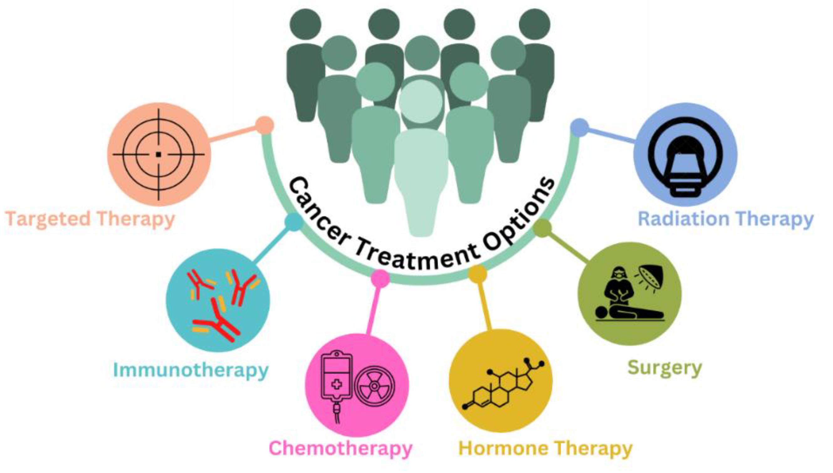
2. Conventional Chemotherapeutic Treatment
3. Obstacles Associated with Conventional Chemotherapeutic Agents
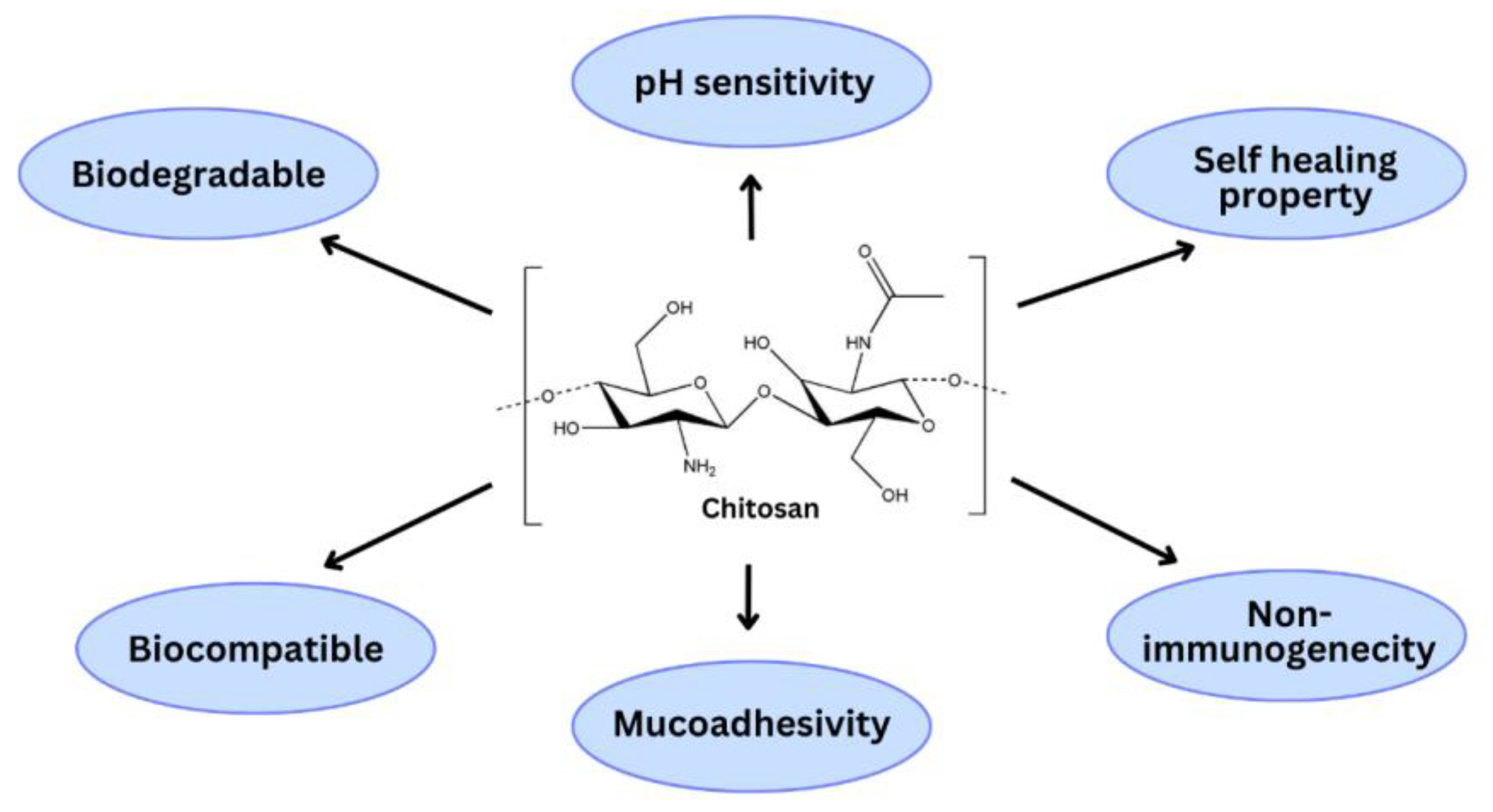
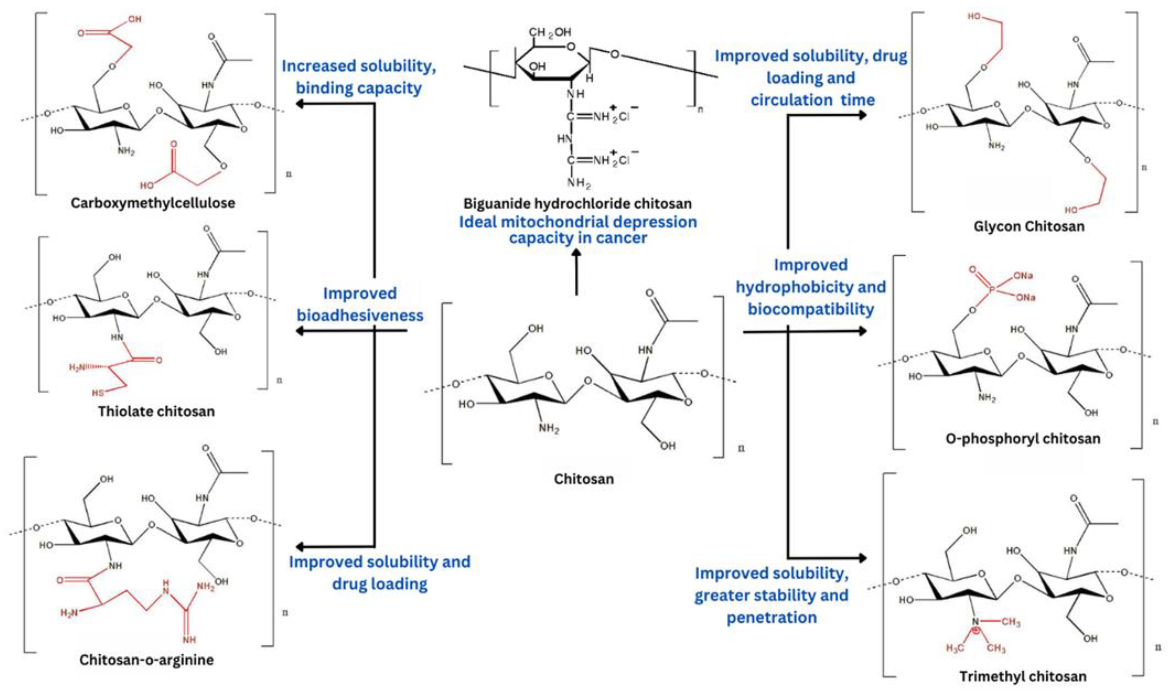
4. Chitosan and Its Derivatives for Anticancer Drug Delivery
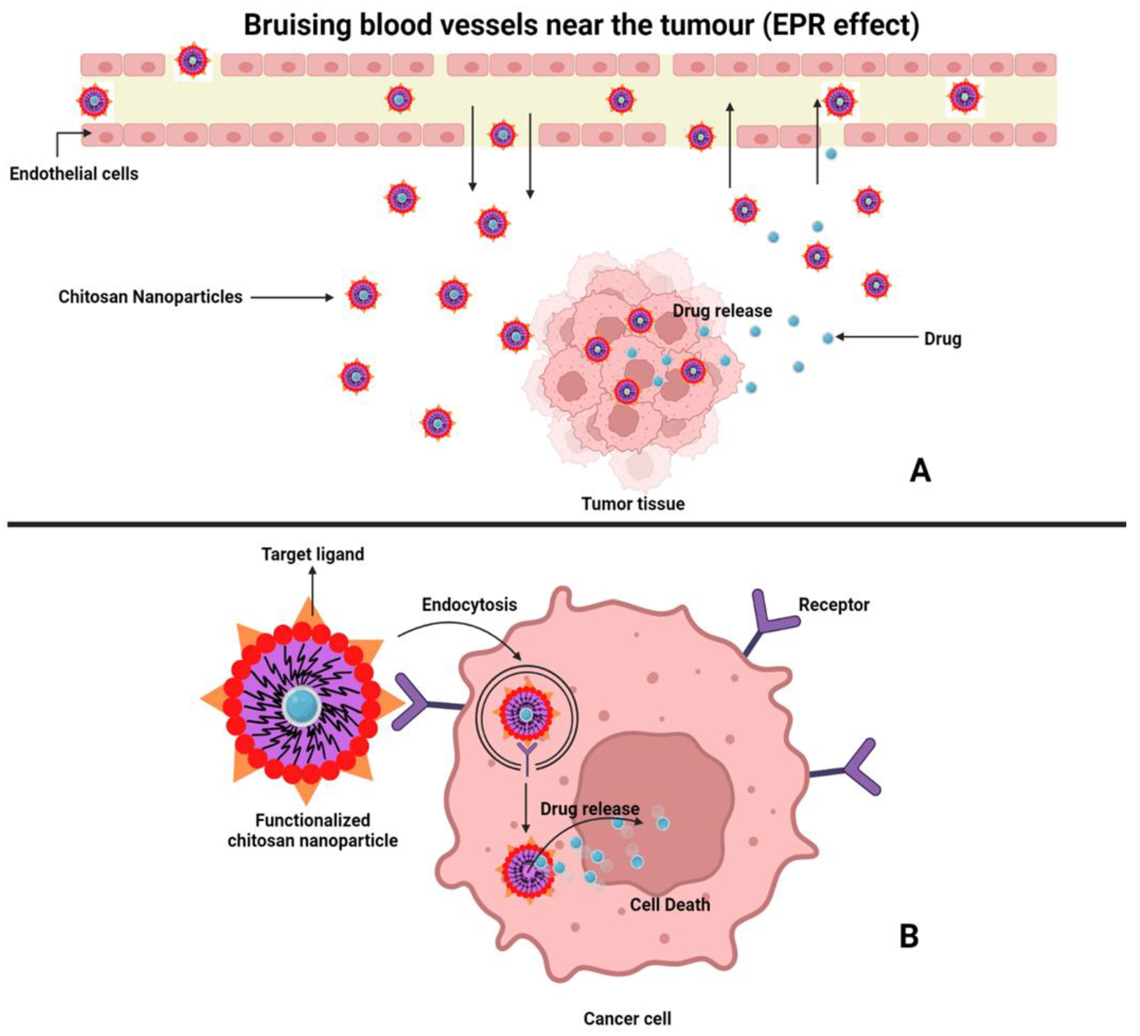
5. Chitosan Nanoparticles as Anticancer Drug Delivery System
6. Chitosan Nanoparticles (CSNPs) for Codelivery of Drugs in Cancer Treatment
7. Surface Modification of Nanocarriers Using Chitosan
8. Conclusions
Author Contributions
Funding
Institutional Review Board Statement
Data Availability Statement
Acknowledgments
Conflicts of Interest
References
- Grover, M.; Behl, T.; Virmani, T.; Sanduja, M.; Makeen, H.A.; Albratty, M.; Alhazmi, H.A.; Meraya, A.M.; Bungau, S.G. Exploration of Cytotoxic Potential of Longifolene/Junipene Isolated from Chrysopogon Zizanioides. Molecules 2022, 27, 5764. [Google Scholar] [CrossRef] [PubMed]
- Joyce Nirmala, M.; Kizhuveetil, U.; Johnson, A.; Balaji, G.; Nagarajan, R.; Muthuvijayan, V. Cancer Nanomedicine: A Review of Nano-Therapeutics and Challenges Ahead. RSC Adv. 2023, 13, 8606–8629. [Google Scholar] [CrossRef] [PubMed]
- Grover, M.; Behl, T.; Virmani, T. Phytochemical Screening, Antioxidant Assay and Cytotoxic Profile for Different Extracts of Chrysopogon Zizanioides Roots. Chem. Biodivers 2021, 18, e2100012. [Google Scholar] [CrossRef] [PubMed]
- Shah, S.C.; Kayamba, V.; Peek, R.M.; Heimburger, D. Cancer Control in Low- and Middle-Income Countries: Is It Time to Consider Screening? J. Glob. Oncol. 2019, 5, JGO.18.00200. [Google Scholar] [CrossRef]
- Brinks, J.; Fowler, A.; Franklin, B.A.; Dulai, J. Lifestyle Modification in Secondary Prevention: Beyond Pharmacotherapy. Am. J. Lifestyle Med. 2017, 11, 137–152. [Google Scholar] [CrossRef]
- Gao, Q.; Feng, J.; Liu, W.; Wen, C.; Wu, Y.; Liao, Q.; Zou, L.; Sui, X.; Xie, T.; Zhang, J.; et al. Opportunities and Challenges for Co-Delivery Nanomedicines Based on Combination of Phytochemicals with Chemotherapeutic Drugs in Cancer Treatment. Adv. Drug Deliv. Rev. 2022, 188, 114445. [Google Scholar] [CrossRef]
- Anand, U.; Dey, A.; Chandel, A.K.S.; Sanyal, R.; Mishra, A.; Pandey, D.K.; De Falco, V.; Upadhyay, A.; Kandimalla, R.; Chaudhary, A.; et al. Cancer Chemotherapy and beyond: Current Status, Drug Candidates, Associated Risks and Progress in Targeted Therapeutics. Genes Dis. 2022, 10, 1367–1401. [Google Scholar] [CrossRef]
- Kumar, G.; Virmani, T.; Sharma, A.; Pathak, K. Codelivery of Phytochemicals with Conventional Anticancer Drugs in Form of Nanocarriers. Pharmaceutics 2023, 15, 889. [Google Scholar] [CrossRef]
- Shakil, M.S.; Mahmud, K.M.; Sayem, M.; Niloy, M.S.; Halder, S.K.; Hossen, M.S.; Uddin, M.F.; Hasan, M.A. Using Chitosan or Chitosan Derivatives in Cancer Therapy. Polysaccharides 2021, 2, 795–816. [Google Scholar] [CrossRef]
- Kumar, G.; Khar, R.K.; Virmani, T.; Jogpal, V.; Virmani, R. Comparative Evaluation of Fast Dissolving Tablet of Atorvastatin Calcium Using Natural and Synthetic Super Disintegrating Agents. Res. J. Pharm. Technol. 2018, 11, 5001. [Google Scholar] [CrossRef]
- Kumar, G.; Virmani, T.; Pathak, K.; Alhalmi, A. A Revolutionary Blueprint for Mitigation of Hypertension via Nanoemulsion. BioMed Res. Int. 2022, 2022, e4109874. [Google Scholar] [CrossRef]
- Virmani, T.; Kumar, G.; Pathak, K. Non-Aqueous Nanoemulsions: An Innovative Lipid-Based Drug Carrier. Available online: https://www.igi-global.com/chapter/non-aqueous-nanoemulsions/www.igi-global.com/chapter/non-aqueous-nanoemulsions/300404 (accessed on 19 April 2022).
- Zahin, N.; Anwar, R.; Tewari, D.; Kabir, M.T.; Sajid, A.; Mathew, B.; Uddin, M.S.; Aleya, L.; Abdel-Daim, M.M. Nanoparticles and Its Biomedical Applications in Health and Diseases: Special Focus on Drug Delivery. Environ. Sci. Pollut. Res. 2020, 27, 19151–19168. [Google Scholar] [CrossRef]
- Sahu, T.; Ratre, Y.K.; Chauhan, S.; Bhaskar, L.V.K.S.; Nair, M.P.; Verma, H.K. Nanotechnology Based Drug Delivery System: Current Strategies and Emerging Therapeutic Potential for Medical Science. J. Drug Deliv. Sci. Technol. 2021, 63, 102487. [Google Scholar] [CrossRef]
- Patra, J.K.; Das, G.; Fraceto, L.F.; Campos, E.V.R.; Rodriguez-Torres, M.d.P.; Acosta-Torres, L.S.; Diaz-Torres, L.A.; Grillo, R.; Swamy, M.K.; Sharma, S.; et al. Nano Based Drug Delivery Systems: Recent Developments and Future Prospects. J Nanobiotechnol. 2018, 16, 71. [Google Scholar] [CrossRef]
- Naqvi, S.; Panghal, A.; Flora, S.J.S. Nanotechnology: A Promising Approach for Delivery of Neuroprotective Drugs. Front. Neurosci. 2020, 14, 494. [Google Scholar] [CrossRef]
- Virmani, T.; Kumar, G.; Virmani, R.; Sharma, A.; Pathak, K. Nanocarrier-Based Approaches to Combat Chronic Obstructive Pulmonary Disease. Nanomedicine 2022, 17, 1833–1854. [Google Scholar] [CrossRef]
- Kumar, G.; Virmani, T.; Pathak, K.; Kamaly, O.A.; Saleh, A. Central Composite Design Implemented Azilsartan Medoxomil Loaded Nanoemulsion to Improve Its Aqueous Solubility and Intestinal Permeability: In Vitro and Ex Vivo Evaluation. Pharmaceuticals 2022, 15, 1343. [Google Scholar] [CrossRef]
- Perumal, S. Polymer Nanoparticles: Synthesis and Applications. Polymers 2022, 14, 5449. [Google Scholar] [CrossRef]
- Zielińska, A.; Carreiró, F.; Oliveira, A.M.; Neves, A.; Pires, B.; Venkatesh, D.N.; Durazzo, A.; Lucarini, M.; Eder, P.; Silva, A.M.; et al. Polymeric Nanoparticles: Production, Characterization, Toxicology and Ecotoxicology. Molecules 2020, 25, 3731. [Google Scholar] [CrossRef]
- Colone, M.; Calcabrini, A.; Stringaro, A. Drug Delivery Systems of Natural Products in Oncology. Molecules 2020, 25, 4560. [Google Scholar] [CrossRef]
- Chang, D.; Ma, Y.; Xu, X.; Xie, J.; Ju, S. Stimuli-Responsive Polymeric Nanoplatforms for Cancer Therapy. Front. Bioeng. Biotechnol. 2021, 9, 707319. [Google Scholar] [CrossRef] [PubMed]
- Virmani, T.; Kumar, G.; Virmani, R.; Sharma, A.; Pathak, K. Xanthan Gum-Based Drug Delivery Systems for Respiratory Diseases. In Natural Polymeric Materials Based Drug Delivery Systems in Lung Diseases; Springer Nature Singapore: Singapore, 2023; pp. 279–295. ISBN 978-981-19765-5-1. [Google Scholar]
- Polat, M.; Polat, H. Recent Advances in Chitosan-Based Systems for Delivery of Anticancer Drugs. In Functional Chitosan: Drug Delivery and Biomedical Applications; Springer: Berlin/Heidelberg, Germany, 2019; pp. 191–228. ISBN 9789811502620. [Google Scholar]
- Herdiana, Y.; Wathoni, N.; Shamsuddin, S.; Joni, I.M.; Muchtaridi, M. Chitosan-Based Nanoparticles of Targeted Drug Delivery System in Breast Cancer Treatment. Polymers 2021, 13, 1717. [Google Scholar] [CrossRef] [PubMed]
- Almutairi, F.M.; Abd-Rabou, A.A.; Mohamed, M.S. Raloxifene-Encapsulated Hyaluronic Acid-Decorated Chitosan Nanoparticles Selectively Induce Apoptosis in Lung Cancer Cells. Bioorganic Med. Chem. 2019, 27, 1629–1638. [Google Scholar] [CrossRef] [PubMed]
- Ullah, S.; Azad, A.K.; Nawaz, A.; Shah, K.U.; Iqbal, M.; Albadrani, G.M.; Al-Joufi, F.A.; Sayed, A.A.; Abdel-Daim, M.M. 5-Fluorouracil-Loaded Folic-Acid-Fabricated Chitosan Nanoparticles for Site-Targeted Drug Delivery Cargo. Polymers 2022, 14, 2010. [Google Scholar] [CrossRef]
- Bosch, F.; Rosich, L. The Contributions of Paul Ehrlich to Pharmacology: A Tribute on the Occasion of the Centenary of His Nobel Prize. Pharmacology 2008, 82, 171–179. [Google Scholar] [CrossRef]
- Arruebo, M.; Vilaboa, N.; Sáez-Gutierrez, B.; Lambea, J.; Tres, A.; Valladares, M.; González-Fernández, Á. Assessment of the Evolution of Cancer Treatment Therapies. Cancers 2011, 3, 3279–3330. [Google Scholar] [CrossRef]
- Dehelean, C.A.; Marcovici, I.; Soica, C.; Mioc, M.; Coricovac, D.; Iurciuc, S.; Cretu, O.M.; Pinzaru, I. Plant-Derived Anticancer Compounds as New Perspectives in Drug Discovery and Alternative Therapy. Molecules 2021, 26, 1109. [Google Scholar] [CrossRef]
- Gupta, V.; Virmani, D.T.; Singh, D.V. A Comparative Diagnostic Account of the Roots of Boerhavia Diffusa Linn. From Four Different Geographical Regions in India. J. Community Pharm. Pract. (JCPP) 2799-1199 2022, 2, 1–6. [Google Scholar] [CrossRef]
- Talib, W.H.; Awajan, D.; Hamed, R.A.; Azzam, A.O.; Mahmod, A.I.; AL-Yasari, I.H. Combination Anticancer Therapies Using Selected Phytochemicals. Molecules 2022, 27, 5452. [Google Scholar] [CrossRef]
- Siddiqui, A.J.; Jahan, S.; Singh, R.; Saxena, J.; Ashraf, S.A.; Khan, A.; Choudhary, R.K.; Balakrishnan, S.; Badraoui, R.; Bardakci, F.; et al. Plants in Anticancer Drug Discovery: From Molecular Mechanism to Chemoprevention. BioMed Res. Int. 2022, 2022, e5425485. [Google Scholar] [CrossRef]
- Alqosaibi, A.I. Nanocarriers for Anticancer Drugs: Challenges and Perspectives. Saudi J. Biol. Sci. 2022, 29, 103298. [Google Scholar] [CrossRef]
- Gyanani, V.; Haley, J.C.; Goswami, R. Challenges of Current Anticancer Treatment Approaches with Focus on Liposomal Drug Delivery Systems. Pharmaceuticals 2021, 14, 835. [Google Scholar] [CrossRef]
- Mansoori, B.; Mohammadi, A.; Davudian, S.; Shirjang, S.; Baradaran, B. The Different Mechanisms of Cancer Drug Resistance: A Brief Review. Adv. Pharm. Bull. 2017, 7, 339–348. [Google Scholar] [CrossRef]
- ALTUN, İ.; SONKAYA, A. The Most Common Side Effects Experienced by Patients Were Receiving First Cycle of Chemotherapy. Iran J. Public Health 2018, 47, 1218–1219. [Google Scholar]
- Edis, Z.; Wang, J.; Waqas, M.K.; Ijaz, M.; Ijaz, M. Nanocarriers-Mediated Drug Delivery Systems for Anticancer Agents: An Overview and Perspectives. Int. J. Nanomed. 2021, 16, 1313–1330. [Google Scholar] [CrossRef]
- Manzari, M.T.; Shamay, Y.; Kiguchi, H.; Rosen, N.; Scaltriti, M.; Heller, D.A. Targeted Drug Delivery Strategies for Precision Medicines. Nat. Rev. Mater. 2021, 6, 351–370. [Google Scholar] [CrossRef]
- Senapati, S.; Mahanta, A.K.; Kumar, S.; Maiti, P. Controlled Drug Delivery Vehicles for Cancer Treatment and Their Performance. Signal Transduct. Target Ther. 2018, 3, 7. [Google Scholar] [CrossRef]
- Xiao, X.; Teng, F.; Shi, C.; Chen, J.; Wu, S.; Wang, B.; Meng, X.; Essiet Imeh, A.; Li, W. Polymeric Nanoparticles—Promising Carriers for Cancer Therapy. Front. Bioeng. Biotechnol. 2022, 10, 1024143. [Google Scholar] [CrossRef]
- Herdiana, Y.; Wathoni, N.; Gozali, D.; Shamsuddin, S.; Muchtaridi, M. Chitosan-Based Nano-Smart Drug Delivery System in Breast Cancer Therapy. Pharmaceutics 2023, 15, 879. [Google Scholar] [CrossRef]
- Karam, M.; Fahs, D.; Maatouk, B.; Safi, B.; Jaffa, A.A.; Mhanna, R. Polymeric Nanoparticles in the Diagnosis and Treatment of Myocardial Infarction: Challenges and Future Prospects. Mater. Today Bio 2022, 14, 100249. [Google Scholar] [CrossRef]
- Díez-Pascual, A.M. Surface Engineering of Nanomaterials with Polymers, Biomolecules, and Small Ligands for Nanomedicine. Materials 2022, 15, 3251. [Google Scholar] [CrossRef] [PubMed]
- Komsthöft, T.; Bovone, G.; Bernhard, S.; Tibbitt, M.W. Polymer Functionalization of Inorganic Nanoparticles for Biomedical Applications. Curr. Opin. Chem. Eng. 2022, 37, 100849. [Google Scholar] [CrossRef]
- Aranaz, I.; Alcántara, A.R.; Civera, M.C.; Arias, C.; Elorza, B.; Heras Caballero, A.; Acosta, N. Chitosan: An Overview of Its Properties and Applications. Polymers 2021, 13, 3256. [Google Scholar] [CrossRef] [PubMed]
- Lim, C.; Hwang, D.S.; Lee, D.W. Intermolecular Interactions of Chitosan: Degree of Acetylation and Molecular Weight. Carbohydr. Polym. 2021, 259, 117782. [Google Scholar] [CrossRef]
- Elgadir, M.A.; Uddin, M.S.; Ferdosh, S.; Adam, A.; Chowdhury, A.J.K.; Sarker, M.Z.I. Impact of Chitosan Composites and Chitosan Nanoparticle Composites on Various Drug Delivery Systems: A Review. J. Food Drug Anal. 2015, 23, 619–629. [Google Scholar] [CrossRef]
- Chen, Y.-Z.; Huang, Y.-K.; Chen, Y.; Ye, Y.-J.; Lou, K.-Y.; Gao, F. Novel Nanoparticles Composed of Chitosan and β-Cyclodextrin Derivatives as Potential Insoluble Drug Carrier. Chin. Chem. Lett. 2015, 26, 909–913. [Google Scholar] [CrossRef]
- Mikušová, V.; Mikuš, P. Advances in Chitosan-Based Nanoparticles for Drug Delivery. Int. J. Mol. Sci. 2021, 22, 9652. [Google Scholar] [CrossRef]
- Jafernik, K.; Ładniak, A.; Blicharska, E.; Czarnek, K.; Ekiert, H.; Wiącek, A.E.; Szopa, A. Chitosan-Based Nanoparticles as Effective Drug Delivery Systems—A Review. Molecules 2023, 28, 1963. [Google Scholar] [CrossRef]
- Baharlouei, P.; Rahman, A. Chitin and Chitosan: Prospective Biomedical Applications in Drug Delivery, Cancer Treatment, and Wound Healing. Mar. Drugs 2022, 20, 460. [Google Scholar] [CrossRef]
- Ways, T.M.; Lau, W.; Khutoryanskiy, V. Chitosan and Its Derivatives for Application in Mucoadhesive Drug Delivery Systems. Polymers 2018, 10, 267. [Google Scholar] [CrossRef]
- Kumar, S.; Deepak, V.; Kumari, M.; Dutta, P. Antibacterial Activity of Diisocyanate-Modified Chitosan for Biomedical Applications. Int. J. Biol. Macromol. 2015, 84, 349–353. [Google Scholar] [CrossRef]
- Xing, L.; Fan, Y.-T.; Zhou, T.-J.; Gong, J.-H.; Cui, L.-H.; Cho, K.-H.; Choi, Y.-J.; Jiang, H.-L.; Cho, C.-S. Chemical Modification of Chitosan for Efficient Vaccine Delivery. Molecules 2018, 23, 229. [Google Scholar] [CrossRef]
- Pellis, A.; Guebitz, G.M.; Nyanhongo, G.S. Chitosan: Sources, Processing and Modification Techniques. Gels 2022, 8, 393. [Google Scholar] [CrossRef]
- Chen, Q.; Qi, Y.; Jiang, Y.; Quan, W.; Luo, H.; Wu, K.; Li, S.; Ouyang, Q. Progress in Research of Chitosan Chemical Modification Technologies and Their Applications. Mar. Drugs 2022, 20, 536. [Google Scholar] [CrossRef]
- Hamedi, H.; Moradi, S.; Hudson, S.M.; Tonelli, A.E. Chitosan Based Hydrogels and Their Applications for Drug Delivery in Wound Dressings: A Review. Carbohydr. Polym. 2018, 199, 445–460. [Google Scholar] [CrossRef]
- Ali, A.; Ahmed, S. A Review on Chitosan and Its Nanocomposites in Drug Delivery. Int. J. Biol. Macromol. 2018, 109, 273–286. [Google Scholar] [CrossRef]
- Safdar, R.; Omar, A.A.; Arunagiri, A.; Regupathi, I.; Thanabalan, M. Potential of Chitosan and Its Derivatives for Controlled Drug Release Applications—A Review. J. Drug Deliv. Sci. Technol. 2019, 49, 642–659. [Google Scholar] [CrossRef]
- Zamboulis, A.; Nanaki, S.; Michailidou, G.; Koumentakou, I.; Lazaridou, M.; Ainali, N.M.; Xanthopoulou, E.; Bikiaris, D.N. Chitosan and Its Derivatives for Ocular Delivery Formulations: Recent Advances and Developments. Polymers 2020, 12, 1519. [Google Scholar] [CrossRef]
- Arya, G.; Gupta, N.; Nimesh, S. 8—Chitosan Nanoparticles for Therapeutic Delivery of Anticancer Drugs. In Polysaccharide Nanoparticles; Micro and Nano Technologies; Venkatesan, J., Kim, S.-K., Anil, S.P.d.R., Eds.; Elsevier: Amsterdam, The Netherlands, 2022; pp. 201–230. ISBN 978-0-12-822351-2. [Google Scholar]
- Kurczewska, J. Chitosan-Based Nanoparticles with Optimized Parameters for Targeted Delivery of a Specific Anticancer Drug—A Comprehensive Review. Pharmaceutics 2023, 15, 503. [Google Scholar] [CrossRef]
- Botelho Da Silva, S.; Krolicka, M.; Van Den Broek, L.A.M.; Frissen, A.E.; Boeriu, C.G. Water-Soluble Chitosan Derivatives and PH-Responsive Hydrogels by Selective C-6 Oxidation Mediated by TEMPO-Laccase Redox System. Carbohydr. Polym. 2018, 186, 299–309. [Google Scholar] [CrossRef]
- Babu, A.; Ramesh, R. Multifaceted Applications of Chitosan in Cancer Drug Delivery and Therapy. Mar. Drugs 2017, 15, 96. [Google Scholar] [CrossRef] [PubMed]
- Jaiswal, S.; Dutta, P.K.; Kumar, S.; Chawla, R. Chitosan Modified by Organo-Functionalities as an Efficient Nanoplatform for Anti-Cancer Drug Delivery Process. J. Drug Deliv. Sci. Technol. 2021, 62, 102407. [Google Scholar] [CrossRef]
- Carroll, E.C.; Jin, L.; Mori, A.; Muñoz-Wolf, N.; Oleszycka, E.; Moran, H.B.T.; Mansouri, S.; McEntee, C.P.; Lambe, E.; Agger, E.M.; et al. The Vaccine Adjuvant Chitosan Promotes Cellular Immunity via DNA Sensor CGAS-STING-Dependent Induction of Type I Interferons. Immunity 2016, 44, 597–608. [Google Scholar] [CrossRef] [PubMed]
- Li, W.; Zhu, X.; Zhou, X.; Wang, X.; Zhai, W.; Li, B.; Du, J.; Li, G.; Sui, X.; Wu, Y.; et al. An Orally Available PD-1/PD-L1 Blocking Peptide OPBP-1-Loaded Trimethyl Chitosan Hydrogel for Cancer Immunotherapy. J. Control. Release 2021, 334, 376–388. [Google Scholar] [CrossRef]
- Esmaily, M.; Masjedi, A.; Hallaj, S.; Nabi Afjadi, M.; Malakotikhah, F.; Ghani, S.; Ahmadi, A.; Sojoodi, M.; Hassannia, H.; Atyabi, F.; et al. Blockade of CTLA-4 Increases Anti-Tumor Response Inducing Potential of Dendritic Cell Vaccine. J Control. Release 2020, 326, 63–74. [Google Scholar] [CrossRef]
- Narmani, A.; Jafari, S.M. Chitosan-Based Nanodelivery Systems for Cancer Therapy: Recent Advances. Carbohydr. Polym. 2021, 272, 118464. [Google Scholar] [CrossRef]
- Abruzzo, A.; Zuccheri, G.; Belluti, F.; Provenzano, S.; Verardi, L.; Bigucci, F.; Cerchiara, T.; Luppi, B.; Calonghi, N. Chitosan Nanoparticles for Lipophilic Anticancer Drug Delivery: Development, Characterization and in Vitro Studies on HT29 Cancer Cells. Colloids Surf. B Biointerfaces 2016, 145, 362–372. [Google Scholar] [CrossRef]
- Chen, G.; Svirskis, D.; Lu, W.; Ying, M.; Huang, Y.; Wen, J. N-Trimethyl Chitosan Nanoparticles and CSKSSDYQC Peptide: N-Trimethyl Chitosan Conjugates Enhance the Oral Bioavailability of Gemcitabine to Treat Breast Cancer. J. Control. Release 2018, 277, 142–153. [Google Scholar] [CrossRef]
- Vllasaliu, D.; Exposito-Harris, R.; Heras, A.; Casettari, L.; Garnett, M.; Illum, L.; Stolnik, S. Tight Junction Modulation by Chitosan Nanoparticles: Comparison with Chitosan Solution. Int. J. Pharm. 2010, 400, 183–193. [Google Scholar] [CrossRef]
- Alhodieb, F.S.; Barkat, M.A.; Barkat, H.A.; Hadi, H.A.; Khan, M.I.; Ashfaq, F.; Rahman, M.A.; Hassan, M.Z.; Alanezi, A.A. Chitosan-Modified Nanocarriers as Carriers for Anticancer Drug Delivery: Promises and Hurdles. Int. J. Biol. Macromol. 2022, 217, 457–469. [Google Scholar] [CrossRef]
- Jha, R.; Mayanovic, R.A. A Review of the Preparation, Characterization, and Applications of Chitosan Nanoparticles in Nanomedicine. Nanomaterials 2023, 13, 1302. [Google Scholar] [CrossRef]
- Hossen, S.; Hossain, M.K.; Basher, M.K.; Mia, M.N.H.; Rahman, M.T.; Uddin, M.J. Smart Nanocarrier-Based Drug Delivery Systems for Cancer Therapy and Toxicity Studies: A Review. J. Adv. Res. 2019, 15, 1–18. [Google Scholar] [CrossRef]
- Abd Ellah, N.H.; Abouelmagd, S.A. Surface Functionalization of Polymeric Nanoparticles for Tumor Drug Delivery: Approaches and Challenges. Expert Opin. Drug Deliv. 2017, 14, 201–214. [Google Scholar] [CrossRef]
- Batlle, R.; Andrés, E.; Gonzalez, L.; Llonch, E.; Igea, A.; Gutierrez-Prat, N.; Berenguer-Llergo, A.; Nebreda, A.R. Regulation of Tumor Angiogenesis and Mesenchymal-Endothelial Transition by P38α through TGF-β and JNK Signaling. Nat. Commun. 2019, 10, 3071. [Google Scholar] [CrossRef]
- Yermak, I.M.; Davydova, V.N.; Volod’ko, A.V. Mucoadhesive Marine Polysaccharides. Mar. Drugs 2022, 20, 522. [Google Scholar] [CrossRef]
- Bae, Y.H.; Park, K. Advanced Drug Delivery 2020 and beyond: Perspectives on the Future. Adv. Drug Deliv. Rev. 2020, 158, 4–16. [Google Scholar] [CrossRef]
- Fang, J.; Islam, W.; Maeda, H. Exploiting the Dynamics of the EPR Effect and Strategies to Improve the Therapeutic Effects of Nanomedicines by Using EPR Effect Enhancers. Adv. Drug Deliv. Rev. 2020, 157, 142–160. [Google Scholar] [CrossRef]
- Mohammed, M.A.; Syeda, J.T.M.; Wasan, K.M.; Wasan, E.K. An Overview of Chitosan Nanoparticles and Its Application in Non-Parenteral Drug Delivery. Pharmaceutics 2017, 9, 53. [Google Scholar] [CrossRef]
- Zhao, X.; Si, J.; Huang, D.; Li, K.; Xin, Y.; Sui, M. Application of Star Poly(Ethylene Glycol) Derivatives in Drug Delivery and Controlled Release. J. Control. Release 2020, 323, 565–577. [Google Scholar] [CrossRef]
- Zhou, Z.; Liu, Y.; Jiang, X.; Zheng, C.; Luo, W.; Xiang, X.; Qi, X.; Shen, J. Metformin Modified Chitosan as a Multi-Functional Adjuvant to Enhance Cisplatin-Based Tumor Chemotherapy Efficacy. Int. J. Biol. Macromol. 2023, 224, 797–809. [Google Scholar] [CrossRef]
- Zhou, Z.; Zheng, C.; Liu, Y.; Luo, W.; Deng, H.; Shen, J. Chitosan Biguanide Induced Mitochondrial Inhibition to Amplify the Efficacy of Oxygen-Sensitive Tumor Therapies. Carbohydr. Polym. 2022, 295, 119878. [Google Scholar] [CrossRef] [PubMed]
- Chen, J.; Zhou, Z.; Zheng, C.; Liu, Y.; Hao, R.; Ji, X.; Xi, Q.; Shen, J.; Li, Z. Chitosan Oligosaccharide Regulates AMPK and STAT1 Pathways Synergistically to Mediate PD-L1 Expression for Cancer Chemoimmunotherapy. Carbohydr. Polym. 2022, 277, 118869. [Google Scholar] [CrossRef] [PubMed]
- Cai, X.; Feng, J.; Chen, F.; Guo, C.; Sun, L.; Li, L. Synergistic Effect of Glycated Chitosan and Photofrin Photodynamic Therapy on Different Breast Tumor Model. Photodiagnosis Photodyn. Ther. 2020, 31, 101842. [Google Scholar] [CrossRef] [PubMed]
- Ding, J.; Guo, Y. Recent Advances in Chitosan and Its Derivatives in Cancer Treatment. Front. Pharm. 2022, 13, 888740. [Google Scholar] [CrossRef]
- Li, X.; Dong, W.; Nalin, A.P.; Wang, Y.; Pan, P.; Xu, B.; Zhang, Y.; Tun, S.; Zhang, J.; Wang, L.-S.; et al. The Natural Product Chitosan Enhances the Anti-Tumor Activity of Natural Killer Cells by Activating Dendritic Cells. Oncoimmunology 2018, 7, e1431085. [Google Scholar] [CrossRef]
- Mortezaee, K.; Narmani, A.; Salehi, M.; Bagheri, H.; Farhood, B.; Haghi-Aminjan, H.; Najafi, M. Synergic Effects of Nanoparticles-Mediated Hyperthermia in Radiotherapy/Chemotherapy of Cancer. Life Sci. 2021, 269, 119020. [Google Scholar] [CrossRef]
- Fernández, M.; Javaid, F.; Chudasama, V. Advances in Targeting the Folate Receptor in the Treatment/Imaging of Cancers. Chem. Sci. 2018, 9, 790–810. [Google Scholar] [CrossRef]
- Narmani, A.; Rezvani, M.; Farhood, B.; Darkhor, P.; Mohammadnejad, J.; Amini, B.; Refahi, S.; Abdi Goushbolagh, N. Folic Acid Functionalized Nanoparticles as Pharmaceutical Carriers in Drug Delivery Systems. Drug Dev. Res. 2019, 80, 404–424. [Google Scholar] [CrossRef]
- Dunn, M.R.; Jimenez, R.M.; Chaput, J.C. Analysis of Aptamer Discovery and Technology. Nat. Rev. Chem. 2017, 1, 0076. [Google Scholar] [CrossRef]
- Tang, Y.; Wu, S.; Lin, J.; Cheng, L.; Zhou, J.; Xie, J.; Huang, K.; Wang, X.; Yu, Y.; Chen, Z.; et al. Nanoparticles Targeted against Cryptococcal Pneumonia by Interactions between Chitosan and Its Peptide Ligand. Nano Lett. 2018, 18, 6207–6213. [Google Scholar] [CrossRef]
- Kamalabadi-Farahani, M.; Vasei, M.; Ahmadbeigi, N.; Ebrahimi-Barough, S.; Soleimani, M.; Roozafzoon, R. Anti-Tumour Effects of TRAIL-Expressing Human Placental Derived Mesenchymal Stem Cells with Curcumin-Loaded Chitosan Nanoparticles in a Mice Model of Triple Negative Breast Cancer. Artif. Cells Nanomed. Biotechnol. 2018, 46, S1011–S1021. [Google Scholar] [CrossRef]
- Kavaz, D.; Kirac, F.; Kirac, M.; Vaseashta, A. Low Releasing Mitomycin C Molecule Encapsulated with Chitosan Nanoparticles for Intravesical Installation. J. Biomater. Nanobiotechnol. 2017, 8, 203–219. [Google Scholar] [CrossRef]
- Komenek, S.; Luesakul, U.; Ekgasit, S.; Vilaivan, T.; Praphairaksit, N.; Puthong, S.; Muangsin, N. Nanogold-Gallate Chitosan-Targeted Pulmonary Delivery for Treatment of Lung Cancer. AAPS PharmSciTech. 2017, 18, 1104–1115. [Google Scholar] [CrossRef]
- Alkhader, E.; Billa, N.; Roberts, C.J. Mucoadhesive Chitosan-Pectinate Nanoparticles for the Delivery of Curcumin to the Colon. AAPS PharmSciTech. 2017, 18, 1009–1018. [Google Scholar] [CrossRef]
- Gupta, U.; Sharma, S.; Khan, I.; Gothwal, A.; Sharma, A.K.; Singh, Y.; Chourasia, M.K.; Kumar, V. Enhanced Apoptotic and Anticancer Potential of Paclitaxel Loaded Biodegradable Nanoparticles Based on Chitosan. Int. J. Biol. Macromol. 2017, 98, 810–819. [Google Scholar] [CrossRef]
- Manivasagan, P.; Bharathiraja, S.; Bui, N.Q.; Lim, I.G.; Oh, J. Paclitaxel-Loaded Chitosan Oligosaccharide-Stabilized Gold Nanoparticles as Novel Agents for Drug Delivery and Photoacoustic Imaging of Cancer Cells. Int. J. Pharm. 2016, 511, 367–379. [Google Scholar] [CrossRef]
- Motawi, T.K.; El-Maraghy, S.A.; ElMeshad, A.N.; Nady, O.M.; Hammam, O.A. Cromolyn Chitosan Nanoparticles as a Novel Protective Approach for Colorectal Cancer. Chem. Biol. Interact. 2017, 275, 1–12. [Google Scholar] [CrossRef]
- Zare, M.; Mohammadi Samani, S.; Sobhani, Z. Enhanced Intestinal Permeation of Doxorubicin Using Chitosan Nanoparticles. Adv. Pharm. Bull. 2018, 8, 411–417. [Google Scholar] [CrossRef]
- Helmi, O.; Elshishiny, F.; Mamdouh, W. Targeted Doxorubicin Delivery and Release within Breast Cancer Environment Using PEGylated Chitosan Nanoparticles Labeled with Monoclonal Antibodies. Int. J. Biol. Macromol. 2021, 184, 325–338. [Google Scholar] [CrossRef]
- Farrag, N.S.; Shetta, A.; Mamdouh, W. Green Tea Essential Oil Encapsulated Chitosan Nanoparticles-Based Radiopharmaceutical as a New Trend for Solid Tumor Theranosis. Int. J. Biol. Macromol. 2021, 186, 811–819. [Google Scholar] [CrossRef]
- Yadav, A.S.; Radharani, N.N.V.; Gorain, M.; Bulbule, A.; Shetti, D.; Roy, G.; Baby, T.; Kundu, G.C. RGD Functionalized Chitosan Nanoparticle Mediated Targeted Delivery of Raloxifene Selectively Suppresses Angiogenesis and Tumor Growth in Breast Cancer. Nanoscale 2020, 12, 10664–10684. [Google Scholar] [CrossRef] [PubMed]
- Murthy, A.; Ravi, P.R.; Kathuria, H.; Vats, R. Self-Assembled Lecithin-Chitosan Nanoparticles Improve the Oral Bioavailability and Alter the Pharmacokinetics of Raloxifene. Int. J. Pharm. 2020, 588, 119731. [Google Scholar] [CrossRef] [PubMed]
- Zhang, W.; Xu, W.; Lan, Y.; He, X.; Liu, K.; Liang, Y. Antitumor Effect of Hyaluronic-Acid-Modified Chitosan Nanoparticles Loaded with SiRNA for Targeted Therapy for Non-Small Cell Lung Cancer. Int. J. Nanomed. 2019, 14, 5287–5301. [Google Scholar] [CrossRef] [PubMed]
- Jain, A.; Thakur, K.; Kush, P.; Jain, U.K. Docetaxel Loaded Chitosan Nanoparticles: Formulation, Characterization and Cytotoxicity Studies. Int. J. Biol. Macromol. 2014, 69, 546–553. [Google Scholar] [CrossRef]
- Mirzaie, Z.H.; Irani, S.; Mirfakhraie, R.; Atyabi, S.M.; Dinarvand, M.; Dinarvand, R.; Varshochian, R.; Atyabi, F. Docetaxel-Chitosan Nanoparticles for Breast Cancer Treatment: Cell Viability and Gene Expression Study. Chem. Biol. Drug Des. 2016, 88, 850–858. [Google Scholar] [CrossRef]
- Hua, S.; Yu, J.; Shang, J.; Zhang, H.; Du, J.; Zhang, Y.; Chen, F.; Zhou, Y.; Liu, F. Effective Tumor-Targeted Delivery of Etoposide Using Chitosan Nanoparticles Conjugated with Folic Acid and Sulfobetaine Methacrylate. RSC Adv. 2016, 6, 91192–91200. [Google Scholar] [CrossRef]
- Barbosa, A.I.; Costa Lima, S.A.; Reis, S. Development of Methotrexate Loaded Fucoidan/Chitosan Nanoparticles with Anti-Inflammatory Potential and Enhanced Skin Permeation. Int. J. Biol. Macromol. 2019, 124, 1115–1122. [Google Scholar] [CrossRef]
- Ekinci, M.; Ilem-Ozdemir, D.; Gundogdu, E.; Asikoglu, M. Methotrexate Loaded Chitosan Nanoparticles: Preparation, Radiolabeling and in Vitro Evaluation for Breast Cancer Diagnosis. J. Drug Deliv. Sci. Technol. 2015, 30, 107–113. [Google Scholar] [CrossRef]
- Sultan, M.H.; Moni, S.S.; Madkhali, O.A.; Bakkari, M.A.; Alshahrani, S.; Alqahtani, S.S.; Alhakamy, N.A.; Mohan, S.; Ghazwani, M.; Bukhary, H.A.; et al. Characterization of Cisplatin-Loaded Chitosan Nanoparticles and Rituximab-Linked Surfaces as Target-Specific Injectable Nano-Formulations for Combating Cancer. Sci. Rep. 2022, 12, 468. [Google Scholar] [CrossRef]
- Sun, L.; Chen, Y.; Zhou, Y.; Guo, D.; Fan, Y.; Guo, F.; Zheng, Y.; Chen, W. Preparation of 5-Fluorouracil-Loaded Chitosan Nanoparticles and Study of the Sustained Release in Vitro and in Vivo. Asian J. Pharm. Sci. 2017, 12, 418–423. [Google Scholar] [CrossRef]
- Patel, G.; Yadav, B.K.N. Study of 5-Fluorouracil Loaded Chitosan Nanoparticles for Treatment of Skin Cancer. Recent Pat. Nanotechnol. 2020, 14, 210–224. [Google Scholar] [CrossRef]
- Shayegh, A.; Khalatbari, F.; Zonoubi, N.; Zarazvand, F.; Monavvari, F.; Hejazinia, H.; Ebrahimi, S.E.S.; Hamedani, M.P.; Ali, V.; Hadadian, S.; et al. Chlorambucil-Chitosan Nano-Conjugate: An Efficient Agent Against Breast Cancer Targeted Therapy. Curr. Drug Deliv. 2021, 18, 721–728. [Google Scholar] [CrossRef]
- Kaur, H.; Mishra, N.; Khurana, B.; Kaur, S.; Arora, D. DoE Based Optimization and Development of Spray-Dried Chitosan-Coated Alginate Microparticles Loaded with Cisplatin for the Treatment of Cervical Cancer. Curr. Mol. Pharm. 2021, 14, 381–398. [Google Scholar] [CrossRef]
- Hemmati, K.; Ahmadi Nasab, N.; Hesaraki, S.; Nezafati, N. In Vitro Evaluation of Curcumin-Loaded Chitosan-Coated Hydroxyapatite Nanocarriers as a Potential System for Effective Treatment of Cancer. J. Biomater. Sci. Polym. Ed. 2021, 32, 1267–1287. [Google Scholar] [CrossRef]
- Nokhodi, F.; Nekoei, M.; Goodarzi, M.T. Hyaluronic Acid-Coated Chitosan Nanoparticles as Targeted-Carrier of Tamoxifen against MCF7 and TMX-Resistant MCF7 Cells. J. Mater. Sci. Mater. Med. 2022, 33, 24. [Google Scholar] [CrossRef]
- Cannavà, C.; De Gaetano, F.; Stancanelli, R.; Venuti, V.; Paladini, G.; Caridi, F.; Ghica, C.; Crupi, V.; Majolino, D.; Ferlazzo, G.; et al. Chitosan-Hyaluronan Nanoparticles for Vinblastine Sulfate Delivery: Characterization and Internalization Studies on K-562 Cells. Pharmaceutics 2022, 14, 942. [Google Scholar] [CrossRef]
- Gooneh-Farahani, S.; Naghib, S.M.; Naimi-Jamal, M.R.; Seyfoori, A. A PH-Sensitive Nanocarrier Based on BSA-Stabilized Graphene-Chitosan Nanocomposite for Sustained and Prolonged Release of Anticancer Agents. Sci. Rep. 2021, 11, 17404. [Google Scholar] [CrossRef]
- Yi, G.; Ling, J.; Jiang, Y.; Lu, Y.; Yang, L.-Y.; Ouyang, X. Fabrication, Characterization, and in Vitro Evaluation of Doxorubicin-Coupled Chitosan Oligosaccharide Nanoparticles. J. Mol. Struct. 2022, 1268, 133688. [Google Scholar] [CrossRef]
- Scheeren, L.E.; Nogueira, D.R.; Macedo, L.B.; Vinardell, M.P.; Mitjans, M.; Infante, M.R.; Rolim, C.M.B. PEGylated and Poloxamer-Modified Chitosan Nanoparticles Incorporating a Lysine-Based Surfactant for PH-Triggered Doxorubicin Release. Colloids Surf. B Biointerfaces 2016, 138, 117–127. [Google Scholar] [CrossRef]
- Varukattu, N.B.; Vivek, R.; Rejeeth, C.; Thangam, R.; Ponraj, T.; Sharma, A.; Kannan, S. Nanostructured PH-Responsive Biocompatible Chitosan Coated Copper Oxide Nanoparticles: A Polymeric Smart Intracellular Delivery System for Doxorubicin in Breast Cancer Cells. Arab. J. Chem. 2020, 13, 2276–2286. [Google Scholar] [CrossRef]
- Luckanagul, J.A.; Pitakchatwong, C.; Ratnatilaka Na Bhuket, P.; Muangnoi, C.; Rojsitthisak, P.; Chirachanchai, S.; Wang, Q.; Rojsitthisak, P. Chitosan-Based Polymer Hybrids for Thermo-Responsive Nanogel Delivery of Curcumin. Carbohydr. Polym. 2018, 181, 1119–1127. [Google Scholar] [CrossRef] [PubMed]
- Hu, Y.; Du, Y.; Liu, N.; Liu, X.; Meng, T.; Cheng, B.; He, J.; You, J.; Yuan, H.; Hu, F. Selective Redox-Responsive Drug Release in Tumor Cells Mediated by Chitosan Based Glycolipid-like Nanocarrier. J. Control. Release 2015, 206, 91–100. [Google Scholar] [CrossRef] [PubMed]
- Xu, W.; Wang, H.; Dong, L.; Zhang, P.; Mu, Y.; Cui, X.; Zhou, J.; Huo, M.; Yin, T. Hyaluronic Acid-Decorated Redox-Sensitive Chitosan Micelles for Tumor-Specific Intracellular Delivery of Gambogic Acid. Int. J. Nanomed. 2019, 14, 4649–4666. [Google Scholar] [CrossRef] [PubMed]
- Qi, M.; Li, G.; Yu, N.; Meng, Y.; Liu, X. Synthesis of Thermo-Sensitive Polyelectrolyte Complex Nanoparticles from CS-g-PNIPAM and SA-g-PNIPAM for Controlled Drug Release. Macromol. Res. 2014, 22, 1004–1011. [Google Scholar] [CrossRef]
- Yan, L.; Crayton, S.H.; Thawani, J.P.; Amirshaghaghi, A.; Tsourkas, A.; Cheng, Z. A PH-Responsive Drug-Delivery Platform Based on Glycol Chitosan–Coated Liposomes. Small 2015, 11, 4870–4874. [Google Scholar] [CrossRef]
- Mansouri, M.; Nazarpak, M.H.; Solouk, A.; Akbari, S.; Hasani-Sadrabadi, M.M. Magnetic Responsive of Paclitaxel Delivery System Based on SPION and Palmitoyl Chitosan. J. Magn. Magn. Mater. 2017, 421, 316–325. [Google Scholar] [CrossRef]
- Zhong, S.; Zhang, H.; Liu, Y.; Wang, G.; Shi, C.; Li, Z.; Feng, Y.; Cui, X. Folic Acid Functionalized Reduction-Responsive Magnetic Chitosan Nanocapsules for Targeted Delivery and Triggered Release of Drugs. Carbohydr. Polym. 2017, 168, 282–289. [Google Scholar] [CrossRef]
- Meng, L.; Huang, W.; Wang, D.; Huang, X.; Zhu, X.; Yan, D. Chitosan-Based Nanocarriers with PH and Light Dual Response for Anticancer Drug Delivery. Biomacromolecules 2013, 14, 2601–2610. [Google Scholar] [CrossRef]
- El-Leithy, E.S.; Hassan, S.A.; Abdel-Rashid, R.S. Tamoxifen Citrate/Coenzyme Q10 as Smart Nanocarriers Bitherapy for Breast Cancer: Cytotoxicity, Genotoxicity, and Antioxidant Activity. J. Drug Deliv. Sci. Technol. 2019, 51, 36–44. [Google Scholar] [CrossRef]
- Li, P.; Wang, Y.; Peng, Z.; She, F.; Kong, L. Development of Chitosan Nanoparticles as Drug Delivery Systems for 5-Fluorouracil and Leucovorin Blends. Carbohydr. Polym. 2011, 85, 698–704. [Google Scholar] [CrossRef]
- Jia, M.; Li, Y.; Yang, X.; Huang, Y.; Wu, H.; Huang, Y.; Lin, J.; Li, Y.; Hou, Z.; Zhang, Q. Development of Both Methotrexate and Mitomycin C Loaded PEGylated Chitosan Nanoparticles for Targeted Drug Codelivery and Synergistic Anticancer Effect. ACS Appl. Mater. Interfaces 2014, 6, 11413–11423. [Google Scholar] [CrossRef]
- Khan, M.M.; Madni, A.; Tahir, N.; Parveen, F.; Khan, S.; Jan, N.; Ali, A.; Abdurrahim, M.; Farooq, U.; Khan, M.I. Co-Delivery of Curcumin and Cisplatin to Enhance Cytotoxicity of Cisplatin Using Lipid-Chitosan Hybrid Nanoparticles. Int. J. Nanomed. 2020, 15, 2207. [Google Scholar] [CrossRef]
- Deng, X.; Cao, M.; Zhang, J.; Hu, K.; Yin, Z.; Zhou, Z.; Xiao, X.; Yang, Y.; Sheng, W.; Wu, Y.; et al. Hyaluronic Acid-Chitosan Nanoparticles for Co-Delivery of MiR-34a and Doxorubicin in Therapy against Triple Negative Breast Cancer. Biomaterials 2014, 35, 4333–4344. [Google Scholar] [CrossRef]
- Chen, G.; Zhao, Y.; Xu, Y.; Zhu, C.; Liu, T.; Wang, K. Chitosan Nanoparticles for Oral Photothermally Enhanced Photodynamic Therapy of Colon Cancer. Int. J. Pharm. 2020, 589, 119763. [Google Scholar] [CrossRef]
- Anitha, A.; Deepa, N.; Chennazhi, K.P.; Lakshmanan, V.-K.; Jayakumar, R. Combinatorial Anticancer Effects of Curcumin and 5-Fluorouracil Loaded Thiolated Chitosan Nanoparticles towards Colon Cancer Treatment. Biochim. Et Biophys. Acta (BBA) Gen. Subj. 2014, 1840, 2730–2743. [Google Scholar] [CrossRef]
- Sadreddini, S.; Safaralizadeh, R.; Baradaran, B.; Aghebati-Maleki, L.; Hosseinpour-Feizi, M.A.; Shanehbandi, D.; Jadidi-Niaragh, F.; Sadreddini, S.; Kafil, H.S.; Younesi, V.; et al. Chitosan Nanoparticles as a Dual Drug/SiRNA Delivery System for Treatment of Colorectal Cancer. Immunol. Lett. 2017, 181, 79–86. [Google Scholar] [CrossRef]
- Mohammadi, Z.; Samadi, F.Y.; Rahmani, S.; Mohammadi, Z. Chitosan-Raloxifene Nanoparticle Containing Doxorubicin as a New Double-Effect Targeting Vehicle for Breast Cancer Therapy. Daru 2020, 28, 433–442. [Google Scholar] [CrossRef]
- Zhu, X.; Yu, Z.; Feng, L.; Deng, L.; Fang, Z.; Liu, Z.; Li, Y.; Wu, X.; Qin, L.; Guo, R.; et al. Chitosan-Based Nanoparticle Co-Delivery of Docetaxel and Curcumin Ameliorates Anti-Tumor Chemoimmunotherapy in Lung Cancer. Carbohydr. Polym. 2021, 268, 118237. [Google Scholar] [CrossRef]
- Fahmy, S.A.; Ramzy, A.; Mandour, A.A.; Nasr, S.; Abdelnaser, A.; Bakowsky, U.; Azzazy, H.M.E.-S. PEGylated Chitosan Nanoparticles Encapsulating Ascorbic Acid and Oxaliplatin Exhibit Dramatic Apoptotic Effects against Breast Cancer Cells. Pharmaceutics 2022, 14, 407. [Google Scholar] [CrossRef]
- Xin, L.; Fan, J.-C.; Le, Y.-G.; Zeng, F.; Cheng, H.; Hu, X.; Cao, J.-Q. Construction of METHFR ShRNA/5-Fluorouracil Co-Loaded Folate-Targeted Chitosan Polymeric Nanoparticles and Its Anti-Carcinoma Effect on Gastric Cells Growth. J. Nanopart. Res. 2016, 18, 105. [Google Scholar] [CrossRef]
- Sebaaly, C.; Trifan, A.; Sieniawska, E.; Greige-Gerges, H. Chitosan-Coating Effect on the Characteristics of Liposomes: A Focus on Bioactive Compounds and Essential Oils: A Review. Processes 2021, 9, 445. [Google Scholar] [CrossRef]
- Priya, S.; Desai, V.M.; Singhvi, G. Surface Modification of Lipid-Based Nanocarriers: A Potential Approach to Enhance Targeted Drug Delivery. ACS Omega 2023, 8, 74–86. [Google Scholar] [CrossRef] [PubMed]
- Ganesan, P.; Ramalingam, P.; Karthivashan, G.; Ko, Y.T.; Choi, D.-K. Recent Developments in Solid Lipid Nanoparticle and Surface-Modified Solid Lipid Nanoparticle Delivery Systems for Oral Delivery of Phyto-Bioactive Compounds in Various Chronic Diseases. IJN 2018, 13, 1569–1583. [Google Scholar] [CrossRef]
- Luesakul, U.; Puthong, S.; Sansanaphongpricha, K.; Muangsin, N. Quaternized Chitosan-Coated Nanoemulsions: A Novel Platform for Improving the Stability, Anti-Inflammatory, Anti-Cancer and Transdermal Properties of Plai Extract. Carbohydr. Polym. 2020, 230, 115625. [Google Scholar] [CrossRef] [PubMed]
- Al-Nemrawi, N.K.; Okour, A.R.; Dave, R.H. Surface Modification of PLGA Nanoparticles Using Chitosan: Effect of Molecular Weight, Concentration, and Degree of Deacetylation. Adv. Polym. Technol. 2018, 37, 3066–3075. [Google Scholar] [CrossRef]
- Hasan, M.; Messaoud, G.B.; Michaux, F.; Tamayol, A.; Kahn, C.J.F.; Belhaj, N.; Linder, M.; Arab-Tehrany, E. Chitosan-Coated Liposomes Encapsulating Curcumin: Study of Lipid–Polysaccharide Interactions and Nanovesicle Behavior. RSC Adv. 2016, 6, 45290–45304. [Google Scholar] [CrossRef]
- Almeida, E.D.P.; Santos Silva, L.A.; de Araujo, G.R.S.; Montalvão, M.M.; Matos, S.S.; da Cunha Gonsalves, J.K.M.; de Souza Nunes, R.; de Meneses, C.T.; Oliveira Araujo, R.G.; Sarmento, V.H.V.; et al. Chitosan-Functionalized Nanostructured Lipid Carriers Containing Chloroaluminum Phthalocyanine for Photodynamic Therapy of Skin Cancer. Eur. J. Pharm. Biopharm. 2022, 179, 221–231. [Google Scholar] [CrossRef]
- Alomrani, A.; Badran, M.; Harisa, G.I.; ALshehry, M.; Alhariri, M.; Alshamsan, A.; Alkholief, M. The Use of Chitosan-Coated Flexible Liposomes as a Remarkable Carrier to Enhance the Antitumor Efficacy of 5-Fluorouracil against Colorectal Cancer. Saudi Pharm. J. 2019, 27, 603–611. [Google Scholar] [CrossRef]
- Bian, Y.; Gao, D.; Liu, Y.; Li, N.; Zhang, X.; Zheng, R.; Wang, Q.; Luo, L.; Dai, K. Preparation and Study on Anti-Tumor Effect of Chitosan-Coated Oleanolic Acid Liposomes. RSC Adv. 2015, 5. [Google Scholar] [CrossRef]
- Hasan, M.; Elkhoury, K.; Belhaj, N.; Kahn, C.; Tamayol, A.; Barberi-Heyob, M.; Arab-Tehrany, E.; Linder, M. Growth-Inhibitory Effect of Chitosan-Coated Liposomes Encapsulating Curcumin on MCF-7 Breast Cancer Cells. Mar. Drugs 2020, 18, 217. [Google Scholar] [CrossRef]
- Dawoud, M. Chitosan Coated Solid Lipid Nanoparticles as Promising Carriers for Docetaxel. J. Drug Deliv. Sci. Technol. 2021, 62, 102409. [Google Scholar] [CrossRef]
- Bhaskaran, N.A.; Jitta, S.R.; Salwa; Cheruku, S.; Kumar, N.; Kumar, L. Orally Delivered Solid Lipid Nanoparticles of Irinotecan Coupled with Chitosan Surface Modification to Treat Colon Cancer: Preparation, in-Vitro and in-Vivo Evaluations. Int. J. Biol. Macromol. 2022, 211, 301–315. [Google Scholar] [CrossRef]
- Wang, J.; Wang, Y.; Meng, X. Chitosan Nanolayered Cisplatin-Loaded Lipid Nanoparticles for Enhanced Anticancer Efficacy in Cervical Cancer. Nanoscale Res. Lett. 2016, 11, 524. [Google Scholar] [CrossRef]
- Truong, T.H.; Alcantara, K.P.; Bulatao, B.P.I.; Sorasitthiyanukarn, F.N.; Muangnoi, C.; Nalinratana, N.; Vajragupta, O.; Rojsitthisak, P.; Rojsitthisak, P. Chitosan-Coated Nanostructured Lipid Carriers for Transdermal Delivery of Tetrahydrocurcumin for Breast Cancer Therapy. Carbohydr. Polym. 2022, 288, 119401. [Google Scholar] [CrossRef]
- Lu, B.; Lv, X.; Le, Y. Chitosan-Modified PLGA Nanoparticles for Control-Released Drug Delivery. Polymers 2019, 11, 304. [Google Scholar] [CrossRef]
- Yang, R.; Yang, S.-G.; Shim, W.-S.; Cui, F.; Cheng, G.; Kim, I.-W.; Kim, D.-D.; Chung, S.-J.; Shim, C.-K. Lung-Specific Delivery of Paclitaxel by Chitosan-Modified PLGA Nanoparticles via Transient Formation of Microaggregates. J. Pharm. Sci. 2009, 98, 970–984. [Google Scholar] [CrossRef]
- Nag, S.; Das Saha, K. Chitosan-Decorated PLGA-NPs Loaded with Tannic Acid/Vitamin E Mitigate Colon Cancer via the NF-ΚB/β-Cat/EMT Pathway. ACS Omega 2021, 6, 28752–28769. [Google Scholar] [CrossRef]
- Badran, M.M.; Alomrani, A.H.; Harisa, G.I.; Ashour, A.E.; Kumar, A.; Yassin, A.E. Novel Docetaxel Chitosan-Coated PLGA/PCL Nanoparticles with Magnified Cytotoxicity and Bioavailability. Biomed. Pharmacother. 2018, 106, 1461–1468. [Google Scholar] [CrossRef]
- Badran, M.M.; Mady, M.M.; Ghannam, M.M.; Shakeel, F. Preparation and Characterization of Polymeric Nanoparticles Surface Modified with Chitosan for Target Treatment of Colorectal Cancer. Int. J. Biol. Macromol. 2017, 95, 643–649. [Google Scholar] [CrossRef]
- Ahmad, N.; Ahmad, R.; Alam, M.A.; Ahmad, F.J.; Amir, M.; Pottoo, F.H.; Sarafroz, M.; Jafar, M.; Umar, K. Daunorubicin Oral Bioavailability Enhancement by Surface Coated Natural Biodegradable Macromolecule Chitosan Based Polymeric Nanoparticles. Int. J. Biol. Macromol. 2019, 128, 825–838. [Google Scholar] [CrossRef]
- Giacone, D.V.; Dartora, V.F.M.C.; de Matos, J.K.R.; Passos, J.S.; Miranda, D.A.G.; de Oliveira, E.A.; Silveira, E.R.; Costa-Lotufo, L.V.; Maria-Engler, S.S.; Lopes, L.B. Effect of Nanoemulsion Modification with Chitosan and Sodium Alginate on the Topical Delivery and Efficacy of the Cytotoxic Agent Piplartine in 2D and 3D Skin Cancer Models. Int. J. Biol. Macromol. 2020, 165, 1055–1065. [Google Scholar] [CrossRef] [PubMed]
- Fathy, M.M.; Mohamed, F.S.; Elbialy, N.; Elshemey, W.M. Multifunctional Chitosan-Capped Gold Nanoparticles for Enhanced Cancer Chemo-Radiotherapy: An Invitro Study. Phys. Med. 2018, 48, 76–83. [Google Scholar] [CrossRef] [PubMed]
- Farmanbar, N.; Mohseni, S.; Darroudi, M. Green Synthesis of Chitosan-Coated Magnetic Nanoparticles for Drug Delivery of Oxaliplatin and Irinotecan against Colorectal Cancer Cells. Polym. Bull. 2022, 79, 10595–10613. [Google Scholar] [CrossRef]


| Category | Sub Class | Examples | Description |
|---|---|---|---|
| Alkylating agent | Nitrogen mustard | Cyclophosphamide, chlorambucil, bendamustine, ifosfamide, mechlorethamine, melphalan | Add an alkyl group to the guanine base of the DNA molecule, which causes inhibition of cross-linking of two strands, preventing replication of DNA overall and leading to cell death. |
| Nitrosoureas | Carmustine, lomustine, streptozocin | ||
| Alkyl sulfonates | Busulfan | ||
| Triazines | Dacarbazine, temozolamide | ||
| Ethylenimines | Thiotepa, altretamine | ||
| Antimetabolites | Folic acid antagonist | methotrexate | Cause inhibition of DNA synthesis by substituting the real metabolites that would be used in the regular metabolism |
| Pyrimidine antagonist | 5-fluorouracil, floxuridine, cytarabine, capecitabine, and gemcitabine | ||
| Purine antagonist | 6-mercaptopurine and 6-thioguanine | ||
| Adenosine deaminase inhibitor | Cladribine, fludarabine, nelarabine, and pentostatin | ||
| Platinum compounds | Cisplatin, carboplatin, oxaliplatin | Cause inhibition of DNA synthesis and transcription via binding of DNA to form interstrand and intrastrand cross-links. | |
| Antimitotic agents | Vinca alkaloids | Vincristine, vinblastine | Cause inhibition of cell growth by causing a stoppage of the mitosis process. |
| Texans | Paclitaxel, docetaxel, carbazitaxel | ||
| Topoisomerase inhibitors | Topoisomerase–inhibitors | Topotecan, irinotecan | Cause inhibition of cell multiplying by stopping DNA replication, encouraging DNA damage, and tempting cell cycle arrest. |
| Topoisomerase-II inhibitors | Etoposide | ||
| Antibiotics | Anthracyclines | Daunorubicin doxorubicin, idarubicinepirubicin, valrubicin | Disrupt DNA via destroying topoisomerase, resulting in the arrest of the growth of cancer cells. |
| Others | Mitoxantrone, bleomycin | ||
| Hormonal drugs | Selective estrogen receptor modifier | Raloxifen, tamoxifen, | Cause the reduction in the speed of the cell cycle in MCF-7 cells, along with serving as proapoptotic factors. |
| Chitosan Based NPs | Characteristic of Chitosan or Modified Chitosan (MW, DD, and DS) | Encapsulated Chemotherapeutic Agent | Type of Cancer | Result Outcomes | Ref. |
|---|---|---|---|---|---|
| Chitosan | MW = 50,000–190,000 Da, DD = 75–85% | Curcumin | Triple negative breast cancer | Provided that concurrent delivery of tumor necrosis factor-related apoptosis-inducing ligand (TRAIL) expressing placental derived mesenchymal stem cells (PDMSCs) and curcumin nanoparticles efficiently brought about apoptosis in cancer cells, leading to significant inhibition of cancer growth. | [95] |
| Chitosan | MW = 50,000–190,000 Da | Mitomycin-C | Bladder cancer | Provided that CSNPs of mitomycin-C had 2.5 folds greater cancer inhibition effect. | [96] |
| Quaternized chitosan gallic acid-folic acid stabilized gold nanoparticles | MW = 500 kDa, DD = 81% | 3,4,5-tribenzyloxybenzoic acid | Lung cancer | Displayed a 10-fold greater cytotoxicity than cisplatin in lung cancer cells and also depicted poor toxicity in normal cells. | [97] |
| Chitosan-pectinate | MW = 50,000–190,000 Da, DD = 75–85% | Curcumin | Colon cancer | Provided that NPs exposed to the different media had a retained matrix in an acidic environment and a distorted matrix in a pectinase-enriched environment. | [98] |
| Chitosan | DD = 75% | Paclitaxel | Triple negative breast cancer | Provided about 1.6 folds reduced IC50 for CSNPs than pure drugs, and hemolytic toxicity was 4 folds reduced than pure drugs. | [99] |
| Chitosan oligosaccharide-stabilized gold nanoparticles | MW = >10 kDa | Paclitaxel | Breast cancer | Showed great cytotoxic potential against MDA-MB-231 cells by inducing apoptosis with enhanced reactive oxygen species generation and changing mitochondrial membrane potential levels. | [100] |
| Chitosan | MW = 50,000–190,000 Da, DD = >75% | Cromolyn | Colorectal cancer | Provided that drug-loaded CSNPs applied an amplified protective anticancer effect via amending tumor pathology, cromolyn solution | [101] |
| Chitosan | MW = 50,000–190,000 Da, DD = >75% | Doxorubicin | Gastric cancer | Provided that the intestinal permeation of the drug was about 90% via loading in CSNPs. | [102] |
| PEGylated CSNPs labelled with monoclonal antibodies | ------- | Doxorubicin | Breast cancer | Provided that anti-human epidermal growth factor-PEGylated doxorubicin-loaded CSNPs were most cytotoxic against MCF-7 cancer cells as compared to L-929 normal cells. | [103] |
| Chitosan | MW = 190–310 kDa, DD = 84.8% | Green tea essential oil | Liver cancer, breast cancer, colon cancer | Provided an extended blood circulation time of 4 h, 20.3 ± 2.1% localization of the encapsulating agent, and targeted cancer cells within 30 min. | [104] |
| RGD functionalized CSNPs | -------- | Raloxifene | Breast cancer | Exhibited improved cellular uptake of CSNPs in αvβ3 integrin-expressing breast cancer cells, induced greater cellular apoptosis, and inhibited breast cancer growth without producing any adverse effect on normal cells. | [105] |
| Self-assembled lecithin CSNPs | MW = 10,000–300,000 Da | Raloxifene | Provided 4.2 folds improved oral bioavailability of prepared NPs and decreased excretion of the drug in stools due to increased mucoadhesion. | [106] | |
| Hyaluronic acid-CSNPs | MW = 35 kDa, DD = 92% | SiRNA | Lung cancer | Hyaluronic acid-decorated CSNPs directly delivered larger quantities of Cy3-labeled siRNA to the tumor sites, causing inhibition of tumor growth by downregulating BCL2, as compared to unmodified NPs loaded with siRNA | [107] |
| Chitosan | DD = 80% | Docetaxel | Breast cancer | Exhibited an improvement of 20% inhibition of growth of MDA-MB-231 cell lines compared to that caused by the free drug. | [108] |
| Chitosan | ---------- | Docetaxel | Breast cancer | Provided the reduced levels of BAX and BCL-2 gene expression in CSNP-treated cells compared to intact cells, while the BAX/BCL-2 ratio was ominously increased compared to the free drug. | [109] |
| Folic acid and poly (sulfobetaine methacrylate) CSNPs | MW = 50,000 Da. DD = 95.2% | Etoposide | Cervical cancer | Provided a strong inhibitory effect on the viability of HeLa cells. On intravenous administration to HeLa tumor-bearing mice, folic acid-conjugated NPs collected more quickly at the cancer site. | [110] |
| Fucoidan/Chitosan NPs | MW = 19,000–310,000 Da | Methotrexate | Skin cancer | Provided improved skin permeation and a significant decrease in pro-inflammatory cytokines formed by activated human monocytes. | [111] |
| Chitosan | MW = 50,000–190,000 Da, DD = >75% | Methotrexate | Breast cancer | Provided that radiolabeled drug-loaded CSNPs were highly uptaken in the cancer cell lines. | [112] |
| CSNPs & CSNPs modified with rituximab | MW = 19,000–310,000 Da, DD = 75% | Cisplatin | Breast cancer | Provided that drug-encapsulated CSNPs were more efficacious as anticancer treatment than CSNPs modified using rituximab due to a lack of specificity. | [113] |
| Chitosan | DD = 86% | 5-Fluorouracil | Gastric cancer | Improved AUC (0−t), MRT (0−t) and t1/2z of drug loaded in CSNPs were found than a solution of 5-fluorouracil. | [114] |
| Chitosan | --------- | 5-Fluorouracil | Skin cancer | Provided improved inhibitory effect on cancer cell lines than drug solutions. | [115] |
| Folic acid-coated CSNPs | MW = 310,000–375,000, DD = 83% | 5-Fluorouracil | Colon cancer | Provided improved cytotoxicity of the NPs on coating with folic acid than uncoated CSNPs. | [27] |
| Chitosan | ------------- | Chlorambucil | Breast cancer | Displayed retained anticancer effect along with low toxic effects. Additionally, it provided better cellular uptake into the MCF-7 cells. | [116] |
| Chitosan coated alginate | ----------- | Cisplatin | Cervical cancer | Provided that chitosan-coated microparticles were more cytotoxic than uncoated microparticles and cisplatin solution. The chitosan-coated microparticles were more permeable than the uncoated microparticles. | [117] |
| Hydroxyapatite coated with CSNPs | DD = 90% | Curcumin | Brain cancer | Provided that both hydroxyapatite and chitosan potentiated the targeting efficiency of curcumin. | [118] |
| Hyaluronic acid coated CSNPs | 50–160 kDa, DD = 85% | Tamoxifen | Breast cancer | Provided that hyaluronic acid-coated CSNPs were more cytotoxic than uncoated CSNPs. | [119] |
| Chitosan-hyaluronan | MW = 50,000–190,000, DD = 75–85% | Vinblastine sulfate | Leukemia | Revealed a rapid internalization of labeled chitosan/hyaluronan NPs on K-562 human myeloid leukemia cells | [120] |
| Type of Stimuli | Nanocarrier | Active Drug | Result Outcomes | Ref. |
|---|---|---|---|---|
| pH-sensitive | BSA-stabilized graphene (BSG)/chitosan nanocomposites | Doxorubicin | Provided that the release of drug from the nanocarrier had a noteworthy effect on the destruction of cancer cells at an acidic pH. | [121] |
| pH-sensitive | Chitosan oligosaccharides NPs | Doxorubicin | Exhibited precise pH responsiveness, depicting improved cytotoxicity. | [122] |
| pH-sensitive | PEGylated and poloxamer modified CSNPs | Doxorubicin | Provided improved ability of doxorubicin loaded in CSNPs to inhibit HeLa cancer cells. | [123] |
| pH-sensitive | Copper oxide NPs | Doxorubicin | Displayed adaptable nature of doxorubicin release at varying pHs. | [124] |
| Thermo sensitive | Chitosan-based polymeric hybrids | Curcumin | Provided that the thermoresponsive performance of each chitosan-based polymeric hybrid nanogel formulation was different owing to the grafted Pnipam length and density. | [125] |
| Redox responsive | Chitosan-based glycolipid nanocarrier | Paclitaxel | Provided a notable prevention of cancer growth with a rather low dose of paclitaxel. | [126] |
| Redox responsive | Chitosan micelles | Gambogic acid | Provided the greatest apoptosis stimulation and cytotoxicity than the non-sensitive and uncoated controls for the A549 NSCLC model. | [127] |
| Temperature responsive | Chitosan-graft-poly (N-isopropyl acrylamide) and sodium alginate-graft-poly(N-isopropyl acrylamide) | 5-Fluorouracil | At a temperature above 37 °C, the hydrophilic to hydrophobic transition of poly(N-isopropylacrylamide) happened to cause an accumulation of NPs. | [128] |
| pH-responsive | Chitosan coated liposomes | Doxorubicin | Exhibited improved cellular uptake and enhanced therapeutic efficiency of the drug due to its Ph-dependent triggering. | [129] |
| Magnetic responsive | Palmitoyl chitosan NPs | Paclitaxel | Provided that the pattern of drug release can be controlled using a magnetic field. | [130] |
| Magnetic responsive | Chitosan nanocapsules | Coumarin 6 | Exhibited a spherical morphology along with outstanding magnetic responsiveness. | [131] |
| pH and light responsive | Polymeric micelles | Camptothecin | Provided enhanced drug release under simultaneous triggering by PH and UV light, resulting in improved cytotoxicity. | [132] |
| Chitosan/ Modified Chitosan | Characteristics of Chitosan or Modified Cgitosan (MW, DD, DS) | Codelivered Drugs | Type of Cancer | Average Particle Size | Result Outcomes | Ref. |
|---|---|---|---|---|---|---|
| Chitosan | MW = 100,000, DD = 85% | 5-fluorouracil and leucovorin | Colorectal cancer | 60 nm | Both 5-fluorouracil and leucovorin had burst releases initially, followed by constant and continuous releases later. | [134] |
| Hyaluronic acid-chitosan NPs | MW = 110 kDa | MiR-34a & doxorubicin | Triple negative breast cancer | 185 nm | Exhibited improved anticancer effects of doxorubicin by conquering the expression of non-pump resistance as well as the anti-apoptosis proto-oncogene Bcl-2. | [137] |
| Chitosan | MW = 50 kDa, DD = 90% | IR780 & 5-Aminolevulinic acid | colon cancer | 180 nm | Provided that drug-encapsulated CSNPs were stable in gastric pH, leading to improvements in oral absorption and deposition in CT-26 cells on oral administration. | [138] |
| Thiolated CSNPs | MW = 100–150 kDa, DD = 80%, DS = 60% | Curcumin & 5-Fluorouracil | Colon cancer | 150 nm | Provided 2.5–3 fold greater anticancer effect and improved drug plasma concentration. | [139] |
| Carboxymethyl dextran CSNPs | MW = 400 kDa, | SiRNA & doxorubicin | Colorectal cancer | 172 nm | Established that treatment with dual therapy in the form of NPs causes significant changes in EMT genes, apoptosis cell death, and inhibition of HCT-116 cells. | [140] |
| Chitosan | MW = 50,000–190,000 Da, DD = more than 90% | Raloxifene& doxorubicin | Breast cancer | 25–35 nm | Provided 60% cancer cell inhibition, as illustrated by the XTT assay on MCF-7 cell lines. | [141] |
| Carboxymethyl chitosan | MW = 33,400 Da, DS = More than 80% | Curcumin & docetaxel | Lung cancer | 190 nm | Improved in vitro and in vivo anticancer effects compared to docetaxel monotherapy and other nanocarriers embedded with docetaxel and curcumin alone. | [142] |
| PEGylated chitosan NPs | MW = 50,000–190,000 Da, DD = 75–85% | Ascorbic acid &oxaliplatin | Breast cancer | 176 nm | PEGylation improved the apoptotic effect of ascorbic acid and oxaliplatin alone and increased the apoptotic effect of both drugs in combination on MCF-7 cells. | [143] |
| Folate targeted CSNPs | MW = 50,000 Da | METHFR shRNA/5-fluorouracil | Gastric cancer | 83.2 nm | Provided enhanced therapeutic effectiveness of 5-fluororuracil and METHFR shRNA in code delivery compared to 5-fluororuracil and METHFR shRNA in solution. | [144] |
| Lipid chitosan hybrid NPs | MW = 50,000–190,000 Da, DD = 75–85% | Curcumin & cisplatin | Ovarian cancer | 225 nm | The cytotoxicity and uptake of both drugs in NPs showed augmented chemosensitization and increased therapeutic efficacy of both drugs. | [136] |
| Type of Nanocarrier | Therapeutic Moiety | Type of Cancer | Average Size | Result Outcomes | Ref. |
|---|---|---|---|---|---|
| Liposomes | 5-Fluorouracil | Colorectal cancer | 212–271 nm | Provided a delayed release of a drug over non-functionalized liposomes and a simple drug solution. exhibited more efficacy in killing cancer cells than non-functionalized liposomes and simple drug solutions. | [152] |
| Liposomes | Oleanolic acid | Breast cancer | 167.44 nm | Exhibited a stronger anticancer effect than the simple solution of drug and uncoated liposomes. | [153] |
| Nanoliposomes | Curcumin | Breast cancer | 120 nm | Improved encapsulation efficiency of 88%, compared to 65% for uncoated nanoliposomes, and exhibited a stronger inhibitory action on breast cancer cells. | [154] |
| SLNs | Docetaxel | Breast cancer | 143–225 nm | Showed a slower drug release of docetaxel than uncoated SLNs. Also provided greater in vitro cytotoxicity and tumor-inhibiting ability than uncoated SLNs. | [155] |
| SLNs | Irinotecan | Colon cancer | 430.77 nm | Displayed improved concentrations of irinotecan in the colon from chitosan-modified SLNs. | [156] |
| SLNs | Cisplatin | Cervical cancer | 190 nm | Exhibited the minimal IC50 value of 0.6125 μg/mL for chitosan-coated SLNs, which was 1.156 μg/mL for uncoated SLNs. It also caused ominously greater apoptosis in cancer cells than uncoated SLNs. | [157] |
| NLCs | Chloroaluminiumphthalocyanine | Skin cancer | 231.5 nm | Improved encapsulation efficiency of 96%, which was 79% for non-functionalized NLCs | [151] |
| NLCs | Tetrahydrocurcumin | Breast cancer | 244 nm | Displayed ominous improvements in cell uptake, in vitro skin permeation, and noteworthy cytotoxicity to MD-MBA-231 breast cancer cells compared to uncoated NLCs of the same drug. | [158] |
| PLGA nanoparticles | Paclitaxel | 132.8 nm | Improved cellular uptake of chitosan-modified PLGA nanoparticles than simple PLGA nanoparticles, whereas cell viability was decreased. | [159] | |
| PLGA nanoparticles | Paclitaxel | Lung cancer | 200–300 nm | Enhanced distribution index of a drug in the lungs. | [160] |
| PLGA nanoparticles | Tannic Acid/Vitamin E | Colon cancer | 140–165 nm | Enhanced content of tannic acid/vitamin E from chitosan functionalized PLGA NPs than simple PLGA NPs and drug solutions. | [161] |
| PLGA/PCL NPs | Docetaxel | Colon cancer | 177.1–287.6 nm | Enhancement in drug cytotoxicity by chitosan-decorated PLGA NPs than undecorated PLGA NPs and drug solutions. | [162] |
| PLGA nanoparticles | 5-Fluorouracil | Colorectal cancer | 188.1–302.2 nm | Displayed significant prevention of HT-29 cells compared to other NPs and drug solutions. | [163] |
| PLGA nanoparticles | Daunorubicin | Breast cancer | 198.3 nm | Provided 10 times greater oral bioavailability than uncoated drug-loaded CSNPs. | [164] |
| Nanoemulsions | Piplartine | Skin cancer | 96–112 nm | Exhibited 2.8-fold greater cytotoxicity of piplartine in chitosan-coated NE than drug solution against melanoma cells. | [165] |
| Gold NPs | Doxorubicin | Breast cancer | 18 nm | Exhibited enhanced chemo-radiotherapeutic effect by reducing the viable cancer cells. | [166] |
| Magnetic NPs | Oxaliplatin & irinotecan | Colorectal cancer | Provided improved cytotoxicity and a reduced IC50 value. | [167] |
| Patent Number | Title of Patent | Description |
|---|---|---|
| CN103520730A | Chitosan oligosaccharide nanoparticles capable of loading hydrophobic anti-tumor medicine | It provided improved targeting properties, slow drug release, and low toxicity. |
| CN112675150B | Preparation method of tumor-targeted drug-loaded nanoparticles based on stibene | It causes tumor targeting, slow release of the drug, and increased stability of the loaded drug. In addition, it can realize fluorescence imaging, photoacoustic imaging, and photo-thermal/chemotherapy synergistic treatment. |
| US20110190399A1 | Curcumin nanoparticles and methods of producing the same | It provided 10 times greater bioavailability of curcumin than a simple solution. |
| CN111214460A | Folic acid-chitosan-nano-selenium tumor-targeted drug delivery system and preparation method thereof | It has the advantages of reduced size, uniform particle size distribution, easy realization of the method, stable property, and strong repeatability |
| KR20120083701A | Nanoparticles are formed by encapsulating an anticancer drug into a glycolchitosan-cholanic acid complex, and a process for the preparation thereof | It provided greater solubility in water, high efficiency, low toxicity, and sterilizing filtration |
| US788372382 | Water-soluble chitosan nanoparticle for delivering an anticancer agent and preparing a method thereof | It provided efficient delivery of paclitaxel by incorporating hydrophilic and hydrophobic functional groups into the highly reactive amine group of the water-soluble chitosan |
| WO2014130866A3 | Targeted buccal delivery comprising cisplatin-loaded chitosan nanoparticles | It provided a diagnostic, therapeutic, or delivery system for local and systemic administration through the oral epithelial cells |
| CN103169665A | Oxaliplatin chitosan nanoparticle and application thereof | It has a reduced grain size, high encapsulation efficiency, a low material price, and is simple |
Disclaimer/Publisher’s Note: The statements, opinions and data contained in all publications are solely those of the individual author(s) and contributor(s) and not of MDPI and/or the editor(s). MDPI and/or the editor(s) disclaim responsibility for any injury to people or property resulting from any ideas, methods, instructions or products referred to in the content. |
© 2023 by the authors. Licensee MDPI, Basel, Switzerland. This article is an open access article distributed under the terms and conditions of the Creative Commons Attribution (CC BY) license (https://creativecommons.org/licenses/by/4.0/).
Share and Cite
Virmani, T.; Kumar, G.; Sharma, A.; Pathak, K.; Akhtar, M.S.; Afzal, O.; Altamimi, A.S.A. Amelioration of Cancer Employing Chitosan, Its Derivatives, and Chitosan-Based Nanoparticles: Recent Updates. Polymers 2023, 15, 2928. https://doi.org/10.3390/polym15132928
Virmani T, Kumar G, Sharma A, Pathak K, Akhtar MS, Afzal O, Altamimi ASA. Amelioration of Cancer Employing Chitosan, Its Derivatives, and Chitosan-Based Nanoparticles: Recent Updates. Polymers. 2023; 15(13):2928. https://doi.org/10.3390/polym15132928
Chicago/Turabian StyleVirmani, Tarun, Girish Kumar, Ashwani Sharma, Kamla Pathak, Md Sayeed Akhtar, Obaid Afzal, and Abdulmalik S. A. Altamimi. 2023. "Amelioration of Cancer Employing Chitosan, Its Derivatives, and Chitosan-Based Nanoparticles: Recent Updates" Polymers 15, no. 13: 2928. https://doi.org/10.3390/polym15132928