
Regioselective Esterification of Cardiac Glycosides Catalyzed by Novozym 435 and Lipase PS in Organic Solvents

Abstract
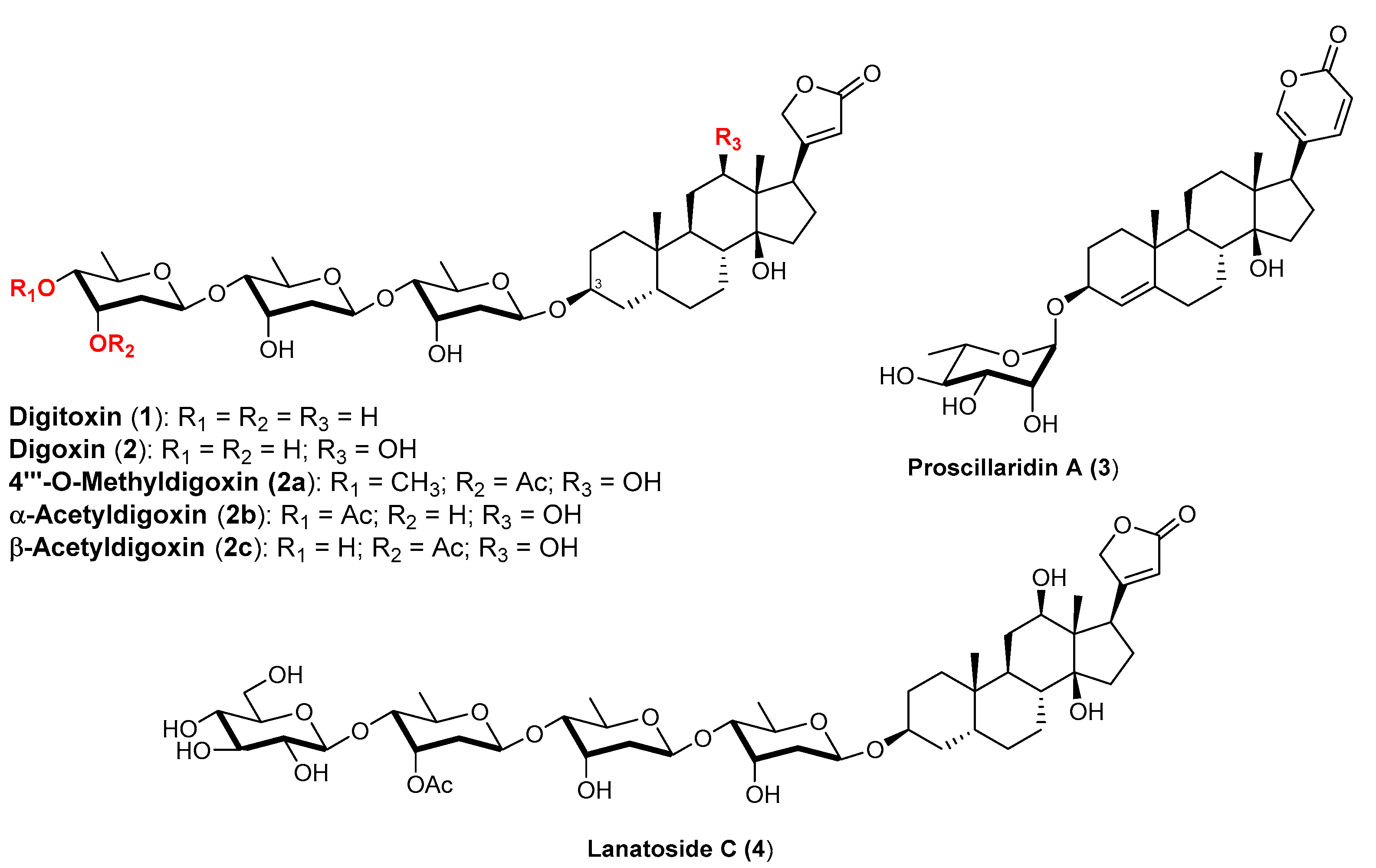
:1. Introduction
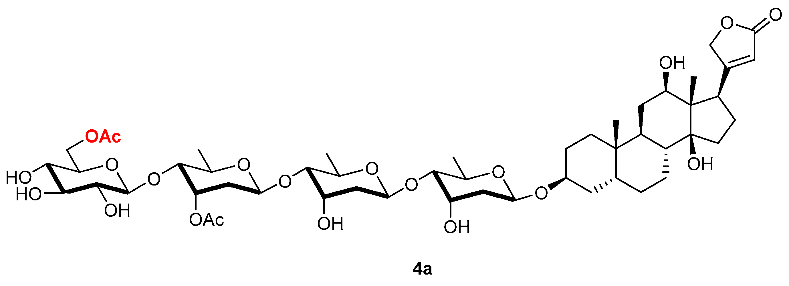
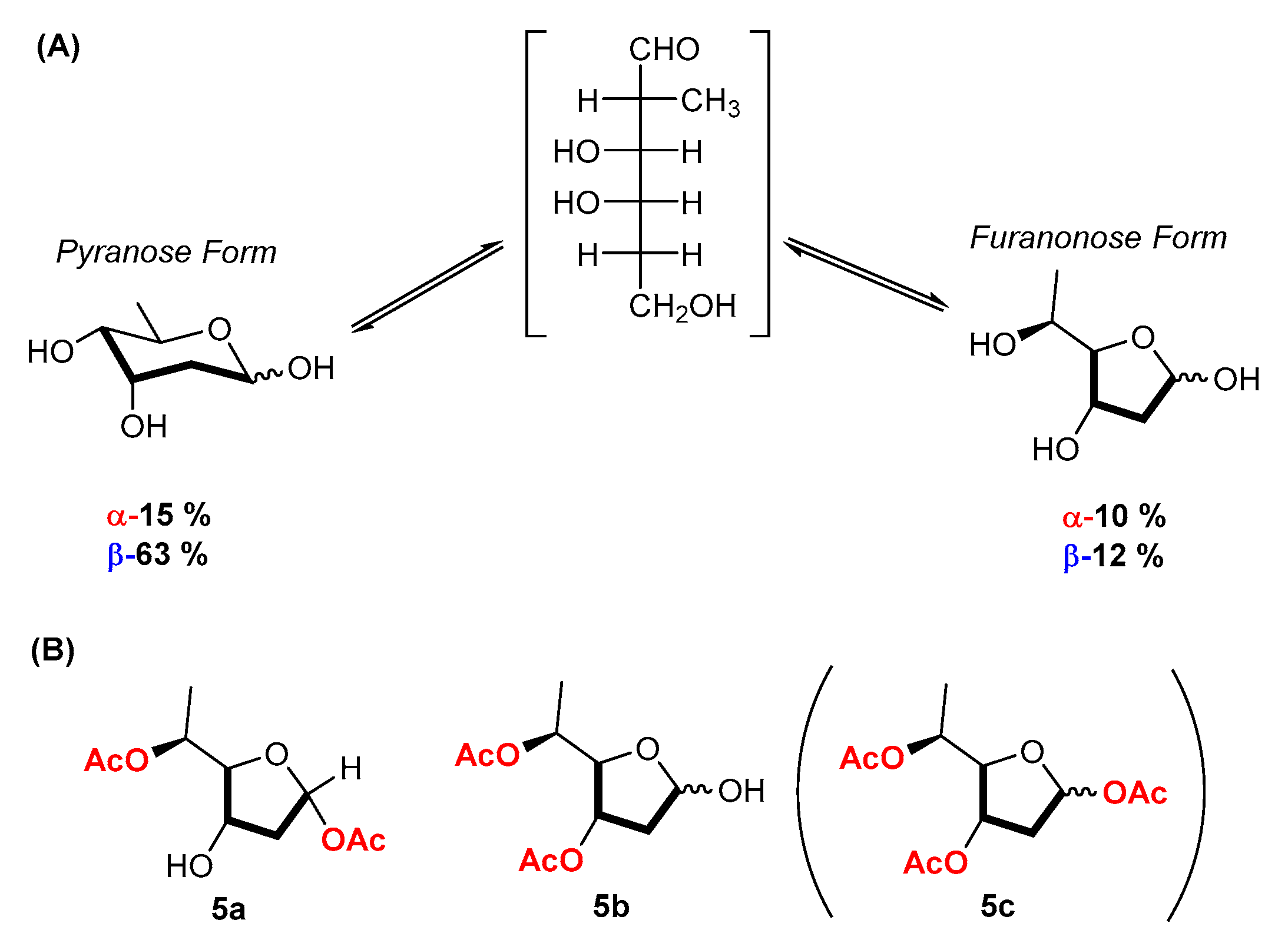
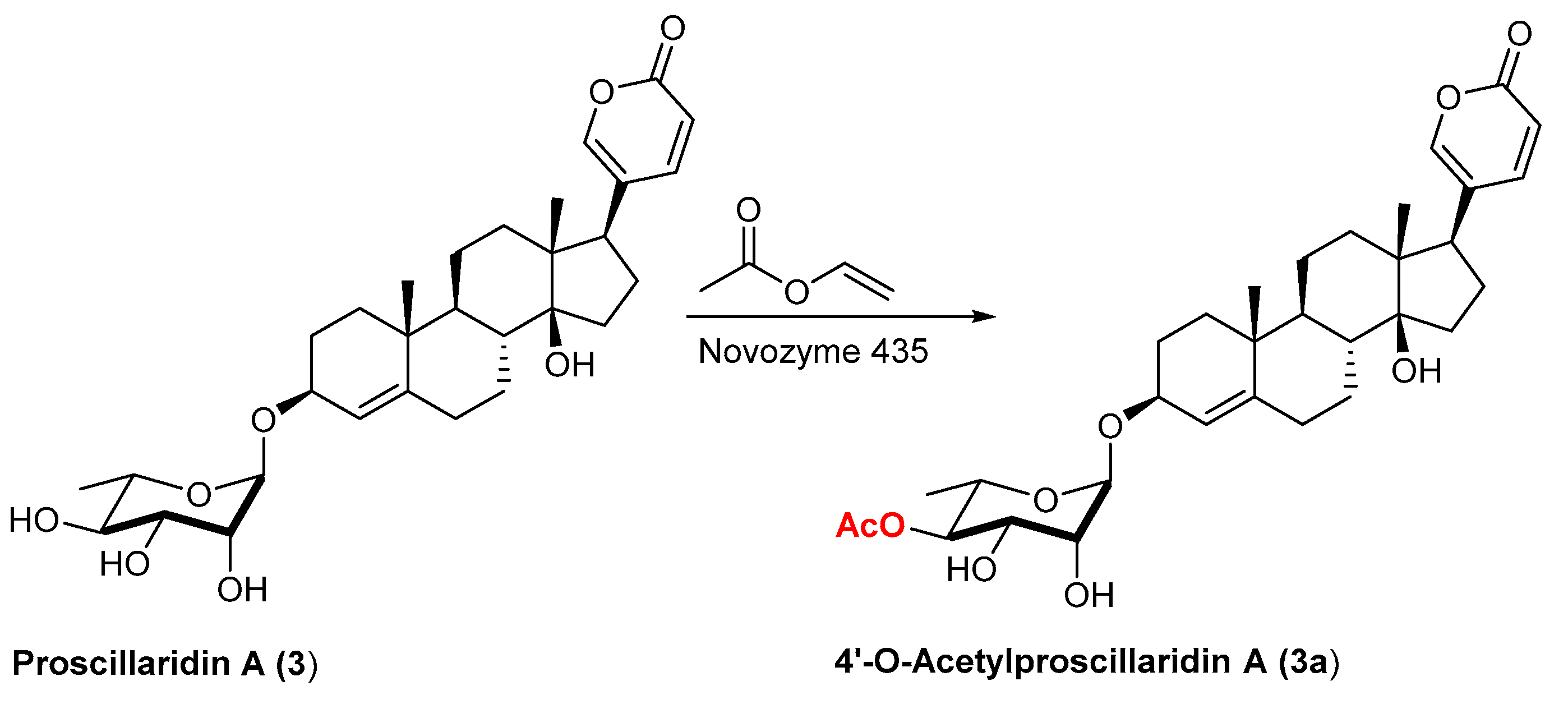
2. Results and Discussion
3. Materials and Methods
3.1. General Information
3.2. Synthesis
- 1H-NMR of D-digitoxose (5).
- 5b (3,5-diacetyl-digitoxose)
4. Conclusions
Supplementary Materials
Author Contributions
Funding
Data Availability Statement
Conflicts of Interest
Abbreviations
References
- Albrecht, H.P. Cardiac Glycosides in Naturally Occurring Glycosides; John Wiley & Sons, Inc.: Chichester, UK, 1999. [Google Scholar]
- Gaignault, J.C.; Bidet, D. Hétérosides Cardiotoniques. Fitoterapia 1988, 59, 259–315. [Google Scholar]
- Melero, C.P.; Medarde, M.; San Feliciano, A. A Short Review on Cardiotonic Steroids and Their Aminoguanidine Analogues. Molecules 2000, 5, 51–81. [Google Scholar] [CrossRef] [Green Version]
- Patel, S. Plant-Derived Cardiac Glycosides: Role in Heart Ailments and Cancer Management. Biomed. Pharmacother. 2016, 84, 1036–1041. [Google Scholar] [CrossRef]
- Ren, J.; Gao, X.; Guo, X.; Wang, N.; Wang, X. Research Progress in Pharmacological Activities and Applications of Cardiotonic Steroids. Front. Pharmacol. 2022, 13, 902459. [Google Scholar] [CrossRef] [PubMed]
- Krstić, D.; Krinulović, K.; Spasojević-Tišma, V.; Joksić, G.; Momić, T.; Vasić, V. Effects of Digoxin and Gitoxin on the Enzymatic Activity and Kinetic Parameters of Na+/K+-ATPase. J. Enzyme Inhib. Med. Chem. 2004, 19, 409–415. [Google Scholar] [CrossRef] [PubMed]
- Cornelius, F.; Mahmmoud, Y.A. Interaction between Cardiotonic Steroids and Na,K-ATPase. Effects of PH and Ouabain-Induced Changes in Enzyme Conformation. Biochemistry 2009, 48, 10056–10065. [Google Scholar] [CrossRef] [PubMed]
- Orlov, S.N.; Tverskoi, A.M.; Sidorenko, S.V.; Smolyaninova, L.V.; Lopina, O.D.; Dulin, N.O.; Klimanova, E.A. Na,K-ATPase as a Target for Endogenous Cardiotonic Steroids: What’s the Evidence? Genes Dis. 2021, 8, 259–271. [Google Scholar] [CrossRef]
- Shah, K.; Chhabra, S.; Singh Chauhan, N. Chemistry and Anticancer Activity of Cardiac Glycosides: A Review. Chem. Biol. Drug. Des. 2022, 100, 364–375. [Google Scholar] [CrossRef]
- Orellana, A.M.; Kinoshita, P.F.; Leite, J.A.; Kawamoto, E.M.; Scavone, C. Cardiotonic Steroids as Modulators of Neuroinflammation. Front. Endocrinol. 2016, 7, 10. [Google Scholar] [CrossRef] [Green Version]
- Schneider, N.; Cerella, C.; Simões, C.M.O.; Diederich, M. Anticancer and Immunogenic Properties of Cardiac Glycosides. Molecules 2017, 22, 1932. [Google Scholar] [CrossRef] [Green Version]
- Škubník, J.; Pavlíčková, V.; Rimpelová, S. Cardiac Glycosides as Immune System Modulators. Biomolecules 2021, 11, 659. [Google Scholar] [CrossRef]
- Kren, V.; Martinkova, L. Glycosides in Medicine: “The Role of Glycosidic Residue in Biological Activity”. Curr. Med. Chem. 2001, 8, 1303–1328. [Google Scholar] [CrossRef]
- Ooi, Y.; Toshihiro, H.; Mituso, N.; Satoh, T. Enzymic Formation of β-Alkyl Glycosides by β-Galactosidase from Aspergillus Oryzae and Its Application to the Synthesis of Chemically Unstable Cardiac Glycosides. Chem. Pharm. Bull. 1984, 35, 1808–1814. [Google Scholar]
- Ooi, Y.; Hashimoto, T.; Mitsuo, N.; Satoh, T. Enzymic Synthesis of Chemically Unstable Cardiac Glycosides by β-Galactosidase from Aspergillus Oryzae. Tetrahedron Lett. 1984, 25, 2241–2244. [Google Scholar] [CrossRef]
- Huang, W.; Wen, C.; Zhou, Z.-R.; Fu, Z.-H.; Katz, A.; Plotnikov, A.; Karlish, S.J.D.; Jiang, R.-W. An Efficient One-Pot Enzymatic Synthesis of Cardiac Glycosides with Varied Sugar Chain Lengths. Adv. Synth. Cat. 2019, 361, 3114–3119. [Google Scholar] [CrossRef]
- Shugrue, C.R.; Miller, S.J. Applications of Nonenzymatic Catalysts to the Alteration of Natural Products. Chem. Rev. 2017, 117, 11894–11951. [Google Scholar] [CrossRef]
- Sun, X.; Lee, H.; Lee, S.; Tan, K.L. Catalyst Recognition of Cis-1,2-Diols Enables Site-Selective Functionalization of Complex Molecules. Nat. Chem. 2013, 5, 790–795. [Google Scholar] [CrossRef]
- Ueda, Y.; Kawabata, T. Organocatalytic Site-Selective Acylation of Carbohydrates and Polyol Compounds. Top. Curr. Chem. 2016, 372, 203–232. [Google Scholar]
- Yoshida, K.; Furuta, T.; Kawabara, T. Perfectly Regioselective Acylation of a Cardiac Glycoside, Digitoxin, via Catalytic Amplification of the Intrinsic Reactivity | Elsevier Enhanced Reader. Tetrahedron Lett. 2010, 51, 4830–4832. [Google Scholar] [CrossRef] [Green Version]
- Danieli, B.; De Bellis, P.; Carrea, G.; Riva, S. Enzyme-Mediated Regioselective Acylations of Flavonoid Disaccharide Monoglycosides. Helv. Chim. Acta 1990, 73, 1837–1844. [Google Scholar] [CrossRef]
- Danieli, B.; Bertario, A.; Carrea, G.; Redigolo, B.; Secundo, F.; Riva, S. Chemo-Enzymatic Synthesis of 6″-O-(3-Arylprop-2-Enoyl) Derivatives of the Flavonol Glucoside Isoquercitrin. Helv. Chim. Acta 1993, 76, 2981–2991. [Google Scholar] [CrossRef]
- Riva, S.; Danieli, B.; Luisetti, M. A Two-Step Efficient Chemoenzymatic Synthesis of Flavonoid Glycoside Malonates. J. Nat. Prod. 1996, 59, 618–621. [Google Scholar] [CrossRef] [PubMed]
- Danieli, B.; De Bellis, P.; Carrea, G.; Riva, S. Regioselective Enzyme-Mediated Acylation of Colchicoside and Thiocolchicoside. Gazz. Chim. Ital. 1991, 121, 123–125. [Google Scholar]
- Danieli, B.; Luisetti, M.; Riva, S.; Bertinotti, A.; Ragg, E.; Scaglioni, L.; Bombardelli, E. Regioselective Enzyme-Mediated Acylation of Polyhydroxy Natural Compounds. A Remarkable, Highly Efficient Preparation of 6′-Acetyl and 6′-O-Carboxyacetyl Ginsenoside Rg1. J. Org. Chem. 1995, 60, 3637–3642. [Google Scholar] [CrossRef]
- Colombo, G.; Riva, S.; Danieli, B. Remote Control of Enzyme Selectivity: The Case of Stevioside and Steviolbioside. Tetrahedron 2004, 60, 741–746. [Google Scholar] [CrossRef]
- Kedra, M.; Kedrowa, S. Clinical evaluation of Proscillaridin A, a glycoside of Scilla maritima. Pol. Tyg. Lek. 1968, 23, 714–716. [Google Scholar]
- Pagliara, R. Clinical use of a new cardioactive glycoside: Proscillaridin A. Case studies. Minerva Med. 1967, 58, 4296–4301. [Google Scholar]
- Krenn, L.; Kopp, B.; Deim, A.; Robien, W.; Kubelka, W. Zum Bufadienolidmuster der roten “Meerzwiebel”. Planta Med. 1994, 60, 63–69. [Google Scholar] [CrossRef]
- Kopp, B.; Krenn, L.; Draxler, M.; Hoyer, A.; Terkola, R.; Vallaster, P.; Robien, W. Bufadienolides from Urginea Maritima from Egypt. Phytochemistry 1996, 42, 513–522. [Google Scholar] [CrossRef]
- Da Costa, E.M.; Armaos, G.; McInnes, G.; Beaudry, A.; Moquin-Beaudry, G.; Bertrand-Lehouillier, V.; Caron, M.; Richer, C.; St-Onge, P.; Johnson, J.R.; et al. Heart Failure Drug Proscillaridin A Targets MYC Overexpressing Leukemia through Global Loss of Lysine Acetylation. J. Exp Clin. Cancer Res. 2019, 38, 251. [Google Scholar] [CrossRef]
- Li, R.-Z.; Fan, X.-X.; Duan, F.-G.; Jiang, Z.-B.; Pan, H.-D.; Luo, L.-X.; Zhou, Y.-L.; Li, Y.; Yao, Y.-J.; Yao, X.-J.; et al. Proscillaridin A Induces Apoptosis and Suppresses Non-Small-Cell Lung Cancer Tumor Growth via Calcium-Induced DR4 Upregulation. Cell Death Dis. 2018, 9, 696. [Google Scholar] [CrossRef] [Green Version]
- Mori, J.; Nagai, S.-I.; Sakakibara, J.; Takeya, K.; Hotta, Y.; Ando, H. Studies on Cardiac Ingredients of Plants. III. Structural Confirmation and Biological Activity of Reduced Proscillaridins. Chem. Pharm. Bull. 1987, 35, 1839–1846. [Google Scholar] [CrossRef] [Green Version]
- Danieli, B.; Luisetti, M.; Sampognaro, G.; Carrea, G.; Riva, S. Regioselective Acylation of Polyhydroxylated Natural Compounds Catalyzed by Candida Antarctica Lipase B (Novozym 435) in Organic Solvents. J. Mol. Catal. B Enzym. 1997, 3, 193–201. [Google Scholar] [CrossRef]
- Monti, D.; Candido, A.; Cruz Silva, M.M.; Křen, V.; Riva, S.; Danieli, B. Biocatalyzed Generation of Molecular Diversity: Selective Modification of the Saponin Asiaticoside. Adv. Synth. Catal. 2005, 347, 1168–1174. [Google Scholar] [CrossRef] [Green Version]
- Hammarström, L.; Smith, C.I.E.; Persson, U. Functional Characterization of Lanatoside-C-Responsive Cells. Scand. J. Immunol. 1978, 8, 263–271. [Google Scholar] [CrossRef] [PubMed]
- Chao, M.-W.; Chen, T.-H.; Haung, H.-L.; Chang, Y.-W.; HangFu, W.-C.; Lee, Y.-C.; Teng, C.-M.; Pan, S.-L. Lanatoside C, a Cardiac Glycoside, Acts through Protein Kinase Cδ to Cause Apoptosis of Human Hepatocellular Carcinoma Cells. Sci. Rep. 2017, 7, 46134. [Google Scholar] [CrossRef] [PubMed]
- Ha, D.P.; Tsai, Y.-L.; Lee, A.S. Suppression of ER-Stress Induction of GRP78 as an Anti-Neoplastic Mechanism of the Cardiac Glycoside Lanatoside C in Pancreatic Cancer. Neoplasia 2021, 23, 1213–1226. [Google Scholar] [CrossRef] [PubMed]
- Vinod, N.; Kim, J.H.; Choi, S.; Lim, I. Combination of I-131-Trastuzumab and Lanatoside C Enhanced Therapeutic Efficacy in HER2 Positive Tumor Model. Sci. Rep. 2021, 11, 12871. [Google Scholar] [CrossRef]
- Duan, Y.; Chen, L.; Shao, J.; Jiang, C.; Zhao, Y.; Li, Y.; Ke, H.; Zhang, R.; Zhu, J.; Yu, M. Lanatoside C Inhibits Human Cervical Cancer Cell Proliferation and Induces Cell Apoptosis by a Reduction of the JAK2/STAT6/SOCS2 Signaling Pathway. Oncol. Lett. 2021, 22, 740. [Google Scholar] [CrossRef]
- Danieli, B.; Luisetti, M.; Steurer, S.; Michelitsch, A.; Likussar, W.; Riva, S.; Reiner, J.; Schubert-Zsilavecz, M. Application of Lipase-Catalyzed Regioselective Esterification in the Preparation of Digitonin Derivatives. J. Nat. Prod. 1999, 62, 670–673. [Google Scholar] [CrossRef]
- Gebhardt, S.; Bihler, S.; Schubert-Zsilavecz, M.; Riva, S.; Monti, D.; Falcone, L.; Danieli, B. Biocatalytic Generation of Molecular Diversity: Modification of Ginsenoside Rb1 by β-1,4-Galactosyltransferase and Candida Antarctica Lipase, Part 4. Helv. Chim. Acta 2002, 85, 1943–1959. [Google Scholar] [CrossRef]
- Danieli, B.; De Bellis, P.; Carrea, G.; Riva, S. Enzyme-Mediated Acylation of Flavonoid Monoglycosides. Heterocycles 1989, 29, 2061. [Google Scholar] [CrossRef]
- Riva, S.; Carrea, G.; Ottolina, G.; Secundo, F.; Danieli, B.; De Bellis, P. Enzymatic Regioselective Acylation of Polyhydroxylated Natural Compounds in Organic Solvents. Ann. N. Y. Acad. Sci. 1990, 613, 712–716. [Google Scholar] [CrossRef]
- Ueda, Y.; Mishiro, K.; Yoshida, K.; Furuta, T.; Kawabata, T. Regioselective Diversification of a Cardiac Glycoside, Lanatoside C, by Organocatalysis. J. Org. Chem. 2012, 77, 7850–7857. [Google Scholar] [CrossRef] [PubMed]
- Riva, S. Enzymatic Modification of the Sugar Moieties of Natural Glycosides. J. Mol. Catal. B Enzym. 2002, 19–20, 43–54. [Google Scholar] [CrossRef]
- Coxon, B. Two-Dimensional Proton Chemical Shift Correlated NMR Spectroscopy of Digitoxose1. J. Carbohydr. Chem. 1984, 3, 525–543. [Google Scholar] [CrossRef]


Disclaimer/Publisher’s Note: The statements, opinions and data contained in all publications are solely those of the individual author(s) and contributor(s) and not of MDPI and/or the editor(s). MDPI and/or the editor(s) disclaim responsibility for any injury to people or property resulting from any ideas, methods, instructions or products referred to in the content. |
© 2023 by the authors. Licensee MDPI, Basel, Switzerland. This article is an open access article distributed under the terms and conditions of the Creative Commons Attribution (CC BY) license (https://creativecommons.org/licenses/by/4.0/).
Share and Cite
Bassanini, I.; Roncaglia, L.; Danieli, B.; Riva, S. Regioselective Esterification of Cardiac Glycosides Catalyzed by Novozym 435 and Lipase PS in Organic Solvents. Catalysts 2023, 13, 819. https://doi.org/10.3390/catal13050819
Bassanini I, Roncaglia L, Danieli B, Riva S. Regioselective Esterification of Cardiac Glycosides Catalyzed by Novozym 435 and Lipase PS in Organic Solvents. Catalysts. 2023; 13(5):819. https://doi.org/10.3390/catal13050819
Chicago/Turabian StyleBassanini, Ivan, Lucia Roncaglia, Bruno Danieli, and Sergio Riva. 2023. "Regioselective Esterification of Cardiac Glycosides Catalyzed by Novozym 435 and Lipase PS in Organic Solvents" Catalysts 13, no. 5: 819. https://doi.org/10.3390/catal13050819