Article

Photoelectrochemical Conversion of Sewage Water into H2 Fuel over the CuFeO2/CuO/Cu Composite Electrode

1
Physics Department, College of Science, Jouf University, Al-Jouf, Sakaka P.O. Box 2014, Saudi Arabia
2
Basic Sciences Research Unit, Jouf University, Sakaka 72388, Saudi Arabia
3
Department of Physics, Faculty of Science, Islamic University of Madinah, Prince Naifbin Abdulaziz, Al Jamiah, Madinah 42351, Saudi Arabia
4
Nanophotonics and Applications Laboratory, Physics Department, Faculty of Science, Beni-Suef University, Beni-Suef 62514, Egypt
5
Physics Department, College of Science, Imam Mohammad Ibn Saud Islamic University (IMSIU), Riyadh 11623, Saudi Arabia
6
College of Engineering and Technology, American University of the Middle East, Egaila 54200, Kuwait
7
Nanoscale Science Program, Department of Chemistry, University of North Carolina at Charlotte, Charlotte, NC 28223, USA
8
Nanomaterials Science Research Laboratory, Chemistry Department, Faculty of Science, Beni-Suef University, Beni-Suef 62514, Egypt
*
Authors to whom correspondence should be addressed.
Catalysts 2023, 13(3), 456; https://doi.org/10.3390/catal13030456
Submission received: 11 January 2023 / Revised: 6 February 2023 / Accepted: 14 February 2023 / Published: 21 February 2023
(This article belongs to the Section Photocatalysis)

Abstract

:
This study describes the synthesis of delafossite, CuFeO2, as a primary photocatalytic material for hydrogen generation. A photoelectrode, CuFeO2/CuO/Cu, was prepared by combusting a Cu foil dipped in FeCl3 in ambient air. This photoelectrode showed excellent optical behavior for the hydrogen generation reaction from sewage water, producing 90 µmol/h of H2. The chemical structure was confirmed through XRD and XPS analyses, and the crystalline rhombohedral shape of CuFeO2 was confirmed using SEM and TEM analyses. With a bandgap of 1.35 ev, the prepared material displayed excellent optical properties. Electrochemical measurements for H2 gas generation were carried out using the CuFeO2/CuO/Cu photoelectrode, comparing the effect of light and dark and monochromatic wavelength light. The electrode exhibited significant enhancement in light compared to dark, with current density (Jph) values of −0.83 and −0.1 mA·cm−2, respectively. The monochromatic light also had a noticeable effect, with the Jph value increasing from −0.45 to −0.79 mA·cm−2 as the wavelength increased from 640 to 390 nm. This system is cheap and durable, making it a promising solution for hydrogen gas fuel generation in the industry.

1. Introduction

Renewable energy has become a vital energy source for people in today’s society, as non-renewable energy sources are becoming scarce. Additionally, these energy sources are known to release dangerous gases, such as SOx, NOx, and COx, which can have adverse effects on the environment and human health [1,2,3].
Hydrogen gas (H2) is a promising alternative energy source with numerous potential applications, including fuel for airplanes and factories and everyday household purposes such as cooking and heating. H2 has a high combustion energy and low cost [4,5,6,7]. It can be produced through different electrolytes, such as strong acids or bases, by relying on the presence of high amounts of H+ or OH ions through an extended mechanism that includes the formation of OH·. This active radical attacks the H2O molecule for additional H2 gas production. However, this reaction has limited economic applications related to the cost of the chemicals, such as NaOH, HCl, or H2SO4, and the corrosion problems caused by the implemented electrolytes, affecting the working electrode.
The working electrode must be a semiconductor material, such as an oxide, sulfide, or nitride [8,9]. Oxides have the advantage of being cost-effective and stable [10,11]. Furthermore, the amount of the produced H2 gas depends on the optical and morphological properties of the semiconductor material, making it crucial to prepare these materials with high surface areas and active sites [12,13,14]. One of the developed methods for increasing H2 production is using plasmonic materials in the electrode construction; copper (Cu) is a promising and cost-effective plasmonic material [15,16]. In the literature, several studies have used Cu as plasmonic material to increase the activity of ZnO as a photocatalytic material [17]. Plasmonic materials enhance the performance of the neighboring semiconductor material (such as a metal oxide) by transferring electrons to the conducting band and inducing a plasmonic resonance that generates an electric field and increases light absorbance [18].
The physical properties of the prepared photocatalytic material play a crucial role in improving the light absorbance and reducing the bandgap. Copper oxide (CuO), with its black color and semiconducting nature, has a bandgap of 0.7 to 1.6 eV, which falls within the visible light absorbance region [19,20]. This behavior enhances its application as a photocatalytic absorbent and in H2 production [21,22]. Li et al. synthesized CuO/Cu through annealing Cu(OH)2 in air conditions at 500 °C [23]. Moreover, Sagadevan et al. synthesized CuO through the combustion process using ascorbic acid as a capping agent at 100–300 °C [24]. Ragupathi et al. synthesized CuO supported with b-C3N4 as photocatalytic material for water splitting reaction; the produced Jph is still very small [25]. Quyen et al. increased the efficiency of the H2 generation of TiO2 by using Cu nanomaterials as plasmonic material [26]. In the same manner, Shen et al. slightly increased H2 generation when g-C3N4 was decorated with CuO, but not to the extent desired [27].
Delafossite, CuFeO2, has a high photocatalytic performance and excellent optical properties, with a bandgap of approximately 1.3 eV [28]. This material can be prepared using various methods, including solvothermal and laser beam techniques. He et al. used CuFeO2 for carbamazepine degradation with an efficiency of 86% [29]. In addition, GC3N4/CuFeO2 has been used as a catalyst for H2O2 degradation through the Fenton reaction [30]. Mao et al. prepared CuFeO2 using the hydrothermal and solar gel for dye removal and water splitting, and the obtained Jph was recorded as 4 × 10⁻⁶ µA cm−2 in 1M NaOH [31]. Baiano et al. investigated the significant photocatalytic reduction of CO2 utilizing CuFeO2 through DFT calculations [32]. Furthermore, CuFeO2 was demonstrated to have photocatalytic antibacterial properties, effectively degrading E. coli, with an improvement in performance when exposed to light [33]. Change et al. used Mg-doped CuFeO2 for photocatalytic degradation of methylene blue and reduction of CO2 to form ethylene glycol under the electrolysis conditions [34].
In the previous studies on H2 generation, the use of additional electrolytes such as HCl or NaOH was required to serve as a source of H+ ions [35,36,37]. Additionally, some studies relied on high-cost preparation techniques [38,39].
In this study, a CuFeO2/CuO delafossite material was synthesized on a Cu metal substrate through a combustion reaction. Subsequently, various analyses were performed to confirm the chemical, optical, and morphological properties of the prepared photoelectrode, CuFeO2/CuO/Cu. The latter was then evaluated for its ability to produce H2 gas through photocatalytic decomposition of sewage water, and the effects of light and dark conditions on H2 production were investigated. The impact of different monochromatic wavelengths on H2 production was also examined, and the amount of H2 produced was calculated over time.

2. Results and Discussion

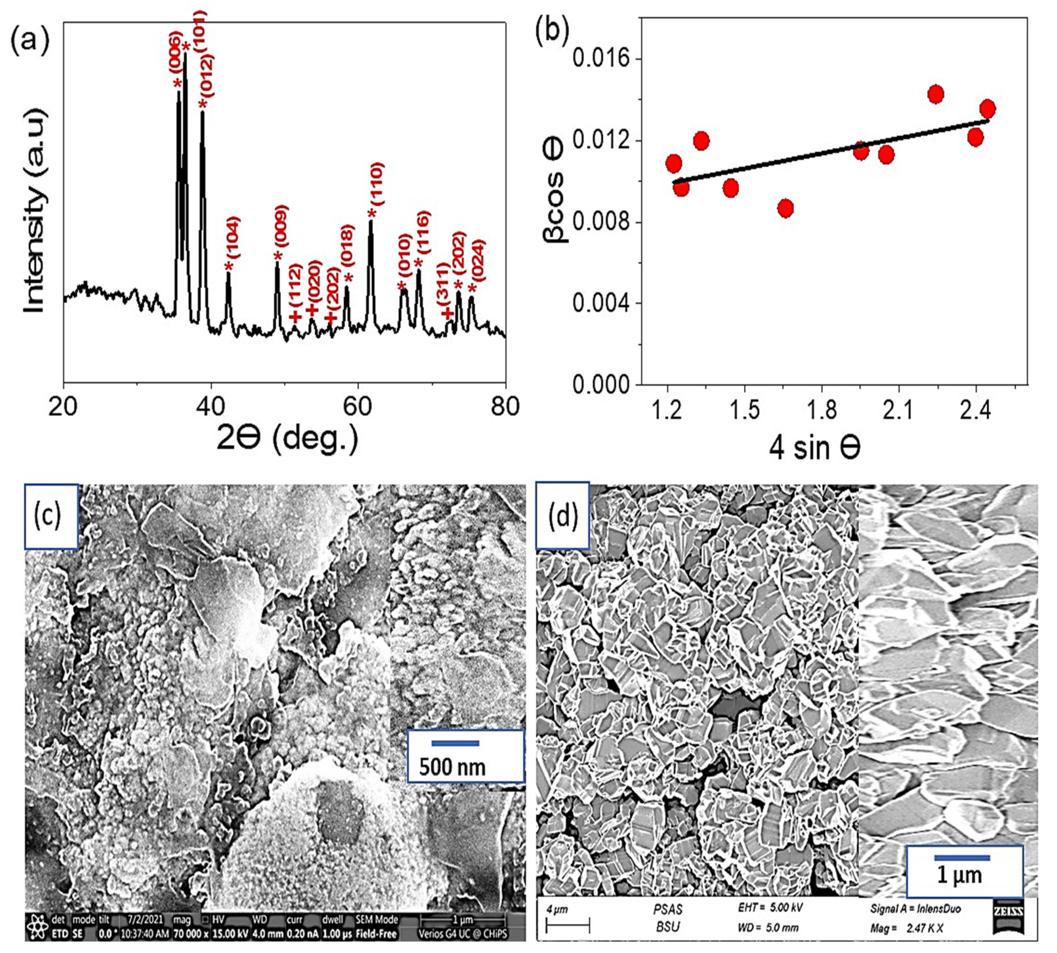
2.1. Characterization of CuFeO2/CuO/Cu Nanomaterials Photoelectrode

The XRD analysis was employed to investigate and verify the crystalline structure of the prepared CuFeO2/CuO/Cu nanomaterials. The results showed a well-demonstrated rhombohedral CuFeO2 phase structure and microstructural parameters of the prepared nanostructured electrode. Figure 1a displays the XRD patterns of the prepared CuFeO2, which show a poly-oriented nature. Fifteen diffraction peaks were observed; eleven of the peaks correspond to the Miller indices (i.e., (hkl) planes) (006), (101), (012),(104), (009), (018), (110), (1010), (116), (202), and (024) of the rhombohedral CuFeO2 structure with a secondary CuO impurity phase. The remaining four peaks correspond to the Miller indices (112), (020), (202), and (311) of the CuO monoclinic phase. The peaks are in agreement with the reference data JCPDS 01-075-2146 [40] for the rhombohedral structure of pure CuFeO2 with R 3   m   space group and JCPDS card no. 48-1548 [41] for the monoclinic phase of CuO.
The broadness of the XRD patterns was exploited to estimate the microstructural parameters, such as crystallite size (D) and microstrain (ε), of the prepared CuFeO2 by using the Williamson–Hall (W–H) approach. The W-H model was applied to the pure peak broadening of the diffraction peaks to calculate the mean values of (D) and (ε). The pure peak broadening (β) was obtained from (Equation (1)) [42,43,44]
β     cos θ = 0.95   λ D     + 4 ε     sin θ
Where 0.95 is the shape factor value for CuFeO2 nanoparticles, and λ is the incident XRD wavelength (~0.15418 nm). In Equation (1), it was considered that the investigated samples have an isotropic nature, and the micro-strain is uniform in all (hkl) crystallographic directions. The crystallite size (D) and the micro-strain (ε) values were estimated by plotting the (βCorrect cos θ) versus (4 sin θ) for each XRD peak, yielding a linear regression where the intercept and slope were calculated (Figure 1b). It was found that the crystalline size and micro-strain values for the CuFeO2 sample are 35 nm and 4.69 × 10⁻³, respectively.
Moreover, the XRD of the CuO nanoparticles is provided in Figure S1; the peaks appearing at 2θ° = 26.55, 35.27, and 41.71° correspond to CuO for the growth direction (110), (111), and (200), respectively. These analyses match the results reported in the literature [45,46,47]. On the other hand, the Cu metal appears at 43.5°, 50.7°, and 74.5° [46,48].
XPS was utilized for further verification of the chemical composition of CuFeO2. The results are presented in Figure S2a, in which Cu, Fe, and O elements are confirmed through their related peaks. Additionally, the spectra of the Cu2p spin-orbital components reveal six peaks in the range of 930 to 970 eV, as shown in Figure S2b. The Fe2P spectra are confirmed through the two peaks at 723 and 726 eV (Figure S2c). The O1s spin-orbital peaks can be found in the 529–533 eV range (Figure S2d).
In Figure 1c, The morphology of CuO can be clearly seen as protruding appendages that greatly enhance the surface area. The magnified image shows that the appendages are randomly distributed across the surface. After forming CuFeO2 (Figure 1d), excellent uniform crystalline rhombohedral shapes appear with porous structure. This highly crystalline structure confirms the findings of the XRD analysis. Given the crystalline nature of the prepared materials, it was expected that their optical properties would be superior [49].
The optical reflectance of CuFeO2 nanoparticles was determined over the wavelength range of 200 to 1200 nm, as shown in Figure 2a. The low reflectance of the CuFeO2 nanoparticles in the visible region confirms their high absorbance in the UV and IR regions. The bandgap of the material was calculated using the Kubelka-Munk equation (Equation (2)) [12], which takes into account reflectance (R), with K and S values that represent the molar absorption coefficient and scattering factor, respectively. The resulting bandgap of this material is 1.35 eV.
K / S = ( 1 R ) 2 2 R .
The TEM image of CuFeO2 is displayed in Figure 2b, which confirms the formation of crystalline nanoparticles with an average size of 50 nm. These results are consistent with those obtained from the SEM analysis. The theoretical images of CuO and CuFeO2, obtained using ImageJ, are presented Figure 2c,d, respectively. Both materials exhibit considerable roughness, but the roughness of the CuFeO2 is more uniform. This feature indicates the suitability of these materials for photocatalytic applications due to their high ability to capture light through their porous structures, acting like a “cave” for the incident photons [3,12].

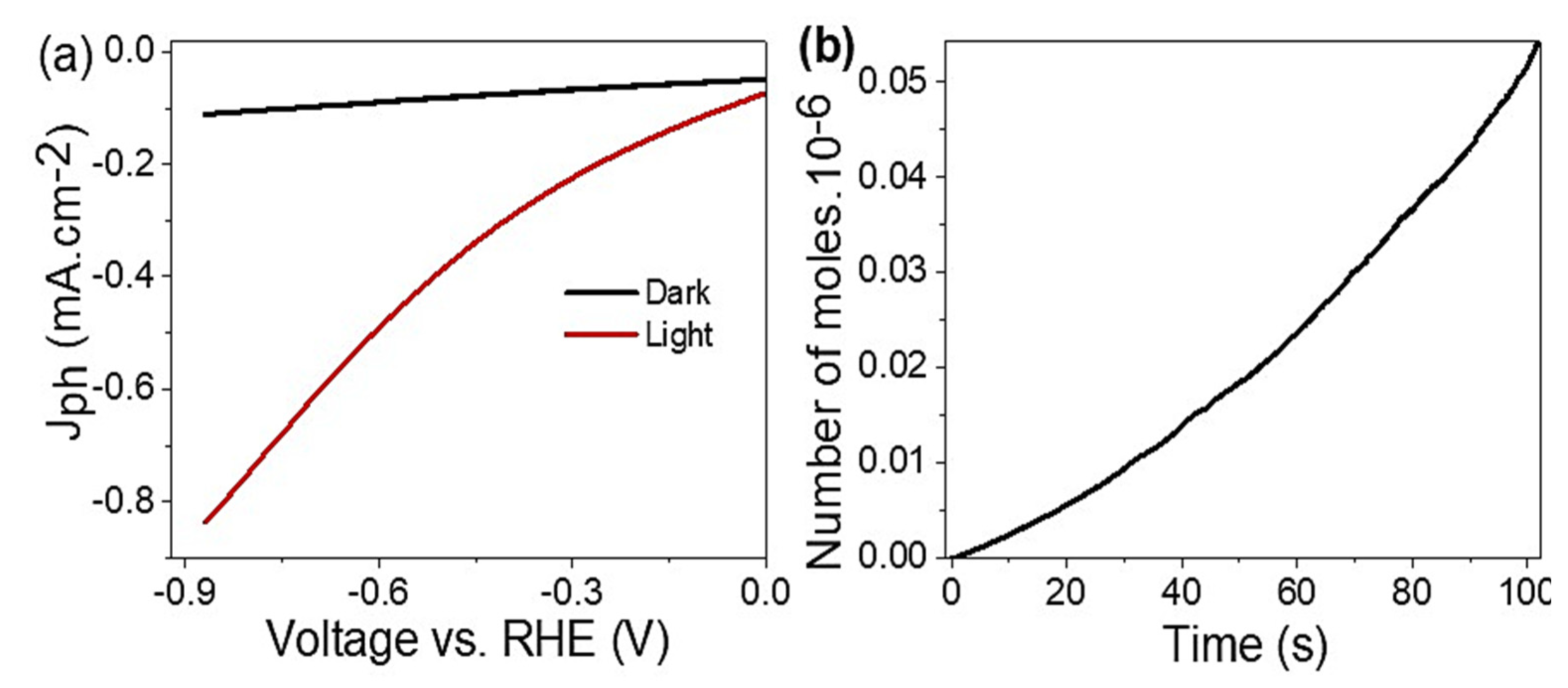
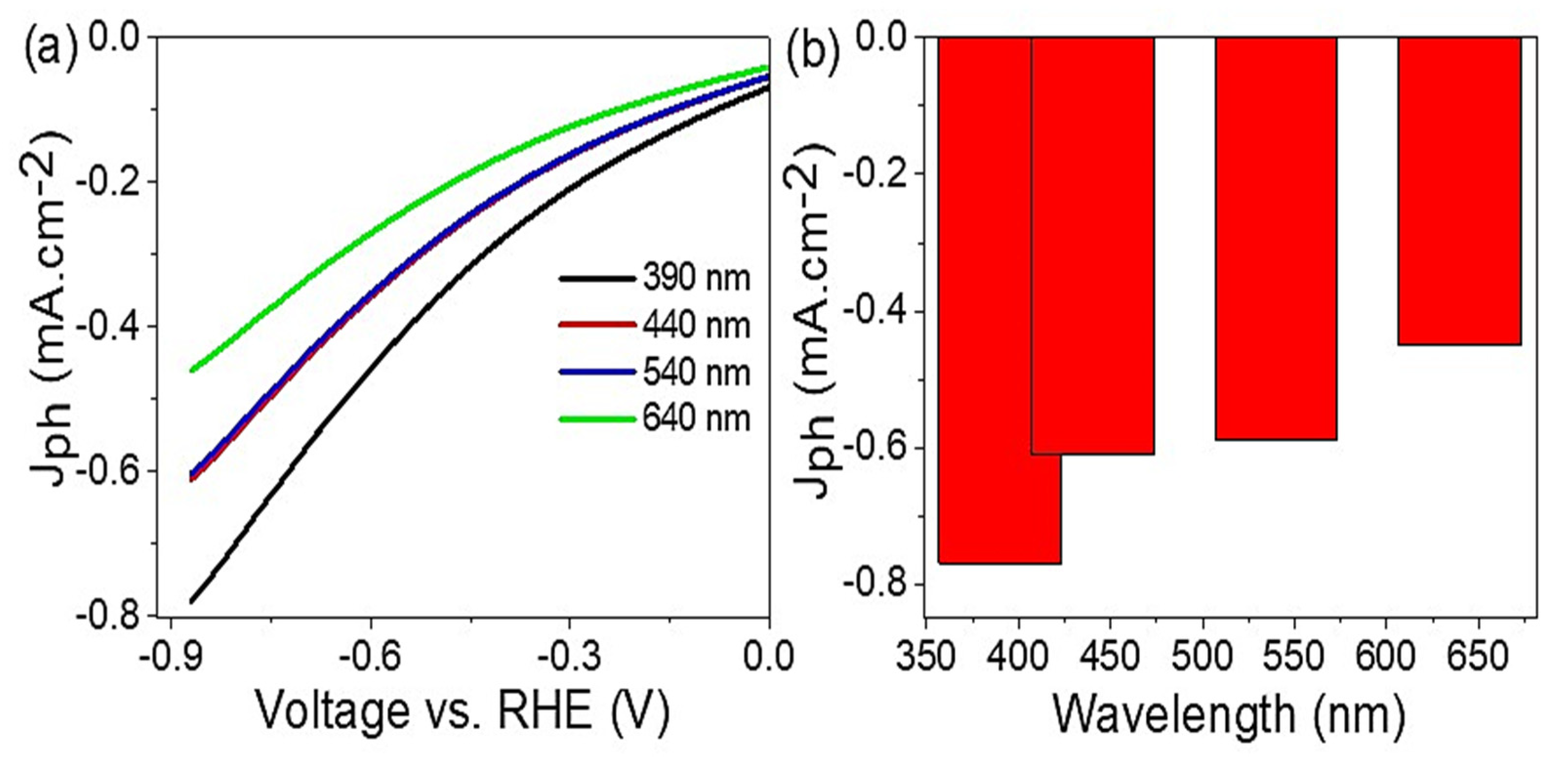
2.2. Photoelectrochemical Water Splitting

The photoelectrochemical water splitting reaction was carried out with a three-electrode cell. The prepared CuFeO2/CuO/Cu photoelectrode served as the working electrode, graphite as the counter electrode, and calomel as the reference electrode. The measurements were conducted using a CHMI608E electrochemical workstation under a 400 W metal halide lamp at a sweep rate of 1 mV/s and a temperature of 25 °C. The measurements were taken both in the dark and under illumination to assess the photocatalytic behavior of the materials.
The impact of light on the performance of CuFeO2/CuO/Cu is depicted in Figure 3a. When illuminated, the current density (Jph) value was increased; the Jph values at 0.88 V in dark and light conditions were −0.10 mA·cm−2 and −0.83 mA·cm−2, respectively. The high surface area of the CuFeO2/CuO/Cu provides a good environment for capturing photons, which leads to the creation of electron-hole pairs. The electrons collected on the surface of CuFeO2 are then transferred to the adjacent electrolyte (sewage water), leading to further reactions and the generation of OH. radicals. The results of this study show that using sewage water as an electrolyte (pH 7.2) is a promising approach, as its chemical composition, outlined in Table 1, acts as a catalyst for the water-splitting reaction.
The addition of Cu increased the efficiency of electron transfer. It also acts as a plasmonic material that enhances light capture, causing an electric field to accumulate around the CuFeO2 materials. This electric field oscillation between Cu and CuFeO2 increases the number of free electrons available to split water into H2 gas. The rate of H2 gas released can be determined from the calculated Jph values.
The H2 production was measured and plotted in Figure 3b. It was found that the H2 production increased with time, reaching 90 µmol/h. This high rate confirms the effectiveness of CuFeO2 as a photocatalyst in water splitting. The active sites of CuFeO2 are activated under light, contributing to its high efficiency. Based on Equation (3), the number of photons was estimated to be 8 × 10²¹ photon/s, which leads to a significant number of electrons being generated and collected on the active sites, and then transferred to the neighbor electrolyte for further water splitting and H2 gas production [50,51]. λ , h, P, and c in Equation (3) are wavelength, Planck constant, light intensity, and light velocity, respectively.
The number of generated H2 moles was determined using Faraday’s law (Equation (4)) [52,53]. This calculation is based on the time change (dt) and the Jph values, with consideration given to the Faraday constant (F).
N = λ P / hc
H 2   mole = 0 t J ph · dt / F
The performance of the prepared CuFeO2/CuO/Cu photoelectrode was evaluated under monochromatic light by studying the effect of different light wavelengths (390–640 nm) on the H2 generation reaction. The results, presented in Figure 4a, show that the Jph values increased from −0.45 to −0.79 mA·cm−2 as the wavelength increased from 390 to 640 nm. Figure 4b shows the produced Jph values at −0.88 V, confirming that the sewage water splitting and H2 gas evolution reactions were enhanced with increasing incident photons.
The conversion of sewage water into H2 gas was achieved through the impact of the incident photons on the CuFeO2/CuO/Cu photoelectrode. The Delafossite material is highly responsive to light illumination, which activates the surface and triggers energy level splitting. Hot electrons are then collected on the surface and transferred to the upper level through resonance energy transfer. The small bandgap, 1.35 eV, is considered promising for electron transfer and confirms the significant optical properties of this material, as seen Figure 2a. The use of Cu as a current collector facilitates this electron transfer process as it can also act as a plasmonic material [52]. The collected electrons are then transferred to the surrounding wastewater solution, generating the current density (Jph) values shown in Figure 3. These Jph values determine the rate of H2 gas produced [51,54,55]. The photocurrent is recorded as −0.11 mA·cm−2. The generation of H2 is initiated by the radiation of light that creates electrons and holes, leading to hot electrons being transferred to the photocathode for reduction and, eventually, H2 production [56].
The IPCE of the photoelectrode, CuFeO2/CuO/Cu, is determined through Equation (5) and found to be 2.4% at 340 nm, which is a favorable result for producing hydrogen from a solution without using additional electrolytes [57].
I P C E = J ph mA · cm 2 · 1240   V · nm P mW · cm 2 ·   λ nm
Finally, a comparison was made between the electrolytes used and the produced Jph values in the literature and those obtained in this study, where sewage water was used as an electrolyte. The results are summarized in Table 2 and show that the CuFeO2/CuO/Cu electrode prepared in this study has one of the highest Jph values, indicating its high efficiency and sensitivity to light. Furthermore, this study’s results are noteworthy as sewage water was used as the electrolyte without needing additional external electrolytes, making it a significant advantage over other electrodes.

3. Materials and Methods

3.1. CuFeO2 Delafossite Nanomaterial Preparation

The CuFeO2 nanomaterial was prepared through a simple combustion reaction. The process began with washing the Cu foil with water, soap, distilled water, and ethanol, followed by immersing it in concentrated H2SO4 (99.9%) for 2–3 min. The foil was then rewashed with distilled water and ethanol. Subsequently, it was immersed in 0.05 M FeCl3 for 15 min on each side and dried at 60 °C for 2 h. The final step involved combusting the substrate at 500 °C for 10 min, forming the CuFeO2/CuO/Cu photoelectrode.

3.2. Characterization

The prepared CuFeO2 materials were characterized using various analysis methods. The chemical structure was examined by x-ray diffraction patterns (XRD, Malvern Panalytical Ltd., Malvern, UK) (λ = 0.15418 nm) (advance diffractometer, Bruker D8) and X-ray photoelectron spectroscopy (XPS, K-ALPHA, Waltham, MA, USA). The morphology was observed by scanning electron microscopy (SEM, S-4800, Hitachi, Japan) and transmitted electron microscopy (TEM, JEOL JEM-2100, Oberkochen, Germany). Finally, the optical properties were analyzed using a double-beam spectrophotometer (Elmer Lamba, Waltham, MA, USA).

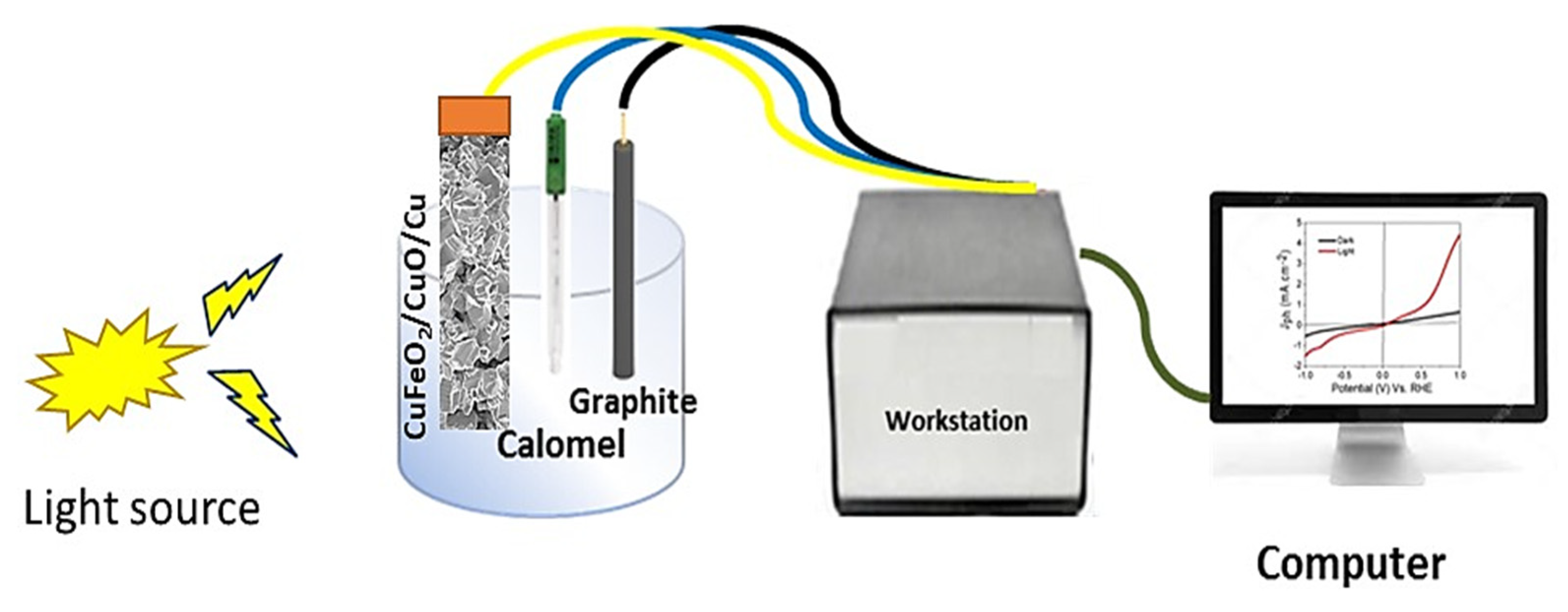
3.3. Electrochemical Measurements

The electrochemical measurements were performed using a CHI608E electrochemical workstation, as illustrated in Figure 5. The prepared CuFeO2/CuO/Cu photoelectrode served as the working electrode (1 cm2), while a calomel and graphite electrodes were used as the reference and counter electrodes, respectively. The measurements were carried out using sewage water with a pH of 7.2. A 400 W metal halide lamp (light intensity) with a photon flux of 500 μmol/s was implemented as the light source. The effect of different light wavelengths (390, 440, 540, and 640 nm) and the impact of alternating light and dark conditions were studied. The produced H2 moles were calculated as a function of time.

4. Conclusions

This study demonstrates the potential of a low-cost photoelectrode made of CuFeO2/CuO/Cu for the generation of H2 gas. Using sewage water as an electrolyte, measurements were conducted using an electrochemical workstation in a three-electrode cell setup. The photoelectrode was found to have excellent optical properties, with a small bandgap of 1.35 eV, making it suitable for photocatalytic reactions. The electrochemical testing under a metal halide lamp was applied; the results showed that the Jph values under light and dark conditions were −0.83 and −0.1 mA·cm−2, respectively. The effect of monochromatic light on the photoelectrode was studied across the range of 390 to 640 nm, resulting in Jph values of −0.45 to −0.79 mA·cm−2. The produced H2 gas was measured to be 90 µmol/h. Additionally, the cost of a 10 × 10 cm2 CuFeO2/CuO/Cu photoelectrode was found to be 0.5 $, making it a cost-effective option for H2 gas production and a candidate for industrial applications.

Supplementary Materials

The following supporting information can be downloaded at: https://www.mdpi.com/article/10.3390/catal13030456/s1, Figure S1: XRD of the CuO nanoparticles. and Figure S2: XPS (a) survey, (b) Cu, (c) Fe, and (d) O elements for the synthesized CuFeO2 nano-material.

Author Contributions

Conceptualization, N.M.A.H., M.S., and M.R.; methodology, N.M.A.H., W.S.M., M.A., M.E., M.F.H., M.S., A.M.A., and M.R.; validation, formal analysis, data curation, N.M.A.H., W.S.M., M.A., M.E., M.F.H., M.S., A.M.A., W.E.M., and M.R.; software, investigation, A.A.A.A., M.S., and M.R.; writing—original draft preparation, writing—review and editing, visualization, W.E.M. and M.R.; supervision, project administration, resources, funding acquisition, N.M.A.H. and M.R. All authors have read and agreed to the published version of the manuscript.

Funding

This research received no external funding.

Data Availability Statement

The data that support the findings of this study are available from the corresponding author upon reasonable request.

Acknowledgments

The authors extend their appreciation to the Deanship of Scientific Research at Jouf University for funding this work through research grant no. (DSR2022-GR-0122).

Conflicts of Interest

The authors declare no conflict of interest.

References

  1. Mohamed, F.; Rabia, M.; Shaban, M. Synthesis and Characterization of Biogenic Iron Oxides of Different Nanomorphologies from Pomegranate Peels for Efficient Solar Hydrogen Production. J. Mater. Res. Technol. 2020, 9, 4255–4271. [Google Scholar] [CrossRef]
  2. Shaban, M.; Ali, S.; Rabia, M. Design and Application of Nanoporous Graphene Oxide Film for CO2, H2, and C2H2 Gases Sensing. J. Mater. Res. Technol. 2019, 8, 4510–4520. [Google Scholar] [CrossRef]
  3. Elsayed, A.M.; Rabia, M.; Shaban, M.; Aly, A.H.; Ahmed, A.M. Preparation of Hexagonal Nanoporous Al2O3/TiO2/TiN as a Novel Photodetector with High Efficiency. Sci. Rep. 2021, 11, 17572. [Google Scholar] [CrossRef]
  4. Nishiyama, H.; Yamada, T.; Nakabayashi, M.; Maehara, Y.; Yamaguchi, M.; Kuromiya, Y.; Nagatsuma, Y.; Tokudome, H.; Akiyama, S.; Watanabe, T.; et al. Photocatalytic Solar Hydrogen Production from Water on a 100-M2 Scale. Nature 2021, 598, 304–307. [Google Scholar] [CrossRef] [PubMed]
  5. Hisatomi, K.D.T. Reaction Systems for Solar Hydrogen Production via Water Splitting with Particulate Semiconductor. Nat. Catal. 2019, 2, 387–399. [Google Scholar] [CrossRef]
  6. Pagliaro, M. Preparing for the Future: Solar Energy and Bioeconomy in the United Arab Emirates. Energy Sci. Eng. 2019, 7, 1451–1457. [Google Scholar] [CrossRef] [Green Version]
  7. Takata, T. Photocatalytic Water Splitting with Quantum Efficiency of Almost Unity. Nature 2020, 581, 411–414. [Google Scholar] [CrossRef] [PubMed]
  8. Kang, Z.; Cheng, Y.; Zheng, Z.; Cheng, F.; Chen, Z.; Li, L.; Tan, X.; Xiong, L.; Zhai, T.; Gao, Y. MoS2-Based Photodetectors Powered by Asymmetric Contact Structure with Large Work Function Difference. Nano-Micro Lett. 2019, 11, 34. [Google Scholar] [CrossRef] [Green Version]
  9. Lee, J.H.; Lee, W.W.; Yang, D.W.; Chang, W.J.; Kwon, S.S.; Park, W. Il Anomalous Photovoltaic Response of Graphene-on-GaN Schottky Photodiodes. ACS Appl. Mater. Interfaces 2018, 10, 14170–14174. [Google Scholar] [CrossRef]
  10. Shaban, M.; Rabia, M.; El-Sayed, A.M.A.; Ahmed, A.; Sayed, S. Photocatalytic Properties of PbS/Graphene Oxide/Polyaniline Electrode for Hydrogen Generation. Sci. Rep. 2017, 7, 14100. [Google Scholar] [CrossRef] [Green Version]
  11. Rabia, M.; Mohamed, H.S.H.; Shaban, M.; Taha, S. Preparation of Polyaniline/PbS Core-Shell Nano/Microcomposite and Its Application for Photocatalytic H2 Electrogeneration from H2O. Sci. Rep. 2018, 8, 1107. [Google Scholar] [CrossRef] [PubMed] [Green Version]
  12. Mohamed, H.S.H.; Rabia, M.; Zhou, X.G.; Qin, X.S.; Khabiri, G.; Shaban, M.; Younus, H.A.; Taha, S.; Hu, Z.Y.; Liu, J.; et al. Phase-Junction Ag/TiO2 Nanocomposite as Photocathode for H2 Generation. J. Mater. Sci. Technol. 2021, 83, 179–187. [Google Scholar] [CrossRef]
  13. Rabia, M.; Shaban, M.; Jibali, B.M.; Abdelkhaliek, A.A. Effect of Annealing Temperature on the Photoactivity of ITO/VO2 (M)/Au Film Electrodes for Water Splitting. J. Nanosci. Nanotechnol. 2020, 20, 4120–4130. [Google Scholar] [CrossRef] [PubMed]
  14. Rabia, M.; Mohamed, S.H.; Zhao, H.; Shaban, M.; Lei, Y.; Ahmed, A.M. TiO2/TiOxNY Hollow Mushrooms-like Nanocomposite Photoanode for Hydrogen Electrogeneration. J. Porous Mater. 2020, 27, 133–139. [Google Scholar] [CrossRef]
  15. Zhang, X.; Li, J.; Yang, W.; Leng, B.; Niu, P.; Jiang, X.; Liu, B. High-Performance Flexible Ultraviolet Photodetectors Based on AZO/ZnO/PVK/PEDOT:PSS Heterostructures Integrated on Human Hair. ACS Appl. Mater. Interfaces 2019, 11, 24459–24467. [Google Scholar] [CrossRef] [PubMed]
  16. Kim, J.; Lee, H.C.; Kim, K.H.; Hwang, M.S.; Park, J.S.; Lee, J.M.; So, J.P.; Choi, J.H.; Kwon, S.H.; Barrelet, C.J.; et al. Photon-Triggered Nanowire Transistors. Nat. Nanotechnol. 2017, 12, 963–968. [Google Scholar] [CrossRef]
  17. Li, M.; Yu, M.; Su, D.; Zhang, J.; Jiang, S.; Wu, J.; Wang, Q.; Liu, S. Ultrahigh Responsivity UV Photodetector Based on Cu Nanostructure/ZnO QD Hybrid Architectures. Small 2019, 15, 1901606. [Google Scholar] [CrossRef]
  18. Almohammedi, A.; Shaban, M.; Mostafa, H.; Rabia, M. Nanoporous TiN/TiO2/Alumina Membrane for Photoelectrochemical Hydrogen Production from Sewage Water. Nanomaterials 2021, 11, 2617. [Google Scholar] [CrossRef]
  19. Schultze, J.W.; Lohrengel, M.M. Stability, Reactivity and Breakdown of Passive Films. Problems of Recent and Future Research. Electrochim. Acta 2000, 45, 2499–2513. [Google Scholar] [CrossRef]
  20. Basnet, P.; Zhao, Y. Tuning the CuxO Nanorod Composition for Efficient Visible Light Induced Photocatalysis. Catal. Sci. Technol. 2016, 6, 2228–2238. [Google Scholar] [CrossRef]
  21. Chiang, C.Y.; Shin, Y.; Aroh, K.; Ehrman, S. Copper Oxide Photocathodes Prepared by a Solution Based Process. Int. J. Hydrogen Energy 2012, 37, 8232–8239. [Google Scholar] [CrossRef]
  22. Chiang, C.-Y.; Chang, M.-H.; Liu, H.-S.; Tai, C.Y.; Ehrman, S. Process Intensification in the Production of Photocatalysts for Solar Hydrogen Generation. Ind. Eng. Chem. Res. 2012, 51, 5207–5215. [Google Scholar] [CrossRef]
  23. Li, C.; Kurniawan, M.; Sun, D.; Tabata, H.; Delaunay, J.J. Nanoporous CuO Layer Modified Cu Electrode for High Performance Enzymatic and Non-Enzymatic Glucose Sensing. Nanot 2015, 26, 015503. [Google Scholar] [CrossRef]
  24. Sagadevan, S.; Vennila, S.; Marlinda, A.R.; Al-Douri, Y.; Rafie Johan, M.; Anita Lett, J. Synthesis and Evaluation of the Structural, Optical, and Antibacterial Properties of Copper Oxide Nanoparticles. Appl. Phys. A Mater. Sci. Process. 2019, 125, 489. [Google Scholar] [CrossRef]
  25. Ragupathi, V.; Raja, M.A.; Panigrahi, P.; Ganapathi Subramaniam, N. CuO/g-C3N4 Nanocomposite as Promising Photocatalyst for Photoelectrochemical Water Splitting. Optik 2020, 208, 164569. [Google Scholar] [CrossRef]
  26. Thi Quyen, V.; Jitae, K.; Thi Huong, P.; Thi Thu Ha, L.; My Thanh, D.; Minh Viet, N.; Quang Thang, P. Copper Doped Titanium Dioxide as a Low-Cost Visible Light Photocatalyst for Water Splitting. Sol. Energy 2021, 218, 150–156. [Google Scholar] [CrossRef]
  27. Chen, J.; Shen, S.; Guo, P.; Wang, M.; Wu, P.; Wang, X.; Guo, L. In-Situ Reduction Synthesis of Nano-Sized Cu2O Particles Modifying g-C3N4 for Enhanced Photocatalytic Hydrogen Production. Appl. Catal. B Environ. 2014, 152–153, 335–341. [Google Scholar] [CrossRef]
  28. Liu, Q.L.; Zhao, Z.Y.; Zhao, R.D.; Yi, J.H. Fundamental Properties of Delafossite CuFeO2 as Photocatalyst for Solar Energy Conversion. J. Alloys Compd. 2020, 819, 153032. [Google Scholar] [CrossRef]
  29. He, B.; Zhao, Z.; Song, L.; Liu, W.; Yang, Y.; Shang, J.; Cheng, X. Highly Efficient Activation of Peroxymonosulfate by the (3R + 2H)-CuFeO2 Nanocomposite Photocatalyst: Intermediate Toxicity, BVS Validation Ionic Migration and Degradation Pathway. Sep. Purif. Technol. 2022, 289, 120729. [Google Scholar] [CrossRef]
  30. Dai, C.; Nie, Y.; Tian, X.; Yang, C.; Hu, Y.; Lin, H.M.; Dionysiou, D.D. Insight into Enhanced Fenton-like Degradation of Antibiotics over CuFeO2 Based Nanocomposite: To Improve the Utilization Efficiency of OH/O 2- via Minimizing Its Migration Distance. Chemosphere 2022, 294, 133743. [Google Scholar] [CrossRef]
  31. Mao, L.; Mohan, S.; Gupta, S.K.; Mao, Y. Multifunctional Delafossite CuFeO2 as Water Splitting Catalyst and Rhodamine B Sensor. Mater. Chem. Phys. 2022, 278, 125643. [Google Scholar] [CrossRef]
  32. Baiano, C.; Schiavo, E.; Gerbaldi, C.; Bella, F.; Meligrana, G.; Talarico, G.; Maddalena, P.; Pavone, M.; Muñoz-García, A.B. Role of Surface Defects in CO2 Adsorption and Activation on CuFeO2 Delafossite Oxide. Mol. Catal. 2020, 496, 111181. [Google Scholar] [CrossRef]
  33. Nien, Y.T.; Chen, Y.Z.; Hsu, Y.R.; Ye, H.J. Enhanced Antibacterial Effect of CuFeO2 Ceramic Powders by Glycine Combustion Process and Visible Light Irradiation. Mater. Chem. Phys. 2022, 276, 125423. [Google Scholar] [CrossRef]
  34. Chang, Y.H.; Wang, H.; Siao, T.F.; Lee, Y.H.; Bai, S.Y.; Liao, C.W.; Zhuang, J.K.; Chiu, T.W.; Kuo, C.H. A New Solution Route for the Synthesis of CuFeO2 and Mg-Doped CuFeO2 as Catalysts for Dye Degradation and CO2 Conversion. J. Alloys Compd. 2021, 854, 157235. [Google Scholar] [CrossRef]
  35. Teixeira, G.F.; Silva Junior, E.; Vilela, R.; Zaghete, M.A.; Colmati, F. Perovskite Structure Associated with Precious Metals: Influence on Heterogenous Catalytic Process. Catalysts 2019, 9, 721. [Google Scholar] [CrossRef] [Green Version]
  36. Mishra, M.; Chun, D.M. α-Fe2O3 as a Photocatalytic Material: A Review. Appl. Catal. A Gen. 2015, 498, 126–141. [Google Scholar] [CrossRef]
  37. Acar, C.; Dincer, I.; Naterer, G.F. Review of Photocatalytic Water-Splitting Methods for Sustainable Hydrogen Production. Int. J. Energy Res. 2016, 40, 1449–1473. [Google Scholar] [CrossRef]
  38. Chiang, C.Y.; Aroh, K.; Franson, N.; Satsangi, V.R.; Dass, S.; Ehrman, S. Copper Oxide Nanoparticle Made by Flame Spray Pyrolysis for Photoelectrochemical Water Splitting—Part II. Photoelectrochemical Study. Int. J. Hydrogen Energy 2011, 36, 15519–15526. [Google Scholar] [CrossRef]
  39. Guo, X.; Diao, P.; Xu, D.; Huang, S.; Yang, Y.; Jin, T.; Wu, Q.; Xiang, M.; Zhang, M. CuO/Pd Composite Photocathodes for Photoelectrochemical Hydrogen Evolution Reaction. Int. J. Hydrogen Energy 2014, 39, 7686–7696. [Google Scholar] [CrossRef]
  40. Cheng, X.; Ding, J.; Wu, Y.; Liu, H.; Dawson, G. The Photocathodic Properties of a Fe2O3 Wrapped CuFeO2 Layer on ITO Glass for Water Splitting. Chem. Phys. 2018, 513, 241–245. [Google Scholar] [CrossRef]
  41. Siddiqui, H.; Parra, M.R.; Qureshi, M.S.; Malik, M.M.; Haque, F.Z. Studies of Structural, Optical, and Electrical Properties Associated with Defects in Sodium-Doped Copper Oxide (CuO/Na) Nanostructures. J. Mater. Sci. 2018, 53, 8826–8843. [Google Scholar] [CrossRef]
  42. Mohamed, W.S.; Alzaid, M.; Abdelbaky, M.S.M.; Amghouz, Z.; García-Granda, S.; Abu-Dief, A.M. Impact of Co2+ Substitution on Microstructure and Magnetic Properties of CoxZn1-XFe2O4 Nanoparticles. Nanomaterials 2019, 9, 1602. [Google Scholar] [CrossRef] [Green Version]
  43. Mohamed, W.S.; Hadia, N.M.A.; Al Bakheet, B.; Alzaid, M.; Abu-Dief, A.M. Impact of Cu2+ Cations Substitution on Structural, Morphological, Optical and Magnetic Properties of Co1-XCuxFe2O4 Nanoparticles Synthesized by a Facile Hydrothermal Approach. Solid State Sci. 2022, 125, 106841. [Google Scholar] [CrossRef]
  44. Mohamed, W.S.; Abu-Dief, A.M. Impact of Rare Earth Europium (RE-Eu3+) Ions Substitution on Microstructural, Optical and Magnetic Properties of CoFe2−xEuxO4 Nanosystems. Ceram. Int. 2020, 46, 16196–16209. [Google Scholar] [CrossRef]
  45. Zúñiga, A.; Fonseca, L.; Souza, J.A.; Rivaldo-Gomez, C.; Pomar, C.D.; Criado, D. Anomalous Ferromagnetic Behavior and Size Effects in CuO Nanowires. J. Magn. Magn. Mater. 2019, 471, 77–81. [Google Scholar] [CrossRef]
  46. Zhan, W.; Chen, Z.; Hu, J.; Chen, X. Vertical CuO Nanowires Array Electrodes: Visible Light Sensitive Photoelectrochemical Biosensor of Ethanol Detection. Mater. Sci. Semicond. Process. 2018, 85, 90–97. [Google Scholar] [CrossRef]
  47. Mahmoodi, A.; Solaymani, S.; Amini, M.; Nezafat, N.B.; Ghoranneviss, M. Structural, Morphological and Antibacterial Characterization of CuO Nanowires. Silicon 2017, 10, 1427–1431. [Google Scholar] [CrossRef]
  48. Kim, J.-H.; Katoch, A.; Choi, S.-W.; Kim, S.S. Growth and Sensing Properties of Networked P-CuO Nanowires. Sens. Actuators B. Chem. 2015, 212, 190–195. [Google Scholar] [CrossRef]
  49. Hadia, N.M.A.; Hajjiah, A.; Elsayed, A.M.; Mohamed, S.H.; Alruqi, M.; Shaban, M.; Alzahrani, F.M.; Abdelazeez, A.A.A.; Rabia, M. Bunch of Grape-Like Shape PANI/Ag2O/Ag Nanocomposite Photocatalyst for Hydrogen Generation from Wastewater. Adsorpt. Sci. Technol. 2022, 2022, 4282485. [Google Scholar] [CrossRef]
  50. Sayyah, S.M.; Shaban, M.; Rabia, M. Electropolymerization of m -Toluidin on Platinum Electrode from Aqueous Acidic Solution and Character of the Obtained Polymer. Adv. Polym. Technol. 2018, 37, 126–136. [Google Scholar] [CrossRef]
  51. Sayyah, S.M.; Shaban, M.; Rabia, M. A High-Sensitivity Potentiometric Mercuric Ion Sensor Based on m-Toluidine Films. IEEE Sens. J. 2016, 16, 1541–1548. [Google Scholar] [CrossRef]
  52. Helmy, A.; Rabia, M.; Shaban, M.; Ashraf, A.M.; Ahmed, S.; Ahmed, A.M. Graphite/Rolled Graphene Oxide/Carbon Nanotube Photoelectrode for Water Splitting of Exhaust Car Solution. Int. J. Energy Res. 2020, 44, 7687–7697. [Google Scholar] [CrossRef]
  53. Mohamed, H.S.H.; Rabia, M.; Shaban, M.; Taha, S. Controlled Synthesis of CdS Nanoflowers Thin Films for H2 Electro-Generation. Mater. Sci. Semicond. Process. 2020, 120, 105307. [Google Scholar] [CrossRef]
  54. Jiang, L.; Zhou, G.; Mi, J.; Wu, Z. Fabrication of Visible-Light-Driven One-Dimensional Anatase TiO2/Ag Heterojunction Plasmonic Photocatalyst. Catal. Commun. 2012, 24, 48–51. [Google Scholar] [CrossRef]
  55. Sayyah, S.M.; Shaban, M.; Rabia, M. M-Toluidine Polymer Film Coated Platinum Electrode as a pH Sensor by Potentiometric Methods. Sens. Lett. 2015, 13, 961–966. [Google Scholar] [CrossRef]
  56. Li, Z.; Qu, Y.; He, G.; Humayun, M.; Chen, S.; Jing, L. Enhanced Visible-Light Activities for PEC Water Reduction of CuO Nanoplates by Coupling with Anatase TiO2 and Mechanism. Appl. Surf. Sci. 2015, 351, 681–685. [Google Scholar] [CrossRef]
  57. Rabia, M.; Hadia, N.M.A.; Farid, O.M.; Abdelazeez, A.A.A.; Mohamed, S.H.; Shaban, M. Poly(m-Toluidine)/Rolled Graphene Oxide Nanocomposite Photocathode for Hydrogen Generation from Wastewater. Int. J. Energy Res. 2022, 46, 11943–11956. [Google Scholar] [CrossRef]
  58. Khalafalla, M.A.H.; Hadia, N.M.A.; Elsayed, A.M.; Alruqi, M.; El Malti, W.; Shaban, M.; Rabia, M. ATO/Polyaniline/PbS Nanocomposite as Highly Efficient Photoelectrode for Hydrogen Production from Wastewater with Theoretical Study for the Water Splitting. Adsorpt. Sci. Technol. 2022, 2022, 5628032. [Google Scholar] [CrossRef]
  59. Huang, X.; Zhang, M.; Sun, R.; Long, G.; Liu, Y.; Zhao, W. Enhanced Hydrogen Evolution from CuOx-C/TiO2 with Multiple Electron Transport Pathways. PLoS ONE 2019, 14, e0215339. [Google Scholar] [CrossRef]
  60. Sherman, B.D.; Ashford, D.L.; Lapides, A.M.; Sheridan, M.V.; Wee, K.-R.; Meyer, T.J. Light-Driven Water Splitting with a Molecular Electroassembly-Based Core/Shell Photoanode. J. Phys. Chem. Lett. 2015, 6, 3213–3217. [Google Scholar] [CrossRef]
  61. Naldoni, A.; Guler, U.; Wang, Z.; Marelli, M.; Malara, F.; Meng, X.; Besteiro, L.V.; Govorov, A.O.; Kildishev, A.V.; Boltasseva, A.; et al. Broadband Hot-Electron Collection for Solar Water Splitting with Plasmonic Titanium Nitride. Adv. Opt. Mater. 2017, 5, 1601031. [Google Scholar] [CrossRef] [Green Version]
  62. Liu, G.; Karuturi, S.K.; Chen, H.; Wang, D.; Ager, J.W.; Simonov, A.N.; Tricoli, A. Enhancement of the Photoelectrochemical Water Splitting by Perovskite BiFeO3 via Interfacial Engineering. Sol. Energy 2020, 202, 198–203. [Google Scholar] [CrossRef]
  63. Wang, Z.; Cao, D.; Wen, L.; Xu, R.; Obergfell, M.; Mi, Y.; Zhan, Z.; Nasori, N.; Demsar, J.; Lei, Y. Manipulation of Charge Transfer and Transport in Plasmonic-Ferroelectric Hybrids for Photoelectrochemical Applications. Nat. Commun. 2016, 7, 10348. [Google Scholar] [CrossRef] [PubMed] [Green Version]
  64. Freeman, E.; Kumar, S.; Thomas, S.R.; Pickering, H.; Fermin, D.J.; Eslava, S. PrFeO3 Photocathodes Prepared Through Spray Pyrolysis. ChemElectroChem 2020, 7, 1365–1372. [Google Scholar] [CrossRef]
  65. Modibane, K.D.; Waleng, N.J.; Ramohlola, K.E.; Maponya, T.C.; Monama, G.R.; Makgopa, K.; Hato, M.J. Poly(3-Aminobenzoic Acid) Decorated with Cobalt Zeolitic Benzimidazolate Framework for Electrochemical Production of Clean Hydrogen. Polymers 2020, 12, 1581. [Google Scholar] [CrossRef]
Figure 1. (a) XRD patterns and (b) W–H plots of CuFeO2. (c) SEM of CuO and (d) CuFeO2 nanomaterial.
Catalysts 13 00456 g001
Figure 2. (a) The optical reflectance and (b) TEM of CuFeO2. ImageJ theoretical of (c) CuO and (d) CuFeO2 nanoparticles.
Catalysts 13 00456 g002
Figure 3. (a) The voltage-current relation in dark and light and (b) the H2 moles produced vs. time for the CuFeO2/CuO/Cu photoelectrode.
Catalysts 13 00456 g003
Figure 4. (a) The effect of monochromatic wavelengths on the prepared CuFeO2/CuO/Cu photoelectrode and (b) the produced Jph values at −0.88 V.
Catalysts 13 00456 g004
Figure 5. The schematic diagram for the H2 generation from sewage water, using the CuFeO2/CuO/Cu photoelectrode.
Catalysts 13 00456 g005
Table 1. The chemical composition of the sewage water used as an electrolyte for H2 gas production.
SpeciesConcentration (mg/L)
Phenols 0.015
F1.0
Al3+3.0
NH35.0
Hg2+0.005
Pb2+0.5
Cd3+0.05
As3+0.05
Cr3+1.0
Cu2+1.5
Ni3+0.1
Fe3+1.5
Mn2+1.0
Zn2+5.0
Ag+0.1
Ba3+2.0
Co2+2.0
Other cations 0.1
Pesticides 0.2
CN−10.1
Industrial washing 0.5
Coli groups 400
Table 2. Comparison of the current study (using sewage water as an electrolyte) with the previous studies in the literature [58].
Photoelectrode ElectrolyteJph (mA/cm2)
g-C3N4-CuO [25]NaOH0.01
CuO-C/TiO2 [59]Glycerol0.012
SnO2/TiO2 [60]Na2S2O30.4
TiN-TiO2 [61]NaOH3.0 × 10−4
BiFeO3 [62]NaOH0.1
Au/Pb(Zr, Ti)O3 [63]NaOH0.06
PrFeO [64] Na2SO40.130
Poly(3-aminobenzoic acid) frame [65]H2SO40.08
CuFeO2/CuO/Cu (present study)Sewage water0.83
Disclaimer/Publisher’s Note: The statements, opinions and data contained in all publications are solely those of the individual author(s) and contributor(s) and not of MDPI and/or the editor(s). MDPI and/or the editor(s) disclaim responsibility for any injury to people or property resulting from any ideas, methods, instructions or products referred to in the content.

