Article

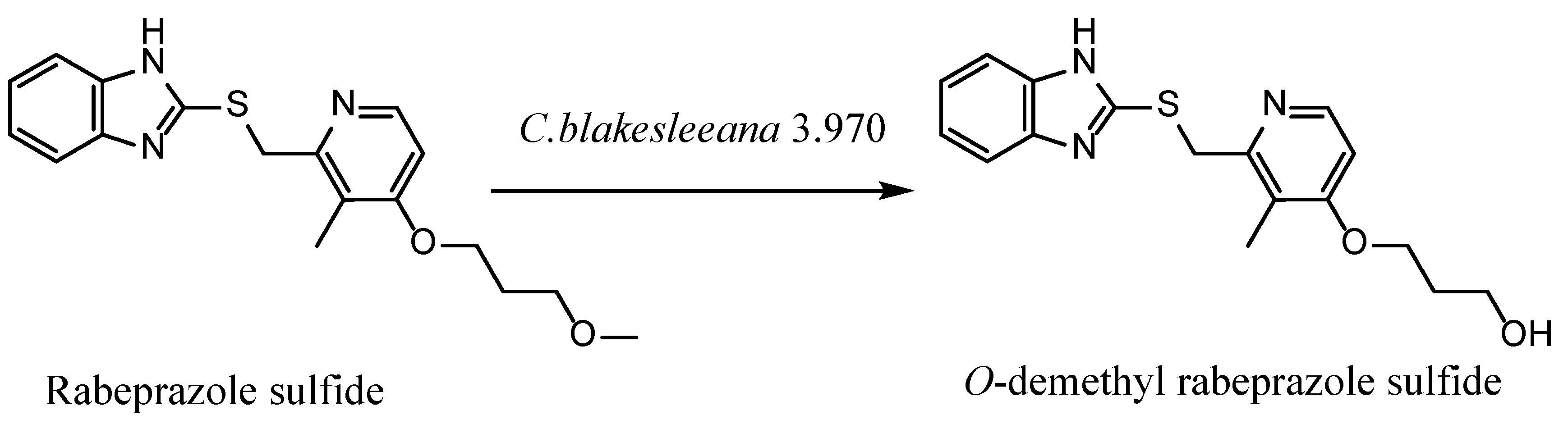
An O-Demethylation Metabolite of Rabeprazole Sulfide by Cunninghamella blakesleeana 3.970 Biotransformation

1
School of Functional Food and Wine, Shenyang Pharmaceutical University, Shenyang 110016, China
2
School of Life Sciences and Biopharmaceuticals, Shenyang Pharmaceutical University, Shenyang 110016, China
3
School of Pharmaceutical Engineering, Shenyang Pharmaceutical University, Shenyang 110016, China
*
Authors to whom correspondence should be addressed.
Catalysts 2023, 13(1), 15; https://doi.org/10.3390/catal13010015
Submission received: 14 November 2022 / Revised: 16 December 2022 / Accepted: 17 December 2022 / Published: 22 December 2022
(This article belongs to the Special Issue Microbial Biocatalysis)

Abstract

:
To explore the potential metabolites from rabeprazole sulfide, seven strains of filamentous fungi were screened for their biotransformation abilities. Among these strains, Cunninghamella blakesleeana 3.970 exhibited the best result. Four different culture media were screened in order to identify the most optimal for subsequent research. Single factors such as the initial pH of culture media, culture time, inoculation volume, and media volume were individually investigated to provide the optimum biotransformation conditions. Then, an orthogonal optimization process using a five-factor, four-level L16(45) experiment was designed and performed. Finally, when the substrate concentration is 3 g/L, one major metabolite was detected with a transformation rate of 72.4%. Isolated by semipreparative HPLC, this metabolite was further detected by ESI-MS and NMR. The final data analysis indicated that the metabolite is O-demethylation rabeprazole sulfide.

1. Introduction

Rabeprazole is a kind of proton pump inhibitor, which has been widely used in the treatment of gastric acid secretion, and can also be applied to eradicate Helicobacter pylori [1]. As a benzimidazole derivative, rabeprazole has a heterocyclic molecule consisting of a pyridine and benzimidazole moiety linked by a methylsulfinyl group. This general structure guarantees the similar pharmacological properties of other proton pump inhibitors. Rabeprazole sulfide (rabeprazole thioether) is an essential intermediate for the production of rabeprazole [2,3]. Meanwhile, sulfide is also one of the metabolites produced by the rabeprazole metabolites in vivo [4,5,6,7,8].
Rabeprazole sulfide is an important imidazole compound, which is not only a precursor of rabeprazole synthesis, but is also one of the main metabolites of rabeprazole in vivo. As the primary metabolite of rabeprazole, rabeprazole sulfide can undergo further metabolic reactions such as demethylation and sulfuric acid binding. These demethylated metabolites often have pharmacological activity. Pharmacologically active metabolites can contribute significantly to the overall therapeutic and adverse effects of drugs. Therefore, to fully understand the mechanism of action of drugs, it is important to recognize the role of active metabolites [9,10,11,12].
Some microorganisms, especially the fungi belonging to Cunninghamella species, possess cytochrome P-450 mono-oxygenase systems analogous to those in mammals [13]. Hence, microbial transformation has been proposed as a complementary in vitro model for mammalian drug metabolism. Cunnihamella is a genus of fungi in the family Cunninghamellaceae, which has been implemented for in vitro biotransformations for a long time [14,15,16,17,18,19,20]. Various reaction types have been reported for these kinds of fungi. Previous studies have demonstrated their hydroxylation [21,22,23,24], epoxidation [25,26,27], amination [28], demethylation [29], and oxidation from sulfide to sulfoxide, which is usually the final step for the production of prazole-type molecules [30,31,32].
Demethylated metabolites often have some unexpected pharmacological effects. Metamizole (dipyrone) is an analgesic with antipyretic and spasmolytic properties, which has been in use for almost 100 years. The primary metabolite of metamizole, 4-methylaminoantipyrine, can be N-demethylated to 4-aminoantipyrine. 4-aminoantipyrine plays a role as a non-steroidal anti-inflammatory drug, a non-narcotic analgesic, an antirheumatic drug, a peripheral nervous system drug, and an EC 1.14.99.1 (prostaglandin-endoperoxide synthase) inhibitor. It is used as a reagent for biochemical reactions, such as the production of peroxides or phenols [33,34]. Papaverine plays a role as a vasodilator agent and an antispasmodic drug. The metabolism of papaverine with Cunninghamella echinulate results in O-demethylation. 4′-O-demethylated papaverine, 3′-O-demethylated papaverine, and 6-O-demethylated papaverine have been isolated. In silico docking studies of these metabolites using crystals of human phosphodiesterase 10a (hPDE10a) revealed that the compounds 4′-O-demethylated papaverine and 6-O-demethylated papaverine possess better docking scores and binding poses with favorable interactions than the native ligand papaverine [35].
In this report, seven strains of filamentous fungi are investigated for their ability to metabolize the rabeprazole sulfide, in order to explore the reaction types of the whole cell transformation. Instead of hunting for the sulfoxide metabolites obtained, an O-demethyl rabeprazole sulfide, a new compound with potential pharmacological activity, was finally isolated and identified (Figure 1), which is the first report of the O-demethyl reaction in rabeprazole sulfide to date.

2. Results and Discussion

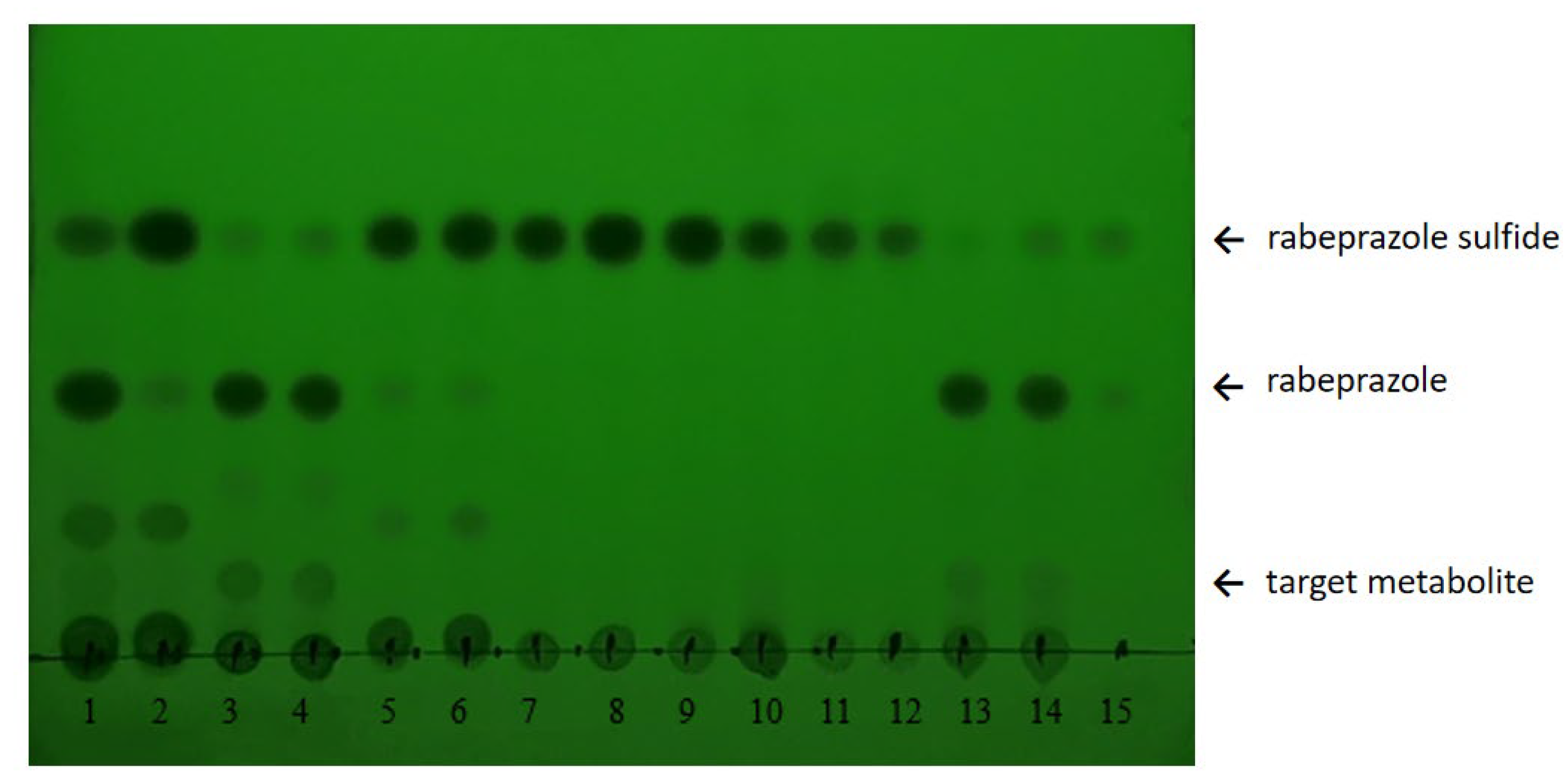
2.1. Whole-Cell Biotransformation Results of Rabeprazole Sulfide

Among the seven strains, Cunninghamella elegans 3.910, Cunninghamella echimnulata 3.967, Cunninghamella blakesleeana 3.970, and Absidia coerulea 41,050 displayed different transformation results for rabeprazole sulfide. Cunninghamella elegans 3.910 and Cunninghamella blakesleeana 3.970 demonstrated a rather significant major metabolite spectrum, whereas Gibberella fujiluroi 40,272, Gibberella sp. 2498, and Caldariomyces fumago 16,373 did not indicate any positive transformation result. According to the transformation TLC results (Figure 2), the fungus Cunninghamella blakesleeana 3.970 converted rabeprazole sulfide to rabeprazole and another, novel compound, and the conversion rate of this new compound was high. Therefore, Cunninghamella blakesleeana 3.970 was selected for further study.

2.2. Optimization of Transformation Media

In order to obtain the transformation products of rabeprazole sulfide, four microbial media were screened. The evaluation criteria were set according to both the growth of the strain and the HPLC detection diagram for the transformation solution. According to Figure S1, fewer mycelium balls were observed in medium 1 and medium 2, and the color of the mycelium ball was a little pink in medium 3, which is not its usual appearance. Only in medium 4 were the numbers of the mycelium balls ambient and the growth of mycelium good.
It can be seen from the HPLC results in Figure S1 that under the same substrate concentration, the ability of transformation media 1, 2, and 3 to transform rabeprazole sulfide is low, and the transformation products are relatively miscellaneous. However, transformation medium 4 had a higher ability to transform rabeprazole sulfide, and only one transformation product was produced. Therefore, based on the results in Figures S1 and S2, medium 4 was selected as the medium for subsequent fermentation.

2.3. Single-Factor Evaluation for Rabeprazole Sulfide Biotransformation

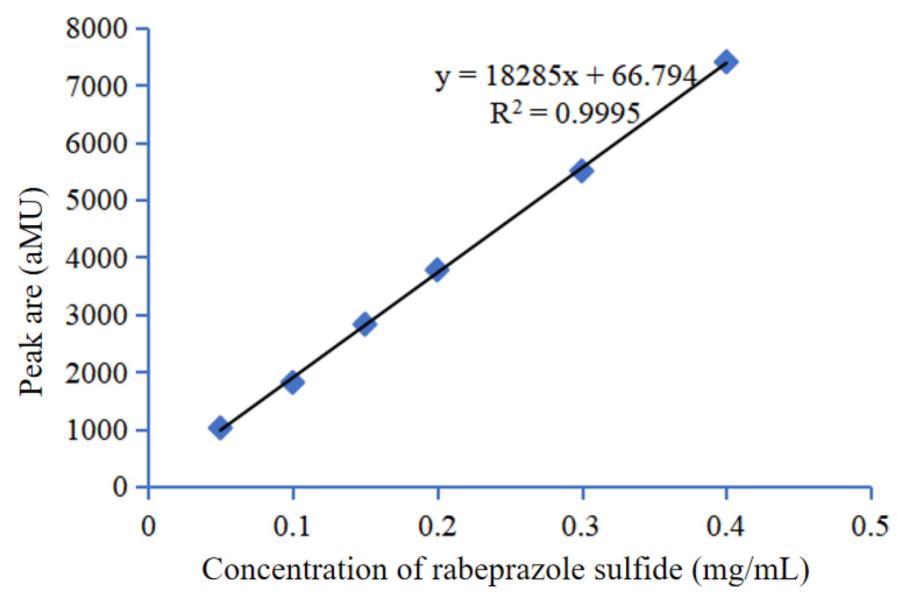
2.3.1. Standard Curve of Rabeprazole Sulfide

Figure 3 indicates that the linear correlation coefficient R2 = 0.9995 and the correlation equation y = 18,285x + 66.794 for the concentration and peak area of rabeprazole sulfide, with a good linear correlation. This standard curve can be used to calculate the conversion rate of rabeprazole sulfide.

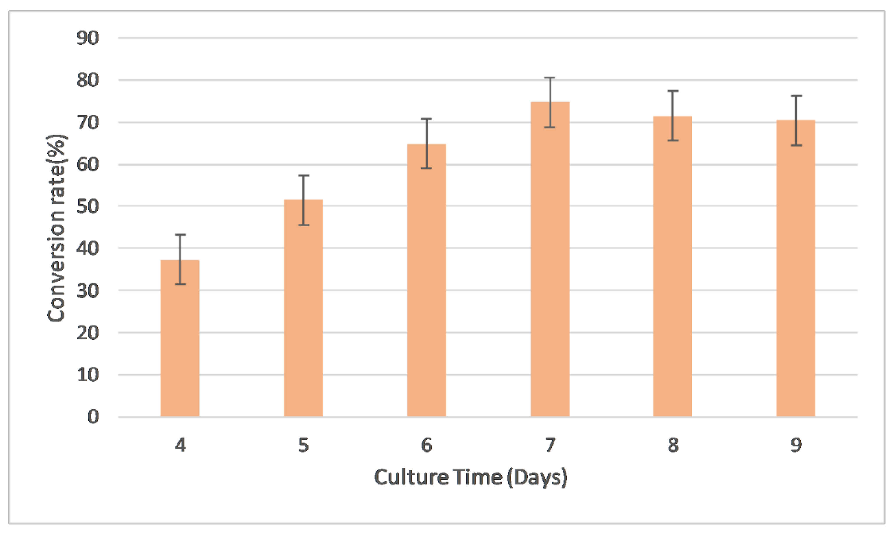
2.3.2. Evaluation of Culture Time

The culture duration of whole-cell catalysis has an important effect on the reaction. If the reaction time is too short, the reaction may not be accomplished. If the reaction time is too long, product inhibition may occur and the total transformation rate may decrease. In this section, we investigate the effect of conversion time on the production of metabolites; the results are shown in Figure 4.
When the transformation was performed at 28 °C, 200 rpm, it could be seen that the culture time ranged between 6 and 9 days. A culture time of 7 days resulted in the optimal conversion rate of 74.71%.

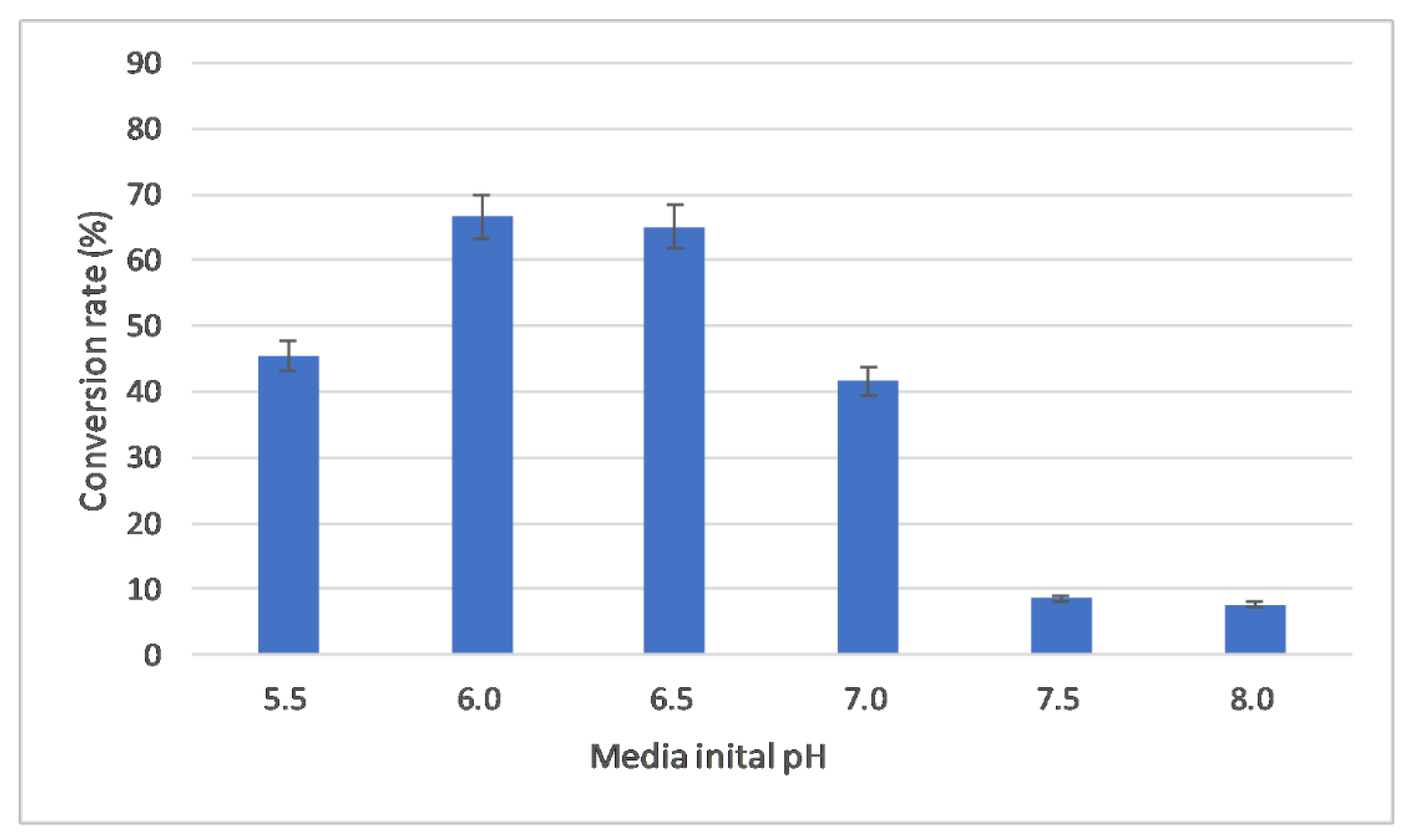
2.3.3. Evaluation of Initial pH

The initial pH of the media is vital for strain growth and transformation. A pH that is too low or high will cause the strains to undergo non-ambient growth and affect their metabolism capability. This is why different strains desire specific initial pH values in their media. In this section, a pH range of 5.5 to 8.0 was selected for the investigation. From Figure 5, it can be seen that both the acid and alkaline conditions are not fit for biotransformation, so the best initial pH is set to 6.0.

2.3.4. Evaluation of Media Volume

The media volume is crucial for the transformation effects, which are related to the dissolved oxygen when the conical flask volume is constant. Media volumes from 30 mL to 120 mL were selected for the investigation. From Figure 6, it can be seen that a media volume of 50 mL displayed the best transformation effect.

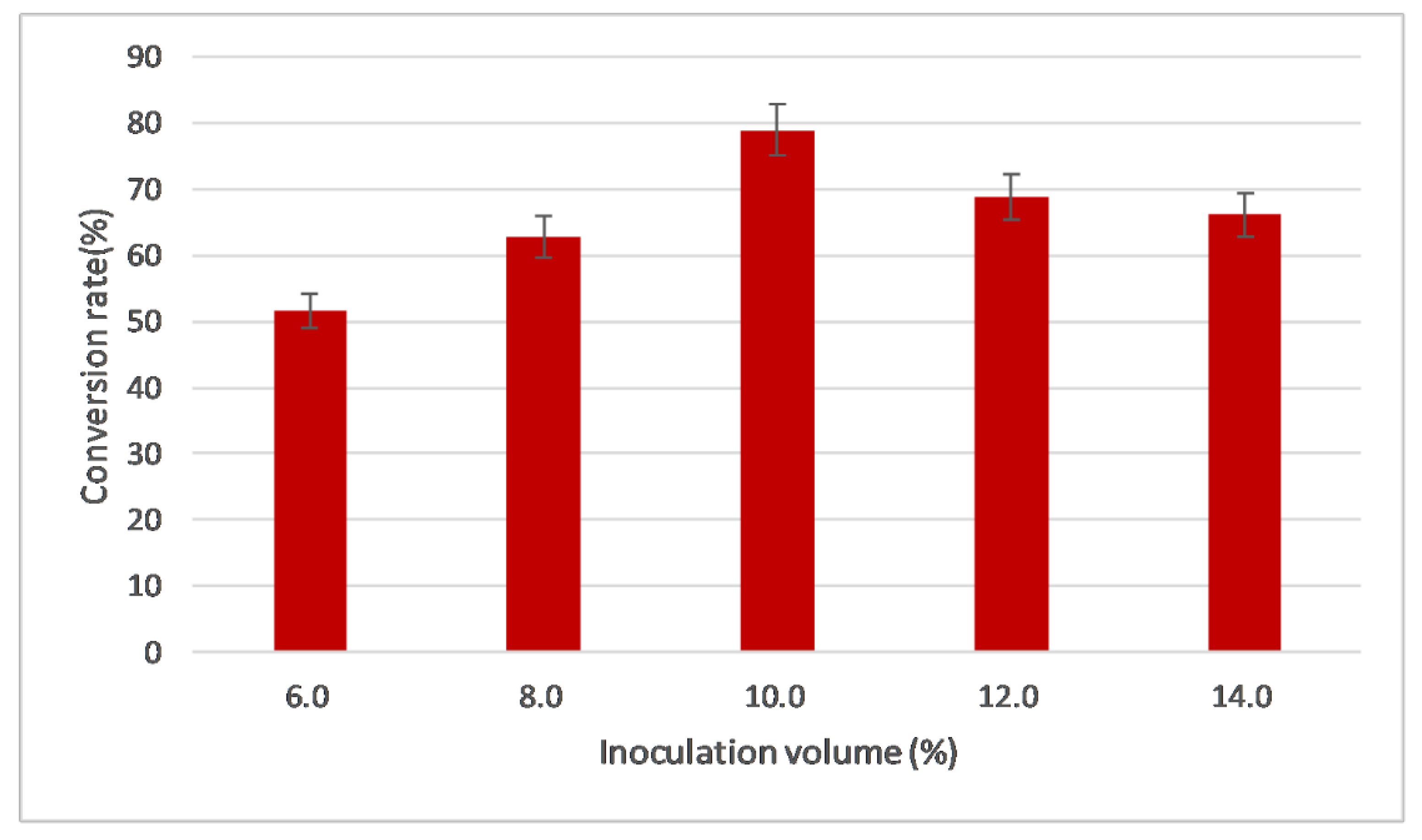
2.3.5. Evaluation of Inoculation Volume

Inoculation volume is a critical factor for the biotransformation process. If the inoculation volume is high, the strains will grow to the exponential phase too early and the nutrients will be depleted quickly, such that the enzymes in the strains may not sufficiently be induced and expressed, and the biotransformation may not be adequate. If the inoculation volume is low, the strains may take too long a time to grow and induce enzymes, which may decrease their metabolism capabilities. Based our previous experiences, inoculation volumes ranging from 6% to 14% were selected for the investigation.
From Figure 7, it can be seen that an inoculation volume of 10% is the best for biotransformation.

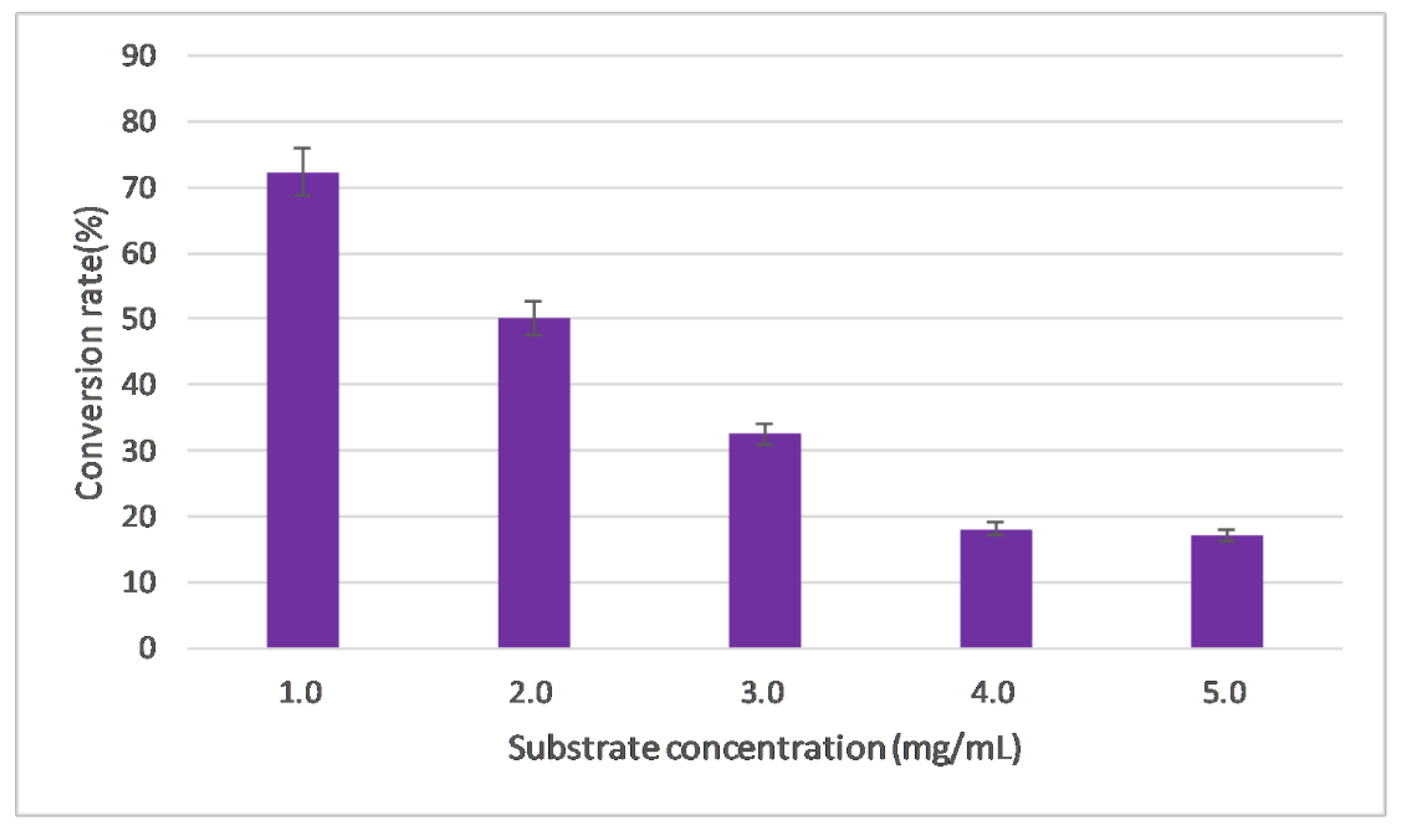
2.3.6. Evaluation of Substrate Concentration

In this study, rabeprazole sulfide is not a natural substrate, which may cause severe inhibition and toxic effects for the strains. Hence, a proper substrate concentration is essential for successful biotransformation. A substrate concentration range from 1 mg/mL to 5 mg/mL was set for the investigation.
From Figure 8, it can be seen from the decreasing conversion rate tendency that 1 mg/mL substrate concentration is the best for biotransformation.

2.4. Orthogonal Optimization

According to the above single-factor investigation results, a 5-factor, 4-level, orthogonal L16(45) optimization process was designed, as shown in Table 1.
After the design and execution of the experiments, it could be seen that the strength of impact of the various factors on biotransformation was in the order D > B > E > C > A, i.e., the most significant influence was that of the initial pH value of the media, as shown in Table 2. When the range analysis was performed, it was found that the best combination was D2B1E3C4A4, so an initial pH of 6.0, inoculation volume of 8%, substrate concentration of 3 mg/mL, media volume of 100 mL, and culture time of 8 days were the best values. These values are consistent between the best results from the orthogonal design and those from the No.13 experimental group (total 16 groups), with a conversion rate of 72.4%. This is almost a double increase in the conversion rate compared with the unoptimized results.

2.5. Structure Identification for the Rabeprazole Sulfide Biotransformation

The isolated metabolite was resolved in DMSO for the MS and NMR analysis. According to the ESI-MS results, showing that the m/z [M+H]+ is 330.25 (Figure S3) and the m/z [M-H] is 328.27 (Figure S4), it could be inferred that the target metabolite has a molecular weight of 329.12, which is only a Dalton loss of 14 compared with the original rabeprazole sulfide with a molecular weight of 343.44. Hence, the loss of a methyl group is assumed.
According to the comparison of the NMR data between the substrate and metabolite, it could be found that a methyl and a carbon signal are absent in the H spectrum and C spectrum. Combined with the MS and NOESY data (Figure S7) indicating a methyl loss, it can be concluded that the target metabolite is the O-dimethyl rabeprazole sulfide (Figure 1). The analysis is given below.
1H-NMR (600 MHz, DMSO-d6) δH: 8.23 (1H, d, J = 5.6 Hz), 7.45 (2H, dd, J1 = 5.8, J2 = 3.2 Hz), 7.11 (2H, dd, J1 = 5.9, J2 = 3.1 Hz), 6.95 (1H, d, J = 5.6 Hz), 4.69 (2H, s), 4.12 (2H, t, J = 6.2 Hz), 3.58 (2H, t, J = 6.2 Hz), 2.21 (3H, s), 1.87–1.91 (2H, m).
13C-NMR (150 MHz, DMSO-d6) δC: 163.27, 155.16, 150.90, 148.24, 121.73, 120.20, 106.78, 65.50, 57.56, 36.74, 32.31, 10.88.

3. Materials and Methods

3.1. Materials and Strains

Rabeprazole sulfide was synthesized in the Department of Pharmaceutical Engineering, Shenyang Pharmaceutical University. Methanol and acetonitrile were purchased from Concord Technology Co., Ltd. (Tianjin, China). Peptone and yeast extract were purchased from HopeBio Co., Ltd. (Qingdao, China). All other chemical reagents were purchased from Yuwang Chemical Co., Ltd. (Shenyang, China).
Cunninghamella blakesleeana 3.970 and Cunninghamella elegans 3.910 were stored in our lab reservoir. Cunninghamella echimnulata 3.967, Absidia coerulea 41,050, Gibberella fujiluroi 40,272, Gibberella sp. 2498, and Caldariomyces fumago 16,373 were purchased from China Center of Industrial Culture Collection (CICC).

3.2. Media and Strain Cultivations

Slant and agar media consisted of potato (200 g), glucose (20 g), agar (20 g), and 1000 mL distilled water. Seed culture media consisted of potato starch (45 g), yeast extract (3 g), corn steep liquor (10 g), CaCO3 (3 g), MgSO4 (0.5 g), and FeSO4 (0.05 g). Four kinds of biotransformation culture media ingredients are listed in Table 3. For all media, the pH was adjusted to 6.5 with 6 M HCl, autoclaved at 115 °C for 30 min, and cooled before use.
Two loops of mycelium from a fresh growth slant were inoculated into the seed culture media and cultured at 28 °C, 200 rpm for 48 h. Then, 5 mL of seed culture was inoculated into the transformation media (50 mL in a 250 mL conical flask, rabeprazole sulfide as substrate) and cultured at 28 °C, 200 rpm for 5 days. Substrate controls were set without inoculating the fungi into the media and strain controls were set without adding the substrate into the media, with all other conditions remaining the same.

3.3. Thin-Layer Chromatography (TLC)

The biotransformation results of the fungal extracts were analyzed by TLC performed on a Merck Silica gel 60 F254 plate (Merck, Darmstadt, Germany) with ethyl acetate:methanol:ammonia water (90:9:1 v/v/v) as the mobile phase, and a UV detection wavelength of 254 nm. A mixture of rabeprazole sulfide and rabeprazole (10 mg) was dissolved in 100 mL of isopropyl alcohol used as a standard solution. For TLC detection, 10 µL of standard solution and fungal extracts was spotted onto TLC plates.

3.4. Extraction of Metabolites and HPLC Detection

Biotransformation cultures were collected, ultrasonically treated for 30 min, and centrifugated at 3000 rpm for 10 min to retrieve the supernatant. Pellets were washed and extracted three times using ethyl acetate. The combined water phase and organic phase were extracted three times with an equal volume of ethyl acetate. The organic phase was then isolated and evaporated by reduced pressure distillation to obtain the raw metabolites. The raw metabolites were dissolved in methanol for further use.
The HPLC detection process for the metabolites was derived from the Pharmacopeia of the People’s Republic of China (2020 Volume II) and optimized according to our practical work. For High-performance liquid chromatography (HPLC), a Shimadzu WondaSil C18 Superb (250 × 4.6 mm × 5 μm) column (Shimadzu Corp., Kyoto, Japan) was used. The mobile phase consisted of 0.015 mol/L Na2HPO4:acetonitrile = 60:40, the detection wavelength was 290 nm, the column temperature was 30 °C, the loading speed was 1 mL/min, and the loading volume was 10 μL.

3.5. Isolation and Identification of Major Metabolite

The isolation of the target metabolite was performed by semipreparative HPLC using an Elite SinoChrom ODS-BP (250 × 10 mm × 5 μm, Dalian, China) consisting of a quaternary pump, a vacuum degasser, a variable wavelength detector, and an autosampler. The mobile phase consisted of acetonitrile and water in different percentages, and the proper fractions were collected.
ESI-MS and NMR analysis were performed under standard conditions.

3.6. Determination of the Standard Curve

Rabeprazole sulfide was accurately weighed, and a solution of 0.40 mg/mL was prepared, diluted step by step with methanol. Standard samples of 0.40, 0.30, 0.20, 0.15, 0.10, and 0.05 mg/mL were prepared and detected with HPLC, and a standard curve was drawn with rabeprazole sulfide concentration as the abscissa and peak area as the ordinate. This standard curve was used to calculate the conversion of rabeprazole sulfide. The total conversion rate was calculated according to the following equation:
Conversion   rate   % = 1 residue   amount   of   raberprazole   sulfide Starting   amount   of   rabeprazole   sulfide

4. Conclusions and Discussion

By screening seven filamentous fungi, rabeprazole sulfide was the most efficiently decomposed into O-dimethyl rabeprazole sulfide by Cunninghamella blakesleeana 3.970, with a conversion rate of 72.4%. This is not a common biotransformation reaction type, with only few studies having suggested the N-demethylation [36,37] and O-dimethyl reactions [38,39].
The original aim of this study was to explore the possibility of whether these fungi could exhibit some oxidase activity so that the rabeprazole sulfide could be transformed into rabeprazole, i.e., to determine whether the thioether structure could be oxidized into the sulfoxide. Despite Cunninghamella species having exhibited various kinds of oxidation in previous works, no oxidation metabolites were identified in this research. Several reports have shown that Absidia and Gibberella species could exhibit oxidase activity as their P450, but positive oxidation metabolites have yet to be generated. More importantly, Caldariomyces fumago was selected for its strong inner chloroperoxidase expression, which exhibited multiple oxidation types, such as epoxidation [40,41], hydroxylation [42,43,44,45,46,47], halogenation [48], etc. However, none of the above reactions were identified in this rabeprazole sulfide biotransformation.
Rabeprazole sulfide is actually not a natural substrate of the fungus, which was mentioned in the previous section on the influence of substrate concentration on the conversion rate. With the increase in substrate concentration, the conversion rate decreases (Figure 8), and a too-high concentration may produce some inhibition. It is speculated that the O-demethylation reaction of rabeprazole sulfide is not completed in one step. It may be that rabeprazole sulfide is first converted into one or several intermediate products and then further converted into O-demethyl rabeprazole sulfide.
Some microorganisms can transform drugs and other xenobiotic compounds in a manner similar to that in mammals, and the utilization of microbial systems as models for mimicking and predicting the metabolism of drugs in humans and animals has received considerable attention. Liu et al. reported that the main metabolic pathways of Cunninghamella blakesleeana to transform verapamil are N-dealkylation, O-demethylation, and sulfate coupling, and the O-demethylated metabolites of verapamil have the same potency as the parent drug [49]. Xie ZY et al. reported that approximately 92.5% of pantoprazole was metabolized to six metabolites by Cunninghamella blakesleeana AS 3.153, one of which was 4′-O-demethyl-pantoprazole thioether [30]. These studies did not deeply explore the mechanism of O-demethylation when Cunninghamella blakesleeana transformed the substrate. Therefore, it is of interest to further study the mechanism of the O-demethylation of Cunninghamella blakesleeana and the pharmacological or biological characteristics of these O-demethylated metabolites.
In addition, further investigation should be undertaken in the hunt for fungi that may oxidize thioether to sulfoxide. Meanwhile, other rational designs for the engineered enzyme to facilitate this reaction are also welcomed.

Supplementary Materials

The following are available online at https://www.mdpi.com/article/10.3390/catal13010015/s1, Figure S1. Growth of Cunninghamella blakesleeana 3.970 in 4 different transformation media. Figure S2. HPLC detection results of Cunninghamella blakesleeana 3.970 in 4 different transformation media. Figure S3. HPLC detection results of rabeprazole sulfide and its metabolites. Figure S4. HPLC detection results of rabeprazole sulfide metabolites for MS and NMR analysis. Figure S5. MS(ESI)m/z[M+H]+ diagram for the metabolite. Figure S6. MS(ESI)m/z[M+H] diagram for the metabolite. Figure S7. 1H NMR diagram for the O-demethyl rabeprazole sulfide. Figure S8. 13C NMR diagram for the O-demethyl rabeprazole sulfide. Figure S9. 1H NOESY diagram for the O-demethyl rabeprazole sulfide.

Author Contributions

M.S. (data curation, investigation); H.Z. (investigation, methodology); J.W. (rabeprazole and its sulfide preparation); W.X. (Weizhuo Xu) and W.X. (Wei Xu) (resources, supervision, writing—review and editing). All authors have read and agreed to the published version of the manuscript.

Funding

This research received no external funding.

Data Availability Statement

Data are available upon reasonable request.

Conflicts of Interest

The authors declare no conflict of interest.

References

  1. FDA. Approval for Aciphex (Rabeprazole Sodium) Tablets. 2002. Available online: https://www.accessdata.fda.gov/drugsatfda_docs/nda/2002/021456_aciphex.cfm (accessed on 25 April 2003).
  2. Duan, Z.; Wang, Y.; Zhang, L.; Cao, X.; Fu, L.; Li, Z.; Zhang, J. An application of continuous flow microreactor in the synthesis and extraction of rabeprazole. Int. J. Chem. React. Eng. 2021, 19, 287–294. [Google Scholar] [CrossRef]
  3. Nishiguchi, S.; Izumi, T.; Kouno, T.; Sukegawa, J.; Ilies, L.; Nakamura, E. Synthesis of Esomeprazole and Related Proton Pump Inhibitors through Iron-Catalyzed Enantioselective Sulfoxidation. ACS Catal. 2018, 8, 9738–9743. [Google Scholar] [CrossRef]
  4. Lu, C.; Jia, Y.; Song, Y.; Li, X.; Sun, Y.; Zhao, J.; Wang, S.; Shi, L.; Wen, A.; Ding, L. Application of a liquid chromatographic/tandem mass spectrometric method to a urinary excretion study of rabeprazole and two of its metabolites in healthy human urine. J. Chromatogr. B 2015, 988, 75–80. [Google Scholar] [CrossRef] [PubMed]
  5. Zannikos, P.N.; Doose, D.R.; Leitz, G.J.; Rusch, S.; Gonzalez, M.D.; Solanki, B.; Haddad, I.; Mulberg, A.E. Pharmacokinetics and Tolerability of Rabeprazole in Children 1 to 11 Years Old with Gastroesophageal Reflux Disease. J. Pediatr. Gastroenterol. Nutr. 2011, 52, 691–701. [Google Scholar] [CrossRef] [PubMed]
  6. Ren, S.; Park, M.-J.; Kim, A.; Lee, B.-J. In vitro metabolic stability of moisture-sensitive rabeprazole in human liver microsomes and its modulation by pharmaceutical excipients. Arch. Pharmacal Res. 2008, 31, 406–413. [Google Scholar] [CrossRef]
  7. Miura, M.; Satoh, S.; Tada, H.; Habuchi, T.; Suzuki, T. Stereoselective metabolism of rabeprazole-thioether to rabeprazole by human liver microsomes. Eur. J. Clin. Pharmacol. 2005, 62, 113–117. [Google Scholar] [CrossRef]
  8. Sharara, A.I. Rabeprazole: The role of proton pump inhibitors in Helicobacter pylori eradication. Expert Rev. Anti-Infect. Ther. 2005, 3, 863–870. [Google Scholar] [CrossRef]
  9. Lun, J.; Ma, S.; Xue, M.; Zhao, P.; Song, Y.; Guo, X. Simultaneous enantiomeric analysis of five proton-pump inhibitors in soil and sediment using a modified QuEChERS method and chiral high performance liquid chromatography coupled with tandem mass spectrometry. Microchem. J. 2020, 160, 105625. [Google Scholar] [CrossRef]
  10. Marelli, S.; Pace, F. Rabeprazole for the treatment of acid-related disorders. Expert Rev. Gastroenterol. Hepatol. 2012, 6, 423–435. [Google Scholar] [CrossRef]
  11. Yousuf, M.; Jamil, W.; Mammadova, M.Y.A.K. Microbial Bioconversion: A Regio-specific Method for Novel Drug Design and Toxicological Study of Metabolites. Curr. Pharm. Biotechnol. 2019, 20, 1156–1162. [Google Scholar] [CrossRef]
  12. Asha, S.; Vidyavathi, M. Cunninghamella—A microbial model for drug metabolism studies—A review. Biotechnol. Adv. 2009, 27, 16–29. [Google Scholar] [CrossRef]
  13. Murphy, C.D. Drug metabolism in microorganisms. Biotechnol. Lett. 2015, 37, 19–28. [Google Scholar] [CrossRef]
  14. Keum, Y.-S.; Lee, Y.-H.; Kim, J.-H. Metabolism of methoxychlor by Cunninghamella elegans ATCC36112. J. Agric Food Chem. 2009, 57, 7931–7937. [Google Scholar] [CrossRef]
  15. Choudhary, M.I.; Khan, N.T.; Musharraf, S.G.; Anjum, S.; Rahman, A.U. Biotransformation of adrenosterone by filamentous fungus, Cunninghamella elegans. Steroids 2007, 72, 923–929. [Google Scholar] [CrossRef]
  16. Hanson, R.L.; Matson, J.A.; Brzozowski, D.B.; LaPorte, T.L.; Springer, D.M.; Patel, R.N. Hydroxylation of Mutilin by Streptomyces griseus and Cunninghamella echinulata. Org. Process Res. Dev. 2002, 6, 482–487. [Google Scholar] [CrossRef]
  17. Ibrahim, A.; Khalifa, S.I.; Khafagi, I.; Youssef, D.T.; Khan, S.; Mesbah, M.; Khan, I. Microbial Metabolism of Biologically Active Secondary Metabolites from Nerium oleander L. Chem. Pharm. Bull. 2008, 56, 1253–1258. [Google Scholar] [CrossRef] [Green Version]
  18. Kouzi, S.A.; Chatterjee, P.; Pezzuto, J.M.; Hamann, M.T. Microbial Transformations of the Antimelanoma Agent Betulinic Acid. J. Nat. Prod. 2000, 63, 1653–1657. [Google Scholar] [CrossRef]
  19. Ning, L.; Zhan, J.; Qu, G.; Zhong, L.; Guo, H.; Bi, K.; Guo, D. Biotransformation of triptolide by Cunninghamella blakesleana. Tetrahedron 2003, 59, 4209–4213. [Google Scholar] [CrossRef]
  20. Zhang, D.; Evans, F.E.; Freeman, J.P.; Duhart, B.; Cerniglia, C.E. Biotransformation of amitriptyline by Cunninghamella elegans. Drug Metab. Dispos. 1995, 23, 1417–1425. [Google Scholar]
  21. Wu, Y.; Lu, Y.; Yi, Y.; Wang, A.; Wang, W.; Yang, M.; Fan, B.; Chen, G. Biotransformation of asiatic acid by Cunninghamella echinulata and Circinella muscae to discover anti-neuroinflammatory derivatives. Nat. Prod. Res. 2022, 36, 1–6. [Google Scholar] [CrossRef]
  22. Khan, M.F.; Murphy, C.D. Cytochrome P450 5208A3 is a promiscuous xenobiotic biotransforming enzyme in Cunninghamella elegans. Enzym. Microb. Technol. 2022, 161, 110102. [Google Scholar] [CrossRef] [PubMed]
  23. Bai, Y.; Zhao, Y.; Gao, X.; Zhang, D.; Ma, Y.; Yang, L.; Sun, P. A Novel Antimalarial Metabolite in Erythrocyte from the Hydroxylation of Dihydroartemisinin by Cunninghamella elegans. Front. Chem. 2022, 10, 850133. [Google Scholar] [CrossRef] [PubMed]
  24. Sousa, I.P.; de SousaTeixeira, M.V.; Freitas, J.A.; Ferreira, A.G.; Pires, L.M.; Santos, R.A.; Heleno, V.C.G.; Furtado, N.A.J.C. Production of More Potent Anti- Candida Labdane Diterpenes by Biotransformation Using Cunninghamella elegans. Chem. Biodivers. 2022, 19, e202100757. [Google Scholar] [CrossRef] [PubMed]
  25. Barrero, A.F.; Oltra, J.E.; Raslan, A.D.S.; Saúde, D.A. Microbial Transformation of Sesquiterpene Lactones by the Fungi Cunninghamella echinulata and Rhizopus oryzae. J. Nat. Prod. 1999, 62, 726–729. [Google Scholar] [CrossRef] [PubMed] [Green Version]
  26. Zhang, D.; Hansen, E.B., Jr.; Deck, J.; Heinze, T.M.; Henderson, A.; Korfmacher, W.A.; Cerniglia, C.E. Fungal transformations of antihistamines: Metabolism of cyproheptadine hydrochloride by Cunninghamella elegans. Xenobiotica 1997, 27, 301–315. [Google Scholar] [CrossRef]
  27. Zhou, L.; Xu, W.; Chen, Y.; Zhao, J.; Yu, N.; Fu, B.; You, S. Stereoselective epoxidation of curcumol and curdione by Cunninghamella elegans AS 3.2028. Catal. Commun. 2012, 28, 191–195. [Google Scholar] [CrossRef]
  28. Grafinger, K.E.; Wilke, A.; König, S.; Weinmann, W. Investigating the ability of the microbial model Cunninghamella elegans for the metabolism of synthetic tryptamines. Drug Test. Anal. 2018, 11, 721–729. [Google Scholar] [CrossRef]
  29. Nykodemová, J.; Šuláková, A.; Palivec, P.; Češková, H.; Rimpelová, S.; Šíchová, K.; Leonhardt, T.; Jurásek, B.; Hájková, K.; Páleníček, T.; et al. 2C-B-Fly-NBOMe Metabolites in Rat Urine, Human Liver Microsomes and C. elegans: Confirmation with Synthesized Analytical Standards. Metabolites 2021, 11, 775. [Google Scholar] [CrossRef]
  30. Xie, Z.Y.; Huang, H.H.; Zhong, D.F. Biotransformation of pantoprazole by the fungus Cunninghamella blakesleeana. Xenobiotica 2005, 35, 467–477. [Google Scholar] [CrossRef]
  31. Sutherland, J.B.; Freeman, J.P.; Heinze, T.M.; Moody, J.D.; Parshikov, I.A.; Williams, A.J.; Zhang, D. Oxidation of phenothiazine and phenoxazine by Cunninghamella elegans. Xenobiotica 2001, 31, 799–809. [Google Scholar] [CrossRef]
  32. Zhang, D.; Freeman, J.P.; Sutherland, J.B.; Walker, A.E.; Yang, Y.; Cerniglia, C.E. Biotransformation of chlorpromazine and methdilazine by Cunninghamella elegans. Appl. Environ. Microbiol. 1996, 62, 798–803. [Google Scholar] [CrossRef]
  33. Bachmann, F.; zu Schwabedissen, H.E.M.; Duthaler, U.; Krähenbühl, S. Cytochrome P450 1A2 is the most important enzyme for hepatic metabolism of the metamizole metabolite 4-methylaminoantipyrine. Br. J. Clin. Pharmacol. 2021, 88, 1885–1896. [Google Scholar] [CrossRef]
  34. Ren, H.; Dhanaraj, P.; Enoch, I.V.; Paulraj, M.S.; Paulraj, M.S. Synthesis and Biological Evaluation of 4-Aminoantipyrine Analogues. Med. Chem. 2022, 18, 26–35. [Google Scholar] [CrossRef]
  35. Eliwa, D.; Albadry, M.A.; Ibrahim AR, S.; Kabbash, A.; Meepagala, K.; Khan, I.A.; El-Aasr, M.; Ross, S.A. Biotransformation of papaverine and in silico docking studies of the metabolites on human phosphodiesterase 10a. Phytochemistry 2021, 183, 112598. [Google Scholar] [CrossRef]
  36. Yang, W.; Jiang, T.; Acosta, D.; Davis, P.J. Microbial models of mammalian metabolism: Involvement of cytochrome P450 in the N-demethylation of N-methylcarbazole by Cunninghamella echinulata. Xenobiotica 1993, 23, 973–982. [Google Scholar] [CrossRef]
  37. Hansen, E.B., Jr.; Cho, B.P.; Korfmacher, W.A.; Cerniglia, C.E. Fungal transformations of antihistamines: Metabolism of brompheniramine, chlorpheniramine, and pheniramine to N-oxide and N-demethylated metabolites by the fungus Cunninghamella elegans. Xenobiotica 1995, 25, 1081–1092. [Google Scholar] [CrossRef]
  38. Fan, H.X.; Zhou, Z.Q.; Peng, J.; Wu, B.J.; Chen, H.R.; Bao, X.F.; Mu, Z.Q.; Jiao, W.H.; Yao, X.S.; Gao, H. A microbial model of mammalian metabolism: Biotransformation of 4,5-dimethoxyl-canthin-6-one using Cunninghamella blakesleeana CGMCC 3.970. Xenobiotica 2017, 47, 284–289. [Google Scholar] [CrossRef]
  39. Ibrahim, A.-R.S.; Galal, A.M.; Ahmed, M.S.; Mossa, G.S. O-Demethylation and Sulfation of 7-Methoxylated Flavanones by Cunninghamella elegans. Chem. Pharm. Bull. 2003, 51, 203–206. [Google Scholar] [CrossRef] [Green Version]
  40. Liu, Y.; Wang, Y.; Jiang, Y.; Hu, M.; Li, S.; Zhai, Q. Biocatalytic synthesis of C3 chiral building blocks by chloroperoxidase-catalyzed enantioselective halo-hydroxylation and epoxidation in the presence of ionic liquids. Biotechnol. Prog. 2015, 31, 724–729. [Google Scholar] [CrossRef]
  41. Morozov, A.N.; Chatfield, D.C. Chloroperoxidase-catalyzed epoxidation of cis-beta-methylstyrene: Distal pocket flexibility tunes catalytic reactivity. J. Phys. Chem. B 2012, 116, 12905–12914. [Google Scholar] [CrossRef] [Green Version]
  42. de Hoog, H.M.; Nallani, M.; Cornelissen, J.J.L.M.; Rowan, A.E.; Nolte, R.J.M.; Arends, I.W.C.E. Biocatalytic oxidation by chloroperoxidase from Caldariomyces fumago in polymersome nanoreactors. Org. Biomol. Chem. 2009, 7, 4604–4610. [Google Scholar] [CrossRef] [PubMed]
  43. Kellner, H.; Pecyna, M.J.; Buchhaupt, M.; Ullrich, R.; Hofrichter, M. Draft Genome Sequence of the Chloroperoxidase-Producing Fungus Caldariomyces fumago Woronichin DSM1256. Genome Announc. 2016, 4, e00774-16. [Google Scholar] [CrossRef] [PubMed] [Green Version]
  44. Dong, X.; Li, H.; Jiang, Y.; Hu, M.; Li, S.; Zhai, Q. Rapid and efficient degradation of bisphenol A by chloroperoxidase from Caldariomyces fumago: Product analysis and ecotoxicity evaluation of the degraded solution. Biotechnol. Lett. 2016, 38, 1483–1491. [Google Scholar] [CrossRef] [PubMed]
  45. Buchhaupt, M.; Hüttmann, S.; Sachs, C.C.; Bormann, S.; Hannappel, A.; Schrader, J. Caldariomyces fumago DSM1256 Contains Two Chloroperoxidase Genes, Both Encoding Secreted and Active Enzymes. J. Mol. Microbiol. Biotechnol. 2015, 25, 237–243. [Google Scholar] [CrossRef] [PubMed]
  46. Buchhaupt, M.; Hüttmann, S.; Schrader, J. White Mutants of Chloroperoxidase-Secreting Caldariomyces fumago as Superior Production Strains, Revealing an Interaction between Pigmentation and Enzyme Secretion. Appl. Environ. Microbiol. 2012, 78, 5923–5925. [Google Scholar] [CrossRef] [Green Version]
  47. Buchhaupt, M.; Ehrich, K.; Hüttmann, S.; Guder, J.; Schrader, J. Over-expression of chloroperoxidase in Caldariomyces fumago. Biotechnol. Lett. 2011, 33, 2225–2231. [Google Scholar] [CrossRef]
  48. Höfler, G.T.; But, A.; Hollmann, F. Haloperoxidases as catalysts in organic synthesis. Org. Biomol. Chem. 2019, 17, 9267–9274. [Google Scholar] [CrossRef] [Green Version]
  49. Sun, L.; Huang, H.-H.; Liu, L.; Zhong, D.-F. Transformation of Verapamil by Cunninghamella blakesleeana. Appl. Environ. Microbiol. 2004, 70, 2722–2727. [Google Scholar] [CrossRef]
Figure 1. Rabeprazole sulfide and its O-demethyl metabolite obtained by the biotransformation of Cunninghamella blakesleeana 3.970.
Catalysts 13 00015 g001
Figure 2. TLC screening results for seven strains of filamentous fungi for rabeprazole sulfide. Lanes 1 and 2, Cunninghamella elegans 3.910, 3 and 4, Cunninghamella echimnulata 3.967, 5 and 6, Absidia coerulea 41,050, 7 and 8, Gibberella sp. 2498, 9 and 10, Caldariomyces fumago 16,373, 11 and 12, Gibberella fujiluroi 40,272, 13 and 14, Cunninghamella blakesleeana 3.970, and 15, standard mixture of rabeprazole sulfide and rabeprazole.
Catalysts 13 00015 g002
Figure 3. Standard curve of rabeprazole sulfide.
Catalysts 13 00015 g003
Figure 4. Effects of culture time on metabolite conversion rate (initial pH = 6.5, media volume 50 mL, inoculation volume 10%, substrate concentration 1 mg/mL).
Catalysts 13 00015 g004
Figure 5. Effects of the initial pH of the media on the metabolite conversion rate (culture time 5 d, media volume 50 mL, inoculation volume 10%, substrate concentration 1 mg/mL).
Catalysts 13 00015 g005
Figure 6. Effects of media volume on metabolite conversion rate (initial pH = 6.5, culture time 5 d, inoculation volume 10%, substrate concentration 1 mg/mL).
Catalysts 13 00015 g006
Figure 7. Effects of inoculation volume on metabolite conversion rate (initial pH = 6.5, culture time 5 d, media volume 50 mL, substrate concentration 1 mg/mL).
Catalysts 13 00015 g007
Figure 8. Effects of substrate concentration on metabolite conversion rate (initial pH = 6.5, culture time 5 d, media volume 50 mL, substrate concentration 1 mg/mL).
Catalysts 13 00015 g008
Table 1. Orthogonal experimental factors and level assignments.
FactorA/Culture Time (Days)B/Inoculation
Volume (%)
C/Media Volume (mL)D/Media Starting pHE/Substrate
Concentration (g/L)
Level
158305.51
2610506.02
3712806.53
48141007.04
Table 2. Orthogonal experimental results and analysis.
Factors
Experiment No.
ABCDEConversion Rate
1111110.352
2122220.315
3133330.348
4144440.201
5212340.213
6221430.124
7234120.336
8243210.566
9313420.347
10324310.171
11331240.135
12342130.343
13414230.724
14423140.053
15432410.094
16441230.033
K11.2161.6360.6441.0841.184
K21.240.6640.9641.8121.032
K30.9960.9121.3120.7641.54
K41.3041.1441.4320.7640.604
k10.3040.4090.1610.2710.296
k20.3100.1660.2410.4530.258
k30.2490.2280.3280.1910.385
k40.3260.2860.3580.1910.151
R(Range analysis)0.0840.2430.1970.2440.234
Best combinationA2B1C4D2E1
Table 3. Four different biotransformation media formulas.
IngredientsNo.1 (g)No.2 (g)No.3 (g)No.4 (g)
Sucrose30 15
Yeast extracts200.5 2
Corn steep liquor5
Peptone 0.555
Glucose 41550
K2HPO4160.514
KH2PO42 5
MgSO40.5 0.50.2
FeSO40.05 0.010.05
KCl 0.50.5
Disclaimer/Publisher’s Note: The statements, opinions and data contained in all publications are solely those of the individual author(s) and contributor(s) and not of MDPI and/or the editor(s). MDPI and/or the editor(s) disclaim responsibility for any injury to people or property resulting from any ideas, methods, instructions or products referred to in the content.

