Review

Nanoparticle Engineered Photocatalytic Paints: A Roadmap to Self-Sterilizing against the Spread of Communicable Diseases

1
Department of Physics, Tuljaram Chaturchand College, Baramati 413102, India
2
Center for Interdisciplanary Research, D. Y. Patil Education Society, Deemed to be University, Kolhapur 416006, India
3
Department of Obstetrics and Gynecology, D. Y. Patil Medical College, Kolhapur 416006, India
4
Centre for Innovative and Applied Research (CIAR), Tuljaram Chaturchand College, Baramati 413102, India
*
Author to whom correspondence should be addressed.
Catalysts 2022, 12(3), 326; https://doi.org/10.3390/catal12030326
Submission received: 10 January 2022 / Revised: 2 March 2022 / Accepted: 7 March 2022 / Published: 11 March 2022
(This article belongs to the Special Issue Photocatalytic Nanomaterials for Abatement of Microorganisms)

Abstract

:
Applications of visible-light photocatalytic engineered nanomaterials in the preparation of smart paints are of recent origin. The authors have revealed a great potential of these new paints for self-sterilizing of the surfaces in hospitals and public places simply with visible light exposure and this is reported for the first time in this review. A recent example of a communicable disease such as COVID-19 is considered. With all precautions and preventions taken as suggested by the World Health Organization (WHO), COVID-19 has remained present for a longer time compared to other diseases. It has affected millions of people worldwide and the significant challenge remains of preventing infections due to SARS-CoV-2. The present review is focused on revealing the cause of this widespread disease and suggests a roadmap to control the spread of disease. It is understood that the transmission of SARS-CoV-2 virus takes place through contact surfaces such as doorknobs, packaging and handrails, which may be responsible for many preventable and nosocomial infections. In addition, due to the potent transmissibility of SARS-CoV-2, its ability to survive for longer periods on common touch surfaces is also an important reason for the spread of COVID-19. The existing antimicrobial cleaning technologies used in hospitals are not suitable, viable or economical to keep public places free from such infections. Hence, in this review, an innovative approach of coating surfaces in public places with visible-light photocatalytic nanocomposite paints has been suggested as a roadmap to self-sterilizing against the spread of communicable diseases. The formulations of different nanoparticle engineered photocatalytic paints with their ability to destroy pathogens using visible light, alongwith the field trials are also summarized and reported in this review. The potential suggestions for controlling the spread of communicable diseases are also listed at the end of the review.

1. Introduction

In recent years, the spread of communicable diseases has created great concern for societal health globally. The spread of the COVID-19 disease is a present example of communicable disease and it is so hazardous that the World Health Organization (WHO) has declared a pandemic. More worse than the severity of the disease is its persistence in the global community for longer than two years. It started in December 2019, still exists even after two years and its eradication is still unpredictable. In light of this situation, our research group which has been involved right from the inception of COVID-19 has taken up the initiative in fighting against this communicable disease based on the backgrounds of nanoscience and nanotechnology [1]. Even before the emergence of COVID-19 disease, our group was working on communicable diseases such as tuberculosis [2,3,4]. The only difference is that tuberculosis is a microbial disease, while COVID-19 is a viral disease caused by SARS-CoV-2 virions (nanobiotic particles). Their methods of spreading and control do differ in principle.
Nanoparticles with significant anticorrosive and antipathogenic qualities have gained popularity in recent years and are in high demand when it comes to the composition of smart paints, which are designed to defend structures against biological and pathogenic attack. In densely populated and coastal places, pollutants and the marine atmosphere cause chemical or electrochemical corrosion. Nanomaterials are used as additives in smart paints since their coatings have great hydrophobicity and self-cleaning capabilities due to their photocatalytic activity [5,6,7]. This may help to reduce surface pollution by decomposing contaminants, hence improving the structural surface quality and look.
Antibacterial compounds are also increasingly being included in a number of coatings in specialized products with the purpose of inhibiting the growth of harmful germs on a variety of surfaces, which would improve the quality of life in both developed and developing countries. Antipathogenic nanoparticles in surface coatings may reduce the activity of a wide spectrum of Gram-positive and Gram-negative bacteria, which is difficult to prevent nowadays [8].
Titanium dioxide (TiO2) and zinc oxide (ZnO) nanoparticles doped with a variety of elements have been extensively studied for their applications as self-cleaning, anticorrosive and antibacterial additives in construction materials, especially in external surfaces, due to the improvement in the aesthetic quality of buildings as well as the reduction in maintenance costs [9,10,11].
Photocatalytic paints are self-cleaning since the slightest incident of light eliminates undesired substances on the surface. However, because its photocatalytic activity is not selective, the polymer paint matrix, as well as impurities, can be destroyed [12]. Most studies have focused on photocatalytic paints produced with anatase nanoparticles due to their high photocatalytic activity. When equally distributed in the film and exposed to light, nanoparticles with a large surface area, on the other hand, swiftly degrade the paint film surrounding them, resulting in photochalking [13].
As a result, the stability and photocatalytic activity of paint films are two critical elements to consider in the development of effective photocatalytic paints. In order for the paint to be effective without hastening the vehicle’s deterioration, a balance must be struck between these two elements [14,15,16]. Many strategies have been offered to improve the stability of acrylic photocatalytic paints. One way to change the photocatalytic action of titanium dioxide is to change the particles shape and size. Because of their greater surface area and high crystallinity, which allows for greater light absorption, the mesoporous TiO2 spheres stand out among the other TiO2 morphologies known. Recently, porous TiO2 microspheres with a large specific surface area were created and exploited in photovoltaic and photocatalytic applications [17,18].
In comparison to photocatalytic paints offering the advantages of TiO2 nanoparticles, hierarchical structures in the form of mesoporous TiO2 microspheres have been added to acrylic paint formulations to produce paints with superior photocatalytic properties and stability [19,20]. The advantage of using a photocatalyst with a large surface area but a micrometric diameter is that the active zone interacts with the paint surface much less, resulting in less photocatalytic degradation of the binder. Only experiments measuring photocatalytic activity and stability under high ageing conditions were conducted. It is also critical to test how smart paints behave in the environments where they may be used.
The majority of previous research has compared TiO2 antibacterial efficiency to bacteria such as E. coli, which is commonly utilized as a test microorganism. Due to structural differences, such as the complexity and thickness of the cell membrane, fungi have been shown to have lower photocatalytic sensitivity than bacteria. Despite this, there are currently no concentrated studies on fungal disinfection using photocatalysis [21]. Fungi are the most common microflora on painted surfaces and they can speed up the deterioration process. Furthermore, they can survive on the surface of the dirt or use the underlying substrate, causing allergies, respiratory problems, asthma and catastrophic health impacts in persons who have their immune systems weakened. Although various publications have tested the antifungal properties of photocatalytic paints using Aspergillus niger and non-pathogen molds as environmental models, these fungal species were not discovered on damaged paint surfaces, and are hence not regarded as paint degraders. Thus, there is some literature available on nanoparticle engineered smart paint coatings for anticorrosive and antifungal properties. However, very little data are available on photocatalytic disinfection with paint coatings for health-care applications [22,23]. Because nitrogen and oxygen have comparable ionic radii, nitrogen doping is a viable approach for modifying TiO2 because it requires less formation energy for substitution. Furthermore, the position of the N 2p state above the valence band narrows the bandgap and the band position is well aligned with respect to the redox potentials. To extend the absorption edge to visible light, nitrogen has been added to a variety of TiO2 nanostructures [24].
The proposed review is in continuation with our efforts towards “Fighting against the spread of COVID-19 disease caused due to the SARS-CoV-2 virus”. COVID-19 disease has persisted for a long time—more than 24 months since December 2019 and the main objective of this review is to reveal the ways and means of the spread of this communicable disease. It is likely that the spread might have been caused due to the existence of SARS-CoV-2 and its delta variant on the surfaces in public places such as lifts, door handles, transports, etc. Another objective of our review is to reveal the limitations of the existing antimicrobial cleaning technologies used in hospitals, which are not suitable, viable or economical to keep public places free from infections. In this situation, we have suggested and described in our review an additional approach of coating the surfaces at public places with visible-light-sensitive photocatalytic nanocomposite paints to disinfect the surfaces even with sunlight [25,26]. This is the roadmap that we have suggested at the end of our review, based on our research findings and the outcome of our project, entitled “Nanoparticle Engineered TiO2-Based Photocatalytic Paint Surface Coatings for Pathogenic Disinfection” with the support of local Indian industries.

2. The Spread of Communicable Diseases: Ways and Means

A communicable disease is one that can be passed from one person to another through a variety of routes, including contact with blood and bodily fluids, inhalation of an airborne virus, or insect bite.
Case reporting is crucial for disease prevention and control program creation and evaluation, as well as the provision of appropriate medical treatment and the detection of common-source epidemics. California law requires healthcare practitioners and laboratories to report over 80 diseases and conditions to their local health department. Only a handful of the recognized communicable diseases include hepatitis A, B and C, influenza, measles, salmonella, food-borne infections and the recently emerging COVID-19 [27]. Depending on the nature of the pathogens, the communicable diseases are grouped into three different classes—viral, fungal and bacterial diseases—and are listed in Table 1.

2.1. The Spread of Communicable Diseases

The disease or infectious agent in tissue determines the spread of communicable diseases. Infectious diseases can spread in a variety of ways, including:
  • Direct contact with an infected person, such as staphylococcus, gonorrhea, HIV, fecal/oral transmission (hepatitis A), or droplets (hepatitis B) (influenza, TB)
  • Contact with a Norwalk virus-infected surface or object, contaminated food (salmonella, E. coli), contaminated blood (HIV, hepatitis B), or contaminated water (HIV, hepatitis B) (cholera).
  • Disease-carrying insect or animal bites (mosquito bites cause malaria and yellow fever; flea bites cause plague).
  • Airborne infectious diseases such as tuberculosis and measles.
COVID-19 is the most recent example of the transmission of a communicable disease. It is transmitted through infectious agents such as SARS-CoV-2. This virus is only a few nanometers in size and cannot be seen with the naked eye, or even an optical microscope. It is also not a living entity, with no ability to regenerate. Nevertheless, its numbers have increased, resulting in a pandemic condition that has lasted for more than two years. This is primarily due to SARS-CoV-2’s proliferation and long-term persistence. In the next sections, the rationale for the virus’s propagation and the method of virus multiplication are discussed [28,29,30].

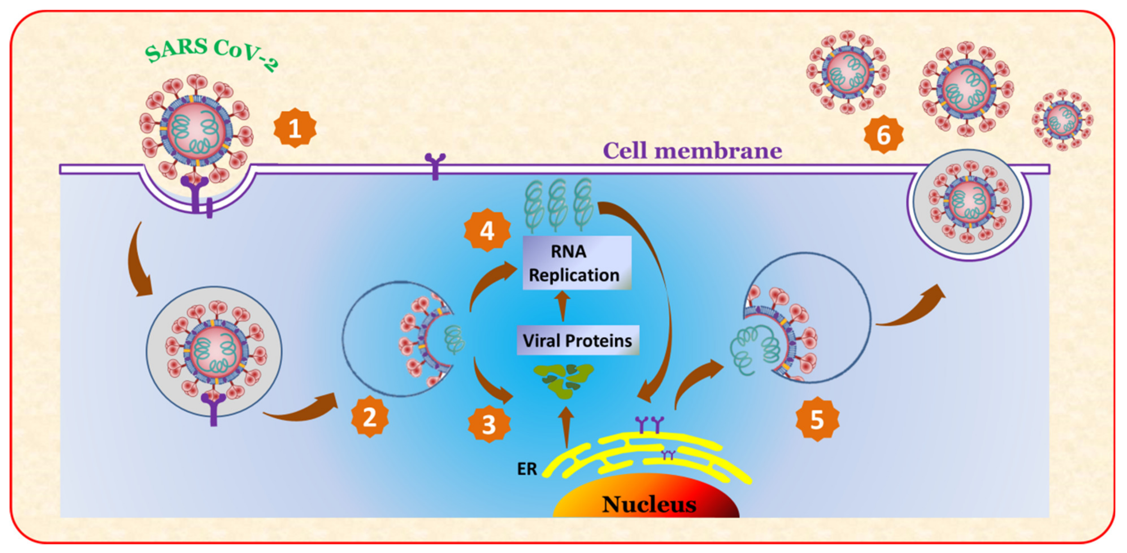
2.1.1. Multiplication of the Virus (SARS CoV-2)

Viruses are non living entities that have a DNA or RNA core and a protective protein coating around them. They are one of the most diverse microorganism families, with 6590 species recognized by the International Committee on Taxonomy of Viruses. Viruses that infect humans usually have a size of 20 to 260 nanometers. However, certain viruses have the ability to proliferate to huge sizes [31,32,33]. The type and natures of viruses, as well as their life cycles, play a major role in their proliferation. The virus life cycles for SARS-CoV-2 are schematically described in Figure 1 and consist of distinct steps.
The virus SARS-CoV-2 grows at the cost of the human biological cell. The human cells are damaged one after another and large numbers of viruses are produced. The sequence of the damage to the cell producing viruses consists of six sequential steps: (1) The virion’s spike protein interacts with ACE2, a cell-surface protein. TMPRSS2, an enzyme, aids virion entry. (2) The virion releases its RNA. (3) Some of this RNA is translated into proteins by the cell’s machinery. (4) Some of these proteins form a replication complex to produce more RNA. (5) Proteins and RNA are assembled into a new virion in the Golgi and released (6) Multiple production of viruses [34,35].
The attachment and penetration of viruses, which is carried out by the virus capsid or envelope proteins, is the initial stage in their proliferation. Viruses replicate, reproduce, and discharge themselves using the machinery of the host cell. Retroviruses, for example, have their own replication enzyme (polymerase or reverse transcriptase), yet they are unable to multiply and proliferate outside of their host cell. Virion assembly begins once enough nucleic acids and proteins are present [36,37].
Virus multiplication is largely determined by the structure of the virus. As a result, it is critical to know what features it might have, such as infectivity, disinfectant susceptibility and replication mode. A virus is usually encased in a protective protein coating called a capsid. The role of the capsid is to safeguard the viral genome. They are generated in a very precise way. Capsids come in three different shapes: icosahedral, helical and complicated. Prokaryotic viruses have a prolate form, which is an elongated icosahedron. Capsid shapes are utilized to aid in virus identification and can reveal information about the virus’ life cycle.

2.1.2. Transmission of Viruses

A variety of factors influence the spread of viral infections in a population. Age, structure, the availability of a new host and the transmission mechanism are all elements to consider. Some viruses can infect a wide range of animals, and cross-species transmission is also possible. Corona viruses, for example, have been found in a variety of birds and animals, including domestic cats and dogs. SARS-CoV, MERS-CoV, and COVID-19 are examples of human CoVoV that can cause respiratory failure [38,39,40,41].
Virus particles are typically transferred between humans through infected persons respiratory, cutaneous or other bodily fluid discharges. The virus can spread directly by aerosols and droplets, or indirectly through contact with contaminated surfaces. Viral contamination of fomites in a community could cause a variety of ailments. Norovirus (a non-enveloped virus) is well known for its ability to survive for several weeks on a variety of surfaces, causing severe epidemics in healthcare facilities [42]. Aerosols and droplets can spread many respiratory viruses, including influenza and SARS-CoV. Aerosol transmission is usually thought to be the most dangerous, since it can spread infectious particles in high concentrations, with few infection control strategies available to halt it. As a result, understanding how long viruses may survive on a surface can help with the development of infection control strategies and the prevention of epidemics [43,44,45].

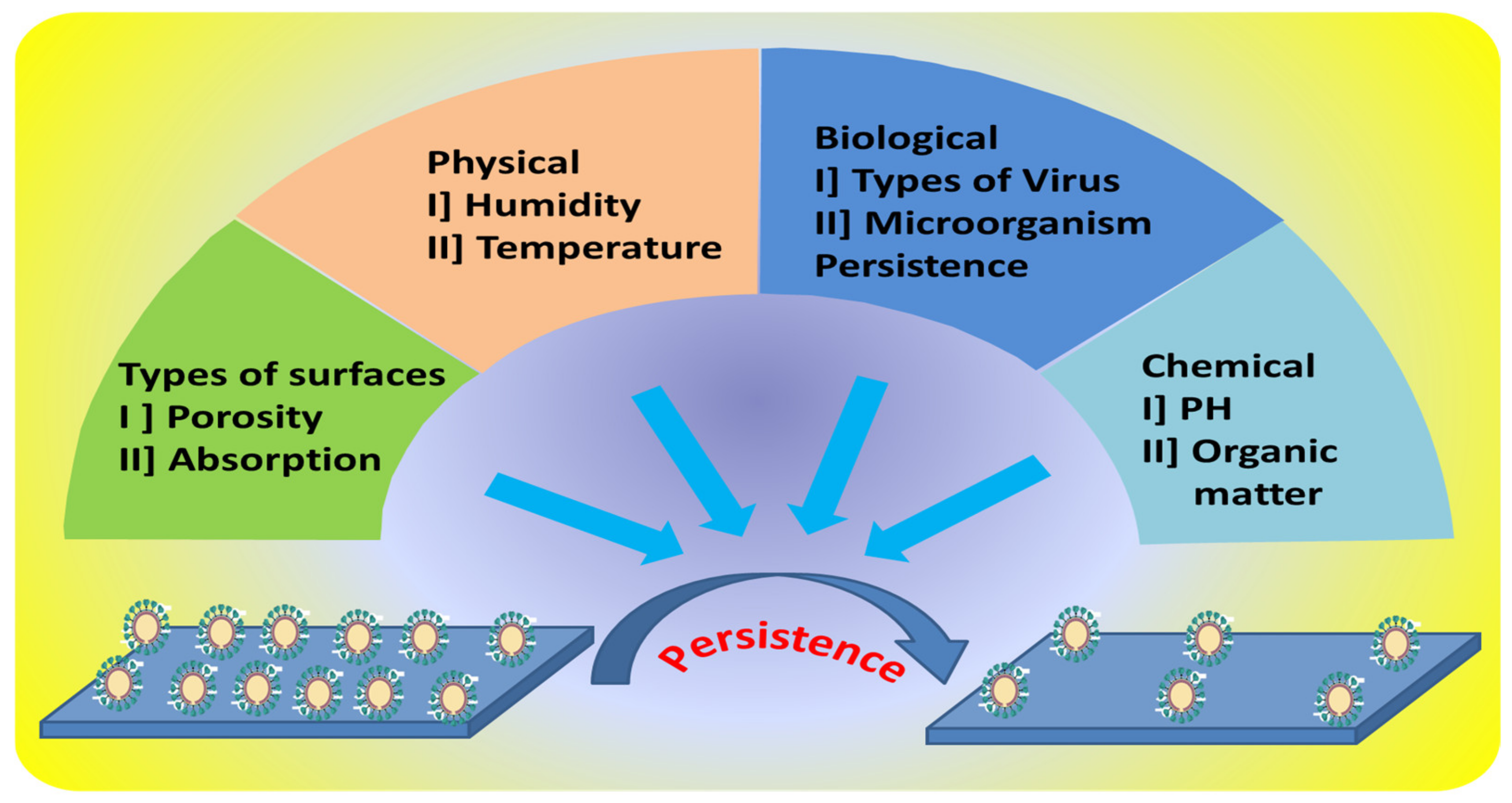
2.1.3. Persistence of Viruses

As the COVID-19 pandemic has continued for more than two years, the persistence of viruses has become a major worry. It is a complicated process that incorporates a variety of surface factors—structural, physical, biological and chemical. Since the discovery of the first virus in 1892, a lot of work has gone into understanding viral survival in different environments and determining the impact of surface characteristics on viral viability. Surface characteristics are influenced by factors such as porosity, topography, absorption, and surface hydrophobicity [46,47,48].
Effective antiviral surfaces may need to be customized to a specific virus type, since each type of virus interacts with a surface differently depending on its structure. Temperature, relative humidity, and how viruses permeate different surfaces are some of the other environmental parameters that influence viral persistence. These additional factors should be considered while developing an antiviral surface [49,50].
The surface parameters that influence virus persistence are schematically described in Figure 2. It may be seen in the diagram that there are four primary characteristics that influence virus persistence on surfaces: (i) physical, abiotic factors, such as relative humidity, temperature, and exposure to light; (ii) type of surface, e.g., roughness; (iii) biological, such as virus structure or the presence of other microorganisms, e.g., microbial biofilms; (iv) chemical, such as pH, presence of reactive ions, adsorption state, organic matter, or presence of specific substances. Many viruses can be sustained and protected by organic material in the environment, such as saliva or mucus droplets. It has been noted that the inclusion of bacteria, lipids and proteins in the viral inoculum can boost the persistence of viral inoculum on low-porosity surfaces such as plastic and stainless steel, as opposed to porous surfaces such as cardboard. The infectious SARS-CoV-2 virus was found to remain on the outer layer of a surgical mask after seven days, while no viable virus was found on treated flat stainless steel and plastic surfaces after seven days [51,52,53].
Here, an attempt has been made to some extent to understand the reason of long lasting persistence of COVID-19 by reviewing the persistence of SARS-CoV-2 on various matrices in different temperature and relative humidity conditions. The data collected from the literature are listed in Table 2. It may be noted that the persistence time of SARS-CoV-2 varies with the nature of the matrix, temperature and relative humidity. It is surprising to note that the SARS-CoV-2 virus persisted for about 21 days on plastics from face shields.

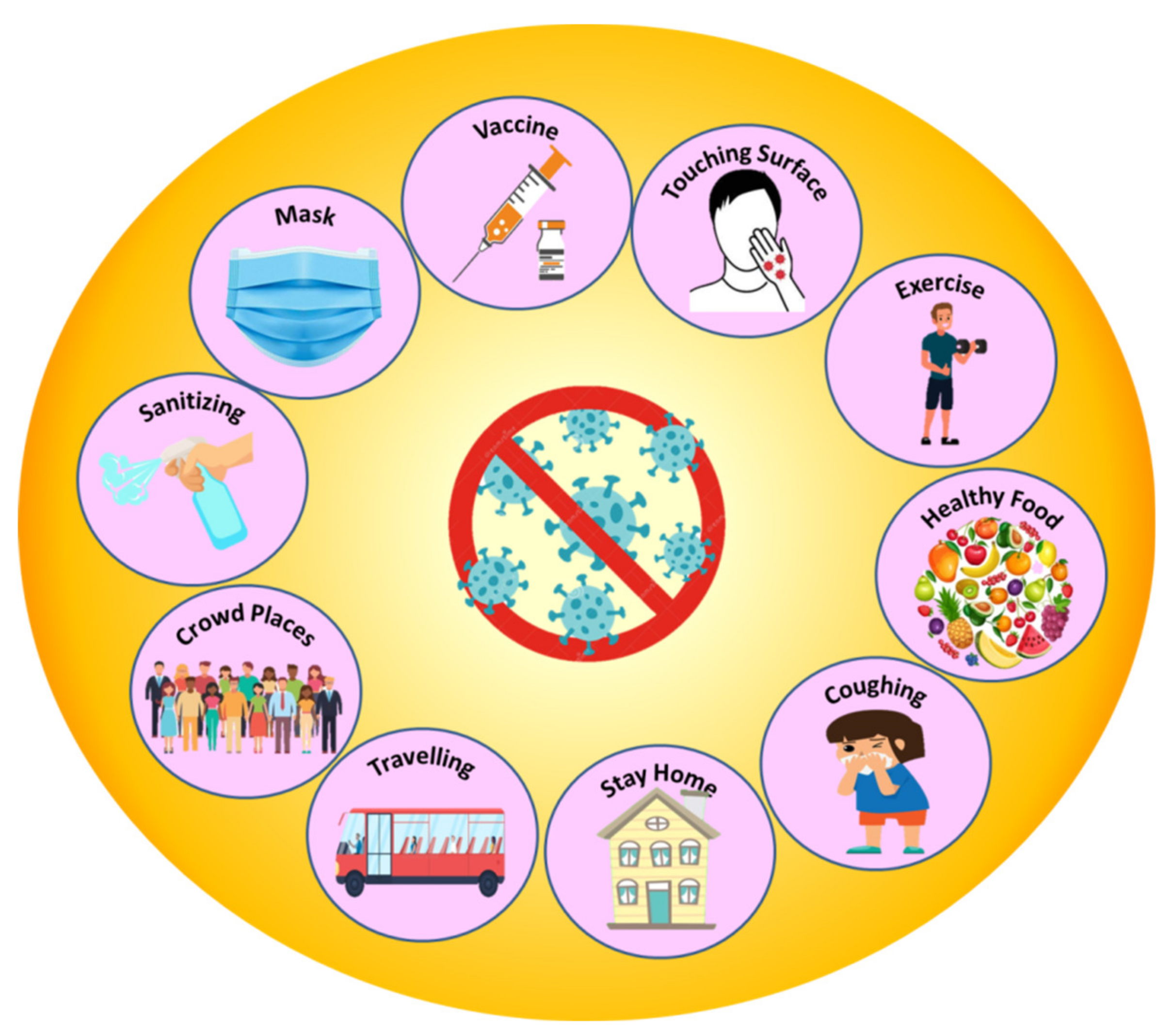
2.2. Controlling the Spread of Communicable Diseases with Disinfection of Pathogens

Pathogen disinfection is a crucial step to avoid the spread of communicable diseases. At the moment, this is mostly accomplished via traditional sterilization methods. However, there are significant limitations and downsides; thus, a new photocatalytic disinfection of pathogens approach is now being developed. In the following sections, we will discuss both traditional and new techniques.

2.2.1. Sterilization of Pathogens and Controlling the Spread of Communicable Diseases

Sterilization is a technique that uses physical or chemical methods to eradicate or eliminate all kinds of microbial life in health-care facilities. In order to prevent the spread of viruses such as SARS-CoV-2 and regulate the COVID-19 sickness, the WHO has given the following instructions: vaccinate yourself, wear a mask and keep a distance of at least 6 feet from others. Wash your hands frequently and avoid crowds and places with poor ventilation. Covering coughs and sneezes is a good idea. Clean and disinfect, keep an eye on your health every day. In order to prevent infectious microorganisms from being transmitted to patients, medical and surgical tools must be disinfected and sterilized [63]. Figure 3 shows a schematic representation of these strategies.

2.2.2. Sterilizing Surface Coatings to Work against the Spread of Communicable Diseases

The spread and persistence of communicable diseases such as COVID-19 have raised many challenges and threats to human society globally. One of the major challenges is the control of the spread of viruses and, in turn, communicable diseases. Some of the examples of applications of surface coatings for pathogenic disinfection are listed in Table 3.
The attachment of pathogens to the surface of a range of materials and devices, such as masks, gowns, clothes, utensils, furniture, frequently handled objects, personal protective equipment (PPE kits), glass, wood, paper and fabric is thought to be the main cause of pathogen dissemination. Doorknobs, stair railings, push plates, handles, drawer pulls, electrical switch plates, plumbing fittings, sinks and elevator floor buttons are all examples of biomedical affected devices. Self-sterilizing materials such as polypropylene, antiviral polymers, the inclusion of metal ions/oxides and functional nanoparticles, copper nanoparticles, metal oxide nanoparticles such as Ag, Au, Cu2O, TiO2, Fe3O4, and graphene are used to coat the surfaces of these substances. Surface coatings developed with these materials act as sterilizing agents and have a long shelf life. As a result, there will be no need for frequent cleaning, no risk of infection and it will be cost effective [64].
TiO2 is the most widely used semiconductor because of its strong oxidizing ability, chemical stability, highly reactivity, ease of preparation, abundance, low cost, low toxicity, chemical inertness and long-term photostability [65].
Table 3. Examples of sterilizing surface coatings against the spread of communicable diseases.
Sr. NoApplication TypeMaterialsReferences
1Mask and PPEPolypropylene[66]
2Mask, gowns and PPEAntiviral polymers, incorporation of metal ions/oxides, and functional nanoparticles[67]
3Clothes, utensils, furniture, regularly touched objects and personal protective equipmentCopper nanoparticles[68]
4Coating, food packaging and textilesPolymeric materials[69]
5Masks and protective clothingNon-woven fabrics, metal-based Nanomaterials such as silver, copper, titanium, gold, and zinc)[70]
6Personal protective equipment (PPE)Polyacrylonitrile (PAN)/zinc oxide (ZnO) hybrid nanocomposite[71]
7Glass, wood, paper and fabricsPolymers, silver, TiO2, and copper-derived chemicals.[72]
8Biomedical devices and protective equipment of medical workers.Cationic polymers, metal coatings and antifouling micro-/nanostructures[73]
9PPE, face masks, gowns, aprons and glovesAntiviral Fabric[74]
10Medical gowns and drapes, PPE, mask or respirators,N,N-dodecyl, methyl-polyurethane[75]
11Face masksMetal oxide nanoparticles such as Ag, Au, Cu2O, TiO2, Fe3O4[76]
12Doorknobs, stair railings, push plates, handles, drawer pulls, electrical switch plates, plumbing fixtures and sinks, and elevator floor buttonsCopper (Cu) and its alloys[77]
13MasksSilver, carbon nanotubes and titanium dioxide nanoparticles[78]
14PPE, masksCopper oxide[79]
15MasksGraphene[80]
16Healthcare workers include protective clothing, pathogenic microbesSilver nanoparticles[81]
17Public transport system such asSilver nanoparticles[82]
18Hospital applicationsAluminum 6063 alloy[83]

3. Photocatalytic Surface Coating/Painting for Self-Sterilizing against Pathogens

Methods for sterilizing surfaces to control the spread of diseases using chemicals in liquid form need to be used frequently. These are suitable only for small-scale applications. For large-area applications, surface coatings are preferred. However, their cost, duration, and maintenance requirements make them inconvenient to use for large-area applications. In view of this, self-sterilizing, long-lasting and maintenance-free methods/techniques for disinfection are in demand to control the spread of viral diseases like COVID-19. The progress and prospects in nanoscience today have been made possible with nanoparticle engineered photocatalytic paints and will be a roadmap to self-sterilizing against the spread of communicable diseases. Recent developments in this field of science and technology are reported and discussed in the following sections.

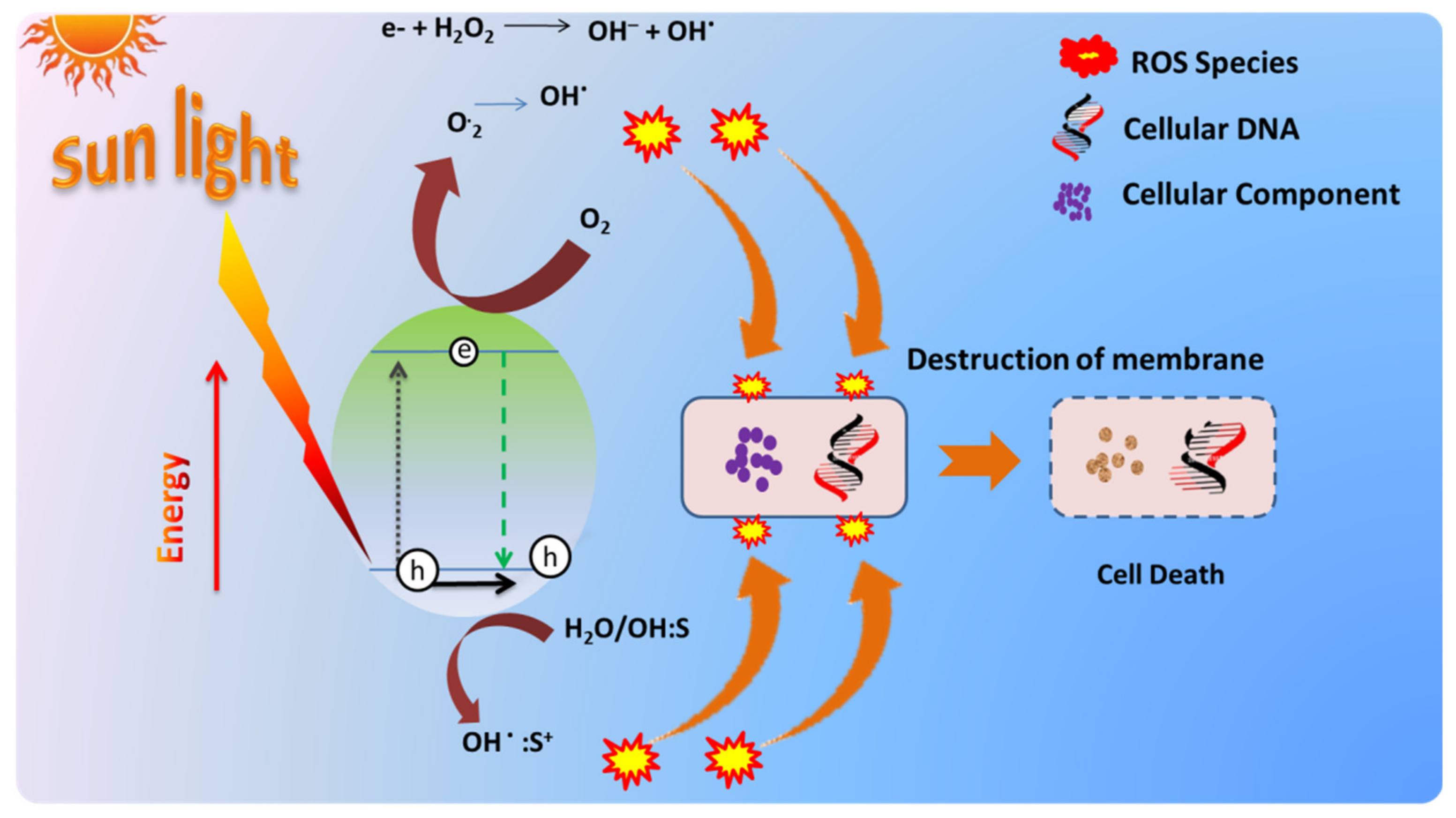
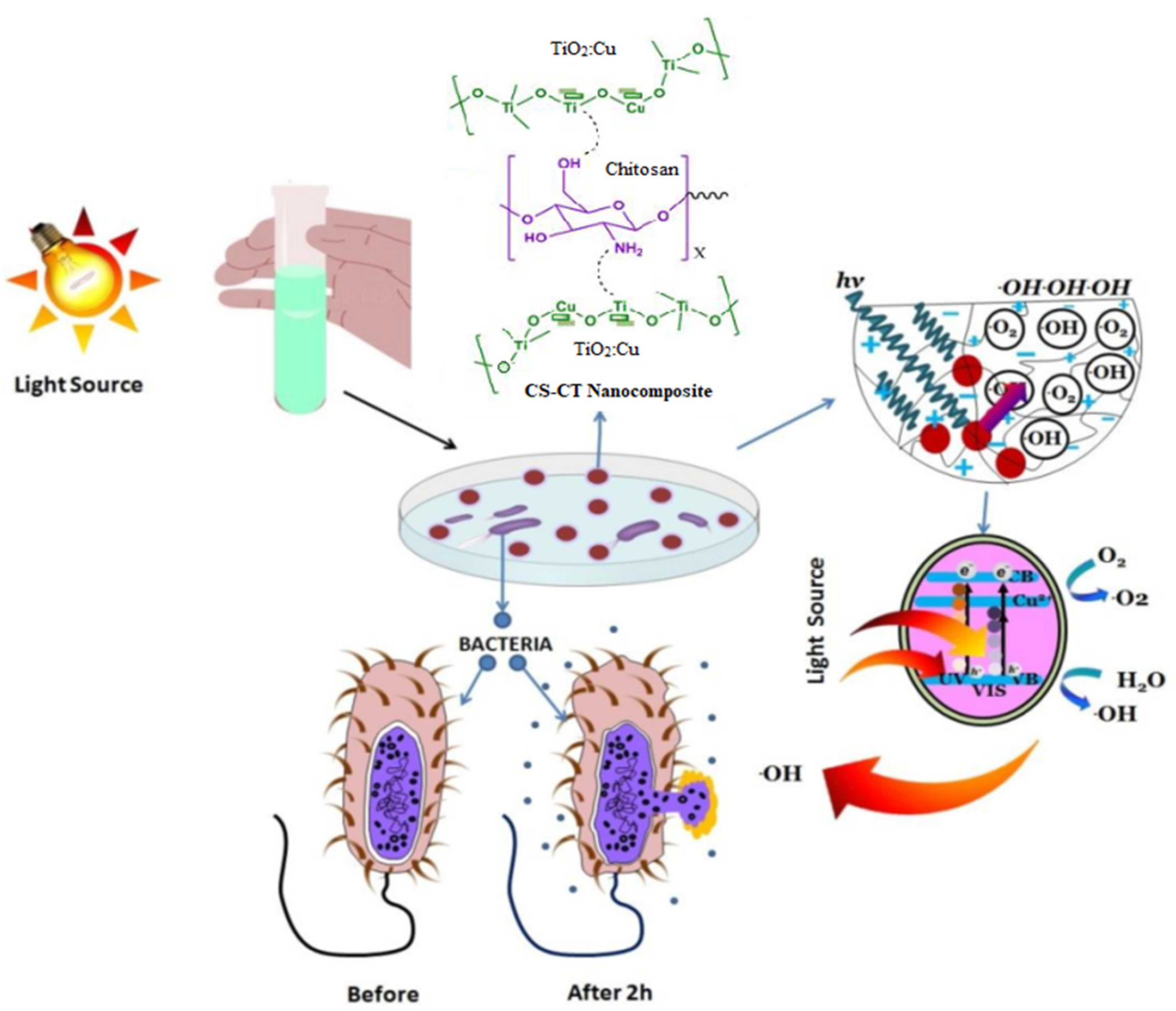
3.1. Self Sterilizing of Pathogens with Photocatalytic Disinfection: Principle and Working

The phenomenon of photocatalysis with semiconducting materials has been known for a long time and its principle is well explained in the literature. When a semiconductor is bombarded with light of a compatible wavelength (energy equal to or greater than the semiconductor’s band gap), the light is absorbed, resulting in the formation of an electron and hole pair.
The excited electron–hole pair generated in the semiconducting material then travels toward the semiconductor’s surface. At the same moment, oxidation and reduction reactions on the semiconductor’s surface take place. Figure 4 shows this conceptually. In the CB, electrons react with oxygen (O2) to create superoxide and hydroperoxide radicals. During the photocatalysis process, reactive oxygen species (ROS) are produced; further, it can induce leakage of minerals, genetic material and protein, and therefore result in cell death and a reduction in microbial load [84].
In this process, ROS oxidizes the proteins, lipids, bacteria, and enzymatic systems of viruses, resulting in the loss of their ability to reproduce due to either damage to other key proteins or damaged membranes, such as the spike protein of SARS-CoV-2. Once the pathogen comes into contact with the photocatalytic surface, it oxidizes key proteins and inactivation takes place, due to the production of ROS [85,86].
The contaminants (viral proteins, genome, and envelope) in the indoor air are photodegraded by the photocatalytic material in general. Antimicrobial photocatalysis can be utilized to disinfect air, water and surfaces [87,88,89].
Adsorption-desorption, electron–hole pair generation, electron pair recombination and chemical reaction are all phases in the activation of photocatalytic nanoparticles present in the nanocomposite. Figure 5 shows the hypothesized mechanism for nanocomposite photocatalytic antibacterial activity.
It has a number of benefits; for example it is a low-cost, easy-to-use, low-maintenance method that allows simultaneous disinfection of a variety of pathogens such as bacteria and viruses.

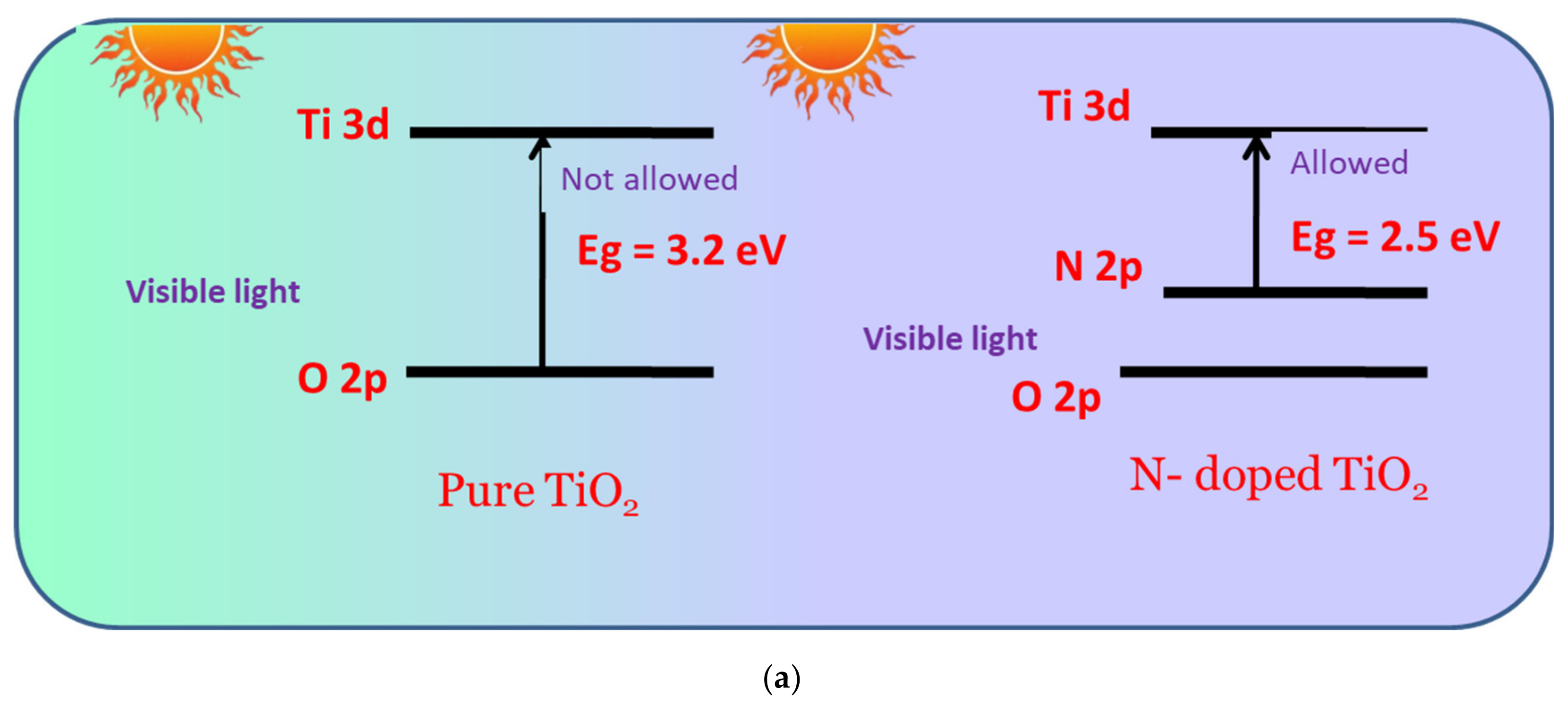
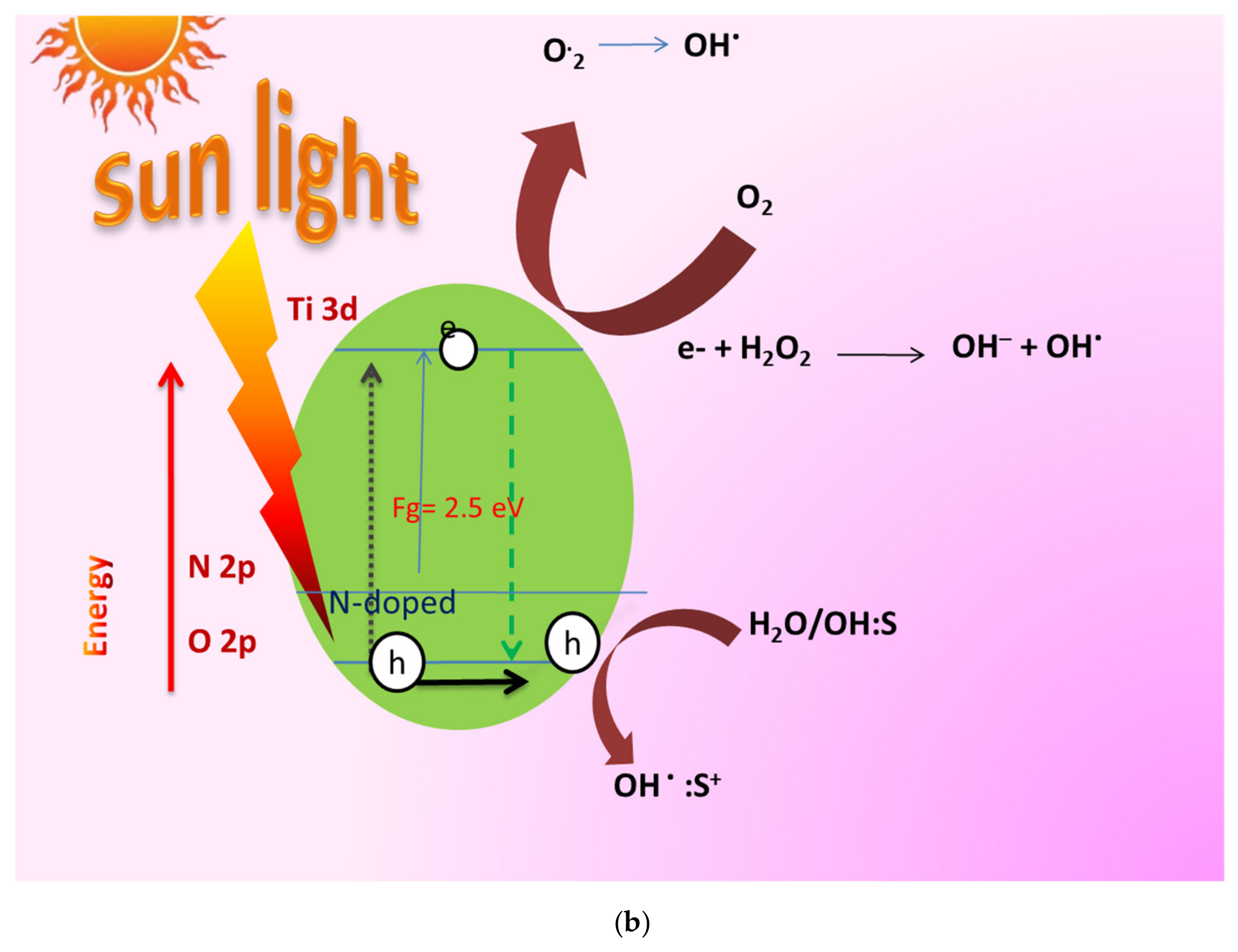
3.2. Engineering of Nanoparticles with Dopants for Visible Light Photocatalytic Disinfection

Various single-semiconductor materials have been examined as a solution for the photocatalytic breakdown of pollutants, including TiO2, Cu2O, ZnO, SnO2, WO3, Fe2O3 and CdS. Due to its activation by UV light, long-term stability and non-toxicity, TiO2 and its hybrid compounds have been researched as viable photocatalytic materials for the destruction of pollutants and viruses, as well as the conversion of CO2 and hydrogen production through water splitting [91].
TiO2 in the anatase crystal phase has a bandgap of 3.2 eV and absorbs UV light with wavelengths shorter than 400 nm, indicating photocatalytic activity. However, due to the wide bandgap and fast recombination of the electron–hole pair, the usage of TiO2 semiconductors as photocatalysts still has a drawback of low absorption and efficiency. When considering indoor use, the use of visible light, lower in energy than UV light is unavoidable. UV light, on the other hand, accounts for less than 10% of solar energy, whereas visible light accounts for roughly 48% [92].
Several methods have been investigated to solve these problems, including: (i) modifying the surface area, crystallinity, and porosity of TiO2 particles; (ii) doping and coating the surface with metals (Se, Cu, Pd, Cd, and Pt); and (iii) combining p-type materials with TiO2 particles to form a p–n heterojunction semiconductor. Combining TiO2 with a semiconductor with a tiny bandgap, such as CdS, CuxO, PbS, or CuS, is a viable technique for boosting visible-light activity or permitting photogenerated electrons to be transported from the conduction band to other semiconductors to prevent electron–hole pair recombination. Copper is recognized as good for mixing with other stable materials since it is highly oxidative and adaptable. Furthermore, past research has shown that copper may be easily produced alone or in combination with other compounds, and that it is stable [93,94].
As a result, various efforts have been undertaken to improve the photocatalytic efficacy of TiO2 using a copper-based material as a p-type semiconductor, such as copper oxide or copper sulfide. Because photocatalysts made of TiO2 can participate in a variety of reactions, including breakdown and reduction, the crystal structure and bandgap must be tailored to maximize performance. Cu–TiO2 nanorods outperformed pure TiO2 photocatalytically in both visible and ultraviolet light.
The majority of the time, thermal plasma treatment is used to prepare TiO2 and combine it with a copper-based semiconductor. Methods include hydrothermal, sol-gel and combustion. Most of the methods discussed above have the drawback of requiring a significant amount of time and energy, as well as a variety of additives and templates for synthesis. A microwave synthesis approach was proposed as a good way to avoid wasting time and energy in this way with quick and uniform heating, especially in the case of a synthesis method using dielectrics such as water.
Furthermore, it is known that lowering the Gibbs energy of activation and boosting hydrolysis can promote the reaction of transition metal ions, with which it is difficult to create a complex under usual conditions [95,96,97]. The bandgap of TiO2 semiconductor has been reduced from 3.2 eV to 2.5 eV due to nitrogen doping, making it sensitive to visible light, as shown in Figure 6b.
Similarly, other dopants, such as Cu2O, C3N4, CdS in TiO2 Au, Ag, N, Pt, etc., are used for photocatalytic disinfections. Accordingly, the band gap will be changed [98,99,100,101,102,103,104,105].

4. Synthesis of TiO2 Engineered Nanoparticles for Visible Light Photocatalytic Disinfections

4.1. TiO2 Nanoparticles: A Potential Candidate for Photocatalytic Paints

Nanosized TiO2 has been studied extensively for decades due to its unusual photocatalytic characteristics. The photocatalytic inactivation of hazardous microbes is gaining popularity as one of the many environmental applications enabled by such a nanoscale material. Many researchers have described the antibacterial activity of commercially available TiO2 NPs in the last few years. Innovative approaches to the production of unique TiO2 based nanomaterials, specifically tailored for photocatalytically assisted inactivation of bacteria and viruses, have recently been published. Such approaches vary in terms of the degree of complexity and production costs, but they allow access to photocatalytic TiO2-based nanomaterials with purposefully selected physical–chemical properties, such as controlled dimensions and geometry, and thus size-dependent characteristics, engineered surface chemistry, enhanced colloidal stability even under different ionic strength conditions, and biocompatibility, which is particularly useful in the fields of health-care and food preservation. A variety of synthetic routes for the preparation of nanosized TiO2 with photocatalytic antimicrobial properties have been proposed in the literature; here, we focus on sol-gel and phytosyntesis, as they have been regarded as promising techniques for the large-scale production of TiO2 NPs, owing to their user-and environment-friendly procedures and low costs. However, for the purpose of brevity, we will only present a tiny sample of the many examples described in the literature and we will refer readers to more detailed articles, particularly in the fields of health-care and food preservation [106].

4.2. Phyto Synthesis of TiO2 Nanoparticles

Researchers have currently focused their attention on the synthesis of nanoparticles using green methods, one of which is the route of biosynthesis. It mainly consists of microbial synthesis and phytosynthesis. In the present review, we have focused on the preparation of nanoparticles by the method of phytosynthesis. The plant species of lemongrass, Annonasquamosa, Leechaikottaikeerai (Pisoniagrandis), Andrographispaniculata and Azadirachtaindica have been considered for phytosynthesis of TiO2 nanoparticles, as these are locally available in plenty and will help to produce nanoparticles on a large scale required for their commercial applications [107,108,109,110,111].
We present a green method for making TiO2 and ZnO nanoparticles that uses lemongrass extract as a capping agent to keep the size of the nanoparticles under control. We discovered that the extracts of phytochemical content aided in the creation of nanoparticles, as evidenced by morphologic data gathered from microscopy pictures. The nanoparticles’ physicochemical properties are comparable to those of nanoparticles generated using standard methods. Finally, the anticorrosive, antibacterial, and self-cleaning properties of TiO2 and ZnO nanoparticles in paints were studied to establish their efficacy.
To remove contaminants, lemongrass leaves were trimmed, rinsed and dried before being hand-crushed. A hundred grams of biomass were placed in cotton bags and solvent-extracted for 6 h with 500 mL of distilled water. After that, the lemongrass extract was stored at 4 °C. Then, in a 250 mL beaker, 100 mL of this extract was combined with 20 mL of the precursor C12H28O4Ti by dropwise addition. The solution was placed in an ultrasonic processor for around 30 min to react. The nanoparticles were rinsed with distilled water and ethanol reagent grade before being recovered by centrifugation at 5000 r/min for 30 min, with the operation repeated multiple times to eliminate any unreacted precursor. The anatase phase of TiO2 nanoparticles was obtained by calcining them at 550 °C for 3 h [107].
Roopan et al. have studied the phyotosynthesis of rutile TiO2 nanoparticles using agricultural waste, such as aqueous extract of Annona squamosal fruit peels, at a lower temperature for the first time with the rapid production of nanoparticles. The UV–Vis spectrophotometer results were promising, with a surface plasmon resonance occurring at 284 nm, indicating its applications for photocatalytic disinfection [108]. The phytosynethesis of TiO2 nanoparticles using the extract of Leechaikottaikeerai (Pisoniagrandis) was achieved for the first time by Vembu et al., and confirmed their formation, shape, crystallinity, and an average size of 34 nm. They also studied the antimicrobial and cytotoxic activities of these nanoparticles [109]. Rajeshkumar et al. have reported phytosynthesis of titanium dioxide (TiO2) nanoparticles using the king of bitter herbal plant Andrographispaniculata. This method is eco-friendly, produces nanoparticles at a low cost, and can provide high-quantity industrial production using biodegradable and reusable natural resources [110]. The plant extracts of Azadirachtaindica were also used by Thakur et al. as the best local source material for the phytosynthesisis of TiO2 nanoparticles. This is a simple, inexpensive and eco-friendly process which reduces the use of toxic chemicals. These nanoparticles exhibited an average size of about 22.97 nm and a broad spectrum of antimicrobial activity against a vast range of pathogens [111].

4.3. Synthesis of Nitrogen Doped TiO2 Nanoparticles by Sol-Gel Technique

The sol-gel technique was used to create TiO2-based photocatalysts with different dopants to make them sensitive for visible light. In this section, we have briefed the attempts of different groups of researchers to dope TiO2 nanoparticles with nitrogen by employing the solgel technique.
Cheng et al. have used tetrabutyl titanate as the basic compound of titanium. To make Ti(OBu)4–C2H5OH solution, 10 mL of tetrabutyl titanate was dissolved in 40 mL of anhydrous ethanol in a usual procedure. Meanwhile, 12 mL of dilute nitric acid (1:5, volume ratio of nitric acid to deionized water) and a small amount of ammonium chloride were added to another 10 mL of anhydrous ethanol to make a C2H5OH–HNO3–water solution. To carry out hydrolysis, the C2H5OH–HNO3 solution was gently added dropwise to the Ti(OBu)4–C2H5OH solution while vigorously stirring. After that, the resulting yellowish, transparent sol was aged for 6 h after being stirred constantly for 2 h. The resulting yellowish, clear sol was next dried for 36 h at 80 °C in an oven and then calcined for 4 h at a specific temperature, with a heating rate of 3 °C per minute [112].
Than et al. synthesized N-doped anatase TiO2 nanoparticles (N-TiO2 NPs) from titanium chloride (TiCl4) and ammonia (NH3) via the sol-gel method. The high visible light activity of the N-TiO2 nanomaterials was recognized in the interstitial nitrogen atoms inside the TiO2 lattice units. These findings could provide a practical pathway capable of large-scale production of a visible-light-activated N-TiO2 photocatalyst. The interstitial nitrogen atoms within the TiO2 lattice units played an important role in generating intermediate energy levels and narrowing the bandgap, thereby enhancing the VLA of the materials [113]. Reda et al. synthesized Cu and N-doped TiO2 photocatalysts using titanium (IV) isopropoxide via a microwave-assisted sol-gel method. For N doping, ammonium nitrate was utilized as a raw source. The doped TiO2 samples showed a higher surface area (253–383 m2/g) than that of the pure one (151 m2/g) and exhibited better light-harvesting in both visible and UV light regions [114]. The nitrogen-doped titanium oxide was successfully synthesized through an EDTA modified sol-gel process by Nassoko et al. Here EDTA is used as source of nitrogen for doping. The results showed that through the simple sol-gel process, nitrogen can be incorporated into the crystal lattice of TiO2, leading to its response to visible light [115]. N-doped titania is synthesized using the various nitrogen sources such as, triethylamine, N,N,N′,N′-tetramethylethane-1,2-diamine, ethyldiamine, 1,2-phenylenediamine, propanolamine, and propylenediamine and TTIP was used as a TiO2 source by Mehdizadeh et al. The bandgap of N/TiO2 has changed from 3.38 eV (pure TiO2) to 3.26 eV for N/TiO2:2.0. It could limit the band gap of titania and its absorption extends to the visible light region; moreover, it might increase the separation efficiency of the photoinduced electron and hole [116].

4.4. Synthesis of CNTs Doped TiO2 Nanoparticles by Sol-Gel Technique

A sol-gel technique was also used to make CNT-doped TiO2 nanoparticles by different groups of researchers to make the paint of photocatalytic disinfection sensitive to visible light.
Akhavan et al. used Titanium (IV) n-butoxide as basic chemical compound of titanium. First, 2.3 mL Titanium (IV) n-butoxide was dissolved in 23 mL ethanol. The solution was then agitated for 30 min to make it homogenous. Another solution was made with 2 mL of nitric acid and the desired amount of CNTs. Drop by drop, the CNT–nitric acid solution was added to the alkoxide–ethanol solution, and the mixture was agitated for 2 h. The molar ratio of H2O to titanium n-butoxide in the final solution was 2.7:1. The solution was left at room temperature for 24 h to age. Dip-coating was used to deposit CNT-doped TiO2 films on glass substrates. The substrate was dragged at a steady speed of 1 mm/s after being soaked in the solution for roughly 1 min. After drying the samples at room temperature for 24 h, they were heated in air for 1 h at 100 °C. Furthermore, parts of the CNT-doped TiO2 thin films were heat treated in air for 1 h at 450 °C [117].
Shafei et al. synthesized TiO2-10 wt. % CNT nanocomposite powder doped with 1 at.% Cu using this sol-gel method. Band gap energy decreased from 3.2 to 3.05 eV by addition of CNTs to the TiO2 and decrease further to 2.85 eV by both the addition of CNT and Cu doping to TiO2 [118]. Mohammad et al. synthesized single-walled carbon nanotubes (SWCNTs) and multiwalled carbon nanotubes (MWCNTs), which were chemically treated with sulfuric acid (H2SO4) and nitric acid (HNO3) in a ratio (3:1). These carbon nanotubes (CNTs) have been coated with Ag-doped TiO2 nanoparticles (NPs) using the sol-gel method. The band gap value of 3.6 eV was obtained from raw MWCNTs, while the corresponding value of the raw-SWCNT sample was 2.94 eV. The addition of TiO2/Ag NPs leads to a decrease in the band gap, 2.69 eV for MWCNTs-TiO2/Ag and 2.6 eV for SWCNTs-TiO2/Ag [119]. Carbon-doped titanium dioxide (TiO2) coating on multiwalled carbon nanotubes (MWCNTs) was prepared by oxidation of titanium carbide (TiC) coated MWCNTs by Cong et al. MWCNTs, which may act as a supporting, absorbent, photo-generated transfer station and carbon-doping source play key roles in narrowing the band gap of TiO2 [120].

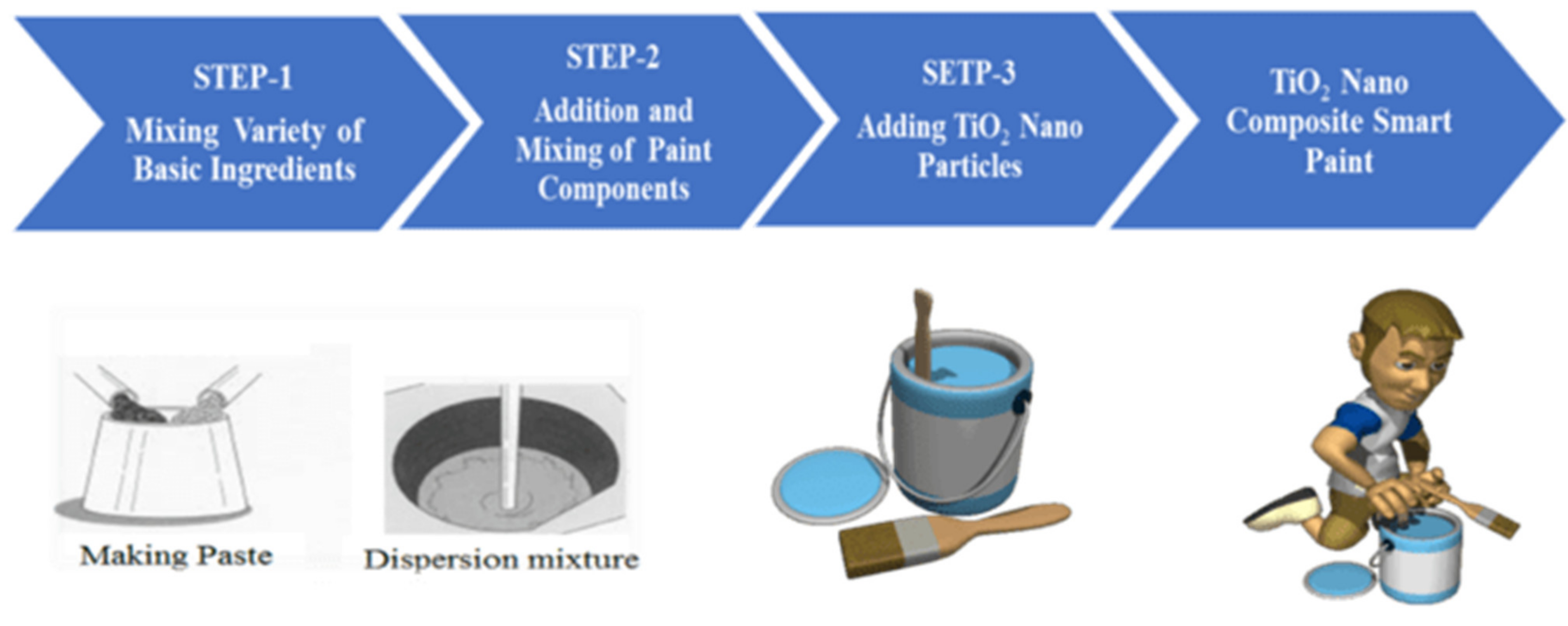
5. Smart Paint Formulations with Nanostructured Additives for Their Photocatalytic Activities

5.1. Updates of Photocatalytic Paints Studied for Pathogenic Disinfections

With the developments in the engineering of nanoparticles with dopants for visible light photocatalytic disinfection, the next step is to use these findings for large-area applications specifically for controlling the spread of communicable diseases such asCOVID-19. In this regard, few research groups including our group have attempted smart paint formulations with nanostructured additives for their photocatalytic activities with an intention to disinfect the pathogens. The survey of such data is of recent origin and the first of its kind in the literature reported in Table 4.

5.2. Formulation of TiO2-Based Oil Paint Samples

Here we report the formulation of three types of nanostructured paints:
(a)
Paint formulated with TiO2 nanoparticles prepared from lemon grass leaves.
(b)
Paint formulations with TiO2 nanoparticles doped with nitrogen prepared by the sol-gel method.
(c)
Paint formulations with TiO2 nanoparticles doped with CNTs by the sol-gel method.
The paint samples were prepared using the percentile mixture of titanium dioxide nanomaterials (TiO2 powder) in the white colored base oil paint with a fraction of the turpentine to obtain a homogenous mixture. The 1%, 3%, 5% and 7% paint samples were prepared and base paint control was used for the testing purpose.
For the paint mix, 30 percent distilled water and 33.4 percent resin BASF ACRONAL were utilized. The dispersing agent BASF DISPEX AA 4146 was used at 0.6 percent w/w, and the total amount of solids, including pigment (TiO2) and extender (CaCO3Cicarelli, >99 percent purity), was kept constant at 36 percent w/w. In the paints, the maximum amount of TiO2 was 18 percent by weight. Other paints were also developed, with the carbon-doped TiO2 quantity varying between 14 and 12 percent w/w. Two paints, on the other hand, were created with different amounts of CaCO3, but the percentage of carbon-doped TiO2 remained constant at 18% w/w. Finally, various pseudo-paints were created by eliminating one of the solid matrix’s components, the extender.
TiO2 and CaCO3 were hand milled and dried at 110 °C before being used in the paints. The solids were then added to a solution of distilled water and dispersing agent, which was mixed at 300 revolutions per minute. To finish the paint, the resin was added in the last phase. Prior to the air decontamination studies, the photocatalytic coatings were subjected to visible radiation lamps. With this method, the epoxy covering the TiO2 particles was damaged, permitting contact between the photocatalyst and the contaminated air [138,139,140].
Photocatalytic TiO2-based Nano-composite Smart Paint Formulation is summarized schematically in Figure 7.
Islam et al. reported the development of photocatalytic paint based on TiO2 and photopolymer resin for the degradation of organic pollutants in water. Photocatalytic paint (consisting of a mixture of TiO2 and photopolymer resin) with a different weight percentage of TiO2 (0–10 wt. %) was prepared in plastic Petri dishes and the degradation of organic pollutants in water was studied [141]. Laisney et al. reported the formulation of photocatalytic paints with TiO2 nanoparticles coated with bio-inspired ligands for the Safer by Design. Photocatalytic paints were formulated with TiO2 NPs and TiO2 NPs coated with Dopa, PEG3350 and PAA. They have successfully modified the surface of a commercial photocatalytic TiO2nanopowder with bio-inspired ligands of different chemical nature (molecules and polymers) for its application in self-cleaning paint [142].
Rosset et al. reported the development of TiO2-based photocatalytic paint. TiO2 NPs synthesized were incorporated into an organic matrix-based paint. The organic-based paint was composed of acrylic, micro-sized TiO2 providing the white color (pigment), calcium carbonate particles, aluminosilicate, water and other additives. The micro-sized TiO2 particles have a mean particle size distribution (PSD) of the primary particles of 240 nm [143]. Basso et al. reported a photocatalytic effect of the addition of TiO2 to acrylic-based paint for passive toluene degradation. Photocatalytic paints were formed by adding TiO2 P25 powder to an acrylic-based paint. Photocatalytic paint samples were prepared by adding TiO2 P25 to the paint in the following proportions (dry basis): 0, 10, 15, 20 and 50 wt. %. Complete dispersion was achieved by stirring different amounts of paint and TiO2 P25 in a glass beaker, using a laboratory overhead stirrer at 300 rpm, for 20 min [144].

5.3. A Field Study of Photocatalytic Nanocomposite Paints for Pathogenic Disinfections

Ren et al. investigated microbial activity on the walls of two Hong Kong schools with and without photocatalytic WOx paint coatings; WOx paints were painted on the semi-outdoor and indoor walls in Tun Mun and Tin Shui Wai schools, and daily chlorine disinfection was performed after class in Tin Shui Wai School. For large-scale field experiments, a technique using the ATP biofluorescence approach was presented. On a control basis, ATP swab samples were collected once a week for three months in areas with and without the WOx paint. After that, the ATP data were processed and plotted in box plots. The median log-scale ATP values of walls with WOx paint were at least 0.5 log lower than those without WOx paint in both schools. In the long run, the WOx paint was more effective than daily chlorine disinfection at reducing microbial activity. The proposed testing technique is determined to be suitable for evaluating the long-term performance of an antimicrobial paint in large-scale field experiments by assessing its microbial activity. In both indoor and semi-outdoor conditions, the WOx paint exhibits long-term efficiency in lowering microbial activity on wall surfaces [145].
The systematic use of the ATP bioluminescence strategy was shown to be beneficial in conducting a field investigation of tracking surface microbial activity on various surfaces in a large-scale disinfection practice to evaluate its practical effectiveness. They have also provided a step-by-step guide for creating a testing strategy for evaluating an antimicrobial paint’s long-term efficacy. This systematic technique can be a valuable and low-cost tool for evaluating the field performance of the large number of disinfection strategies that claim to be successful in the long term, particularly those that arose during the COVID-19 period. The findings from the two primary schools showed that the RAZE photocatalyst paint was successful in lowering microbial activity on wall surfaces in both indoor and semi-outdoor situations. In settings where low microbial activity is required, such as hospitals, schools, and kindergartens, RAZE paint has a lot of potential. To meet stricter disinfection requirements, more work is needed to improve its efficacy when alternative disinfection procedures, such as chlorine disinfection, are used. Furthermore, the differences in log-scale ATP values in the aforementioned scenario-based analysis highlighted the need for more research into the effects of disinfection strategies, such as chlorine disinfection with RAZE paint and chlorine disinfection alone, on microbial species evolution using, for example, metagenomic analysis. Such research will aid the community in better understanding how to choose more appropriate disinfection procedures to safeguard our living environment while avoiding the potential for microbial species to become stronger and more resistant [146,147].
The proposed testing protocol is suitable to evaluate the long-term performance of an antimicrobial paint by analyzing its microbial activity in large-scale field tests. The TiO2 doped paint shows long-term effectiveness in reducing microbial activity on wall surfaces in both indoor and semi-outdoor environments.

6. Conclusions

Based on the critical survey of the relevant literature referred in this review and our experience in fighting against communicable diseases, during the past few years, the following conclusions can be drawn:
(1)
COVID-19 disease has remained persistent for longer than 24 months since December 2019, and it has affected millions of people worldwide.
(2)
There remains the significant challenge of preventing infections due to the spread of the SARS-CoV2 viron as it is a bio-nanoparticle. Apart from its biological structures, the nano structural properties do change drastically with their shape and size as per the knowledge of nanoscience, and thus its eradication is still uncertain.
(3)
The transmission of SARS-CoV2 vireses also takes place through contact surfaces such as doorknobs, packaging and handrails in public places. This may be responsible for many preventable and nosocomial infections.
(4)
The spread of communicable diseases may be attributed to different phenomena such as the multiplication, transmission and persistence of viruses.
(5)
The persistence of viruses such as SARS-CoV-2 in/on different 22 environmental matrices has been researched and presented for the first time collectively in this review. It may be noted that the spread of communicable diseases may be predominantly attributed to the phenomenon of persistence of viruses on different substrates for different durations.
(6)
The existing antimicrobial cleaning technologies used in hospitals are not suitable, viable or economical to keep public places free from such infections.
(7)
As an alternative to existing sterilization techniques, photocatalytic pathogen disinfectant surface coatings/paintings have been developed in recent years.
(8)
Smart paint formulations with a variety of nanostructured additives for their photocatalytic pathogen disinfectant activities have been attempted in recent years, but in a very limited way. Reports on a variety of photocatalytic paints studied for their pathogen disinfection potential are reviewed and listed together for first time to make their comparison easier.
(9)
The protection of public places with smart paint photocatalytic pathogenic disinfectant coatings may help to control the spread of communicable diseases such as COVID-19.

7. Future Suggestions

Health-care is of prime importance for humanity globally. Communicable diseases such as COVID-19 have raised a great threat due to infectious virions like SARS-CoV-2. The means to control such virions are discussed in this review. Based on the contents of the review and present needs, the following are key suggestions to build a roadmap to self-sterilizing against the spread of communicable diseases:
(1)
Visible-light-sensitive nanoparticle engineered photocatalytic paints need to be formulated and tested for their pathogen disinfectant activity infield applications in public places.
(2)
The durability, viability and impact on the environment of the photocatalytic paint coatings in open public places need to studied.
(3)
The economic viability of the methods of surface sterilization with conventional techniques and with the newly suggested visible-light photocatalytic disinfection of pathogens needs to be compared and the suitability of new method should be worked out.
(4)
A roadmap to self-sterilizing against the spread of communicable diseases is still at a conceptual level and needs to be given more thought before its implementation on a large scale.

Author Contributions

Conceptualization, S.H.P.; methodology, M.M.D.; validation, R.K.S.; formal analysis, V.S.M.; investigation, S.H.P.; resources, M.M.D.; data curation, R.K.S.; writing—original draft preparation, V.S.M. and S.H.P.; writing—review and editing, V.S.M. and S.H.P.; visualization, M.M.D.; supervision, S.H.P.; project administration, V.S.M.; funding acquisition, S.H.P. All authors have read and agreed to the published version of the manuscript.

Funding

This research received no external funding.

Data Availability Statement

Not applicable.

Acknowledgments

The authors are grateful to C. V. Murumkar and P. C. Pingale from T.C. Col-lege, Baramati and R. K. Mudgal, Shimpa Shrama and D.Y.Patil Education Society, deemed to be university, Kolhapur for their constant encouragement and support during this work.

Conflicts of Interest

The authors declare no conflict of interest.

References

  1. Pawar, S.H. Progress and Prospects in Nanoscience Today; Nova Science Publishers: Hauppauge, NY, USA, 2020. [Google Scholar]
  2. Sawant, D.V.; Desai, M.M.; Patil, R.S.; Pawar, S.H. Evolution of nanotech assisted PCR diagnosis of Mycobacterium tuberculosis and its assessment with convential methods. Int. J. Pharm. Pharm. Sci. 2018, 10, 133–137. [Google Scholar] [CrossRef] [Green Version]
  3. Sawant, D.V.; Bohara, R.A.; Patil, R.S.; Pawar, S.H. Detection of Mycobacterium tuberculosis from pulmonary sputum sample using SPION mediated DNA extraction method. Res. J. Life Sci. Bioinform. Pharm. Chem. Sci. 2018, 4, 4. [Google Scholar]
  4. Deepak, V.S.; Shivaji, H.P. Studies on gold nanobiosensor for early diagnosis of Mycobacterium tuberculosis. Int. J. Pharm. Biol. Sci. 2019, 9, 77–82. [Google Scholar]
  5. Armelin, E.; Liesa, F.; Estrany, F. Marine paint formulations: Conducting polymers as anticorrosive additives. Prog. Org. Coat. 2007, 59, 46–52. [Google Scholar] [CrossRef]
  6. Tornero, A.C.F.; Blasco, M.G.; Azqueta, M.C. Antimicrobial ecological waterborne paint based on novel hybrid nanoparticles of zinc oxide partially coated with silver. Prog. Org. Coat. 2018, 121, 130–141. [Google Scholar] [CrossRef]
  7. Yazid, S.A.; Mohd, Z.; Mohamad, J. Effect of titanium (IV) isopropoxide molarity on the crystallinity and photocatalytic activity of titanium dioxide thin film deposited via green sol-gel route. J. Mater. Res. Technol. 2018, 8, 1434–1439. [Google Scholar] [CrossRef]
  8. Wang, L.; Hu, C.; Shao, L. The antimicrobial activity of nanoparticles: Present situation and prospects for the future. Int. J. Nanomed. 2017, 12, 1227–1249. [Google Scholar] [CrossRef] [Green Version]
  9. Abbas, M.; Iftikhar, H.; Malik, M.H.; Nazir, A. Surface Coatings of TiO2 Nanoparticles onto the Designed Fabrics for Enhanced Self-Cleaning Properties. Coatings 2018, 8, 35. [Google Scholar] [CrossRef] [Green Version]
  10. Mo, C.; Zhang, Y.; Wang, F.; Mo, Q. A Simple Process for Fabricating Organic/TiO2 Super-Hydrophobic and Anti-Corrosion Coating. Int. J. Electrochem. Sci. 2015, 10, 7380–7391. [Google Scholar]
  11. Rabajczyk, A.; Zielecka, M.; Klapsa, W.; Dziechciarz, A. Self-Cleaning Coatings and Surfaces of Modern Building Materials for the Removal of Some Air Pollutants. Materials 2021, 14, 2161. [Google Scholar] [CrossRef]
  12. Diamanti, M.V.; Del Curto, B.; Ormellese, M.; Pedeferri, M.P. Photocatalytic and self-cleaning activity of colored mortars containing TiO2. Constr. Build. Mater. 2013, 46, 167–174. [Google Scholar] [CrossRef]
  13. Amorim, S.M.; Suave, J.; Andrade, L.; Mendes, A.M.; José, H.J.; Moreira, R.F.P.M. Towards an efficient and durable self-cleaning acrylic paint containing mesoporous TiO2 microspheres. Prog. Org. Coat. 2018, 118, 48–56. [Google Scholar] [CrossRef]
  14. Ling, Y.; Zhang, C.; Wu, J.; Xu, W.; Qi, Y.; He, P. Enhanced photocatalytic activity of TiO2 by micrometer-scale flower-like morphology for gaseous elemental mercury removal. Catal. Commun. 2018, 116, 91–95. [Google Scholar] [CrossRef]
  15. Song, H.; Cheng, K.; Guo, H.; Wang, F.; Wang, J.; Zhu, N.; Bai, M.; Wang, X. Effect of ethylene glycol concentration on the morphology and catalytic properties of TiO2 nanotubes. Catal. Commun. 2017, 97, 23–26. [Google Scholar] [CrossRef]
  16. Scalarone, D.; Lazzari, M.; Chiantore, O. Acrylic protective coatings modified with titanium dioxide nanoparticles: Comparative study of stability under irradiation. Polym. Degrad. Stab. 2019, 97, 2136–2142. [Google Scholar] [CrossRef]
  17. Lan, K.; Wang, R.; Zhang, W.; Zhao, Z.; Elzatahry, A.; Zhang, X.; Liu, Y.; Al-Dhayan, D.; Xia, Y.; Zhao, D. Mesoporous TiO2 Microspheres with Precisely Controlled Crystallites and Architectures. Chemistry 2018, 4, 1–15. [Google Scholar] [CrossRef] [Green Version]
  18. Zhao, J.; Liao, C.; Liu, J.; Shen, X.; Tong, H. Development of mesoporous titanium dioxide hybrid poly (vinylidene fluoride) ultrafiltration membranes with photocatalytic properties. J. Appl. Polym. Sci. 2016, 133, 43427. [Google Scholar] [CrossRef]
  19. Hochmannova, L.; Vytrasova, J. Photocatalytic and antimicrobial effects of interior paints. Prog. Org. Coat. 2010, 67, 1–5. [Google Scholar] [CrossRef]
  20. Auvinen, J.; Wirtanen, L. The influence of photocatalytic interior paints on indoor air quality. Atmos. Environ. 2008, 42, 4101–4112. [Google Scholar] [CrossRef]
  21. Rincon, A.; Pulgarin, C. Photocatalytical inactivation of E. coli: Effect of (continuous–intermittent) light intensity and of (suspended–fixed) TiO2 concentration. Appl. Catal. B Environ. 2003, 44, 263. [Google Scholar] [CrossRef]
  22. Gaylarde, C.C.; Morton, L.H.G.; Loh, K.; Shirakawa, M.A. Biodeterioration of external architectural paint films—A review. Int. Biodeterior. Biodegrad. 2011, 65, 1189–1198. [Google Scholar] [CrossRef]
  23. Pittol, M.; Tomacheski, D.; Simões, D.N.; Ribeiro, V.F.; Santana, R.M.C. Antimicrobial performance of thermoplastic elastomers containing zinc pyrithione and silver nanoparticles. Mater. Res. 2017, 20, 1266–1273. [Google Scholar] [CrossRef] [Green Version]
  24. Kwon, J.; Choi, K.; Schreck, M.; Liu, T.; Tervoort, E.; Niederberger, M. Gas-Phase Nitrogen Doping of Monolithic TiO2 Nanoparticle-Based Aerogels for Efficient Visible Light-Driven Photocatalytic H2 Production. ACS Appl. Mater. Interfaces 2021, 13, 53691–53701. [Google Scholar] [CrossRef]
  25. Darade, M.M.; Sawant, D.V.; Sharma, R.K.; Pawar, S.H. An Additional Approach to Control the Spread of COVID-19 with Photocatalytic Disinfection by Nanocomposite Painting. Int. J. Sci. Eng. Res. 2021, 12. Available online: https://www.ijser.org/research-paper-publishing-october-2021.aspx (accessed on 5 March 2022).
  26. Stephanie, J. Dancer, Controlling Hospital-Acquired Infection: Focus on the Role of the Environment and New Technologies for Decontamination. Clin. Microbiol. Rev. 2014, 27, 665–690. [Google Scholar]
  27. Zhang, R.; Li, Y.; Zhang, A.L.; Wang, Y.; Molina, M.J. Identifying airborne transmission as the dominant route for the spread of COVID-19. Proc. Natl. Acad. Sci. USA 2020, 117, 202009637. [Google Scholar]
  28. Vazquez-Munoz, R.; Lopez-Ribot, J.L. Nanotechnology as an Alternative to Reduce the Spread of COVID-19. Challenges 2020, 11, 15. [Google Scholar] [CrossRef]
  29. Stadnytskyi, V.; Bax, C.E.; Bax, A.; Anfinrud, P. The airborne lifetime of small speech droplets and their potential importance in SARS-CoV-2 transmission. Proc. Natl. Acad. Sci. USA 2020, 117, 11875–11877. [Google Scholar] [CrossRef]
  30. Morawska, L.; Milton, D.K. It is time to address airborne transmission of COVID-19. Clin. Infect. Dis. 2020, 71, 2311–2313. [Google Scholar] [CrossRef]
  31. Colson, P.; La Scola, B.; Levasseur, A.; Caetano-Anollés, G.; Raoult, D. Mimivirus: Leading the way in the discovery of giant viruses of amoebae. Nat. Rev. Microbiol. 2017, 15, 243–254. [Google Scholar] [CrossRef]
  32. Ng, T.F.F.; Chen, L.-F.; Zhou, Y.; Shapiro, B.; Stiller, M.; Heintzman, P.D.; Varsani, A.; Kondov, N.O.; Wong, W.; Deng, X.; et al. Preservation of viral genomes in 700-y-old caribou feces from a subarctic ice patch. Proc. Natl. Acad. Sci. USA. 2014, 111, 16842–16847. [Google Scholar] [CrossRef] [Green Version]
  33. Malone, B.; Urakova, N.; Snijder, E.; Campbe, E. Structures and functions of coronavirus replication–transcription complexes and their relevance for SARS-CoV-2 drug design. Nat. Rev. Mol. Cell Biol. 2022, 23, 21. [Google Scholar] [CrossRef]
  34. Song, Z.; Xu, Y.; Bao, L.; Zhang, L.; Yu, P.; Qu, Y.; Zhu, H.; Zhao, W.; Han, Y.; Qin, C. From SARS to MERS, Thrusting Coronaviruses into the Spotlight. Viruses 2019, 11, 59. [Google Scholar] [CrossRef] [Green Version]
  35. Ward, P. ‘COVID-19/SARS-CoV-2 Pandemic’. Faculty of Pharmaceutical Medicine Blog. 6 April 2020. Available online: https://www.fpm.org.uk/blog/covid-19-sars-cov-2-pandemic/ (accessed on 4 December 2021).
  36. Moelling, K.; Broecker, F. Viruses and evolution-viruses first? A personal perspective. Front. Microbiol. 2019, 10, 523. [Google Scholar] [CrossRef] [Green Version]
  37. Retroviruses. Overview of Reverse Transcription; Coffin, J.M., Hughes, S.H., Varmus, H.E., Eds.; Laboratory Press: Cold Spring Harbor, NY, USA, 1997; Available online: https://www.ncbi.nlm.nih.gov/books/NBK19376/ (accessed on 26 February 2022).
  38. Rakowska, P.D.; Tiddia, M.; Faruqui, N.; Bankier, C.; Pei, Y.; Pollard, A.J.; Zhang, J.; Gilmore, I.S. Antiviral surfaces and coatings and their mechanisms of action. Commun. Mater. 2021, 2, 53. [Google Scholar] [CrossRef]
  39. Cui, J.; Li, F.; Shi, Z.L. Origin and evolution of pathogenic corona viruses. Nat. Rev. Microbiol. 2019, 17, 181–192. [Google Scholar] [CrossRef] [Green Version]
  40. Tekes, G.; Thiel, H.-J. Feline Corona viruses: Pathogenesis of Feline Infectious Peritonitis. Adv. Virus Res. 2016, 96, 193–218. [Google Scholar]
  41. Pica, N.; Bouvier, N.M. Environmental factors affecting the transmission of respiratory viruses. Curr. Opin. Virol. 2012, 2, 90–95. [Google Scholar] [CrossRef]
  42. Zhou, L.; Samuel; Ayeh, K.; Chidambaram, V.; Petros; Karakousis, C. Modes of transmission of SARS-CoV-2 and evidence for preventive behavioral interventions. BMC Infect. Dis. 2021, 21, 496. [Google Scholar] [CrossRef]
  43. Meselson, M. Droplets and aerosols in the transmission of SARS-CoV-2. N. Eng. J. Med. 2020, 382, 2063. [Google Scholar] [CrossRef]
  44. Weber, T.P.; Stilianakis, N.I. Inactivation of influenza A viruses in the environment and modes of transmission: A critical review. J. Infect. 2008, 57, 361–373. [Google Scholar] [CrossRef]
  45. Kambhampati, A.; Koopmans, M.; Lopman, B.A. Burden of norovirus in healthcare facilities and strategies for outbreak control. J. Hosp. Infect. 2015, 89, 296–301. [Google Scholar] [CrossRef] [Green Version]
  46. Wolfel, R.; Corman, V.M.; Guggemos, W.; Seilmaier, M.; Zange, S.; Muller, M.A.; Niemeyer, D.; Jones, T.C.; Vollmar, P.; Rothe, C. Virological assessment of hospitalized patients with COVID-2019. Nature 2020, 581, 465–469. [Google Scholar] [CrossRef] [Green Version]
  47. Firquet, S. Survival of enveloped and non-enveloped viruses on inanimate surfaces. Microbes Environ. 2015, 30, 140–144. [Google Scholar] [CrossRef] [Green Version]
  48. Desimmie, B.A.; Raru, Y.Y.; Awadh, H.M.; He, P.; Teka, S.; Willenburg, K.S. Insights into SARS-CoV-2 Persistence and Its Relevance. Viruses 2021, 13, 1025. [Google Scholar] [CrossRef]
  49. Lai, M.Y.Y.; Cheng, P.K.C.; Lim, W.W.L. Survival of severe acute respiratory syndrome Corona virus. Clin. Infect. Dis. 2005, 41, e67–e71. [Google Scholar] [CrossRef] [Green Version]
  50. Chattopadhyay, D.; Chattopadhyay, S.; Lyon, W.G.; Wilson, J.T. Effect of surfactants on the survival and sorption of viruses. Environ. Sci. Technol. 2002, 36, 4017–4024. [Google Scholar] [CrossRef]
  51. Vasickova, P.; Pavlik, I.; Verani, M.; Carducci, A. Issues concerning survival of viruses on surfaces. Food Environ. Virol. 2010, 2, 24–34. [Google Scholar] [CrossRef]
  52. Cai, J.; Sun, W.; Huang, J.; Gamber, M.; Wu, J.; He, G. Indirect virus transmission in cluster of COVID-19 cases, Wenzhou, China. Emerg. Infect. Dis. 2020, 26, 1343–1345. [Google Scholar] [CrossRef]
  53. Meyerowitz, E.A.; Richterman, A.; Gandhi, R.T.; Sax, P.E. Transmission of SARS-CoV-2: A review of viral, host, and environmental factors. Ann. Intern. Med. 2021, 174, 69–79. [Google Scholar] [CrossRef]
  54. Patel, M.; Chaubey, A.K.; Charles, U.P., Jr.; Mlsna, T.; Mohan, D. Coronavirus (SARS-CoV-2) in the Environment: Occurrence, Persistence, Analysis in Aquatic Systems and Possible Management. Sci Total Environ. 2021, 765, 765142698. [Google Scholar] [CrossRef] [PubMed]
  55. Bivins, A.; Greaves, J.; Fischer, R.; Yinda, K.C.; Ahmed, W.; Kitajima, M.; Vincent; Munster, J.; Bibby, K. Persistence of SARS-CoV-2 in Water and Wastewater. Environ. Sci. Technol. Lett. 2020, 7, 937–942. [Google Scholar] [CrossRef]
  56. van Doremalen, N.; Bushmaker, T.; Morris, D.H.; Holbrook, M.G.; Gamble, A.; Williamson, B.N. Aerosol and surface stability of SARS-CoV-2 as compared with SARS-CoV-2. N. Engl. J. Med. 2020, 382, 1564–1567. [Google Scholar] [CrossRef] [PubMed]
  57. Fears, A.C.; Klimstra, W.B.; Duprex, P.; Hartman, A.; Weaver, S.C.; Plante, K.C.; Mirchandani, D.; Plante, J.A.; Aguilar, P.V.; Fernández, D.; et al. Comparative dynamic aerosol efficiencies of three emergent corona viruses and the unusual persistence of SARS-CoV-2 in aerosol suspensions. medRxiv 2020. [Google Scholar] [CrossRef] [Green Version]
  58. Chin, A.W.H.; Chu, J.T.S.; Perera, M.R.A.; Hui, K.P.Y.; Yen, H.; Chan, M.C.W. Stability of SARSCoV-2 in different environmental conditions. Lancet Microbe 2020, 1, 10. [Google Scholar] [CrossRef]
  59. Kasloff, S.B.; Strong, J.E.; Funk, D.; Cutts, T.A. Stability of SARS-CoV-2 on Critical Personal Protective Equipment. medRxiv 2020. [CrossRef]
  60. Riddell, S.; Goldie, S.; Hill, A.; Eagles, D.; Drew, T.W. The effect of temperature on persistence of SARS-CoV-2 on common surfaces. Virol. J. 2020, 17, 145. [Google Scholar] [CrossRef]
  61. Pottage, T.; Garratt, I.; Onianwa, O.; Spencer, A.; Paton, S.; Verlander, N.Q.; Dunning, J.; Bennett, A. A comparison of persistence of SARS-CoV-2 variants on stainless steel. J. Hosp. Infect. 2021, 114, 163–166. [Google Scholar] [CrossRef]
  62. Morris, D.H.; Yinda, K.C.; Gamble, A.; Rossine, F.W.; Huang, Q.; Bushmaker, T.; Fischer, R.J.; Matson, M.J.; van Doremalen, N.; Vikesland, P.J.; et al. The effect of temperature and humidity on the stability of SARS-CoV-2 and other enveloped viruses. bioRxiv 2020. [Google Scholar] [CrossRef]
  63. Cozad, A.; Jones, R.D. Disinfection and the prevention of infectious disease. Am. J. Infect. Control. 2003, 31, 243–254. [Google Scholar] [CrossRef] [Green Version]
  64. Humberto, P. Antimicrobial Polymers with Metal Nanoparticles. Int. J. Mol. Sci. 2015, 16, 2099–2116. [Google Scholar]
  65. Lee, K.; Yoon, H.; Ahn, C.; Park, J.; Jeon, S. Strategies to improve the photocatalytic activity of TiO2: 3D nanostructuring and heterostructuring with graphitic carbon nanomaterials. Nanoscale 2019, 11, 7025–7040. [Google Scholar] [CrossRef]
  66. Kris, O.; Dowd, K.; Nair, M.; Parnia, F.; Snehamol, M.; Grant, J.; Moran, R.; Bartlett, J.; Bird, J.; Pillai, S.C. Face Masks and Respirators in the Fight Against the COVID-19 Pandemic: A Review of Current Materials, Advances and Future Perspectives. Materials 2020, 13, 3363. [Google Scholar]
  67. Pemmada, R.; Zhu, X.; Dash, M.; Zhou, Y.; Ramakrishna, S.; Peng, X.; Thomas, V.; Jain, S.; Nanda, H.S. Science-Based Strategies of Antiviral Coatings with Viricidal Properties for the COVID-19 Like Pandemics. Materials 2020, 13, 4041. [Google Scholar] [CrossRef]
  68. Meguid, S.A.; Elzaabalawy, A. Potential of combating transmission of COVID-19 using novel self-cleaning superhydrophobic surfaces: Part I—Protection strategies against fomites. Int. J. Mech. Mater. Des. 2020, 16, 423–431. [Google Scholar] [CrossRef]
  69. Mallakpour, S.; Azadi, E.; Hussain, C.M. Recent breakthroughs of antibacterial and antiviral protective polymeric materials during COVID-19 pandemic and after pandemic: Coating, packaging and textile applications. Curr. Opin. Colloid Interface Sci. 2021, 55, 101480. [Google Scholar] [CrossRef]
  70. Zhou, J.; Hu, Z.; Zabihi, F.; Chen, Z.; Zhu, M. Progress and Perspective of Antiviral Protective Material. Adv. Fiber. Mater. 2020, 2, 123–139. [Google Scholar] [CrossRef]
  71. Salam, A.; Hassan, T.; Jabri, T.; Riaz, S.; Khan, A.; Iqbal, K.M.; Khan, S.U.; Wasim, M.; Shah, M.R.; Khan, M.Q.; et al. Electrospun Nanofiber-Based Viroblock/ZnO/PAN Hybrid Antiviral Nanocomposite for Personal Protective Applications. Nanomaterials 2021, 11, 2208. [Google Scholar] [CrossRef]
  72. Balasubramaniam, B.; Prateek; Ranjan, S.; Saraf, M.; Kar, P.; Singh, S.P.; Thakur, V.K.; Singh, A.; Gupta, R.K. Antibacterial and Antiviral Functional Materials: Chemistry and Biological Activity toward Tackling COVID-19-like Pandemics. ACS Pharmacol. Transl. Sci. 2021, 4, 8–54. [Google Scholar] [CrossRef]
  73. Erkoc, P.; Ulucan-Karnak, F. Nanotechnology-Based Antimicrobial and Antiviral Surface Coating Strategies. Prosthesis 2021, 3, 5. [Google Scholar] [CrossRef]
  74. Raza, Z.A.; Taqi, M.; Tariq, M.R. Antibacterial agents applied as antivirals in textilebased PPE: A narrative review. J. Text. Inst. 2021, 113, 515–526. [Google Scholar] [CrossRef]
  75. Karim, N.; Afroj, S.; Lloyd, K.; Oaten, L.C.; Andreeva, D.V.; Carr, C.; Farmery, A.D.; Kim, I.; Kostya, N.S. Sustainable Personal Protective Clothing for Healthcare Applications: A Review. ACS Nano 2020, 14, 12313–12340. [Google Scholar] [CrossRef]
  76. Mallakpour, S.; Azadi, E.; Hussain, C.M. The latest strategies in the fight against the COVID-19 pandemic: The role of metal and metal oxide nanoparticles. New J. Chem. 2021, 45, 6167. [Google Scholar] [CrossRef]
  77. Govind, V.; Bharadwaj, S.; Ganesh, M.R.S.; Vishnu, J.; Karthik, V.S.; Shankar, B.; Rajesh, R. Antiviral properties of copper and its alloys to inactivate COVID-19 virus: A review. Biometals 2021, 34, 1217–1235. [Google Scholar] [CrossRef]
  78. Chiome, T.J.; Srinivasan, A. Use of antiviral nanocoating in personal protective wear. Int. J. Health Allied Sci. 2020, 9, S62–S67. [Google Scholar]
  79. Borkow, G.; Zhou, S.S.; Page, T.; Gabbay, J.A. Novel Anti-Influenza Copper Oxide Containing Respiratory Face Mask. PLoS ONE 2010, 5, e11295. [Google Scholar] [CrossRef] [PubMed] [Green Version]
  80. Zhong, H.; Zhu, Z.; Lin, J.; Cheung, C.F.; Vivien, L.L.; Yan, F.; Chan, C.-Y.; Li, G. Reusable and Recyclable Graphene Masks with Outstanding Superhydrophobic and Photothermal Performances. ACS Nano 2020, 14, 6213–6221. [Google Scholar] [CrossRef] [PubMed]
  81. Nakamura, S.; Sato, M.; Sato, Y.; Ando, N.; Takayama, T.; Fujita, M.; Ishihara, M. Synthesis and Application of Silver Nanoparticles (Ag NPs) for the Prevention of Infection in Healthcare Workers. Int. J. Mol. Sci. 2019, 20, 3620. [Google Scholar] [CrossRef] [PubMed] [Green Version]
  82. Pollini, M.; Paladini, F.; Licciulli, A.; Maffezzoli, A.; Sannino, A.; Nicolais, L. Antibacterial natural leather for application in the public transport system. J. Coat. Technol. Res. 2013, 10, 239–245. [Google Scholar] [CrossRef]
  83. Hasan, J.; Xu, Y.; Yarlagadda, T.; Schuetz, M.; Spann, K.; Yarlagadda, P.K.D.V. Antiviral and Antibacterial Nanostructured Surfaces with Excellent Mechanical Properties for Hospital Applications. ACS Biomater. Sci. Eng. 2020, 6, 3608–3618. [Google Scholar] [CrossRef]
  84. Murugesan, P.; Moses, J.A.; Anandharamakrishnan, C. Photocatalytic disinfection efficiency of 2D structure graphitic carbon nitride-based nanocomposites: A review. J. Mater. Sci. 2019, 54, 12206–12235. [Google Scholar] [CrossRef]
  85. Rtimi, S.; Sanjines, R.; Andrzejczuk, M.; Pulgarin, C.; Kulik, A.; Kiwi, J. Innovative transparent non-scattering TiO2 bactericide thin films inducing increased E. coli cell wall fluidity. Surf. Coat. Technol. 2014, 254, 333–343. [Google Scholar] [CrossRef]
  86. Djurišić, A.B.; Leung, Y.H.; Ng, A.M.C.; Xu, X.Y.; Lee, P.K.H.; Degger, N.; Wu, R.S.S. Toxicity of metal oxide nanoparticles: Mechanisms, characterization, and avoiding experimental artefacts. Small 2015, 11, 26–44. [Google Scholar] [CrossRef]
  87. Kim, J.Y.; Lee, C.; Cho, M.; Yoon, J. Enhanced inactivation of E. coli and MS-2 phage by silver ions combined with UV-A and visible light irradiation. Water Res. 2008, 42, 356–362. [Google Scholar] [CrossRef]
  88. Xia, D.; Hu, L.; Wang, Y.; Bohong, X. Immobilization of facet-engineered Ag3PO4 on mesoporous Al2O3 for efficient industrial waste gas purification with indoor LED illumination. Appl. Catal. B Environ. 2019, 256, 117811. [Google Scholar] [CrossRef]
  89. Zhao, H.N.; Guan, X.Y.; Zhang, F. Rational design of a bismuth oxyiodide (Bi/BiO1- xI) catalyst for synergistic photothermal and photocatalytic inactivation of pathogenic bacteria in water. J. Mater. Sci. 2021, 100, 110–119. [Google Scholar]
  90. Raut, A.; Yadav, H.; Gnanamani, A.; Pushpavanam, S.; Pawar, S. Synthesis and characterization of chitosan-TiO2: Cu nanocomposite and their enhanced antimicrobial activity with visible light. Colloids Surf. B Biointerfaces 2016, 148, 566–575. [Google Scholar] [CrossRef]
  91. Hosseini-Sarvari, M.; Jafari, F.; Mohajeri, A.; Hassani, N. Cu2O/TiO2 nanoparticles as visible light photocatalyst concerning C(sp2)-P bond formation. Catal. Sci. Technol. 2013, 8, 1–3. [Google Scholar] [CrossRef]
  92. Cao, Y.; Zi, T.; Zhao, X.; Liu, C.; Ren, Q.; Fang, J.; Li, W.; Li, A.-D. Enhanced visible light photocatalytic activity ofFe2O3modifed TiO2 prepared by atomic layer deposition. Sci. Rep. 2020, 10, 13437. [Google Scholar] [CrossRef]
  93. Patil, S.; Shivaraj, B.; Patil, B.; Ganganagappa, N.; Reddy, K.R.; Anjanapura; Raghu, V.; Reddy, C.V. Recent progress in metal-doped TiO2, non-metal doped/codoped TiO2 and TiO2 nanostructured hybrids for enhanced photocatalysis. Int. J. Hydrog. 2020, 45, 7764–7778. [Google Scholar]
  94. Zhou, J.; Tian, G.; Chen, Y.; Wang, J.-Q.; Cao, X.; Shi, Y.; Pan, K.; Fu, H. Synthesis of hierarchical TiO2 nanoflower with anatase rutile heterojunction as Ag support for efficient visible-light photocatalytic activity. Dalton Trans. 2013, 42, 11242e51. [Google Scholar] [CrossRef] [PubMed]
  95. Coto, M.; Divitini, G.; Dey, A.; Krishnamurthy, S.; Ullah, N.; Ducati, C.; Kumar, R.V. Tuning the properties of a black TiO2-Ag visible light photocatalyst produced by a rapid one-pot chemical reduction. Mater. Today Chem. 2017, 4, 142e9. [Google Scholar] [CrossRef]
  96. Park, B.G.; Lee, C.-H.; Chung, K.-H. Visible Light Photocatalytic Activity of Thin Film Coated on Polycarbonate Surface with N- and Ni-Co doped TiO2 Photocatalyst. Catalysts 2020, 10, 1237. [Google Scholar] [CrossRef]
  97. Etacheria, V.; di Valentin, C.; Schneider, J.; Bahnemannd, D.; Suresh, C.P. Visible-light activation of TiO2 photocatalysts: Advances in theory and experiments. J. Photochem. Photobiol. C Photochem. Rev. 2015, 25, 1–29. [Google Scholar] [CrossRef] [Green Version]
  98. Chuaicham, C.; Xiong, Y.; Sekar, K.; Weinan, C.; Zhang, L.; Ohtani, B.; Dabo, I.; Sasaki, K. A promising Zn-Ti layered double hydroxide/Fe-bearing montmorillonite composite as an efficient photocatalyst for Cr (VI) reduction: Insight into the role of Fe impurity in montmorillonite. Appl. Surf. Sci. 2021, 546, 148835. [Google Scholar] [CrossRef]
  99. Chuaicham, C.; Sekar, K.; Xiong, Y.; Balakumar, V.; Mittraphab, Y.; Shimizu, K.; Ohtani, B.; Dabo, I.; Sasaki, K. Single-step synthesis of oxygen-doped hollow porous graphitic carbon nitride for photocatalytic ciprofloxacin decomposition. Chem. Eng. J. 2021, 425, 130502. [Google Scholar] [CrossRef]
  100. Hu, C.; Guo, J.; Qu, J.; Hu, X. Photocatalytic Degradation of Pathogenic Bacteria with AgI/TiO2 under Visible Light Irradiation. Langmuir 2007, 23, 4982–4987. [Google Scholar] [CrossRef]
  101. Guiying, L.; Nie, X.; Chen, J.; Jiang, Q.; An, T.; Wong, P.K.; Zhang, H.; Zhao, H.; Yamashita, H. Enhanced visible-light-driven photocatalytic inactivation of E. coli using g-C3N4/TiO2 hybrid photocatalyst synthesized using a hydrothermal-calcination approach. Water Res. 2015, 86, 17–24. [Google Scholar]
  102. Ghodsi, S.; Esrafli, A.; Sobhi, H.R.; Kalantary, R.R.; Gholami, M.; Maleki, R. Synthesis and application of g-C3N4/ Fe3O4/Ag nanocomposite for the efficient photocatalytic inactivation of Escherichia coli and Bacillus subtilis bacteria in aqueous solutions. AMB Express 2021, 11, 161. [Google Scholar] [CrossRef]
  103. Saravanan, A.; Kumar, P.S.; Jeevanantham, S.; Karishma, S.; Kiruthika, A.R. Photocatalytic disinfection of micro-organisms: Mechanisms and applications. Environ. Technol. Innov. 2021, 24, 101909. [Google Scholar] [CrossRef]
  104. Wang, W.; Huang, G.; Jimmy, C.Y.; Wong, P.K. Advances in photocatalytic disinfection of bacteria: Development of photocatalysts and mechanisms. J. Environ. Sci. 2015, 34, 232–247. [Google Scholar] [CrossRef]
  105. An, T.; Zhao, H.; Wong, P.K. Advances in Photocatalytic Disinfection; Springer: Berlin, Germany, 2017; ISBN 978-3-662-53494-6. [Google Scholar]
  106. Irshad, M.A.; Nawaz, R.; ur Rehman, M.Z.; Adrees, M.; Rizwan, M.; Ali, S.; Ahmad, S.; Tasleem, S. Synthesis characterization and advanced sustainable applications of titanium dioxide nanoparticles: A review. Ecotoxicol. Environ. Saf. 2021, 212, 111978. [Google Scholar] [CrossRef]
  107. Solano, R.; Patiño-Ruiz, D.; Herrera, A. Preparation of modified paints with nano-structured additives and its potential applications. Nanomater. Nanotechnol. 2020, 10, 1–17. [Google Scholar] [CrossRef]
  108. Roopan, S.M.; Bharathi, A.; Prabhakarn, A.; Rahuman, A.A.; Velayutham, K.; Rajakumar, G.; Padmaja, R.D.; Lekshmi, M.; Madhumitha, G. Efficient phyto-synthesis and structural characterization of rutile TiO2 nanoparticles using Annona squamosa peel extract. Spectrochim. Acta A Mol. Biomol. Spectrosc. 2012, 98, 86–90. [Google Scholar] [CrossRef]
  109. Vembu, S.; Vijayakumar, S.; Nilavukkarasi, M.; Vidhya, E.; Punitha, V.N. Phytosynthesis of TiO2 nanoparticles in diverse applications: What is the exact mechanism of action? Sensors 2022, 3, 100161. [Google Scholar] [CrossRef]
  110. Rajeshkumar, S.; Santhoshkumar, J.; Jule, L.T.; Ramaswamy, K. Phytosynthesis of Titanium Dioxide Nanoparticles Using King of Bitter Andrographis paniculata and Its Embryonic Toxicology Evaluation and Biomedical Potential. Bioinorg. Chem. Appl. 2021, 2021, 1–11. [Google Scholar] [CrossRef]
  111. Thakur, B.K.; Kumar, A.; Kumar, D. Green synthesis of titanium dioxide nanoparticles using Azadirachta indica leaf extract and evaluation of their antibacterial activity. S. Afr. J. Bot. 2019, 124, 223–227. [Google Scholar] [CrossRef]
  112. Cheng, X.; Yu, X.; Xing, Z.; Yang, L. Synthesis and characterization of N-doped TiO2 and its enhanced visible-light photocatalytic activity. Arab. J. Chem. 2016, 9, S1706–S1711. [Google Scholar] [CrossRef]
  113. Than, L.D.; Luong, N.S.; Ngo, V.D.; Tien, N.M.; Dung, T.N.; Nghia, N.M.; Nguyen, L.T.; Vu, T.T.; Lam, T.D. Highly Visible Light Activity of Nitrogen Doped TiO2 Prepared by Sol–Gel Approach. J. Electron. Mater. 2017, 46, 1. [Google Scholar] [CrossRef]
  114. Reda, S.M.; Khairy, M.; Mousa, M.A. Photocatalytic Activity of Nitrogen and Copper Doped TiO2 Nanoparticles Prepared by Microwave-Assisted Sol-Gel Process. Arabian J. Chem. 2020, 13, 86–95. [Google Scholar] [CrossRef]
  115. Nassoko, D.; Li, Y.-F.; Wang, H.; Li, J.-L.; Li, Y.-Z.; Yu, Y. Nitrogen-doped TiO2 nanoparticles by using EDTA as nitrogen source and soft template: Simple preparation, mesoporous structure, and photocatalytic activity under visible light. J. Alloys Compd. 2012, 540, 228–235. [Google Scholar] [CrossRef]
  116. Mehdizadeh, P.; Tavangar, Z.; Shabani, N.; Hamadanian, M. Visible Light Activity of Nitrogen-Doped TiO2 by Sol-Gel Method Using Various Nitrogen Sources. J. Nanostruct. Spring. 2020, 10, 307–316. [Google Scholar]
  117. Akhavan, O.; Azimirad, R.; Safa, S.; Larijani, M.M. Visible light photo-induced antibacterial activity of CNT–doped TiO2 thin films with various CNT contents. J. Mater. Chem. J. Mater. Chem. 2010, 20, 7386–7392. [Google Scholar] [CrossRef]
  118. Shafei, A.; Sheibani, S. Visible light photocatalytic activity of Cu doped TiO2-CNT nanocomposite powder prepared by sol-gel method. Mater. Res. Bulletion 2019, 110, 198–206. [Google Scholar] [CrossRef]
  119. Mohammad, R.M.; Ahmed, D.S.; Mohammed, M.K.A. Synthesis of Ag-doped TiO2 nanoparticles coated with carbon nanotubes by the sol–gel method and their antibacterial activities. J. Sol-Gel Sci. Technol. 2019, 90, 498–509. [Google Scholar] [CrossRef]
  120. Cong, Y.; Li, X.; Qin, Y.; Dong, Z.; Guanming, Y.; Cui, Z.; Lai, X. Carbon-doped TiO2 coating on multiwalled carbon nanotubes with higher visible light photocatalytic activity. Appl. Catal. B 2011, 107, 128–134. [Google Scholar] [CrossRef]
  121. Markowska-Szczupak, A.; Ulfig, K.; Grzmil, B.U.; Morawski, A.W. A preliminary study on antifungal effect of TiO2-based paints in natural indoor light. Pol. J. Chem. Technol. 2010, 12, 53–57. [Google Scholar] [CrossRef] [Green Version]
  122. de Amorim, S.M.; Sapatieri, J.C.; Moritz, D.E.; di Domenico, M.; Laqua, L.A.d.C.; Moura-Nickel, C.D.; Falcão Aragão, G.M.; Moreira, R.d.F.P.M. Antifungal and Photocatalytic Activity of Smart Paint Containing Porous Microspheres of TiO2. Mater. Res. 2019, 22, 6. [Google Scholar] [CrossRef]
  123. Kumaravel, V.; Nair, K.M.; Mathew, S.; Bartlett, J.; Kennedy, J.E.; Manning, H.G.; Whelan, B.J.; Leyland, N.S.; Pillai, S.C. Antimicrobial TiO2 nanocomposite coatings for surfaces, dental and orthopaedic implants. Chem. Eng. 2021, 416, 129071. [Google Scholar] [CrossRef]
  124. Jašková, V.; Hochmannová, L.; Vytřasová, J. TiO2 and ZnO Nanoparticles in Photocatalytic and Hygienic Coatings. Int. J. Photoenergy. 2013, 2013, 1–6. [Google Scholar] [CrossRef] [Green Version]
  125. Sousa, V.M.; Manaia, C.M.; Mendes, A.; Nunes, O. Photoinactivation of various antibiotic resistant strains of Escherichia coli using a paint coat. J. Photochem. Photobiol. 2013, 251, 148–153. [Google Scholar] [CrossRef]
  126. Zacarıas, S.M.; Marchetti, S.; Alfano, O.M.; de los Milagros Ballari, M. Photocatalytic paint for fungi growth control under different environmental conditions and irradiation sources. J. Photochem. Photobiol. A Chem. 2018, 364, 76–87. [Google Scholar] [CrossRef]
  127. Cheng, C.; Sun, D.; Chu, W.; Tseng, Y.; Ho, H.; Wang, J.; Chung, P.; Chen, J.; Tsai, P.; Lin, N.; et al. The effects of the bacterial interaction with visible-light responsive titania photocatalyst on the bactericidal performance. J. Biomed. Sci. 2009, 16, 7. [Google Scholar] [CrossRef] [PubMed] [Green Version]
  128. Reddy, P.V.L.; Kavitha, B.; Reddy, P.A.K.; Kim, K.-H. TiO2-based photocatalytic disinfection of microbes in aqueous media: A review. Environ. Res. 2017, 154, 296–303. [Google Scholar] [CrossRef]
  129. Markowska-Szczupak, A.; Ulfig, K.; Morawski, A.W. The application of titanium dioxide for deactivation of bioparticulates: An overview. Catal. Today 2011, 169, 249–257. [Google Scholar] [CrossRef]
  130. Lotfizadeh, H.; Rezazadeh, S.; Fathollahi, M.R. The effect of silver nanoparticles on the automotive-based paint drying process: An experimental study. Int. J. Adv. Multidiscip. Eng. Sci. 2018, 2, 7–14. [Google Scholar]
  131. Bonnefond, A.; González, E.; Asua, J.; Leiza, J.R.; Ieva, E.; Brinati, G.; Carella, S.; Marrani, A.; Veneroni, A.; Kiwi, J.; et al. Stable Photocatalytic Paints Prepared from Hybrid Core-Shell Fluorinated/Acrylic/TiO2 Waterborne Dispersions. Crystals 2016, 6, 136. [Google Scholar] [CrossRef] [Green Version]
  132. Khaiboullina, S.; Uppal, T.; Dhabarde, N.; Subramanian, V.R.; Verma, S.C. Inactivation of Human Coronavirus by Titania Nanoparticle Coatings and UVC Radiation: Throwing Light on SARS-CoV-2. Viruses 2021, 13, 19. [Google Scholar] [CrossRef]
  133. Koli, V.B.; Dhodamani, A.G.; Delekar, S.D.; Pawar, S.H. In situ sol-gel synthesis of anatase TiO2-MWCNTs nanocomposites and their photocatalytic applications. J. Photochem. Photobiol. 2017, 333, 40–48. [Google Scholar] [CrossRef]
  134. Yadav, H.M.; Otari, S.V.; Koli, V.B.; Mali, S.S.; Hong, C.K.; Pawar, S.H.; Delekar, S.D. Preparation and characterization of copper-doped anatase TiO2 nanoparticles with visible light photocatalytic antibacterial activity. J. Photochem. Photobiol. 2014, 280, 32–38. [Google Scholar] [CrossRef]
  135. Zuccheri, T.; Colonna, M.; Stefanini, I.; Santini, C.; di Gioia, D. Bactericidal Activity of Aqueous Acrylic Paint Dispersion for Wooden Substrates Based on TiO2 Nanoparticles Activated by Fluorescent Light. Materials 2013, 6, 3270–3283. [Google Scholar] [CrossRef]
  136. Darade, M.M.; Pawar, S.H.; Pawaskar, S.R. Antifungal activities and photo disinfection of TiO2 nano composite paints for health care in building. Int. J. Sci. Eng. Res. 2021, 12, 10–15. [Google Scholar]
  137. Bonetta, S.; Bonetta, S.; Motta, F.; Strini, A.; Carraro, E. Photocatalytic bacterial inactivation by TiO2-coated surfaces. AMB Express 2013, 3, 59. [Google Scholar] [CrossRef] [Green Version]
  138. Salvadores, F.; Reli, M.; Alfano, O.M.; Kočí, K.; de los Milagros Ballari, M. Efficiencies Evaluation of Photocatalytic Paints Under Indoor and Outdoor Air Conditions. Front. Chem. 2020, 8, 551710. [Google Scholar] [CrossRef]
  139. Seven, O. Solar photocatalytic disinfection of a group of bacteria and fungi aqueous suspensions with TiO2, ZnO and Sahara Desert dust. J. Photochem. Photobiol. 2004, 165, 103–107. [Google Scholar] [CrossRef]
  140. Darade, M. Antimicrobial Activities of TiO2 Nano-Powder Based Surface Coatings for Health Care Applications. J. Emerg. Technol. Innov. Res. 2019, 6, 1006–1013. [Google Scholar]
  141. van Driel, B.A.; van den Berg, K.J.; Smout, M.; Dekker, N.; Kooyman, P.J.; Dik, J. Investigating the efect of artists’ paint formulation on degradation rates of TiO2-based oil paints. Herit. Sci. 2018, 6, 21. [Google Scholar] [CrossRef] [Green Version]
  142. Morsch, S.; van Driel, B.A.; van den Berg, K.J.; Dik, J. Investigating the Photocatalytic Degradation of Oil Paint using ATR-IR and AFM-IR. Appl. Mater. Interfaces 2017, 9, 10169–10179. [Google Scholar] [CrossRef] [Green Version]
  143. van Driel, B.A.; Wezendonk, T.A.; van den Berg, K.J.; Kooyman, P.J.; Gascon, J.; Dik, J. Determination of early warning signs for photocatalytic degradation of titanium white oil paints by means of surface analysis. Spectrochim. Acta A Mol. Biomol. Spectrosc. 2017, 172, 100–108. [Google Scholar] [CrossRef] [Green Version]
  144. Basso, A.; Battisti, A.P.; Moreira, R.d.F.P.M.; Jose, H.J. Photocatalytic effect of addition of TiO2 to acrylic-based paint for passive toluene degradation. Environ. Technol. 2018, 41, 1568–1579. [Google Scholar] [CrossRef]
  145. Ren, Y.; Cai, J.; Cheung, H.; Shao, H.; Au, K.; Chow, T.; Wen, W.; Ling, L.; Chen, S. Controlling microbial activity on walls by a photocatalytic nanocomposite paint: A field study. Am. J. Infect. Control 2021, 21, 1–8. [Google Scholar] [CrossRef]
  146. Lindblad, M.; Tano, E.; Lindahl, C.; Huss, F. Ultraviolet-C Decontamination of a hospital room: Amount of UV light needed. Burns 2020, 46, 842–849. [Google Scholar] [CrossRef]
  147. Jelden, K.C.; Gibbs, S.G.; Smith, P.W.; Hewlett, A.L.; Iwen, P.C.; Schmid, K.K.; Lowe, J.J. Comparison of hospital room surface disinfection using a novel ultraviolet germicidal irradiation (UVGI) generator. J. Occup. Environ. Hyg. 2016, 13, 690–698. [Google Scholar] [CrossRef]
Figure 1. Principle of Multiplication of SARS-CoV-2 virus.
Catalysts 12 00326 g001
Figure 2. Surface properties influencing the persistence of viruses.
Catalysts 12 00326 g002
Figure 3. Present methods for controlling the spread of communicable diseases.
Catalysts 12 00326 g003
Figure 4. The fundamental mechanism of photocatalysis on a semiconductor particle surface for microbial treatment.
Catalysts 12 00326 g004
Figure 5. A possible mechanism for photocatalytic antibacterial activity resulting from light stimulation of nanocomposites [90]. Reproduced from Raut et al.
Catalysts 12 00326 g005
Figure 6. Schematic representation of the energy level diagrams of bare TiO2 and N-doped TiO2 (a) and reaction mechanism of the degradation of organic dyes/pathogens in the presence of N-doped TiO2 (b).
Catalysts 12 00326 g006aCatalysts 12 00326 g006b
Figure 7. Schematics of steps involved in formulation of nanocomposite paint.
Catalysts 12 00326 g007
Table 1. List of communicable diseases spread with different pathogens.
Sr. NoViral Diseases Fungal DiseasesBacterial Diseases
12019-nCoVFungal nail infectionsCRE
2EbolaRing warmMSRA
3Enterovirus D68Vaginal CandidiasisPertussis
4FluCandida Infections of the mouth, throat &esophagusShigellosis
5Hantavirus-Tuberculosis
6Hepatitis A--
7Hepatitis B--
8HIV/AIDS--
9Measles--
10Rabies--
11Sexually Transmitted Disease--
12West Nile Virus--
13Zika--
Table 2. Persistence of SARS-CoV-2 in/on different environmental matrices [54].
Sr. NoMatrix Temp °CRelative Humidity (%)Persistence100 % Decay TimeReferences
1Tap waterRT-1.7 d-[55]
2Cardboard21–23651 d2 d[56]
3Copper21–23654 h8 h[56]
4Stainless steel21–23653 d4 d[56]
5AerosolRT-16 h-[57]
6Cloth 22651 d2 d[58]
7Paper226530 min3 d[58]
8Plastic 22654 d7 d[58]
9Cotton2035–401 h4 h[59]
10Gloves (Nitrile)2035–407 d7 d[59]
11Plastics from face shields2035–4021 d21 d[59]
12Tyvek2035–4014 d21 d[59]
13Cotton cloth 40-24 h-[60]
14Stainless steel19577 d-[61]
15Plastic (polypropylene)27651.5 h-[62]
Table 4. The Summary of Photocatalytic Paints used in pathogenic disinfections.
Sr. No.Paint UsedMaterialDopant UsedPathogenic DisinfectionSource of LightBand Gap Energy & Particle SizeReferences
MicrobialFungalViral
1KEIM Ecosil ME, Titanium FA, Photo Silicate and Silicate DTiO2--Trichoderma viride, Aspergillus niger, Coonemeria crustacea, Eurotiumherbariorum, and Dactylomycessp-UV light -[121]
2Acrylic paintTiO2--Monascusruber,-UV light945 nm [122]
3Photocatalytic paintTiO2Metals:copper (Cu), silver (Ag), manganese (Mn)
Non-metals:
fluorine (F), calcium (Ca), phosphorus (P)
Escherichia coli, methicillin-resistant Staphylococcus aureus, Pseudomonas aeruginosa, Bacillus subtilis, Legionella pneumophila, Staphylococcus aureus, Streptococcus mutans, T2 bacteriophage, H1N1, HCoVNL63, vesicular stomatitis virus, bovine coronavirus.--UU-Visible light 3.5 eV
20–30 nm
[123]
4Acrylic paint TiO2/ZnO-Escherichia coli, Pseudomonas aeruginosa, and Staphylococcus aureus, Penicillium chrysogenum and Aspergillus niger--UV light -[124]
5Photocatalytic paint TiO2-Escherichia coli--UV-light3.2 eV[125]
6Photocatalytic paint TiO2Carbon-Aspergillus niger-UV & Visible-[126]
7Photocatalytic paintTiO2CarbonA. baumanniiand S. flexneri--UV & Visible-[127]
8Photocatalytic paint TiO2-E. coliC. parapsilosisMS2UV light -[128]
9Photocatalytic paint TiO2-- SARS-CoV-2UV light -[129]
10Photocatalytic paint ZnOAgStaphylococcus aureus, Pseudomona spp., Salmonella spp., Bacillus subtilis, Listeria monocytogenes--Visible35 nm [130]
11Photocatalytic paint TiO2-Escherichia coli--UV light-[131]
12Photocatalytic paintTiO2-Escherichia coli, methicillin-resistant Staphylococcus aureus, Pseudomonas aeruginosa, Bacillus subtilis, Legionella pneumophila, Staphylococcus aureus, Streptococcus mutans-H1N1, HCoVNL63, vesicular stomatitis virus, bovine coronavirus.UV-light-[132]
13Photocatalytic paintTiO2Carbon nanotubesBacillussubtilis--UV-Visible3 eV[133]
14Photocatalytic paintTiO2Copper Escherichia coli and Staphylococcus aureus--Visible light 2.89 eV[134]
15Acrylic paint TiO2-Escherichia coli, Staphylococcus aureus and Pseudomonas aeruginosa--UV light 22.6 nm [135]
16Photocatalytic paintTiO2--Aspergillus niger and Aspergillus oryzae-UV light-[136]
17Photocatalytic paintTiO2-Escherichia coli, Staphylococcus aureus, Pseudomonas putida and Listeria innocua- UV light-[137]
Publisher’s Note: MDPI stays neutral with regard to jurisdictional claims in published maps and institutional affiliations.

