Article

Polyoxometalate Template-Based Synthetic Strategy to Prepare Ni, Mo Co-Doped CdS for Efficient Photocatalytic Hydrogen Evolution from Water Splitting

1
Research Center for Combustion and Environment Technology, Shanghai Jiao Tong University, Shanghai 200240, China
2
Center of Hydrogen Science, Shanghai Jiao Tong University, NO. 800, Dongchuan Road, Shanghai 200240, China
*
Author to whom correspondence should be addressed.
Catalysts 2020, 10(12), 1478; https://doi.org/10.3390/catal10121478
Submission received: 2 December 2020 / Revised: 13 December 2020 / Accepted: 15 December 2020 / Published: 17 December 2020
(This article belongs to the Special Issue Commemorative Issue in Honor of Professor Akira Fujishima)

Abstract

:
In this work, a novel polyoxometalate template-based strategy was applied to construct the bi-metal-doped CdS photocatalysts. NiMo6 polyoxometalate template precursor was applied for the preparation of Ni, Mo co-doped CdS photocatalysts (NiMo-CdS). The structures of the materials were explored by XRD, SEM, HRTEM, HAADF, element mapping, XPS, Raman spectrum and UV-vis DRS. Moreover, the results of the UV-vis spectrum showed that NiMo-CdS exhibited an enhanced performance on light absorption. The results of photocatalytic hydrogen evolution from water splitting demonstrated that the NiMo-CdS showed higher efficiency on hydrogen evolution than noble-metal Pt-doped CdS. The reason could be ascribed to the enhanced light absorption ability and charge separation after Ni and Mo were introduced, which could also act as co-catalysts. The apparent quantum yield (AQY) efficiency could reach 42% at 365 nm. This work proposed a novel and inexpensive method to synthesize the bi-metal (Ni, Mo) decorated CdS photocatalysts for efficient hydrogen evolution from water splitting.

1. Introduction

Photocatalytic water splitting has been proposed since Honda and Fujishima first reported it in 1972 [1]. Afterward, it ignited a majority of interests in photocatalytic water splitting for hydrogen production. Past decades have witnessed the development of photocatalysis on the materials and the intrinsic mechanisms [2,3,4,5]. Among them, TiO2 was regarded as one of the most traditional materials [6,7,8]. However, the limitations constrained its application since it could only absorb the UV-light that occupied only 4% of the full arc solar spectrum. Furthermore, the low carrier mobility also makes efficiency lower than 1%. Therefore, it was urgent to explore the novel materials which could absorb the irradiation light from a wide range, like visible light, or even infrared light.
CdS, which could absorb visible light, has suitable energy band structures for photocatalytic hydrogen production from water splitting. In recent years, several works have reported CdS-based materials for hydrogen evolution [9,10,11,12]. The strategies could be mainly summarized as morphology design, solid solution method as well as heterojunction construction. Consequently, a majority of composites, like CdS/Co9S8 [13], ZnS/CdS [14], MnS/CdS [15], g-C3N4/CdS [16] and BiVO4/CdS [17], have been synthesized successfully and applied for photocatalytic hydrogen production, which was enhanced evidently after utilizing the mentioned strategies.
For promoting the photocatalytic activity, another critical factor was the co-catalysts in CdS-based photocatalysts, which also played an essential role in the procedure of photogenerated carrier transfer. MoS2, which mainly has two different phases of 1T and 2H [18], was considered as the efficient co-catalyst in CdS-based materials [9,11]. Although large works have devoted much effort to the MoS2-CdS-based materials, some researchers proposed that 1T MoS2-CdS has a superior performance than 2H MoS2-CdS. Therefore, obtaining a higher concentration of 1T-MoS2 in MoS2 co-catalysts was an urgent work and has ignited interest in the design of the photocatalysts [19,20,21].
Our previous study also investigated the properties of Ni-based co-catalysts with CdS as the photocatalysts, like Ni, NiO and Ni(OH)2. The results showed that NiO species were tightly dispersed on the surface of CdS, which could enhance the photocatalytic performance [22,23,24]. Afterward, the 1T and 2H mixed phase of MoS2 coupled with CdS as the photocatalyst also was synthesized [11]. The results exhibited an excellent activity on photocatalytic hydrogen evolution. However, suppose that the Ni-based co-catalysts and 1T-MoS2 with a higher concentration could serve as the co-catalysts simultaneously, it will be favorable for the enhancement of the charge separation and light absorption and thus, improve the photocatalytic activity.
Recently, Huang et al. has presented a novel method to stabilize the 1T-MoS2, which gained an ascendant performance in electrochemical hydrogen evolution [10]. In this work, the polyoxometalate was used as a template to synthesize the NiO/1T-MoS2 composites. If this synthetic strategy could be applied in the construction of photocatalysts, the Ni, Mo co-doped CdS could be fulfilled via a simple method. However, relative works were rare. Consequently, in this work, NiO/1T-MoS2 as a co-catalyst was prepared via the polyoxometalate template-based method in one-step. The results exhibited that the MoS2 was mainly composed of the 1T phase and thus the photocatalytic ability was evidently improved. The AQY efficiency could reach 42% at under 365 nm. This work proposed a novel method for the construction of co-catalysts with non-noble metal in particulate photocatalytic water splitting, especially for the 1T-MoS2 preparation strategy.

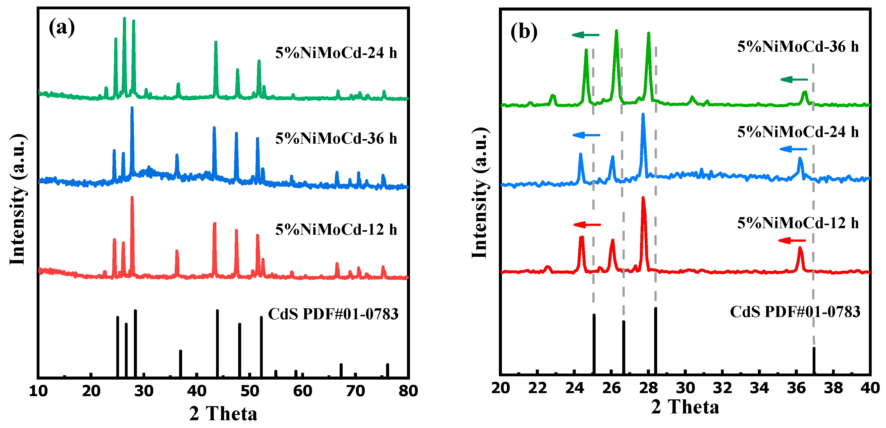
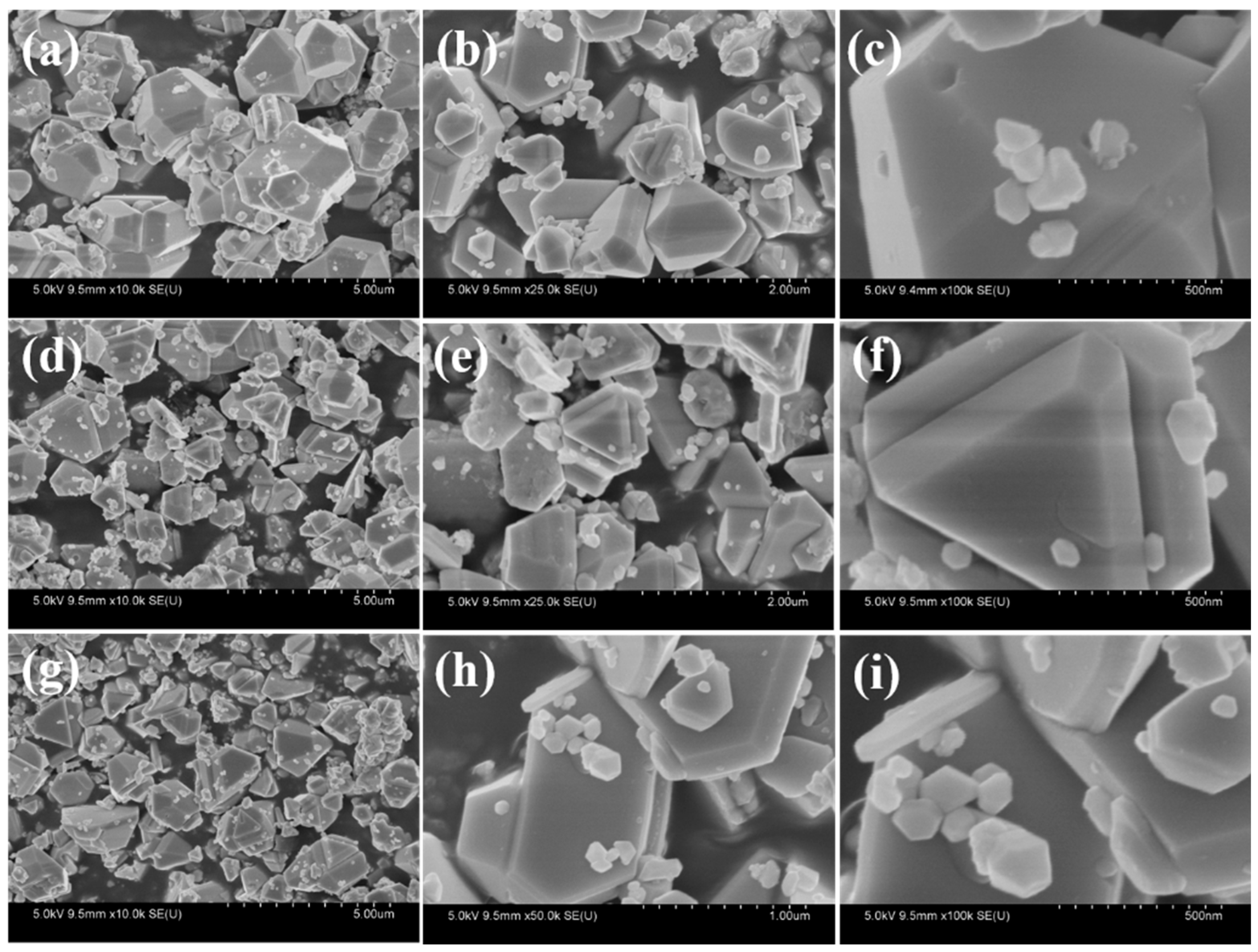
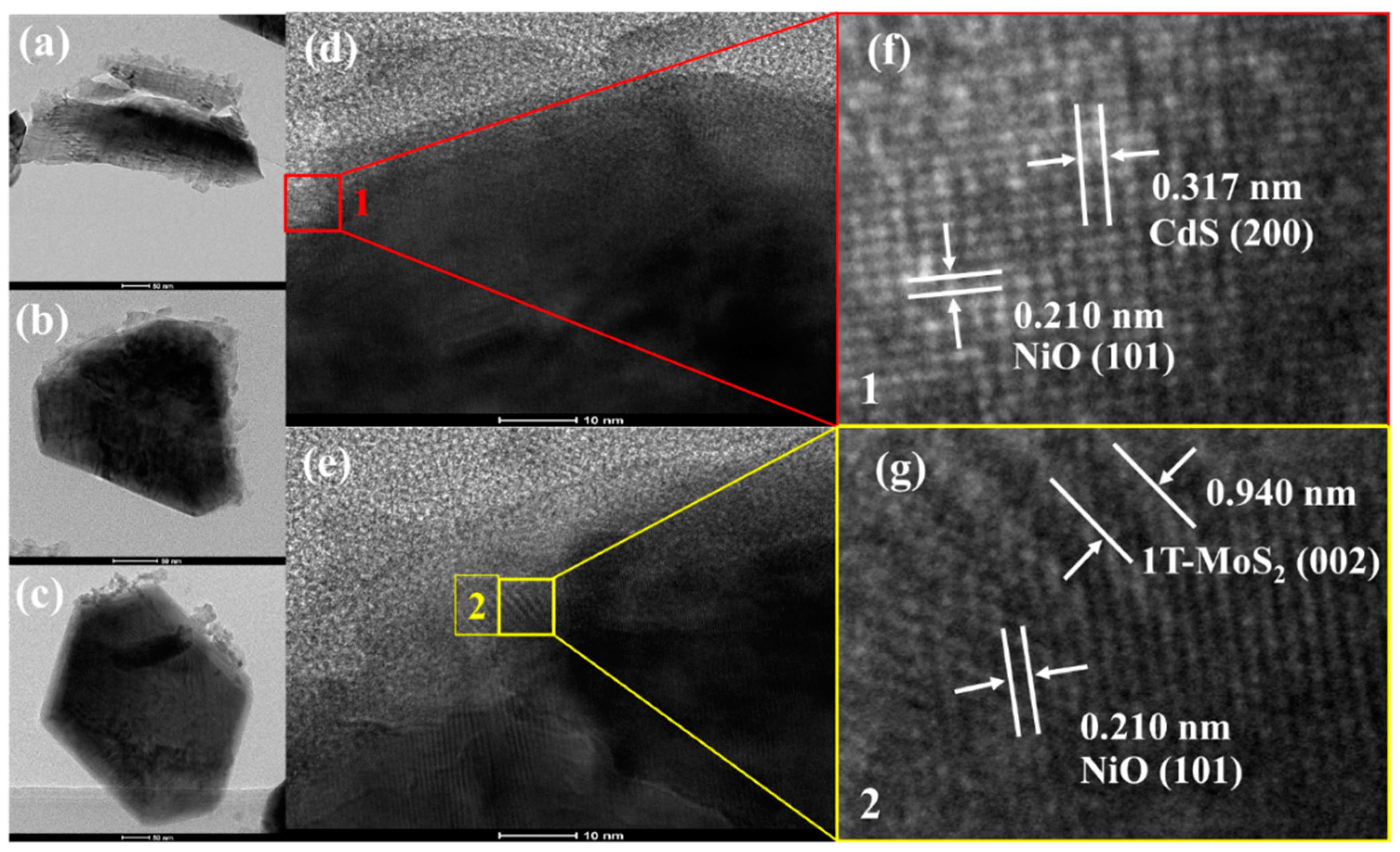
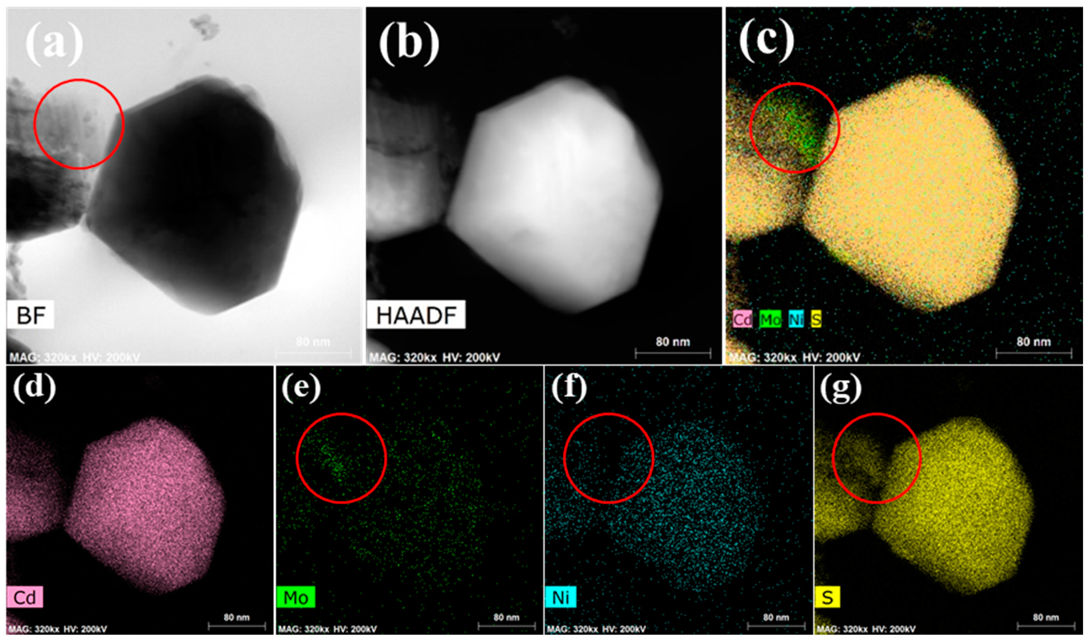
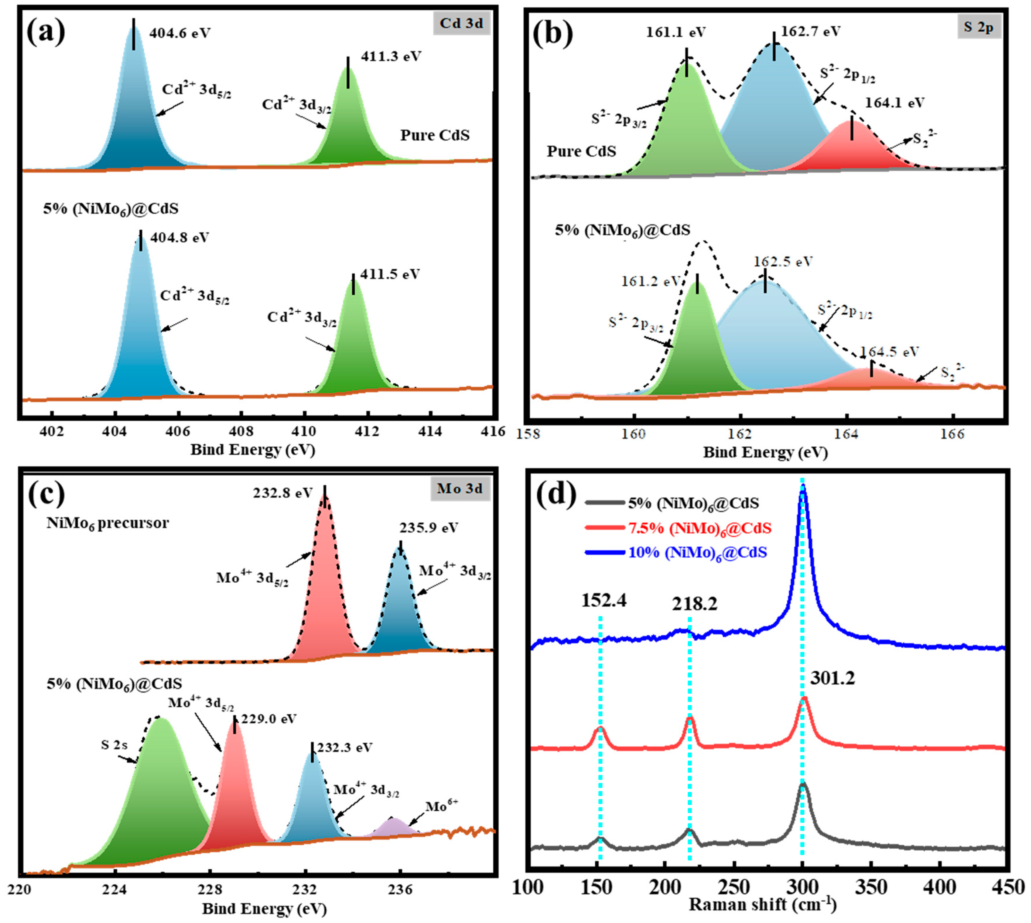
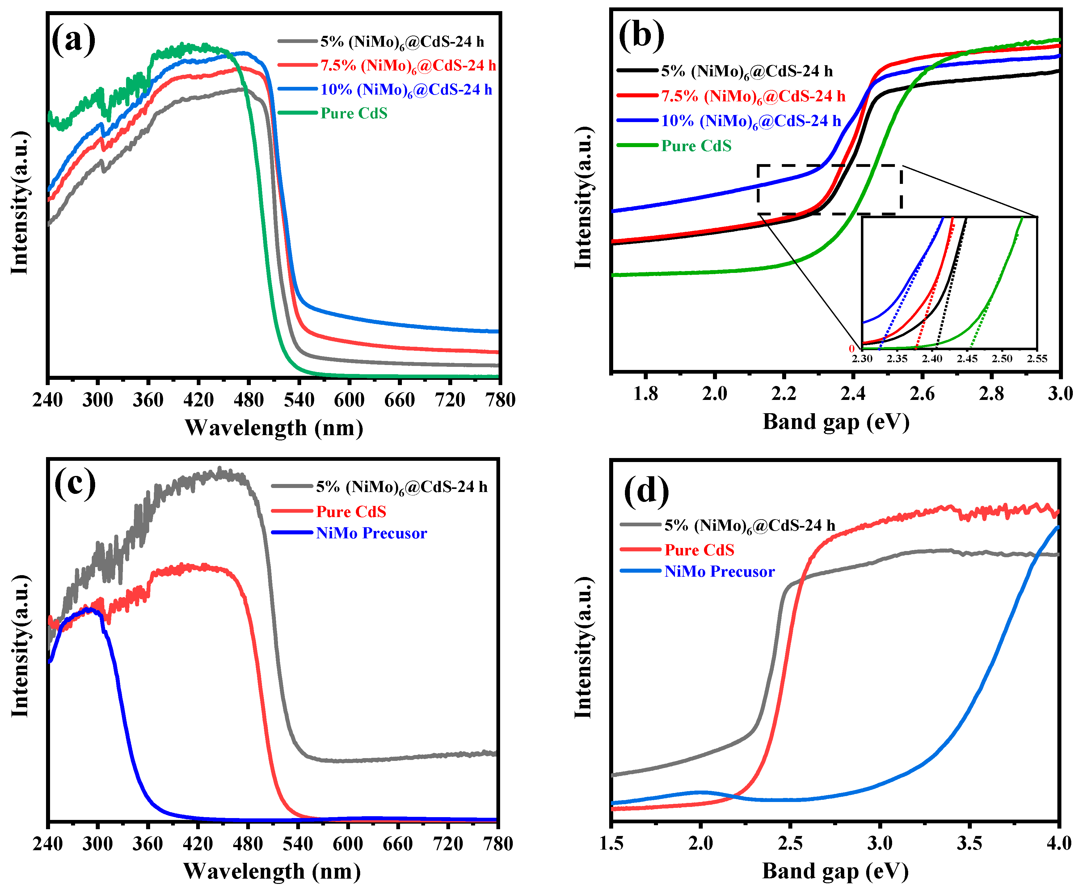
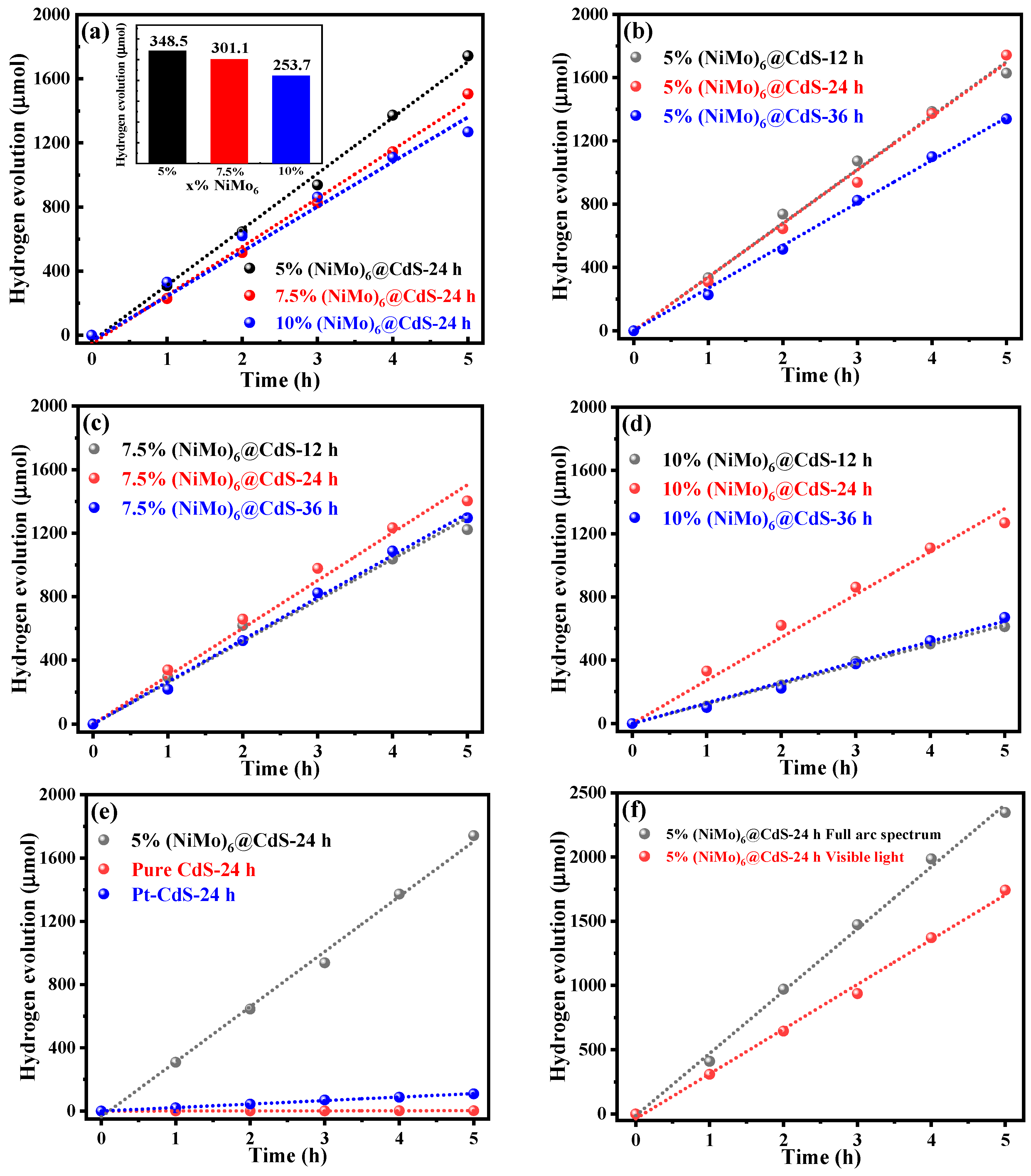
2. Results and Discussions

The powder X-ray diffraction (XRD) patterns of x%(NiMo6)@CdS-τ (x = 5, 7.5, 10, τ = 12, 24, 36 h) composites are shown in Figure 1a–f, respectively. Although, after modification with variable amounts of NiMo6 precursor and variable hydrothermal time, all XRD patterns turned out to be similar and all peaks of pristine CdS could be obviously detected in Figure 1a,c,e, according to Jade card No.01-0783. It implies that the introduction of Ni and Mo shows little influence on the bulk CdS structure. Despite this, Ni, Mo co-doping increases the lattice constant of the CdS structure, resulting in a small peak shift towards a lower angle, as shown in Figure 1b,d,f. Nonetheless, the crystalline structure of Ni and Mo species was still uncertain from the above XRD patterns, and hereafter, identified by X-ray photoelectron spectrum (XPS) measurements that will be discussed later.
The morphology characterizations of x%(NiMo6)@CdS-24 h were investigated by scanning electron microscopy (SEM) and high-resolution transmission electron microscopy (HRTEM) as depicted in Figure 2a–i and Figure 3a–g, respectively. It can be seen in Figure 2 that polyhedral CdS particles exposing terrace facets on the corners were decorated with stacked thin slices. Comparing Figure 2a,d,g, the average crystal size of CdS, which was estimated from SEM images, exhibited a decreasing trend with the increasing hydrothermal time. In Figure 3 and Figure 4a,b, a detailed understanding of crystal structure and morphology was provided by HRTEM measurements equipped with bright field (BF) and high-angle annular dark-field (HAADF) modes. As illustrated in Figure 3a–c, bulk polyhedral CdS particles were loaded with stacked structures that were speculated as NiO/MoS2 layers according to Reference [10]. Furthermore, in Figure 3d,e, two specified areas highlighted by the square were targeted at the stacked layers (Figure 3g) and the interfacial region (Figure 3f), respectively. More specifically, the lattice distances in these two regions were measured. In the interfacial region (red square box 1), two lattice fringes with the space of 0.210 and 0.317 nm, corresponding to the (200) plane of NiO (Jade card No.47-1049) and (101) plane of CdS (Jade card No. 65-3414) respectively, were observed in Figure 3f. The amplified HRTEM image of the stacked structure (yellow square box 2) in Figure 3g showed the coexistence of 0.210 and 0.940 nm spacing lattice fringes [25], which were referred to as NiO and 1T-MoS2, respectively. Through the identification of lattice constant in amplified HRTEM images, a preliminary conclusion that the composite of 1T-MoS2 doping with NiO was decorated as the layer structure on bulk CdS particle has been raised so far. Moreover, the chemical composition and element distribution of stacked structures were further determined by element mapping. It is worth noting in Figure 4 that the stacked structures marked in a circle were mainly composed of Mo and S elements, whereas no Cd element was found. Besides, the Ni element was also observed in the red circle and bulk particle simultaneously, which can be considered that Ni atoms dope in MoS2 and bulk CdS particle with a uniform distribution. The HRTEM mapping results also supported the speculation of NiO/1T- MoS2 structure.
For the purpose of unveiling the definite crystalline structure of NiMo-containing stacks on the CdS surface, the 5%(NiMo6)@CdS-24 h sample was probed by X-ray photoelectron spectrum (XPS) measurements. As indicated in Figure 5a, two characteristic signals referred to as Cd2+ 3d5/2 and Cd2+ 3d3/2, of which binding energy was located at 404.8 and 411.5 eV respectively, both exhibited a 0.2 eV blueshift in comparison with pure CdS [9,18,26]. The S 2p spectrum in Figure 5b was deconvolved into three characteristic peaks appearing at 161.2, 162.5 and 164.5 eV, which can be assigned to S2− 2p3/2, S2− 2p1/2 [9,18,27] and S22− [28], respectively. Similarly, compared with pure CdS, these peaks in the S 2p spectrum showed a slight blueshift ranging from 0.1 to 0.4 eV. The blue shift of Cd 3d and S 2p spectrum resulted from the decoration of NiMo-containing composite that slightly changed the chemical state of bulk CdS.
The Mo 3d spectrum in Figure 5c was characterized by three main peaks referred to as Mo4+ 3d5/2, Mo4+ 3d3/2 and Mo6+, implying the existence of Mo4+ species in (NiMo6)@CdS. Furthermore, the peaks of Mo4+ 3d5/2 and Mo4+ 3d3/2 appeared at 229.0 and 232.3 eV respectively, which were in accordance with bare 1T-MoS2 published in previous reports [9,18] and no additional Mo4+ signal was observed. That said, it is 1T-MoS2 rather than 2H-MoS2 or other Mo-containing compounds that were modified on bulk CdS particles, exactly falling in line with the anticipation through this template-based synthetic strategy.
On the contrary, according to our group’s previous work [11], 1T and 2H phases of MoS2 can coexist as the mixed-phase in MoS2@CdS composites. It seems to inevitably generate a mixed phase of MoS2 while modifying CdS photocatalyst [9,26]. By contrast, the above observation of pure 1T-MoS2 phase could be ascribed to the dopant Ni, which might facilitate the transformation from the coexistence of 1T- and 2H-MoS2 to a mere 1T-MoS2 phase. According to the previous study, NiMo6 precursor was introduced as the template to fabricate the NiO/1T-MoS2 composite structure that was decorated on CdS particles, where 1T-MoS2 was stabilized by NiO species [10]. Meanwhile, due to the n-type doping, Mo atoms donate electrons to Ni atoms (n-type dopants) and act as the positive charge centers, resulting in the upshift in Mo 3d spectrum, as same to the similar upshift in Cd 3d spectrum. Besides, the appearance of Mo6+ species can be attributed to partial oxidation of Mo4+ in the synthesis procedure [10,18,29]. The above findings indicate an intriguing possibility that NiO/1T-MoS2@CdS photocatalysts were successfully prepared by the introduction of NiMo6 precursors into the CdS photocatalyst.
Raman spectra were conducted to obtain further insight into the phase of MoS2 on the surface of CdS. As illustrated in Figure 5d, two intensive peaks of the Raman shift which appeared at 152.4 and 218.2 cm−1 [30] were detected, respectively. It further substantiates the existence of the 1T-MoS2 phase, whereas no observation of the 2H-MoS2 phase was found. It keeps in agreement with the XPS analysis discussed above. Surprisingly no obvious peak referred to 1T-MoS2 was observed in 10%(NiMo6)@CdS sample, which inferred that 1T-MoS2 was almost covered by excess NiO once the amount of NiMo6 precursor increased over 10%.
As the first step of the three main steps in photocatalysis, the optical absorption of photocatalysts is of necessity to enhance photocatalytic performance [31], which can be reflected by UV-vis diffuse reflection spectrum (UV-vis DRS). As illustrated in Figure 6a, pure CdS and 10%(NiMo6)@CdS-24 h samples had an approximate band edge position at nearly 515 and 555 nm, respectively. The absorption band edge positions of 5% and 7.5%(NiMo6)@CdS-24 h samples were at around 540 nm. According to the Tauc plot method, the band gap of semiconductor catalysts was calculated. As presented in Figure 6b, the 10%(NiMo6)@CdS-24 h sample had a band gap at around 2.24 eV, which was narrower than 5% (2.32 eV), 7.5%(NiMo6)@CdS-24 h samples (2.28 eV) and pure CdS (2.40 eV). Moreover, the light absorption ability of 5%(NiMo6)@CdS-24 h composite was superior to the pristine CdS photocatalyst and NiMo6 precursor in Figure 6c,d. Therefore, the modification with NiO/1T-MoS2 on bulk CdS photocatalyst broadened the absorption of the light wavelength range [27,32]. The enhancement of absorption ability can be attributed to the heterojunction among NiO, 1T-MoS2 and CdS [32,33]. Additionally, the tailing region of light absorption was intensified, which could be ascribed to the increasing amount of MoS2 with a broad absorption range in visible light [34,35].
Accumulated hydrogen evolution of x%(NiMo6)@CdS-τ samples was recorded as illustrated in Figure 7a–f. Typically, loading noble metal Pt as a co-catalyst on CdS photocatalyst enhances the photocatalytic performance. As shown in Figure 7e, (NiMo6)@CdS samples with non-noble metal all displayed a remarkable improvement in photocatalytic activity, which was comparable or even superior to the optimal Pt-CdS in the literature [36]. With regard to the hydrothermal time, τ, in Figure 7b–d, the (NiMo6)@CdS-24 h sample was endowed with the best photocatalytic performance. It infers that the (NiMo6)@CdS-24 h sample was prepared in suitable particle size and modified with a uniform distribution of NiO/1T-MoS2 after moderate hydrothermal time of 24 h, which accounts for its outstanding activity compared with the other hydrothermal times. Furthermore, as we can see in Figure 7a, the 5%(NiMo6)@CdS sample exhibited the highest photocatalytic activity with a hydrogen evolution rate of 348.5 μmol/h, whereas 7.5% and 10%(NiMo6)@CdS samples showed a relatively lower rate of 301.1 and 253.7 μmol/h, under the same hydrothermal time of 24 h. This difference of photocatalytic performance between variable precursor amounts can be explained by the influence of the modification with the special layer structure of NiO/1T-MoS2, which is clarified as mentioned below. Additionally, combined with the analysis of optical absorption, Figure 7f showed that the 5%(NiMo6)@CdS sample performed an excellent photocatalytic activity within the visible light region. Photocatalytic stability was evaluated by intermittent test sequences and each sequence consisted of the consecutive hydrogen measurement for 5 h. After one test sequence, the photocatalysts were reused through suction filtration and drying in a vacuum. Then, the photocatalytic reactor with photocatalysts inside was evacuated again and then irradiated for the next round. As illustrated in Figure 8a, the 5%(NiMo6)@CdS-24 h composite maintained a relatively stable photocatalytic performance through three cycles. In the last stability test cycle, the average hydrogen evolution per hour had decreased to approximately 90% compared to the initial cycle. In Figure 8b, the XRD pattern of the after-test sample was conducted in comparison with the before-test one. As illustrated in Figure 8b, there was no obvious difference in the characteristic peaks, which indicated that the photo-corrosion phenomenon was not observed from the XRD result and the main structure of CdS was maintained. Therefore, the photocatalytic performance reduction can be attributed to the mass loss of photocatalysts in the suction filtration and drying process. As mentioned above, the introduction of NiO species could stabilize the 1T-MoS2 structure, which could probably result in the stable photocatalytic activity of NiMo-CdS samples. However, the intrinsic stabilization mechanism of NiMo-CdS photocatalysts needs to be further explored in future research.
The influence mechanism of the modification with NiO/1T-MoS2 on pristine CdS photocatalyst can be elucidated from two primary perspectives. Through the polyoxometalate template-based synthetic strategy with NiMo6 precursor [10], NiO/1T-MoS2 was successfully loaded on the bulk CdS surface and stacked together to eventually form the thick layer structures. On the one hand, due to the superior absorption ability of 1T-MoS2 [33,34,35], the thicker layer structure containing more NiO/1T-MoS2 can absorb a larger proportion of the incident light, which in turn suppresses the excitation of photo-induced electron and hole pairs inside CdS photocatalysts and further diminishes the total photocatalytic efficiency of the NiO/1T-MoS2@CdS composite. In other words, the excessive coverage of NiO/1T-MoS2 restrains the optical absorption of bulk CdS photocatalyst and hence the total photocatalytic performance is bound to reduce to some extent. On the other side, the effective photocatalysis reacts in the prerequisite that the photo-excited carriers separated inside the bulk CdS particle successfully transfer to the surface of Ni/1T-MoS2 layer structures, where the interfacial chemical reaction occurs. It is reasonable to speculate that the 1T-MoS2 layer structure as its excellent conductivity [37,38] enhances the transportation of electrons through its suitable layer structure and simultaneously inhibits the combination of photo-excited charges in CdS particles. NiO species, distributed uniformly on the surface of both CdS particles and 1T-MoS2 layers, exhibit high activity in proton reduction [22,24]. Therefore, NiO and 1T-MoS2 loaded as bi-metal co-catalysts both play a vital role in promoting proton reduction, ultimately accelerating the hydrogen evolution. Having said that, the substantial interaction between NiO/1T-MoS2 with bulk CdS need to be further investigated with in-depth studies, and the migration mechanism of photo-induced electrons inside NiO/1T-MoS2 layers have not yet been fully clarified.

3. Experimental Methods and Characterizations

3.1. Materials

All chemicals were obtained as analytic reagent chemicals unless otherwise noted. Ammonium molybdate tetrahydrate ((NH4)6Mo7O24·4H2O), nickel nitrate hexahydrate (Ni(NO3)2·6H2O) and cadmium nitrate tetrahydrate (Cd(NO3)2·4H2O) were bought from Beijing InnoChem technology Co., Ltd. (Beijing, China). Thioacetamide (CH3CSNH2) was purchased from Sinopharm Chemical Reagent Co., Ltd. (Beijing, China). Ultra-pure water with a resistivity of 18.2 MΩ·cm was used throughout all of the experiments.

3.2. Synthesis of NiMo6 Precursor (NH4)4[NiH6Mo6O24]·5H2O

The NiMo6 precursor (NH4)4[NiH6Mo6O24]·5H2O was prepared based on the modified experimental procedure in the previous report [10]. 4.2 mmol of (NH4)6Mo7O24·4H2O was dissolved in 80 mL ultra-pure water and the solution was heated to 80 °C and then stirred for 1 h, which was referred to as Mo-Solution. 4 mmol of Ni(NO)3·6H2O was dissolved in 20 mL ultra-pure water, which was referred to as Ni-Solution. Next, the latter Ni-Solution was added dropwise to the former Mo-Solution and was stirred for 5 h at 80 °C. Subsequently, 80 mL ultra-pure water was added, after which the solution was heated to 100 °C and stirred for 1 h. Finally, after the solution was naturally cooled down to room temperature, the obtained NiMo6 products were washed several times by centrifugation with distilled water and dried at 80 °C for 12 h under vacuum.

3.3. Synthesis of x%(NiMo6)@CdS-τ(τ = 12, 24, 36 h)

(NiMo6)@CdS particles with variable amounts of NiMo6 were fabricated by a conventional hydrothermal reaction method. 36 mmol of CH3CSNH2 was dissolved into 50 mL ultra-pure water and the solution was sonicated for 5 min. After sonication, the solution was stirred for 1 h. Then, 5 mmol of Cd(NO3)2·4H2O and variable amounts of NiMo6 precursor (x% × 5 mmol NiMo6) were introduced into the CH3CSNH2 aqueous solution, respectively. After continuous magnetic stirring for 6 h, the thorough stirring solution was transferred into a 100 mL Teflon-lined stainless-steel autoclave and heated at 200 °C for periods ranging from 12 to 36 h. After being washed 5~6 times by centrifugation with distilled water and dried at 80 °C for 12 h under vacuum, the brownish-yellow x%(NiMo6)@CdS-τ products were obtained, where x and τ represent the mole fraction of NiMo6 precursor and hydrothermal time, respectively.

3.4. Synthesis of Pt-CdS

Pt-CdS photocatalyst was prepared by the photo-deposition method. Typically, with H2PtCl6 as a precursor, the co-catalyst Pt with a mass ratio of 0.2% compared to the samples, was loaded on pure CdS photocatalyst via the photo-deposition method, in the presence of 80 mL of methanol solution (25 vol%). After the suction filtration, all solid samples were dried at 80 °C for 12 h in a vacuum and then the Pt-CdS photocatalyst was collected and obtained as the experimental control group.

3.5. Characterizations

To identify the crystalline phase and the composition of the obtained photocatalysts, powder X-ray diffraction patterns were measured on an X-ray diffractometer (D8 Advance, Bruker, Karlsruhe, Germany) equipped with Cu-Kα as the X-ray source at 40 kV voltage and 15 mA current. The diffraction angle (2θ) range was from 10° to 80° with a scan speed of 5°/min. The powder X-ray diffraction patterns were further analyzed by Jade 6.0 software. The surface morphology of obtained photocatalysts was unveiled by scanning electron microscopy (SEM) (JSM-6700, JEOL, Tokyo, Japan) and high-resolution field-emission transmission electron microscopy (HRTEM) with element mapping (TALOS F200X, FEI, Hillsborough, OR, USA). The chemical composition and electronic state of obtained photocatalysts were derived from X-ray photoelectron spectroscopy (XPS) conducted by an X-ray photoelectron spectrometer (AXIS UltraDLD, Kratos, Manchester, UK) with Mg-Kα radiation. Raman spectra measurement was performed on a dispersive Raman microscope (Senterra R200-L, Bruker Optics, Karlsruhe, Germany) with the excitation wavelength of 520 nm. The optical absorption property of obtained photocatalysts was reflected by UV-vis diffuse reflection spectra (DRS) on a UV-Vis spectrophotometer (UV-2450, Shimadzu, Kyoto, Japan) with BaSO4 as the reference.

3.6. Photocatalytic Activity Measurements

The particulate photocatalytic hydrogen evolution was conducted in a 300 mL Pyrex glass reactor under irradiation under visible light, using a 300 W Xe-lamp (CEL-HXF300, Beijing China Education Au-light Co., Ltd., Beijing, China) equipped with an optical filter ranging from 420 to 780 nm, followed with a closed gas circuit system connected with a vacuum pump and online gas chromatography (Huaai GC9160, Huaai, Shanghai, China). 50 mg of (NiMo6)@CdS sample was dispersed in an aqueous solution (100 mL) containing 20 vol% 0.05/0.07 mol/L Na2S/Na2SO3 through the 10 min sonication. The photocatalytic reactor was pumped to vacuum to evacuate the dissolved air in the solution. The photocatalysts were in suspension with continuous magnetic stirring while being exposed to light irradiation. The amount of hydrogen evolved per hour was determined by the online gas chromatography equipped with argon as a carrier gas.

4. Conclusions

In summary, a series of Ni, Mo co-doped CdS photocatalysts with non-noble metal such as Pt, have been prepared by polyoxometalate template-based synthetic strategy with NiMo6 precursor. 1T-MoS2 stabilized by the introduction of NiO together with stacking layer structures was successfully decorated on the terrace facets of bulk CdS particles. Due to the modification of the special layer structure, NiO/1T-MoS2@CdS composites exhibited a greater photocatalytic performance than pristine Pt-loaded CdS, among which the 5%(NiMo6)@CdS sample stood out for its best hydrogen evolution activity with approximately 348.5 μmol/h, in the presence of Na2S/Na2SO3. The AQY efficiency could reach to 42% at 365 nm. The above results illustrated that NiO and 1T-MoS2 have a synergistic effect on enhancing the separation efficiency of photoexcited electron and hole pairs in CdS and facilitating the interfacial proton reduction, namely hydrogen evolution, at the surface-active site. However, the more detailed charge transfer procedure through the NiO/1T-MoS2 layer structure still needs clarifying in future research.

Author Contributions

J.Y.—Validation, Writing—original draft, Conceptualization, Visualization, Investigation and Data curation; Z.W.—Formal analysis; M.X.—Methodology; Z.J.—Supervision; W.S.—Writing—review and editing, Project administration and Funding acquisition. All authors have read and agreed to the published version of the manuscript.

Funding

This research was supported by the National Key Basic Research and Development Program (2018YFB1502001), the National Natural Science Foundation of China (21773153), Centre of Hydrogen Science, Shanghai Jiao Tong University, China.

Acknowledgments

We show our gratitude to the engineers and technicians in Instrumental Analysis Center, Shanghai Jiao Tong University, China.

Conflicts of Interest

The authors declared that they have no conflict of interest.

References

  1. Fujishima, A.; Honda, K. Electrochemical photolysis of water at a semiconductor electrode. Nature 1972, 238, 37–38. [Google Scholar] [CrossRef]
  2. Hitoki, G.; Takata, T.; Kondo, J.N.; Hara, M.; Kobayashi, H.; Domen, K. An oxynitride, TaON, as an efficient water oxidation photocatalyst under visible light irradiation (λ ≤ 500 nm). Chem. Commun. 2002, 16, 1698–1699. [Google Scholar] [CrossRef]
  3. Wang, X.C.; Maeda, K.; Thomas, A.; Takanabe, K.; Xin, G.; Carlsson, J.M.; Domen, K.; Antonietti, M. A metal-free polymeric photocatalyst for hydrogen production from water under visible light. Nat. Mater. 2008, 8, 76–80. [Google Scholar] [CrossRef]
  4. Wagner, F.T.; Somorjai, G.A. Photocatalytic and photoelectrochemical hydrogen production on strontium titanate single crystals. J. Am. Chem. Soc. 1980, 102, 5494–5502. [Google Scholar] [CrossRef] [Green Version]
  5. Tokunaga, S.; Kato, H.; Kudo, A. Selective Preparation of monoclinic and tetragonal BiVO4 with scheelite structure and their photocatalytic properties. Chem. Mater. 2001, 13, 4624–4628. [Google Scholar] [CrossRef]
  6. Yuan, J.; Chen, M.X.; Shi, J.W.; Shangguan, W.F. Preparations and photocatalytic hydrogen evolution of N-doped TiO2 from urea and titanium tetrachloride. Int. J. Hydrogen Energy 2006, 31, 1326–1331. [Google Scholar] [CrossRef]
  7. Schneider, J.; Matsuoka, M.; Takeuchi, M.; Zhang, J.; Horiuchi, Y.; Anpo, M.; Bahnemann, D.W. Understanding TiO2 photocatalysis: Mechanisms and materials. Chem. Rev. 2014, 114, 9919–9986. [Google Scholar] [CrossRef]
  8. Hashimoto, K.; Irie, H.; Fujishima, A. TiO2 photocatalysis: A historical overview and future prospects. Jpn. J. Appl. Phys. 2005, 44, 8269–8285. [Google Scholar] [CrossRef]
  9. Xu, M.Q.; Wei, Z.D.; Liu, J.Y.; Guo, W.Q.; Zhu, Y.; Chi, J.S.; Jiang, Z.; Shangguan, W.F. One-pot synthesized visible-light-responsive MoS2@CdS nanosheets-on- nanospheres for hydrogen evolution from the antibiotic wastewater: Waste to energy insight. Int. J. Hydrogen Energy 2019, 44, 21577–21587. [Google Scholar] [CrossRef]
  10. Wei, Z.D.; Xu, M.Q.; Liu, J.Y.; Guo, W.Q.; Jiang, Z.; Shangguan, W.F. Simultaneous visible-light-induced hydrogen production enhancement and antibiotic wastewater degradation using MoS2@ZnxCd1−xS: Solid-solution-assisted photocatalysis. Chin. J. Catal. 2020, 41, 103–113. [Google Scholar] [CrossRef]
  11. Kumar, D.P.; Hong, S.; Reddy, D.A.; Kim, T.K. Noble metal-free ultrathin MoS2 nanosheet-decorated CdS nanorods as an efficient photocatalyst for spectacular hydrogen evolution under solar light irradiation. J. Mater. Chem. A 2016, 4, 18551–18558. [Google Scholar] [CrossRef]
  12. Huang, Y.C.; Sun, Y.H.; Zheng, X.L.; Aoki, T.; Pattengale, B.; Huang, J.E.; He, X.; Bian, W.; Younan, S.; Williams, N.; et al. Atomically engineering activation sites onto metallic 1T-MoS2 catalysts for enhanced electrochemical hydrogen evolution. Nat. Commun. 2019, 10, 982. [Google Scholar] [CrossRef]
  13. Qiu, B.C.; Zhu, Q.H.; Du, M.M.; Fan, L.G.; Xing, M.Y.; Zhang, J.L. Efficient solar light harvesting CdS/Co9S8 hollow cubes for Z-scheme photocatalytic water splitting. Angew. Chem. Int. Ed. 2017, 56, 2684–2688. [Google Scholar] [CrossRef]
  14. Xu, X.J.; Hu, L.F.; Gao, N.; Liu, S.X.; Wageh, S.; Al-Ghamdi, A.A.; Alshahrie, A.; Fang, X.S. Controlled growth from ZnS nanoparticles to ZnS-CdS nanoparticle hybrids with enhanced photoactivity. Adv. Funct. Mater. 2015, 25, 445–454. [Google Scholar] [CrossRef]
  15. Fang, X.Y.; Cui, L.F.; Pu, T.T.; Song, J.L.; Zhang, X.D. Core-shell CdS@MnS nanorods as highly efficient photocatalysts for visible light driven hydrogen evolution. Appl. Surf. Sci. 2018, 457, 863–869. [Google Scholar] [CrossRef]
  16. Zhang, J.; Wang, Y.H.; Jin, J.; Zhang, J.; Lin, Z.; Huang, F.; Yu, J.G. Efficient visible-light photocatalytic hydrogen evolution and enhanced photostability of core/shell CdS/g-C3N4 nanowires. ACS Appl. Mater. Interfaces 2013, 5, 10317–10324. [Google Scholar] [CrossRef]
  17. Wu, X.Q.; Zhao, J.; Wang, L.P.; Han, M.M.; Zhang, M.L.; Wang, H.B.; Huang, H.; Liu, Y.; Kang, Z.H. Carbon dots as solid-state electron mediator for BiVO4/CDs/CdS Z-scheme photocatalyst working under visible light. Appl. Catal. B Environ. 2017, 206, 501–509. [Google Scholar] [CrossRef]
  18. Chang, K.; Hai, X.; Pang, H.; Zhang, H.B.; Shi, L.; Liu, G.G.; Liu, H.M.; Zhao, G.X.; Li, M.; Ye, J.H. Targeted synthesis of 2H- and 1T-phase MoS2 monolayers for catalytic hydrogen evolution. Adv. Mater. 2016, 28, 10033–10041. [Google Scholar] [CrossRef]
  19. Zhang, X.H.; Li, N.; Wu, J.; Zheng, Y.Z.; Tao, X. Defect-rich O-incorporated 1T-MoS2 nanosheets for remarkably enhanced visible-light photocatalytic H2 evolution over CdS: The impact of enriched defects. Appl. Catal. B Environ. 2018, 229, 227–236. [Google Scholar] [CrossRef]
  20. Liu, Q.; Shang, Q.C.; Khalil, A.; Fang, Q.; Chen, S.M.; He, Q.; Xiang, T.; Liu, D.B.; Zhang, Q.; Luo, Y.; et al. In situ integration of a metallic 1T-MoS2/CdS heterostructure as a means to promote visible-light-driven photocatalytic hydrogen evolution. ChemCatChem 2016, 8, 2614–2619. [Google Scholar] [CrossRef]
  21. Lian, Z.Q.; Liu, Y.C.; Liu, H.; Zhou, H.L.; Chang, Z.D.; Li, W.J. Fabrication of CdS@1T-MoS2 core-shell nanostructure for enhanced visible-light-driven photocatalytic H2 evolution from water splitting. J. Taiwan Inst. Chem. E 2019, 105, 57–64. [Google Scholar] [CrossRef]
  22. Qiao, S.S.; Feng, C.; Guo, Y.; Chen, T.X.; Akram, N.; Zhang, Y.; Wang, W.; Yue, F.; Wang, J.D. CdS nanoparticles modified Ni@NiO spheres as photocatalyst for oxygen production in water oxidation system and hydrogen production in water reduction system. Chem. Eng. J. 2020, 395, 125068. [Google Scholar] [CrossRef]
  23. Khan, Z.; Khannam, M.; Vinothkumar, N.; De, M.; Qureshi, M. Hierarchical 3D NiO–CdS heteroarchitecture for efficient visible light photocatalytic hydrogen generation. J. Mater. Chem. 2012, 22, 12090. [Google Scholar] [CrossRef]
  24. Chen, X.P.; Chen, W.; Gao, H.Y.; Yang, Y.; Shangguan, W.F. In situ photodeposition of NiOx on CdS for hydrogen production under visible light: Enhanced activity by controlling solution environment. Appl. Catal. B Environ. 2014, 152–153, 68–72. [Google Scholar] [CrossRef]
  25. Li, Z.Y.; Fan, R.Y.; Hu, Z.; Li, W.C.; Zhou, H.J.; Kang, S.D.; Zhang, Y.X.; Zhang, H.M.; Wang, G.Z. Ethanol introduced synthesis of ultrastable 1T-MoS2 for removal of Cr(VI). J. Hazard. Mater. 2020, 394, 122525. [Google Scholar] [CrossRef]
  26. Yin, X.L.; Li, L.L.; Jiang, W.J.; Zhang, Y.; Zhang, X.; Wan, L.J.; Hu, J.S. MoS2/CdS nanosheets-on-nanorod heterostructure for highly efficient photocatalytic H2 generation under visible light irradiation. ACS Appl. Mater. Interfaces 2016, 8, 15258–15266. [Google Scholar] [CrossRef]
  27. Han, B.; Liu, S.; Zhang, N.; Xu, Y.J.; Tang, Z.R. One-dimensional CdS@MoS2 core-shell nanowires for boosted photocatalytic hydrogen evolution under visible light. Appl. Catal. B Environ. 2017, 202, 298–304. [Google Scholar] [CrossRef]
  28. Colvin, V.L.; Goldstein, A.N.; Alivisatos, A.P. Semiconductor nanocrystals covalently bound to metal surfaces with self-assembled monolayers. J. Am. Chem. Soc. 1992, 114, 5221–5230. [Google Scholar] [CrossRef]
  29. Wang, L.L.; Liu, X.; Luo, J.M.; Duan, X.D.; Crittenden, J.; Liu, C.B.; Zhang, S.Q.; Pei, Y.; Zeng, Y.X.; Duan, X.F. Self-Optimization of the active site of molybdenum disulfide by an irreversible phase transition during photocatalytic hydrogen evolution. Angew. Chem. Int. Ed. 2017, 56, 7610–7614. [Google Scholar] [CrossRef]
  30. Yang, D.; Sandoval, S.J.; Divigalpitiya, W.M.; Irwin, J.C.; Frindt, R.F. Structure of single-molecular-layer MoS2. Phys. Rev. B 1991, 43, 12053. [Google Scholar] [CrossRef]
  31. Chen, S.S.; Takata, T.; Domen, K. Particulate photocatalysts for overall water splitting. Nat. Rev. Mater. 2017, 2, 1–17. [Google Scholar] [CrossRef]
  32. Yang, M.Q.; Han, C.; Xu, Y.J. Insight into the effect of highly dispersed MoS2 versus layer-structured MoS2 on the photocorrosion and photoactivity of CdS in graphene–CdS–MoS2 composites. J. Phys. Chem. C 2015, 119, 27234–27246. [Google Scholar] [CrossRef]
  33. Lee, H.S.; Min, S.W.; Chang, Y.G.; Park, M.K.; Nam, T.; Kim, H.; Kim, J.H.; Ryu, S.; Im, S. MoS₂ nanosheet phototransistors with thickness-modulated optical energy gap. Nano Lett. 2012, 12, 3695–3700. [Google Scholar] [CrossRef]
  34. Li, H.; Lu, G.; Yin, Z.Y.; He, Q.Y.; Li, H.; Zhang, Q.; Zhang, H. Optical identification of single- and few-layer MoS₂ sheets. Small 2012, 8, 682–686. [Google Scholar] [CrossRef]
  35. Jia, G.Y.; Liu, Y.; Gong, J.Y.; Lei, D.Y.; Wang, D.L.; Huang, Z.X. Excitonic quantum confinement modified optical conductivity of monolayer and few-layered MoS2. J. Mater. Chem. C 2016, 4, 8822–8828. [Google Scholar] [CrossRef] [Green Version]
  36. Xin, G.; Yu, B.; Xia, Y.J.; Hu, T.; Liu, L.M.; Li, C.F. Highly efficient deposition method of platinum over CdS for H2 evolution under visible light. J. Phys. Chem. C 2014, 118, 21928–21934. [Google Scholar] [CrossRef]
  37. Fivaz, R.; Mooser, E. Mobility of charge carriers in semiconducting layer structures. Phys. Rev. 1967, 163, 743. [Google Scholar] [CrossRef]
  38. Souder, A.; Brodie, D.E. Electrical contacts and conductivity of MoS2 layer structures. Can. J. Phys. 1971, 49, 2565–2571. [Google Scholar] [CrossRef]
Figure 1. (af) XRD patterns of CdS modified with x%NiMo6 precursor after different hydrothermal time, τ. Full patterns ranging from 10° to 80°: (a) x = 5, (c) x = 7.5 and (e) x = 10, and amplified patterns ranging from 20° to 40°: (b) x = 5, (d) x = 7.5 and (f) x = 10.
Catalysts 10 01478 g001aCatalysts 10 01478 g001b
Figure 2. SEM images of (ac) 5%, (df) 7.5% and (gi) 10% (NiMo6)@CdS-24 h composites.
Catalysts 10 01478 g002
Figure 3. HRTEM images of 5%(NiMo6)@CdS-24 h composite. (ac) HRTEM images with a scale bar of 50 nm. (d,e) HRTEM images with a scale bar of 10 nm. (f,g) Amplified specific regions in (f) red square box 1 and (g) yellow square box 2 derived from (d,e), respectively.
Catalysts 10 01478 g003
Figure 4. (a) BF image, (b) HAADF image and element mapping of (d) Cd, (e) Mo, (f) Ni, (g) S and (c) overall maps of 5%(NiMo6)@CdS-24 h composite.
Catalysts 10 01478 g004
Figure 5. X-ray photoelectron spectrum of 5%(NiMo6)@CdS-24 h composite, (a) Cd spectrum, (b) S spectrum, (c) Mo spectrum. (d) Raman spectra of x%(NiMo6)@CdS-24 h composites.
Catalysts 10 01478 g005
Figure 6. (a,c) UV-vis DRS and (b,d) band gap of x%(NiMo6)@CdS-24 h composites compared with pure CdS and NiMo6 precursor.
Catalysts 10 01478 g006
Figure 7. (af) Photocatalytic hydrogen evolution performance: 50 mg catalysts dispersed in 100 mL aqueous solution containing 20 vol% of 0.05/0.07 mol/L Na2S/Na2SO3. (a) Variable x% amounts of NiMo6, (bd) with variable hydrothermal time of 12, 24 and 36 h in series of (b) 5%, (c) 7.5% and (d) 10%(NiMo6)@CdS composites. (f) Comparison between full arc spectrum and visible spectrum. All the hydrogen evolution measurements were conducted under irradiation within the visible spectrum, except for (f).
Catalysts 10 01478 g007
Figure 8. (a) Photocatalytic stability tests and (b) XRD patterns of the 5%(NiMo6)@CdS-24 h composite before and after photocatalytic measurements.
Catalysts 10 01478 g008
Publisher’s Note: MDPI stays neutral with regard to jurisdictional claims in published maps and institutional affiliations.

