Next Issue
Volume 13, January-2
Previous Issue
Volume 12, December


Cancers, Volume 13, Issue 1 (January-1 2021) – 160 articles

Cover Story (view full-size image): 
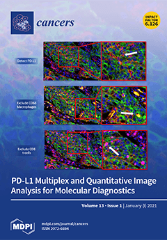
The molecular diagnostic assessment of the PD-L1 test is challenging. Multiplex immunofluorescence and whole-slide digital pathological image analysis have transformed the ability to analyze clinical biomarkers. The Precision Medicine Centre of Excellence at Queen’s University Belfast, which conducts reflex testing of all non-small cell lung cancer cases for the entire population of Northern Ireland, has developed a validated multiplex immunofluorescent biomarker panel for the assessment of PD-L1. Whole-slide digital pathology fluorescent imaging, coupled with bespoke image analysis, enabled the comprehensive extraction of phenotypic data intrinsic to patient samples. The adoption of multiplex immunofluorescence and a digital pathology workflow could triage the vast majority of PD-L1 molecular diagnostic cases and augment the pathologist assessment. View this paper
  • Issues are regarded as officially published after their release is announced to the table of contents alert mailing list.
  • You may sign up for e-mail alerts to receive table of contents of newly released issues.
  • PDF is the official format for papers published in both, html and pdf forms. To view the papers in pdf format, click on the "PDF Full-text" link, and use the free Adobe Reader to open them.
Order results
Result details
Section
Select all
Export citation of selected articles as:
12 pages, 1663 KiB  
Article
Alternating Lenvatinib and Trans-Arterial Therapy Prolongs Overall Survival in Patients with Inter-Mediate Stage HepatoCellular Carcinoma: A Propensity Score Matching Study
by Shigeo Shimose, Hideki Iwamoto, Masatoshi Tanaka, Takashi Niizeki, Tomotake Shirono, Yu Noda, Naoki Kamachi, Shusuke Okamura, Masahito Nakano, Hideya Suga, Taizo Yamaguchi, Takumi Kawaguchi, Ryoko Kuromatsu, Kazunori Noguchi, Hironori Koga and Takuji Torimura
Cancers 2021, 13(1), 160; https://doi.org/10.3390/cancers13010160 - 05 Jan 2021
Cited by 40 | Viewed by 5148
Abstract
We aimed to evaluate the impact of alternating lenvatinib (LEN) and trans-arterial therapy (AT) in patients with intermediate-stage hepatocellular carcinoma (HCC) after propensity score matching (PSM). This retrospective study enrolled 113 patients with intermediate-stage HCC treated LEN. Patients were classified into the AT [...] Read more.
We aimed to evaluate the impact of alternating lenvatinib (LEN) and trans-arterial therapy (AT) in patients with intermediate-stage hepatocellular carcinoma (HCC) after propensity score matching (PSM). This retrospective study enrolled 113 patients with intermediate-stage HCC treated LEN. Patients were classified into the AT (n = 41) or non-AT group (n = 72) according to the post LEN treatment. Overall survival (OS) was calculated using the Kaplan–Meier method and analyzed using a log-rank test after PSM. Factors associated with AT were evaluated using a decision tree analysis. After PSM, there were no significant differences in age, sex, etiology, or albumin-bilirubin (ALBI) score/grade between groups. The survival rate of the AT group was significantly higher than that of the non-AT group (median survival time; not reached vs. 16.3 months, P = 0.01). Independent factors associated with OS were AT and ALBI grade 1 in the Cox regression analysis. In the decision tree analysis, age and ALBI were the first and second splitting variables for AT. In this study, we show that AT may improve prognosis in patients with intermediate-stage HCC. Moreover, alternating LEN and trans-arterial therapy may be recommended for patients below 70 years of age with ALBI grade 1. Full article
(This article belongs to the Special Issue Prognosis and Treatment of Hepatocellular Carcinoma)
Show Figures

Figure 1

16 pages, 1752 KiB  
Review
Ways into Understanding HIF Inhibition
by Tina Schönberger, Joachim Fandrey and Katrin Prost-Fingerle
Cancers 2021, 13(1), 159; https://doi.org/10.3390/cancers13010159 - 05 Jan 2021
Cited by 24 | Viewed by 7547
Abstract
Hypoxia is a key characteristic of tumor tissue. Cancer cells adapt to low oxygen by activating hypoxia-inducible factors (HIFs), ensuring their survival and continued growth despite this hostile environment. Therefore, the inhibition of HIFs and their target genes is a promising and emerging [...] Read more.
Hypoxia is a key characteristic of tumor tissue. Cancer cells adapt to low oxygen by activating hypoxia-inducible factors (HIFs), ensuring their survival and continued growth despite this hostile environment. Therefore, the inhibition of HIFs and their target genes is a promising and emerging field of cancer research. Several drug candidates target protein–protein interactions or transcription mechanisms of the HIF pathway in order to interfere with activation of this pathway, which is deregulated in a wide range of solid and liquid cancers. Although some inhibitors are already in clinical trials, open questions remain with respect to their modes of action. New imaging technologies using luminescent and fluorescent methods or nanobodies to complement widely used approaches such as chromatin immunoprecipitation may help to answer some of these questions. In this review, we aim to summarize current inhibitor classes targeting the HIF pathway and to provide an overview of in vitro and in vivo techniques that could improve the understanding of inhibitor mechanisms. Unravelling the distinct principles regarding how inhibitors work is an indispensable step for efficient clinical applications and safety of anticancer compounds. Full article
(This article belongs to the Special Issue Inhibition of HIFs as an Anti-Cancer Strategy)
Show Figures

Figure 1

16 pages, 3488 KiB  
Article
Novel Epigenetic Eight-Gene Signature Predictive of Poor Prognosis and MSI-Like Phenotype in Human Metastatic Colorectal Carcinomas
by Valentina Condelli, Giovanni Calice, Alessandra Cassano, Michele Basso, Maria Grazia Rodriquenz, Angela Zupa, Francesca Maddalena, Fabiana Crispo, Michele Pietrafesa, Michele Aieta, Alessandro Sgambato, Giampaolo Tortora, Pietro Zoppoli and Matteo Landriscina
Cancers 2021, 13(1), 158; https://doi.org/10.3390/cancers13010158 - 05 Jan 2021
Cited by 15 | Viewed by 4012
Abstract
Epigenetics is involved in tumor progression and drug resistance in human colorectal carcinoma (CRC). This study addressed the hypothesis that the DNA methylation profiling may predict the clinical behavior of metastatic CRCs (mCRCs). The global methylation profile of two human mCRC subgroups with [...] Read more.
Epigenetics is involved in tumor progression and drug resistance in human colorectal carcinoma (CRC). This study addressed the hypothesis that the DNA methylation profiling may predict the clinical behavior of metastatic CRCs (mCRCs). The global methylation profile of two human mCRC subgroups with significantly different outcome was analyzed and compared with gene expression and methylation data from The Cancer Genome Atlas COlon ADenocarcinoma (TCGA COAD) and the NCBI GENE expression Omnibus repository (GEO) GSE48684 mCRCs datasets to identify a prognostic signature of functionally methylated genes. A novel epigenetic signature of eight hypermethylated genes was characterized that was able to identify mCRCs with poor prognosis, which had a CpG-island methylator phenotype (CIMP)-high and microsatellite instability (MSI)-like phenotype. Interestingly, methylation events were enriched in genes located on the q-arm of chromosomes 13 and 20, two chromosomal regions with gain/loss alterations associated with adenoma-to-carcinoma progression. Finally, the expression of the eight-genes signature and MSI-enriching genes was confirmed in oxaliplatin- and irinotecan-resistant CRC cell lines. These data reveal that the hypermethylation of specific genes may provide prognostic information that is able to identify a subgroup of mCRCs with poor prognosis. Full article
Show Figures

Graphical abstract

14 pages, 4561 KiB  
Review
The Role of Cannabinoids as Anticancer Agents in Pediatric Oncology
by Clara Andradas, Alexandra Truong, Jacob Byrne and Raelene Endersby
Cancers 2021, 13(1), 157; https://doi.org/10.3390/cancers13010157 - 05 Jan 2021
Cited by 11 | Viewed by 7334
Abstract
Cannabinoids are a group of chemicals that bind to receptors in the human body and, in turn, modulate the endocannabinoid system (ECS). They can be endogenously produced, synthetic, or derived from the plant Cannabis sativa L. Research over the past several decades [...] Read more.
Cannabinoids are a group of chemicals that bind to receptors in the human body and, in turn, modulate the endocannabinoid system (ECS). They can be endogenously produced, synthetic, or derived from the plant Cannabis sativa L. Research over the past several decades has shown that the ECS is a cellular communication network essential to maintain multiple biological functions and the homeostasis of the body. Indeed, cannabinoids have been shown to influence a wide variety of biological effects, including memory, pain, reproduction, bone remodeling or immunity, to name a few. Unsurprisingly, given these broad physiological effects, alterations of the ECS have been found in different diseases, including cancer. In recent years, the medical use of cannabis has been approved in different countries for a variety of human conditions. However, the use of these compounds, specifically as anticancer agents, remains controversial. Studies have shown that cannabinoids do have anticancer activity in different tumor types such as breast cancer, melanoma, lymphoma and adult brain cancer. Specifically, phytocannabinoids Δ9-tetrahydrocannabinol (THC) and cannabidiol (CBD) has been shown to induce apoptosis and inhibit proliferation of adult cancer cells, as well as modulate angiogenesis and metastasis. Despite increasing evidence that cannabinoids elicit antitumor effects in adult cancers, there is minimal data available on their effects in children or in pediatric cancers despite public and clinical demand for information. Here we describe a comprehensive and critical review of what is known about the effects of cannabinoids on pediatric cancers, highlight current gaps in knowledge and identify the critical issues that need addressing before considering these promising but controversial drugs for use in pediatric oncology. Full article
(This article belongs to the Special Issue Cannabinoids and Cancer)
Show Figures

Graphical abstract

12 pages, 840 KiB  
Article
Prolonged Diagnostic Intervals as Marker of Missed Diagnostic Opportunities in Bladder and Kidney Cancer Patients with Alarm Features: A Longitudinal Linked Data Study
by Yin Zhou, Fiona M. Walter, Hardeep Singh, William Hamilton, Gary A. Abel and Georgios Lyratzopoulos
Cancers 2021, 13(1), 156; https://doi.org/10.3390/cancers13010156 - 05 Jan 2021
Cited by 12 | Viewed by 3674
Abstract
Background: In England, patients who meet National Institute for Health and Care Excellence (NICE) guideline criteria for suspected cancer should receive a specialist assessment within 14 days. We examined how quickly bladder and kidney cancer patients who met fast-track referral criteria were actually [...] Read more.
Background: In England, patients who meet National Institute for Health and Care Excellence (NICE) guideline criteria for suspected cancer should receive a specialist assessment within 14 days. We examined how quickly bladder and kidney cancer patients who met fast-track referral criteria were actually diagnosed. Methods: We used linked primary care and cancer registration data on bladder and kidney cancer patients who met fast-track referral criteria and examined the time from their first presentation with alarm features to diagnosis. Using logistic regression we examined factors most likely to be associated with non-timely diagnosis (defined as intervals exceeding 90 days), adjusting for age, sex and cancer type, positing that such occurrences represent missed opportunity for timely referral, possibly due to sub-optimal guideline adherence. Results: 28%, 42% and 31% of all urological cancer patients reported no, one or two or more relevant symptoms respectively in the year before diagnosis. Of the 2105 patients with alarm features warranting fast-track assessment, 1373 (65%) presented with unexplained haematuria, 382 (18%) with recurrent urinary tract infections (UTIs), 303 (14%) with visible haematuria, and 45 (2%) with an abdominal mass. 27% overall, and 24%, 45%, 18% and 27% of each group respectively, had a non-timely diagnosis. Presentation with recurrent UTI was associated with longest median diagnostic interval (median 83 days, IQR 43–151) and visible haematuria with the shortest (median 50 days, IQR 30–79). After adjustment, presentation with recurrent UTIs, being in the youngest or oldest age group, female sex, and diagnosis of kidney and upper tract urothelial cancer, were associated with greater odds of non-timely diagnosis. Conclusion: More than a quarter of patients presenting with fast-track referral features did not achieve a timely diagnosis, suggesting inadequate guideline adherence for some patients. The findings highlight a substantial number of opportunities for expediting the diagnosis of patients with bladder or kidney cancers. Full article
(This article belongs to the Section Cancer Causes, Screening and Diagnosis)
18 pages, 6739 KiB  
Article
Immunogenomic Gene Signature of Cell-Death Associated Genes with Prognostic Implications in Lung Cancer
by Pankaj Ahluwalia, Meenakshi Ahluwalia, Ashis K. Mondal, Nikhil Sahajpal, Vamsi Kota, Mumtaz V. Rojiani, Amyn M. Rojiani and Ravindra Kolhe
Cancers 2021, 13(1), 155; https://doi.org/10.3390/cancers13010155 - 05 Jan 2021
Cited by 36 | Viewed by 5968
Abstract
Lung cancer is one of the leading causes of death worldwide. Cell death pathways such as autophagy, apoptosis, and necrosis can provide useful clinical and immunological insights that can assist in the design of personalized therapeutics. In this study, variations in the expression [...] Read more.
Lung cancer is one of the leading causes of death worldwide. Cell death pathways such as autophagy, apoptosis, and necrosis can provide useful clinical and immunological insights that can assist in the design of personalized therapeutics. In this study, variations in the expression of genes involved in cell death pathways and resulting infiltration of immune cells were explored in lung adenocarcinoma (The Cancer Genome Atlas: TCGA, lung adenocarcinoma (LUAD), 510 patients). Firstly, genes involved in autophagy (n = 34 genes), apoptosis (n = 66 genes), and necrosis (n = 32 genes) were analyzed to assess the prognostic significance in lung cancer. The significant genes were used to develop the cell death index (CDI) of 21 genes which clustered patients based on high risk (high CDI) and low risk (low CDI). The survival analysis using the Kaplan–Meier curve differentiated patients based on overall survival (40.4 months vs. 76.2 months), progression-free survival (26.2 months vs. 48.6 months), and disease-free survival (62.2 months vs. 158.2 months) (Log-rank test, p < 0.01). Cox proportional hazard model significantly associated patients in high CDI group with a higher risk of mortality (Hazard Ratio: H.R 1.75, 95% CI: 1.28–2.45, p < 0.001). Differential gene expression analysis using principal component analysis (PCA) identified genes with the highest fold change forming distinct clusters. To analyze the immune parameters in two risk groups, cytokines expression (n = 265 genes) analysis revealed the highest association of IL-15RA and IL 15 (> 1.5-fold, p < 0.01) with the high-risk group. The microenvironment cell-population (MCP)-counter algorithm identified the higher infiltration of CD8+ T cells, macrophages, and lower infiltration of neutrophils with the high-risk group. Interestingly, this group also showed a higher expression of immune checkpoint molecules CD-274 (PD-L1), CTLA-4, and T cell exhaustion genes (HAVCR2, TIGIT, LAG3, PDCD1, CXCL13, and LYN) (p < 0.01). Furthermore, functional enrichment analysis identified significant perturbations in immune pathways in the higher risk group. This study highlights the presence of an immunocompromised microenvironment indicated by the higher infiltration of cytotoxic T cells along with the presence of checkpoint molecules and T cell exhaustion genes. These patients at higher risk might be more suitable to benefit from PD-L1 blockade or other checkpoint blockade immunotherapies. Full article
(This article belongs to the Special Issue Non-small Cell Lung Cancer--Tumor Biology)
Show Figures

Figure 1

24 pages, 8160 KiB  
Article
Relation of Metal-Binding Property and Selective Toxicity of 8-Hydroxyquinoline Derived Mannich Bases Targeting Multidrug Resistant Cancer Cells
by Veronika F.S. Pape, Anikó Gaál, István Szatmári, Nóra Kucsma, Norbert Szoboszlai, Christina Streli, Ferenc Fülöp, Éva A. Enyedy and Gergely Szakács
Cancers 2021, 13(1), 154; https://doi.org/10.3390/cancers13010154 - 05 Jan 2021
Cited by 9 | Viewed by 3264
Abstract
Resistance to chemotherapeutic agents is a major obstacle in cancer treatment. A recently proposed strategy is to target the collateral sensitivity of multidrug resistant (MDR) cancer. Paradoxically, the toxicity of certain metal chelating agents is increased, rather than decreased, by the function of [...] Read more.
Resistance to chemotherapeutic agents is a major obstacle in cancer treatment. A recently proposed strategy is to target the collateral sensitivity of multidrug resistant (MDR) cancer. Paradoxically, the toxicity of certain metal chelating agents is increased, rather than decreased, by the function of P-glycoprotein (Pgp), which is known to confer resistance by effluxing chemotherapeutic compounds from cancer cells. We have recently characterized and compared the solution’s chemical properties including ligand protonation and the metal binding properties of a set of structurally related 8-hydroxyquinoline derived Mannich bases. Here we characterize the impact of the solution stability and redox activity of their iron(III) and copper(II) complexes on MDR-selective toxicity. Our results show that the MDR-selective anticancer activity of the studied 8-hydroxyquinoline derived Mannich bases is associated with the iron deprivation of MDR cells and the preferential formation of redox-active copper(II) complexes, which undergo intracellular redox-cycling to induce oxidative stress. Full article
Show Figures

Graphical abstract

19 pages, 9547 KiB  
Article
Co-Overexpression of TWIST1-CSF1 Is a Common Event in Metastatic Oral Cancer and Drives Biologically Aggressive Phenotype
by Sabrina Daniela da Silva, Fabio Albuquerque Marchi, Jie Su, Long Yang, Ludmila Valverde, Jessica Hier, Krikor Bijian, Michael Hier, Alex Mlynarek, Luiz Paulo Kowalski and Moulay A. Alaoui-Jamali
Cancers 2021, 13(1), 153; https://doi.org/10.3390/cancers13010153 - 05 Jan 2021
Cited by 12 | Viewed by 3182
Abstract
Invasive oral squamous cell carcinoma (OSCC) is often ulcerated and heavily infiltrated by pro-inflammatory cells. We conducted a genome-wide profiling of tissues from OSCC patients (early versus advanced stages) with 10 years follow-up. Co-amplification and co-overexpression of TWIST1, a transcriptional activator of [...] Read more.
Invasive oral squamous cell carcinoma (OSCC) is often ulcerated and heavily infiltrated by pro-inflammatory cells. We conducted a genome-wide profiling of tissues from OSCC patients (early versus advanced stages) with 10 years follow-up. Co-amplification and co-overexpression of TWIST1, a transcriptional activator of epithelial-mesenchymal-transition (EMT), and colony-stimulating factor-1 (CSF1), a major chemotactic agent for tumor-associated macrophages (TAMs), were observed in metastatic OSCC cases. The overexpression of these markers strongly predicted poor patient survival (log-rank test, p = 0.0035 and p = 0.0219). Protein analysis confirmed the enhanced expression of TWIST1 and CSF1 in metastatic tissues. In preclinical models using OSCC cell lines, macrophages, and an in vivo matrigel plug assay, we demonstrated that TWIST1 gene overexpression induces the activation of CSF1 while TWIST1 gene silencing down-regulates CSF1 preventing OSCC invasion. Furthermore, excessive macrophage activation and polarization was observed in co-culture system involving OSCC cells overexpressing TWIST1. In summary, this study provides insight into the cooperation between TWIST1 transcription factor and CSF1 to promote OSCC invasiveness and opens up the potential therapeutic utility of currently developed antibodies and small molecules targeting cancer-associated macrophages. Full article
Show Figures

Figure 1

13 pages, 600 KiB  
Review
The Emerging Role of Nerves and Glia in Colorectal Cancer
by Simone L. Schonkeren, Meike S. Thijssen, Nathalie Vaes, Werend Boesmans and Veerle Melotte
Cancers 2021, 13(1), 152; https://doi.org/10.3390/cancers13010152 - 05 Jan 2021
Cited by 26 | Viewed by 7155
Abstract
The role of the nervous system as a contributor in the tumor microenvironment has been recognized in different cancer types, including colorectal cancer (CRC). The gastrointestinal tract is a highly innervated organ system, which is not only innervated by the autonomic nervous system, [...] Read more.
The role of the nervous system as a contributor in the tumor microenvironment has been recognized in different cancer types, including colorectal cancer (CRC). The gastrointestinal tract is a highly innervated organ system, which is not only innervated by the autonomic nervous system, but also contains an extensive nervous system of its own; the enteric nervous system (ENS). The ENS is important for gut function and homeostasis by regulating processes such as fluid absorption, blood flow, and gut motility. Dysfunction of the ENS has been linked with multiple gastrointestinal diseases, such as Hirschsprung disease and inflammatory bowel disease, and even with neurodegenerative disorders. How the extrinsic and intrinsic innervation of the gut contributes to CRC is not fully understood, although a mutual relationship between cancer cells and nerves has been described. Nerves enhance cancer progression through the secretion of neurotransmitters and neuropeptides, and cancer cells are capable of stimulating nerve growth. This review summarizes and discusses the nervous system innervation of the gastrointestinal tract and how it can influence carcinogenesis, and vice versa. Lastly, the therapeutic potential of these novel insights is discussed. Full article
(This article belongs to the Special Issue Tumor Evolution: Progression, Metastasis and Therapeutic Response)
Show Figures

Figure 1

14 pages, 532 KiB  
Review
Mechanisms of Resistance to KRASG12C Inhibitors
by Victoria Dunnett-Kane, Pantelis Nicola, Fiona Blackhall and Colin Lindsay
Cancers 2021, 13(1), 151; https://doi.org/10.3390/cancers13010151 - 05 Jan 2021
Cited by 74 | Viewed by 11516
Abstract
KRAS is one of the most common human oncogenes, but concerted efforts to produce direct inhibitors have largely failed, earning KRAS the title of “undruggable”. Recent efforts to produce subtype specific inhibitors have been more successful, and several KRASG12C inhibitors have reached [...] Read more.
KRAS is one of the most common human oncogenes, but concerted efforts to produce direct inhibitors have largely failed, earning KRAS the title of “undruggable”. Recent efforts to produce subtype specific inhibitors have been more successful, and several KRASG12C inhibitors have reached clinical trials, including adagrasib and sotorasib, which have shown early evidence of efficacy in patients. Lessons from other inhibitors of the RAS pathway suggest that the effect of these drugs will be limited in vivo by the development of drug resistance, and pre-clinical studies of G12C inhibitors have identified evidence of this. In this review we discuss the current evidence for G12C inhibitors, the mechanisms of resistance to G12C inhibitors and potential approaches to overcome them. We discuss possible targets of combination therapy, including SHP2, receptor tyrosine kinases, downstream effectors and PD1/PDL1, and review the ongoing clinical trials investigating these inhibitors. Full article
(This article belongs to the Special Issue Targeting KRAS: Elucidating Mechanisms of Sensitivity and Resistance)
Show Figures

Figure 1

12 pages, 1306 KiB  
Article
Specific and Sensitive Detection of Neuroblastoma mRNA Markers by Multiplex RT-qPCR
by Lieke M. J. van Zogchel, Lily Zappeij-Kannegieter, Ahmad Javadi, Marjolein Lugtigheid, Nina U. Gelineau, Nathalie S. M. Lak, Danny A. Zwijnenburg, Jan Koster, Janine Stutterheim, C. Ellen van der Schoot and Godelieve A. M. Tytgat
Cancers 2021, 13(1), 150; https://doi.org/10.3390/cancers13010150 - 05 Jan 2021
Cited by 12 | Viewed by 3413
Abstract
mRNA RT-qPCR is shown to be a very sensitive technique to detect minimal residual disease (MRD) in patients with neuroblastoma. Multiple mRNA markers are known to detect heterogeneous neuroblastoma cells in bone marrow (BM) or blood from patients. However, the limited volumes of [...] Read more.
mRNA RT-qPCR is shown to be a very sensitive technique to detect minimal residual disease (MRD) in patients with neuroblastoma. Multiple mRNA markers are known to detect heterogeneous neuroblastoma cells in bone marrow (BM) or blood from patients. However, the limited volumes of BM and blood available can hamper the detection of multiple markers. To make optimal use of these samples, we developed a multiplex RT-qPCR for the detection of MRD in neuroblastoma. GUSB and PHOX2B were tested as single markers. The adrenergic markers TH, GAP43, CHRNA3 and DBH and mesenchymal markers POSTN, PRRX1 and FMO3 were tested in multiplex. Using control blood and BM, we established new thresholds for positivity. Comparison of multiplex and singleplex RT-qPCR results from 21 blood and 24 BM samples from neuroblastoma patients demonstrated a comparable sensitivity. With this multiplex RT-qPCR, we are able to test seven different neuroblastoma mRNA markers, which overcomes tumor heterogeneity and improves sensitivity of MRD detection, even in those samples of low RNA quantity. With resources and time being saved, reduction in sample volume and consumables can assist in the introduction of MRD by RT-qPCR into clinical practice. Full article
(This article belongs to the Special Issue Minimal Residual Disease of Cancers)
Show Figures

Graphical abstract

12 pages, 1691 KiB  
Article
Molecular Changes in Retinoblastoma beyond RB1: Findings from Next-Generation Sequencing
by Jasmine H. Francis, Allison L. Richards, Diana L. Mandelker, Michael F. Berger, Michael F. Walsh, Ira J. Dunkel, Mark T. A. Donoghue and David H. Abramson
Cancers 2021, 13(1), 149; https://doi.org/10.3390/cancers13010149 - 05 Jan 2021
Cited by 27 | Viewed by 4381
Abstract
This investigation uses hybridization capture-based next-generation sequencing to deepen our understanding of genetics that underlie retinoblastoma. Eighty-three enucleated retinoblastoma specimens were evaluated using a MSK-IMPACT clinical next-generation sequencing panel to evaluate both somatic and germline alterations. Somatic copy number variations (CNVs) were also [...] Read more.
This investigation uses hybridization capture-based next-generation sequencing to deepen our understanding of genetics that underlie retinoblastoma. Eighty-three enucleated retinoblastoma specimens were evaluated using a MSK-IMPACT clinical next-generation sequencing panel to evaluate both somatic and germline alterations. Somatic copy number variations (CNVs) were also identified. Genetic profiles were correlated to clinicopathologic characteristics. RB1 inactivation was found in 79 (97.5%) patients. All specimens had additional molecular alterations. The most common non-RB1 gene alteration was BCOR in 19 (22.9%). Five (11.0%) had pathogenic germline mutations in other non-RB1 cancer predisposition genes. Significant clinicopathologic correlations included: vitreous seeds associated with 1q gains and 16q loss of heterozygosity (BH-corrected p-value = 0.008, 0.004; OR = 12.6, 26.7, respectively). BCOR mutations were associated with poor prognosis, specifically metastases-free survival (MFS) (nominal p-value 0.03). Furthermore, retinoblastoma patients can have non-RB1 germline mutations in other cancer-associated genes. No two specimens had the identical genetic profile, emphasizing the individuality of tumors with the same clinical diagnosis. Full article
(This article belongs to the Section Methods and Technologies Development)
Show Figures

Figure 1

20 pages, 2891 KiB  
Article
In Vivo Optical Metabolic Imaging of Long-Chain Fatty Acid Uptake in Orthotopic Models of Triple-Negative Breast Cancer
by Megan C. Madonna, Joy E. Duer, Joyce V. Lee, Jeremy Williams, Baris Avsaroglu, Caigang Zhu, Riley Deutsch, Roujia Wang, Brian T. Crouch, Matthew D. Hirschey, Andrei Goga and Nirmala Ramanujam
Cancers 2021, 13(1), 148; https://doi.org/10.3390/cancers13010148 - 05 Jan 2021
Cited by 17 | Viewed by 4944
Abstract
Targeting a tumor’s metabolic dependencies is a clinically actionable therapeutic approach; however, identifying subtypes of tumors likely to respond remains difficult. The use of lipids as a nutrient source is of particular importance, especially in breast cancer. Imaging techniques offer the opportunity to [...] Read more.
Targeting a tumor’s metabolic dependencies is a clinically actionable therapeutic approach; however, identifying subtypes of tumors likely to respond remains difficult. The use of lipids as a nutrient source is of particular importance, especially in breast cancer. Imaging techniques offer the opportunity to quantify nutrient use in preclinical tumor models to guide development of new drugs that restrict uptake or utilization of these nutrients. We describe a fast and dynamic approach to image fatty acid uptake in vivo and demonstrate its relevance to study both tumor metabolic reprogramming directly, as well as the effectiveness of drugs targeting lipid metabolism. Specifically, we developed a quantitative optical approach to spatially and longitudinally map the kinetics of long-chain fatty acid uptake in in vivo murine models of breast cancer using a fluorescently labeled palmitate molecule, Bodipy FL c16. We chose intra-vital microscopy of mammary tumor windows to validate our approach in two orthotopic breast cancer models: a MYC-overexpressing, transgenic, triple-negative breast cancer (TNBC) model and a murine model of the 4T1 family. Following injection, Bodipy FL c16 fluorescence increased and reached its maximum after approximately 30 min, with the signal remaining stable during the 30–80 min post-injection period. We used the fluorescence at 60 min (Bodipy60), the mid-point in the plateau region, as a summary parameter to quantify Bodipy FL c16 fluorescence in subsequent experiments. Using our imaging platform, we observed a two- to four-fold decrease in fatty acid uptake in response to the downregulation of the MYC oncogene, consistent with findings from in vitro metabolic assays. In contrast, our imaging studies report an increase in fatty acid uptake with tumor aggressiveness (6NR, 4T07, and 4T1), and uptake was significantly decreased after treatment with a fatty acid transport inhibitor, perphenazine, in both normal mammary pads and in the most aggressive 4T1 tumor model. Our approach fills an important gap between in vitro assays providing rich metabolic information at static time points and imaging approaches visualizing metabolism in whole organs at a reduced resolution. Full article
(This article belongs to the Special Issue Imaging Cancer Metabolism)
Show Figures

Graphical abstract

18 pages, 2694 KiB  
Article
Human Plasma Metabolomics for Biomarker Discovery: Targeting the Molecular Subtypes in Breast Cancer
by Leticia Díaz-Beltrán, Carmen González-Olmedo, Natalia Luque-Caro, Caridad Díaz, Ariadna Martín-Blázquez, Mónica Fernández-Navarro, Ana Laura Ortega-Granados, Fernando Gálvez-Montosa, Francisca Vicente, José Pérez del Palacio and Pedro Sánchez-Rovira
Cancers 2021, 13(1), 147; https://doi.org/10.3390/cancers13010147 - 05 Jan 2021
Cited by 20 | Viewed by 4077
Abstract
Purpose: The aim of this study is to identify differential metabolomic signatures in plasma samples of distinct subtypes of breast cancer patients that could be used in clinical practice as diagnostic biomarkers for these molecular phenotypes and to provide a more individualized and [...] Read more.
Purpose: The aim of this study is to identify differential metabolomic signatures in plasma samples of distinct subtypes of breast cancer patients that could be used in clinical practice as diagnostic biomarkers for these molecular phenotypes and to provide a more individualized and accurate therapeutic procedure. Methods: Untargeted LC-HRMS metabolomics approach in positive and negative electrospray ionization mode was used to analyze plasma samples from LA, LB, HER2+ and TN breast cancer patients and healthy controls in order to determine specific metabolomic profiles through univariate and multivariate statistical data analysis. Results: We tentatively identified altered metabolites displaying concentration variations among the four breast cancer molecular subtypes. We found a biomarker panel of 5 candidates in LA, 7 in LB, 5 in HER2 and 3 in TN that were able to discriminate each breast cancer subtype with a false discovery range corrected p-value < 0.05 and a fold-change cutoff value > 1.3. The model clinical value was evaluated with the AUROC, providing diagnostic capacities above 0.85. Conclusion: Our study identifies metabolic profiling differences in molecular phenotypes of breast cancer. This may represent a key step towards therapy improvement in personalized medicine and prioritization of tailored therapeutic intervention strategies. Full article
Show Figures

Figure 1

23 pages, 678 KiB  
Review
Stromal Protein-Mediated Immune Regulation in Digestive Cancers
by Pia Gamradt, Christelle De La Fouchardière and Ana Hennino
Cancers 2021, 13(1), 146; https://doi.org/10.3390/cancers13010146 - 05 Jan 2021
Cited by 17 | Viewed by 4264
Abstract
The stromal tumor microenvironment (TME) consists of immune cells, vascular and neural structures, cancer-associated fibroblasts (CAFs), as well as extracellular matrix (ECM), and favors immune escape mechanisms promoting the initiation and progression of digestive cancers. Numerous ECM proteins released by stromal and tumor [...] Read more.
The stromal tumor microenvironment (TME) consists of immune cells, vascular and neural structures, cancer-associated fibroblasts (CAFs), as well as extracellular matrix (ECM), and favors immune escape mechanisms promoting the initiation and progression of digestive cancers. Numerous ECM proteins released by stromal and tumor cells are crucial in providing physical rigidity to the TME, though they are also key regulators of the immune response against cancer cells by interacting directly with immune cells or engaging with immune regulatory molecules. Here, we discuss current knowledge of stromal proteins in digestive cancers including pancreatic cancer, colorectal cancer, and gastric cancer, focusing on their functions in inhibiting tumor immunity and enabling drug resistance. Moreover, we will discuss the implication of stromal proteins as therapeutic targets to unleash efficient immunotherapy-based treatments. Full article
Show Figures

Figure 1

10 pages, 379 KiB  
Article
Use of Palliative Chemotherapy and ICU Admissions in Gastric and Esophageal Cancer Patients in the Last Phase of Life: A Nationwide Observational Study
by Joost Besseling, Jan Reitsma, Judith A. Van Erkelens, Maike H. J. Schepens, Michiel P. C. Siroen, Cathelijne M. P. Ziedses des Plantes, Mark I. van Berge Henegouwen, Laurens V. Beerepoot, Theo Van Voorthuizen, Lia Van Zuylen, Rob H. A. Verhoeven and Hanneke van Laarhoven
Cancers 2021, 13(1), 145; https://doi.org/10.3390/cancers13010145 - 05 Jan 2021
Cited by 9 | Viewed by 2821
Abstract
Since intensive care unit (ICU) admission and chemotherapy use near death impair the quality of life, we studied the prevalence of both and their correlation with hospital volume in incurable gastroesophageal cancer patients as both impair the quality of life. We analyzed all [...] Read more.
Since intensive care unit (ICU) admission and chemotherapy use near death impair the quality of life, we studied the prevalence of both and their correlation with hospital volume in incurable gastroesophageal cancer patients as both impair the quality of life. We analyzed all Dutch patients with incurable gastroesophageal cancer who died in 2017–2018. National insurance claims data were used to determine the prevalence of ICU admission and chemotherapy use (stratified on previous chemotherapy treatment) at three and one month(s) before death. We calculated correlations between hospital volume (i.e., the number of included patients per hospital) and both outcomes. We included 3748 patients (mean age: 71.4 years; 71.4% male). The prevalence of ICU admission and chemotherapy use were, respectively, 5.6% and 21.2% at three months and 4.2% and 8.0% at one month before death. Chemotherapy use at three and one months before death was, respectively, 4.3 times (48.0% vs. 11.2%) and 3.7 times higher (15.7% vs. 4.3%), comparing patients with previous chemotherapy treatment to those without. Hospital volume was negatively correlated with chemotherapy use in the final month (rweighted = −0.23, p = 0.04). ICU admission and chemotherapy use were relatively infrequent. Oncologists in high-volume hospitals may be better equipped in selecting patients most likely to benefit from chemotherapy. Full article
Show Figures

Figure 1

17 pages, 860 KiB  
Review
NPM-ALK: A Driver of Lymphoma Pathogenesis and a Therapeutic Target
by Elissa Andraos, Joséphine Dignac and Fabienne Meggetto
Cancers 2021, 13(1), 144; https://doi.org/10.3390/cancers13010144 - 05 Jan 2021
Cited by 13 | Viewed by 3178
Abstract
Initially discovered in anaplastic large cell lymphoma (ALCL), the ALK anaplastic lymphoma kinase is a tyrosine kinase which is affected in lymphomas by oncogenic translocations, mainly NPM-ALK. To date, chemotherapy remains a viable option in ALCL patients with ALK translocations as it leads [...] Read more.
Initially discovered in anaplastic large cell lymphoma (ALCL), the ALK anaplastic lymphoma kinase is a tyrosine kinase which is affected in lymphomas by oncogenic translocations, mainly NPM-ALK. To date, chemotherapy remains a viable option in ALCL patients with ALK translocations as it leads to remission rates of approximately 80%. However, the remaining patients do not respond to chemotherapy and some patients have drug-resistant relapses. It is therefore crucial to identify new and better treatment options. Nowadays, different classes of ALK tyrosine kinase inhibitors (TKI) are available and used exclusively for EML4-ALK (+) lung cancers. In fact, the significant toxicities of most ALK inhibitors explain the delay in their use in ALCL patients, who are predominantly children. Moreover, some ALCL patients do not respond to Crizotinib, the first generation TKI, or develop an acquired resistance months following an initial response. Combination therapy with ALK inhibitors in ALCL is the current challenge. Full article
(This article belongs to the Special Issue New Therapeutic Developments in Hematological Malignancies)
Show Figures

Figure 1

17 pages, 2907 KiB  
Systematic Review
Functional Signatures in Non-Small-Cell Lung Cancer: A Systematic Review and Meta-Analysis of Sex-Based Differences in Transcriptomic Studies
by Irene Pérez-Díez, Marta R. Hidalgo, Pablo Malmierca-Merlo, Zoraida Andreu, Sergio Romera-Giner, Rosa Farràs, María de la Iglesia-Vayá, Mariano Provencio, Atocha Romero and Francisco García-García
Cancers 2021, 13(1), 143; https://doi.org/10.3390/cancers13010143 - 05 Jan 2021
Cited by 10 | Viewed by 4218
Abstract
While studies have established the existence of differences in the epidemiological and clinical patterns of lung adenocarcinoma between male and female patients, we know relatively little regarding the molecular mechanisms underlying such sex-based differences. In this study, we explore said differences through a [...] Read more.
While studies have established the existence of differences in the epidemiological and clinical patterns of lung adenocarcinoma between male and female patients, we know relatively little regarding the molecular mechanisms underlying such sex-based differences. In this study, we explore said differences through a meta-analysis of transcriptomic data. We performed a meta-analysis of the functional profiling of nine public datasets that included 1366 samples from Gene Expression Omnibus and The Cancer Genome Atlas databases. Meta-analysis results from data merged, normalized, and corrected for batch effect show an enrichment for Gene Ontology terms and Kyoto Encyclopedia of Genes and Genomes pathways related to the immune response, nucleic acid metabolism, and purinergic signaling. We discovered the overrepresentation of terms associated with the immune response, particularly with the acute inflammatory response, and purinergic signaling in female lung adenocarcinoma patients, which could influence reported clinical differences. Further evaluations of the identified differential biological processes and pathways could lead to the discovery of new biomarkers and therapeutic targets. Our findings also emphasize the relevance of sex-specific analyses in biomedicine, which represents a crucial aspect influencing biological variability in disease. Full article
(This article belongs to the Special Issue Diagnostic and Predictive Biomarkers in Lung Cancer)
Show Figures

Graphical abstract

17 pages, 715 KiB  
Review
The Network of Cytokines in Brain Metastases
by Jawad Fares, Alex Cordero, Deepak Kanojia and Maciej S. Lesniak
Cancers 2021, 13(1), 142; https://doi.org/10.3390/cancers13010142 - 05 Jan 2021
Cited by 21 | Viewed by 4226
Abstract
Brain metastases are the most common of all intracranial tumors and a major cause of death in patients with cancer. Cytokines, including chemokines, interferons, interleukins, lymphokines, and tumor necrosis factors are key regulators in the formation of brain metastases. They regulate the infiltration [...] Read more.
Brain metastases are the most common of all intracranial tumors and a major cause of death in patients with cancer. Cytokines, including chemokines, interferons, interleukins, lymphokines, and tumor necrosis factors are key regulators in the formation of brain metastases. They regulate the infiltration of different cellular subsets into the tumor microenvironment and affect the therapeutic outcomes in patients. Elucidating the cancer cell-cytokine interactions in the setting of brain metastases is crucial for the development of more accurate diagnostics and efficacious therapies. In this review, we focus on cytokines that are found in the tumor microenvironment of brain metastases and elaborate on their trends of expression, regulation, and roles in cellular recruitment and tumorigenesis. We also explore how cytokines can alter the anti-tumor response in the context of brain metastases and discuss ways through which cytokine networks can be manipulated for diagnosis and treatment. Full article
(This article belongs to the Special Issue Cytokines in Cancer Immunotherapy)
Show Figures

Figure 1

16 pages, 2189 KiB  
Article
Global Burden, Risk Factors, and Trends of Esophageal Cancer: An Analysis of Cancer Registries from 48 Countries
by Junjie Huang, Anastasios Koulaouzidis, Wojciech Marlicz, Veeleah Lok, Cedric Chu, Chun Ho Ngai, Lin Zhang, Ping Chen, Shanjuan Wang, Jinqiu Yuan, Xiang-Qian Lao, Shelly L.A. Tse, Wanghong Xu, Zhi-Jie Zheng, Shao-Hua Xie and Martin C.S. Wong
Cancers 2021, 13(1), 141; https://doi.org/10.3390/cancers13010141 - 05 Jan 2021
Cited by 105 | Viewed by 10931
Abstract
This study aimed to examine the global burden, risk factors, and trends of esophageal cancer based on age, sex, and histological subtype. The data were retrieved from cancer registries database from 48 countries in the period 1980–2017. Temporal patterns of incidence and mortality [...] Read more.
This study aimed to examine the global burden, risk factors, and trends of esophageal cancer based on age, sex, and histological subtype. The data were retrieved from cancer registries database from 48 countries in the period 1980–2017. Temporal patterns of incidence and mortality were evaluated by average annual percent change (AAPC) using joinpoint regression. Associations with risk factors were examined by linear regression. The highest incidence of esophageal cancer was observed in Eastern Asia. The highest incidence of adenocarcinoma (AC) was found in the Netherlands, the United Kingdom, and Ireland. A higher AC/squamous cell carcinoma (SCC) incidence ratio was associated with a higher prevalence of obesity and elevated cholesterol. We observed an incidence increase (including AC and SCC) in some countries, with the Czech Republic (female: AAPC 4.66), Spain (female: 3.41), Norway (male: 3.10), Japan (female: 2.18), Thailand (male: 2.17), the Netherlands (male: 2.11; female: 1.88), and Canada (male: 1.51) showing the most significant increase. Countries with increasing mortality included Thailand (male: 5.24), Austria (female: 3.67), Latvia (male: 2.33), and Portugal (male: 1.12). Although the incidence of esophageal cancer showed an overall decreasing trend, an increasing trend was observed in some countries with high AC/SCC incidence ratios. More preventive measures are needed for these countries. Full article
Show Figures

Graphical abstract

13 pages, 1092 KiB  
Article
Circulating Tumor Cells and Bevacizumab Pharmacokinetics during Neoadjuvant Treatment Combining Chemotherapy and Bevacizumab for Early Breast Cancer: Ancillary Analysis of the AVASTEM Trial
by Renaud Sabatier, Jean-Yves Pierga, Hervé Curé, Rakan Abulnaja, Eric Lambaudie, François-Clément Bidard, Jean-Marc Extra, Patrick Sfumato and Anthony Gonçalves
Cancers 2021, 13(1), 140; https://doi.org/10.3390/cancers13010140 - 05 Jan 2021
Cited by 3 | Viewed by 2529
Abstract
The phase II AVASTEM trial explored the impact of chemotherapy-bevacizumab combination on breast cancer stem cells in the neoadjuvant setting. We aimed to identify biological features associated with preoperative chemotherapy efficacy and prognosis by analyses of circulating tumor cells (CTCs) and bevacizumab pharmacokinetics [...] Read more.
The phase II AVASTEM trial explored the impact of chemotherapy-bevacizumab combination on breast cancer stem cells in the neoadjuvant setting. We aimed to identify biological features associated with preoperative chemotherapy efficacy and prognosis by analyses of circulating tumor cells (CTCs) and bevacizumab pharmacokinetics (PK). The main objective was to assess the prognostic (relapse-free survival and overall survival) and predictive (pathological complete response, pCR) values of CTCs (CellSearch technology) and bevacizumab PK (ELISA). Seventy-five patients were included. Out of them 50 received bevacizumab-chemotherapy and 25 received chemotherapy alone. CTC results were available for 60 patients and PK data for 29 patients in the experimental arm. The absence of CTC at inclusion was correlated to better outcome. Five-years overall survival (OS) was 91% for CTC-negative patients vs. 54% for CTC-positive cases (HR = 6.21; 95%CI (1.75–22.06), p = 0.001, log-rank test). Similar results were observed for RFS with 5 y-RFS of 78% vs. 44% (HR = 3.51; 95%CI (1.17–10.52), p = 0.017, log-rank test). However, CTC status at baseline was not predictive of pCR (p = 0.74). CTC status after one cycle was not a significant prognostic factor (HR = 1.56; 95%CI (0.19–12.67); p = 0.68 for OS and HR = 2.76; 95%CI (0.60–12.61); p = 0.17 for RFS, log-rank test). Bevacizumab serum levels could not predict pCR and survival. PK values were not associated with treatment-related toxicities. In conclusion, CTCs detection at baseline is a prognostic marker for breast cancer receiving a neoadjuvant chemotherapy-bevacizumab combination independently of tumor response. Full article
(This article belongs to the Special Issue Neoadjuvant Systemic Therapy in Early Breast Cancer)
Show Figures

Figure 1

14 pages, 1316 KiB  
Article
RNA-Based Assay for Next-Generation Sequencing of Clinically Relevant Gene Fusions in Non-Small Cell Lung Cancer
by Caterina De Luca, Francesco Pepe, Antonino Iaccarino, Pasquale Pisapia, Luisella Righi, Angela Listì, Lorenza Greco, Gianluca Gragnano, Severo Campione, Gianfranco De Dominicis, Fabio Pagni, Roberta Sgariglia, Mariantonia Nacchio, Rossella Tufano, Floriana Conticelli, Elena Vigliar, Claudio Bellevicine, Diego Luigi Cortinovis, Silvia Novello, Miguel Angel Molina-Vila, Rafael Rosell, Giancarlo Troncone and Umberto Malapelleadd Show full author list remove Hide full author list
Cancers 2021, 13(1), 139; https://doi.org/10.3390/cancers13010139 - 04 Jan 2021
Cited by 18 | Viewed by 3965
Abstract
Gene fusions represent novel predictive biomarkers for advanced non-small cell lung cancer (NSCLC). In this study, we validated a narrow NGS gene panel able to cover therapeutically-relevant gene fusions and splicing events in advanced-stage NSCLC patients. To this aim, we first assessed minimal [...] Read more.
Gene fusions represent novel predictive biomarkers for advanced non-small cell lung cancer (NSCLC). In this study, we validated a narrow NGS gene panel able to cover therapeutically-relevant gene fusions and splicing events in advanced-stage NSCLC patients. To this aim, we first assessed minimal complementary DNA (cDNA) input and the limit of detection (LoD) in different cell lines. Then, to evaluate the feasibility of applying our panel to routine clinical samples, we retrospectively selected archived lung adenocarcinoma histological and cytological (cell blocks) samples. Overall, our SiRe RNA fusion panel was able to detect all fusions and a splicing event harbored in a RNA pool diluted up to 2 ng/µL. It also successfully analyzed 46 (95.8%) out of 48 samples. Among these, 43 (93.5%) out of 46 samples reproduced the same results as those obtained with conventional techniques. Intriguingly, the three discordant results were confirmed by a CE-IVD automated real-time polymerase chain reaction (RT-PCR) analysis (Easy PGX platform, Diatech Pharmacogenetics, Jesi, Italy). Based on these findings, we conclude that our new SiRe RNA fusion panel is a valid and robust tool for the detection of clinically relevant gene fusions and splicing events in advanced NSCLC. Full article
(This article belongs to the Special Issue Diagnostic and Predictive Biomarkers in Lung Cancer)
Show Figures

Figure 1

15 pages, 2890 KiB  
Article
Loss of Tid1/DNAJA3 Co-Chaperone Promotes Progression and Recurrence of Hepatocellular Carcinoma after Surgical Resection: A Novel Model to Stratify Risk of Recurrence
by Kuan-Yang Chen, Yi-Hsiang Huang, Wan-Huai Teo, Ching-Wen Chang, Yu-Syuan Chen, Yi-Chen Yeh, Chieh-Ju Lee and Jeng-Fan Lo
Cancers 2021, 13(1), 138; https://doi.org/10.3390/cancers13010138 - 04 Jan 2021
Cited by 6 | Viewed by 2886
Abstract
Tid1, a mitochondrial co-chaperone protein, acts as a tumor suppressor in various cancer types. However, the role of Tid1 in hepatocellular carcinoma (HCC) remains unclear. First, we found that a low endogenous Tid1 protein level was observed in poorly differentiated HCC cell lines. [...] Read more.
Tid1, a mitochondrial co-chaperone protein, acts as a tumor suppressor in various cancer types. However, the role of Tid1 in hepatocellular carcinoma (HCC) remains unclear. First, we found that a low endogenous Tid1 protein level was observed in poorly differentiated HCC cell lines. Further, upregulation/downregulation of Tid1 abrogated/promoted the malignancy of human HCC cell lines, respectively. Interestingly, Tid1 negatively modulated the protein level of Nrf2. Tissue assays from 210 surgically resected HCC patients were examined by immunohistochemistry (IHC) analyses. The protein levels of Tid1 in the normal and tumor part of liver tissues were correlated with the clinical outcome of the 210 HCC cases. In multivariate analysis, we discovered that tumor size > 5 cm, multiple tumors, presence of vascular invasion, low Tid1 expression in the non-tumor part, and high Nrf2 expression in the non-tumor part were significant factors associated with worse recurrence-free survival (RFS). A scoring system by integrating the five clinical and pathological factors predicts the RFS among HCC patients after surgical resection. Together, Tid1, serving as a tumor suppressor, has a prognostic role for surgically resected HCC to predict RFS. Full article
(This article belongs to the Special Issue Bio-Pathological Markers in the Diagnosis and Therapy of Cancer)
Show Figures

Graphical abstract

15 pages, 1701 KiB  
Review
The Evolutionary Landscape of Treatment for BRAFV600E Mutant Metastatic Colorectal Cancer
by Gianluca Mauri, Erica Bonazzina, Alessio Amatu, Federica Tosi, Katia Bencardino, Viviana Gori, Daniela Massihnia, Tiziana Cipani, Francesco Spina, Silvia Ghezzi, Salvatore Siena and Andrea Sartore-Bianchi
Cancers 2021, 13(1), 137; https://doi.org/10.3390/cancers13010137 - 04 Jan 2021
Cited by 41 | Viewed by 6749
Abstract
The BRAFV600E mutation is found in 8–10% of metastatic colorectal cancer (mCRC) patients and it is recognized as a poor prognostic factor with a median overall survival inferior to 20 months. At present, besides immune checkpoint inhibitors (CPIs) for those tumors with [...] Read more.
The BRAFV600E mutation is found in 8–10% of metastatic colorectal cancer (mCRC) patients and it is recognized as a poor prognostic factor with a median overall survival inferior to 20 months. At present, besides immune checkpoint inhibitors (CPIs) for those tumors with concomitant MSI-H status, recommended treatment options include cytotoxic chemotherapy + anti-VEGF in the first line setting, and a combination of EGFR and a BRAF inhibitor (cetuximab plus encorafenib) in second line. However, even with the latter targeted approach, acquired resistance limits the possibility of more than an incremental benefit and survival is still dismal. In this review, we discuss current treatment options for this subset of patients and perform a systematic review of ongoing clinical trials. Overall, we identified six emerging strategies: targeting MAPK pathway (monotherapy or combinations), targeting MAPK pathway combined with cytotoxic agents, intensive cytotoxic regimen combinations, targeted agents combined with CPIs, oxidative stress induction, and cytotoxic agents combined with antiangiogenic drugs and CPIs. In the future, the integration of new therapeutic strategies targeting key players in the BRAFV600E oncogenic pathways with current treatment approach based on cytotoxic chemotherapy and surgery is likely to redefine the treatment landscape of these CRC patients. Full article
Show Figures

Figure 1

19 pages, 7056 KiB  
Article
Ursolic Acid and Its Nanoparticles Are Potentiators of Oncolytic Measles Virotherapy against Breast Cancer Cells
by Ching-Hsuan Liu, Shu Hui Wong, Chen-Jei Tai, Cheng-Jeng Tai, Yu-Chi Pan, Hsue-Yin Hsu, Christopher D. Richardson and Liang-Tzung Lin
Cancers 2021, 13(1), 136; https://doi.org/10.3390/cancers13010136 - 04 Jan 2021
Cited by 13 | Viewed by 3649
Abstract
Oncolytic viruses (OVs) and phytochemical ursolic acid (UA) are two efficacious therapeutic candidates in development against breast cancer, the deadliest women’s cancer worldwide. However, as single agents, OVs and UA have limited clinical efficacies. As a common strategy of enhancing monotherapeutic anticancer efficacy, [...] Read more.
Oncolytic viruses (OVs) and phytochemical ursolic acid (UA) are two efficacious therapeutic candidates in development against breast cancer, the deadliest women’s cancer worldwide. However, as single agents, OVs and UA have limited clinical efficacies. As a common strategy of enhancing monotherapeutic anticancer efficacy, we explored the combinatorial chemovirotherapeutic approach of combining oncolytic measles virus (MV), which targets the breast tumor marker Nectin-4, and the anticancer UA against breast adenocarcinoma. Our findings revealed that in vitro co-treatment with UA synergistically potentiated the killing of human breast cancer cells by oncolytic MV, without UA interfering the various steps of the viral infection. Mechanistic studies revealed that the synergistic outcome from the combined treatment was mediated through UA’s potentiation of apoptotic killing by MV. To circumvent UA’s poor solubility and bioavailability and strengthen its clinical applicability, we further developed UA nanoparticles (UA-NP) by nanoemulsification. Compared to the non-formulated UA, UA-NP exhibited improved drug dissolution property and similarly synergized with oncolytic MV in inducing apoptotic breast cancer cell death. This oncolytic potentiation was partly attributed to the enhanced autophagic flux induced by the UA-NP and MV combined treatment. Finally, the synergistic effect from the UA-NP and MV combination was also observed in BT-474 and MDA-MB-468 breast cancer cells. Our study thus highlights the potential value of oncolytic MV and UA-based chemovirotherapy for further development as a treatment strategy against breast cancer, and the feasibility of employing nanoformulation to enhance UA’s applicability. Full article
Show Figures

Graphical abstract

17 pages, 614 KiB  
Article
Can National Tests from the Last Year of Compulsory School Be Used to Obtain More Detailed Information about Academic Performance in Children Treated for Brain Tumours? A Nationwide, Population-Based Study from Sweden
by Malin Lönnerblad, Eva Berglund, Ingrid van’t Hooft and Klas Blomgren
Cancers 2021, 13(1), 135; https://doi.org/10.3390/cancers13010135 - 04 Jan 2021
Cited by 5 | Viewed by 2217
Abstract
Children treated for brain tumours often have late-appearing complications that may affect their school performance. Uneven skill profiles may help reveal late complications that can be compensated for but otherwise remain undetected. We investigated Swedish national school tests of oral, reading and writing [...] Read more.
Children treated for brain tumours often have late-appearing complications that may affect their school performance. Uneven skill profiles may help reveal late complications that can be compensated for but otherwise remain undetected. We investigated Swedish national school tests of oral, reading and writing skills in the first foreign language (English), the mother tongue (Swedish) and mathematics. Data were obtained from The Swedish Childhood Cancer Registry and Statistics Sweden. The results from 475 children diagnosed with a brain tumour before their 15th birthday and 2197 matched controls showed that children treated for brain tumours evinced more difficulties with national tests than controls in almost all subtests, especially in the subject English, and that they may perform better on oral than written tasks. There were larger differences between female cases and controls than between male cases and controls; age at diagnosis played a significant role for some subtests, whereas tumour grade did not. Missing information from national tests proved to be a strong predictor of poor academic performance. Our results show that regular educational follow-ups, as a complement to neuropsychological follow-ups, are important for all children treated for brain tumours, regardless of sex, age at diagnosis or tumour grade. Full article
(This article belongs to the Special Issue Pediatric Brain Tumors)
Show Figures

Figure 1

30 pages, 2929 KiB  
Review
The Role of Antigen Processing and Presentation in Cancer and the Efficacy of Immune Checkpoint Inhibitor Immunotherapy
by Anastasia Mpakali and Efstratios Stratikos
Cancers 2021, 13(1), 134; https://doi.org/10.3390/cancers13010134 - 04 Jan 2021
Cited by 65 | Viewed by 7296
Abstract
Recent clinical successes of cancer immunotherapy using immune checkpoint inhibitors (ICIs) are rapidly changing the landscape of cancer treatment. Regardless of initial impressive clinical results though, the therapeutic benefit of ICIs appears to be limited to a subset of patients and tumor types. [...] Read more.
Recent clinical successes of cancer immunotherapy using immune checkpoint inhibitors (ICIs) are rapidly changing the landscape of cancer treatment. Regardless of initial impressive clinical results though, the therapeutic benefit of ICIs appears to be limited to a subset of patients and tumor types. Recent analyses have revealed that the potency of ICI therapies depends on the efficient presentation of tumor-specific antigens by cancer cells and professional antigen presenting cells. Here, we review current knowledge on the role of antigen presentation in cancer. We focus on intracellular antigen processing and presentation by Major Histocompatibility class I (MHCI) molecules and how it can affect cancer immune evasion. Finally, we discuss the pharmacological tractability of manipulating intracellular antigen processing as a complementary approach to enhance tumor immunogenicity and the effectiveness of ICI immunotherapy. Full article
Show Figures

Figure 1

17 pages, 2871 KiB  
Review
The p53 Pathway and Metabolism: The Tree That Hides the Forest
by Airelle Lahalle, Matthieu Lacroix, Carlo De Blasio, Madi Y. Cissé, Laetitia K. Linares and Laurent Le Cam
Cancers 2021, 13(1), 133; https://doi.org/10.3390/cancers13010133 - 04 Jan 2021
Cited by 29 | Viewed by 4844
Abstract
The p53 pathway is functionally inactivated in most, if not all, human cancers. The p53 protein is a central effector of numerous stress-related molecular cascades. p53 controls a safeguard mechanism that prevents accumulation of abnormal cells and their transformation by regulating DNA repair, [...] Read more.
The p53 pathway is functionally inactivated in most, if not all, human cancers. The p53 protein is a central effector of numerous stress-related molecular cascades. p53 controls a safeguard mechanism that prevents accumulation of abnormal cells and their transformation by regulating DNA repair, cell cycle progression, cell death, or senescence. The multiple cellular processes regulated by p53 were more recently extended to the control of metabolism and many studies support the notion that perturbations of p53-associated metabolic activities are linked to cancer development, as well as to other pathophysiological conditions including aging, type II diabetes, and liver disease. Although much less documented than p53 metabolic activities, converging lines of evidence indicate that other key components of this tumor suppressor pathway are also involved in cellular metabolism through p53-dependent as well as p53-independent mechanisms. Thus, at least from a metabolic standpoint, the p53 pathway must be considered as a non-linear pathway, but the complex metabolic network controlled by these p53 regulators and the mechanisms by which their activities are coordinated with p53 metabolic functions remain poorly understood. In this review, we highlight some of the metabolic pathways controlled by several central components of the p53 pathway and their role in tissue homeostasis, metabolic diseases, and cancer. Full article
(This article belongs to the Special Issue Metabolic Pathways and Redox Homeostasis in Cancer)
Show Figures

Figure 1

21 pages, 959 KiB  
Review
Hypoplastic Myelodysplastic Syndromes: Just an Overlap Syndrome?
by Bruno Fattizzo, Fabio Serpenti, Wilma Barcellini and Chiara Caprioli
Cancers 2021, 13(1), 132; https://doi.org/10.3390/cancers13010132 - 03 Jan 2021
Cited by 22 | Viewed by 8291
Abstract
Myelodysplasias with hypocellular bone marrow (hMDS) represent about 10–15% of MDS and are defined by reduced bone marrow cellularity (i.e., <25% or an inappropriately reduced cellularity for their age in young patients). Their diagnosis is still an object of debate and has not [...] Read more.
Myelodysplasias with hypocellular bone marrow (hMDS) represent about 10–15% of MDS and are defined by reduced bone marrow cellularity (i.e., <25% or an inappropriately reduced cellularity for their age in young patients). Their diagnosis is still an object of debate and has not been clearly established in the recent WHO classification. Clinical and morphological overlaps with both normo/hypercellular MDS and aplastic anemia include cytopenias, the presence of marrow hypocellularity and dysplasia, and cytogenetic and molecular alterations. Activation of the immune system against the hematopoietic precursors, typical of aplastic anemia, is reckoned even in hMDS and may account for the response to immunosuppressive treatment. Finally, the hMDS outcome seems more favorable than that of normo/hypercellular MDS patients. In this review, we analyze the available literature on hMDS, focusing on clinical, immunological, and molecular features. We show that hMDS pathogenesis and clinical presentation are peculiar, albeit in-between aplastic anemia (AA) and normo/hypercellular MDS. Two different hMDS phenotypes may be encountered: one featured by inflammation and immune activation, with increased cytotoxic T cells, increased T and B regulatory cells, and better response to immunosuppression; and the other, resembling MDS, where T and B regulatory/suppressor cells prevail, leading to genetic clonal selection and an increased risk of leukemic evolution. The identification of the prevailing hMDS phenotype might assist treatment choice, inform prognosis, and suggest personalized monitoring. Full article
(This article belongs to the Special Issue Recent Advances in Myelodysplastic Syndrome)
Show Figures

Figure 1

16 pages, 855 KiB  
Review
Immune Checkpoint Inhibitors for the Treatment of Bladder Cancer
by Antonio Lopez-Beltran, Alessia Cimadamore, Ana Blanca, Francesco Massari, Nuno Vau, Marina Scarpelli, Liang Cheng and Rodolfo Montironi
Cancers 2021, 13(1), 131; https://doi.org/10.3390/cancers13010131 - 03 Jan 2021
Cited by 145 | Viewed by 9016
Abstract
A number of immune checkpoint inhibitors (ICIs) have been approved as first-line therapy in case of cisplatin-ineligible patients or as second-line therapy for patients with metastatic urothelial carcinoma (mUC) of the bladder. About 30% of patients with mUC will respond to ICIs immunotherapy. [...] Read more.
A number of immune checkpoint inhibitors (ICIs) have been approved as first-line therapy in case of cisplatin-ineligible patients or as second-line therapy for patients with metastatic urothelial carcinoma (mUC) of the bladder. About 30% of patients with mUC will respond to ICIs immunotherapy. Programmed death-ligand 1 (PD-L1) expression detected by immunohistochemistry seems to predict response to immune checkpoint inhibitors in patients with mUC as supported by the objective response rate (ORR) and overall survival (OS) associated with the response observed in most clinical trials. Pembrolizumab, an anti-PD-1 antibody, demonstrated better OS respective to chemotherapy in a randomized phase 3 study for second-line treatment of mUC. Nivolumab, a PD-1 antibody, also demonstrated an OS benefit when compared to controls. Atezolizumab, Durvalumab, and Avelumab antibodies targeting PD-L1 have also received approval as second-line treatments for mUC with durable response for more than 1 year in selected patients. Atezolizumab and Pembrolizumab also received approval for first-line treatment of patients that are ineligible for cisplatin. A focus on the utility of ICIs in the adjuvant or neoadjuvant setting, or as combination with chemotherapy, is the basis of some ongoing trials. The identification of a clinically useful biomarker, single or in association, to determine the optimal ICIs treatment for patients with mUC is very much needed as emphasized by the current literature. In this review, we examined relevant clinical trial results with ICIs in patients with mUC alone or as part of drug combinations; emphasis is also placed on the adjuvant and neoadjuvant setting. The current landscape of selected biomarkers of response to ICIs including anti-PD-L1 immunohistochemistry is also briefly reviewed. Full article
(This article belongs to the Special Issue Breakthroughs in Cancer-Related Immunotherapy)
Show Figures

Figure 1

Previous Issue
Back to TopTop