
Exhaled Biomarkers for Point-of-Care Diagnosis: Recent Advances and New Challenges in Breathomics
 and
and
Abstract
:1. Introduction
1.1. The Path of Using Exhaled Volatile Compounds in Medicine
1.2. Gaso-Transmitters in Exhaled Breath
1.3. Biological and Artificial Olfaction Systems to Assess Exhaled Volatiles
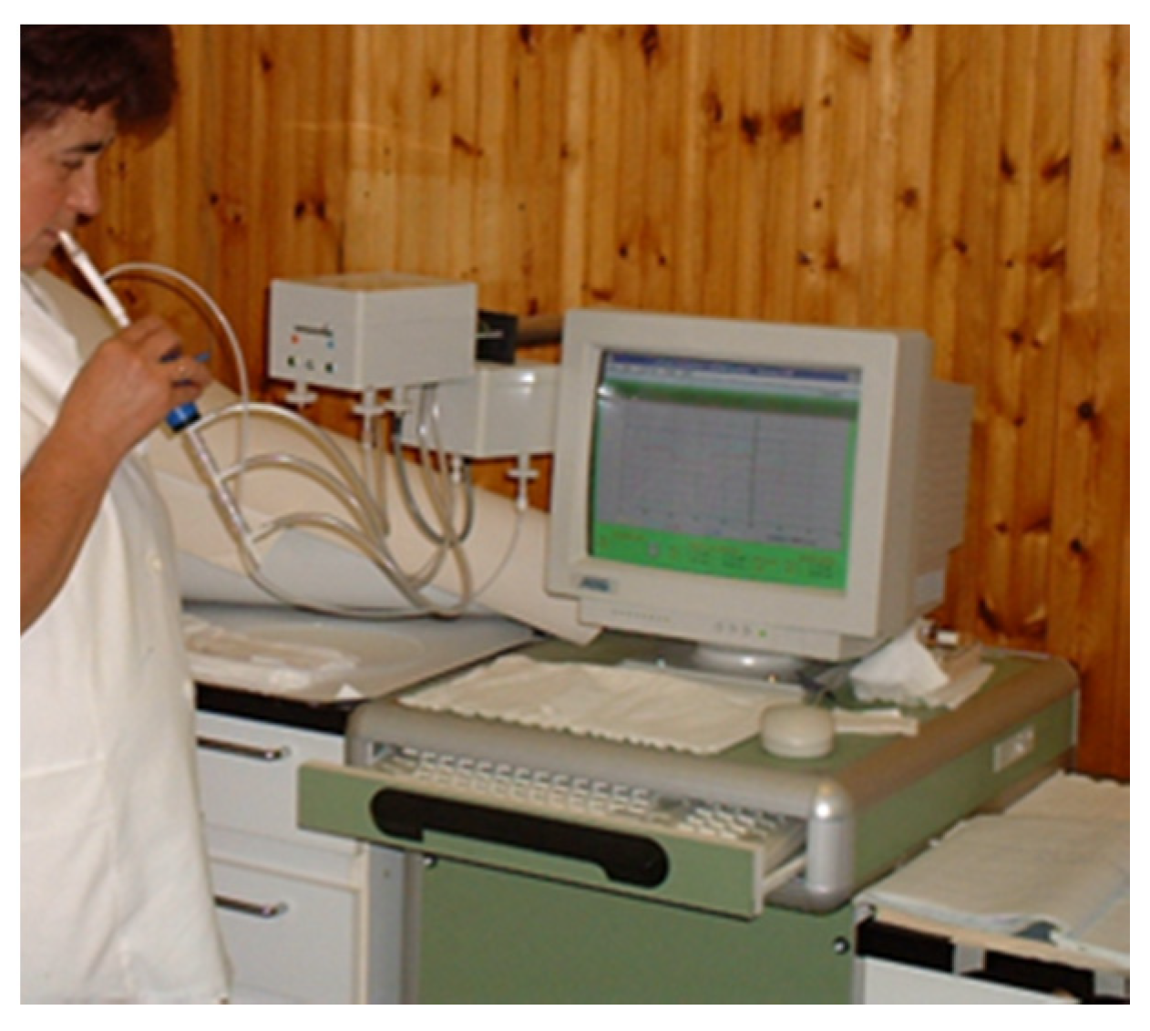
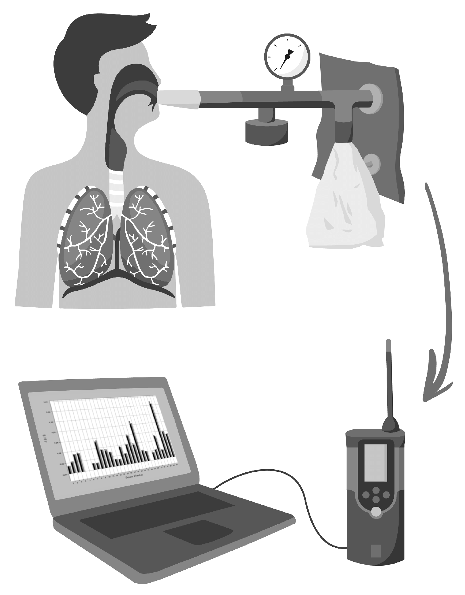
1.4. Methodological Issues Related to Breath Sampling
2. Exhaled Gaso-Transmitters
2.1. Exhaled Nitric Oxide

| Conditions/Diseases | FeNO Levels | References |
|---|---|---|
| Physical exercise | ↓ | [80,81] |
| Pregnancy | → | [67,68,82] |
| Smoking | ↓ | [66,83,84,85,86,87,88] |
| Pulmonary diseases | ||
| Stable COPD | →↑ | [89,90,91,92,93,94] |
| Severe COPD | ↓ | [95] |
| COPD exacerbation | ↑ | [96] |
| Cystic fibrosis | ↓ | [97,98] |
| Primary ciliary dyskinesia | ↓ | [99,100] |
| Bronchial asthma (esp. with eosinophilic airway inflammation) | ↑ | [65,66,68,82,101,102] |
| Interstitial lung disease | ↑ | [103] |
| Lung cancer | ↑ | [104,105,106] |
| Pulmonary tuberculosis | → | [107] |
| Pulmonary infections after lung transplantation | ↑ | [108] |
| Asbestos-related diseases | ↑ | [109,110] |
| Cardiovascular diseases | ||
| Heart failure | ↑ | [111] |
| Atherosclerotic risk | ↓ | [112] |
| Pulmonary hypertension | ↓ | [113,114] |
| COVID-19 infection (CONTROVERSIAL!) | ||
| Acute severe infection | ↓ | [115,116] |
| Lung parenchimal involvement, prognosis | ↑ | [117,118] |
| Post-COVID syndrome | ↓ | [115] |
| Inflammatory diseases | ||
| Psoriasis, psoriatic arthritis | ↑ | [119,120] |
| Systemic sclerosis | ↑ | [121] |
| Inflammatory bowel disease | → | [122,123] |
| (Eosinophilic) esophagitis | → | [124,125] |
2.2. Exhaled Carbon Monoxide
2.3. Exhaled Hydrogen Sulfide
| Measurement Technique | Sample Type Used | Detection Range or Limit | Reference |
|---|---|---|---|
| Colorimetric detection of H2S using an etching-resistant effect on silver nanoprisms | Gaseous H2S released from garlic and Na2S solution in phosphate-buffered saline dilution series | Linear range from 1.03 to 32.9 μM H2S μM | Ahn, Y.J. et. al. [180] |
| Conclusion: Ag NPRs-coated H2S sensing paper demonstrated high selectivity, good sensitivity and good reproducibility and stability, together with a fast response time. Possible tool for on-site colorimetric detection of free H2S gas for exhaled breath analysis. | |||
| Spectroscopic techniques for H2S measurement in gaseous mixes: FMS, 2f -WMS, OA-ICOS, PAS | VSC gas containing H2S | LOD from 500 ppb to 8.4 ppm | Ciaffoni, L. et. al. [181] |
| Conclusion: Laser-based spectroscopic sensors are possibilities for accurate breath diagnostics in clinical environment. | |||
| Amperometric detection of H2S gas in N2 gaseous mix | 10 ppm H2S in 99.95% pure N2 gas | Linear range from 75 ppb to 820 ppb | Gatty, H.K. et. al. [182] |
| Conclusion: The ppb-level detection capacity, combined fast response and high sensitivity to H2S makes the sensor potentially suitable for oral breath monitoring with a miniaturized handheld instrument. | |||
| A paper-based fluorescent sensor for in situ gaseous H2S determination | Gaseous mix of H2S and purified air | LOD of 3 ppb | Petruci, J.F. et. al. [183] |
| Conclusion: An automated portable sensor for in situ determination of H2S gas that can be utilized in a clinical environment. | |||
| Interscan RM-17 sulphide detectors | H2S gas exhaled from human subjects who received iv. Na2S solution. | LOD of 10 ppb–5 ppm | Toombs, C.F. et. al. [184] |
| Conclusion: The aim of the study was to prove that exhaled H2S represents a detectable route of elimination in the human body after iv. infusion of Na2S solution. | |||
| Ion sensitive electrodes for H2S detection | Mammalian plasma | LOD of 100 nM, detection range: 1–100 μM | Xu, T. et. al. [185] |
| Conclusion: There is broad range of applications of the ISE-based H2S sensor, but the system requires high maintenance from the operator. | |||
| Polarographic H2S sensors | Plasma or mammalian tissue homogenates | LOD from 2 μM down to 5 nM in certain methods | Xu, T. et. al. [185] |
| Conclusion: Compared to ISE-based H2S sensor, method detects H2S without an external electrical potential, has a simple structure, good reproducibility, a short response time and contains fewer noble metals. A great disadvantage of the liquid electrolyte sensors is that they leak easily, are prone to dry up and have a large residual current. | |||
| Enzyme-based electrochemical H2S biosensors | Environmental water | LOD of 1 ppm in gas phase, LOD of 0.3 μM, linear response in the range of 1.09–16.3 μM in H2S solution | Xu, T. et. al. [185] |
| Conclusion: The enzyme-based H2S biosensor shows great advantages of selectivity and sensitivity. However, one of the most regrettable characteristics of the enzyme-inhibition-based biosensors is the different inhibition degrees caused by different inhibitors. Moreover, this kind of sensor also highly depends on the pH concentration due to its inhibition on the activity of enzyme, making it difficult to apply to in vivo H2S detection. | |||
| Exhaled H2S on test paper with an ultrasensitive and time-gated luminescent probe | Breath exhaled by the mice | Semi-quantification of gaseous H2S in the range of 10–30 ppm | Zhang, R. et. al. [186] |
| Conclusion: The test paper imprinted by the complex probe ink can visualize clearly the trace H2S gas exhaled by the mouse. | |||
3. Exhaled Hydrogen Peroxide
4. Breathomics—Breath Fingerprinting
4.1. Spectrometry-Based Measurements
| Measurement Technique | No. of Detected Exhaled Compounds | Subjects | Findings | Reference |
|---|---|---|---|---|
| Computer-assisted GC/MS | 150 peaks | Healthy (n = 17) and lung cancer patients (n = 14) | 49 peaks differed between groups | Gordon SM. et al. 1985 [3] |
| Computer-assisted GC/MS | 22 selected for further analysis | Patients undergoing bronchoscopy because of chest radiograph abnormalities (n = 108) | 22 VOCs discriminated between patients with and without lung cancer | Phillips M. et al. 1999 [4] |
| Computer-assisted GC/MS | 13 VOCs selected for further analysis | NSCLC (n = 36); COPD (n = 25); asymptomatic smoker (n = 35) and non-smokers (n = 50) controls | A logistic regression model using the concentration of the 13 VOCs classified 82.5% of subjects correctly | Poli D. et al. 2005 [243] |
| Computer-assisted GC/MS | 12 selected VOCs were studied | CF patients with stable (n = 15) condition and exacerbation (n = 5); healthy controls (n = 20) | The alveolar gradient for pentane was higher in CF patients (with highest values in patients with pulmonary exacerbations) and inversely proportional to FEV1; (0.73 versus 0.24 ppb). CF patients exhibited a lower output of dimethyl sulphide | Barker M. et al. 2006 [247] |
| High-resolution MS | N.A. | Young non-smoking healthy adults (n = 10) | 65 and 55 major compounds identified in positive and negative ion mode, respectively; diurnal changes in VOC spectra described | Xu L. et al. 2022 [236] |
| Multicapillary column/ion mobility spectrometer | N.A. | Patients with MPM (n = 52); healthy controls (n = 52) and asbestos workers without symptoms (n = 59) and benign asbestos-related diseases (n = 41); patients with non-asbestos-related lung diseases (n = 70) and lung cancer (n = 56) | Discrimination of patients with MPM from healthy, asymptomatic asbestos-exposed subjects, and from patients with other lung diseases and cancer with high specificity and sensitivity | Lamote K. et al. 2017 [233] |
| Multicapillary column/ion mobility spectrometer | 35 of the peaks were identified in all subjects | Healthy subjects (n = 18). Simultaneous measurement of inhaled air and exhaled breath | Results facilitate the calculation of alveolar gradients and selection of truly endogenous VOCs | Westhoff M. et al. 2022 [234] |
| Fourier-transform infrared (FTIR) spectroscopy | N.A. | Emergency patients tested for SARS-CoV-2 (n = 297) | With the aid of an artificial intelligence algorithm, SARS-CoV-2 positivity is detected with high specificity and sensitivity based on exhaled breath samples | Shlomo I.B. et al. 2022 [241] |
| Selected ion flow tube mass spectrometry | 116 specific human breath biomarker VOCs | Patients with lung cancer (n = 148), healthy controls (n = 168) | Based on VOC pattern, a prediction model of high accuracy was developed to predict lung cancer | Tsou P. H. et al. 2021 [245] |
| GC–ToF–MS | N.A. | Paediatric patients with persistent asthma (n = 96) | 15 VOCs were selected as good predictors, and were used to build a prediction model that could discriminate between persistently controlled and uncontrolled asthma with high accuracy | Van Vliet D. et al. 2016 [248] |
| Proton transfer reaction ToF-MS | N.A. | Patients tested for SARS-CoV-2 infection: symptomatic positives (n = 270), asymptomatic positives (n = 27) and negatives (n = 840) | Based on VOC pattern, a prediction model of positivity is developed with specificity and sensitivity similar to conventional test methods | Liangou A. et al. 2021 [228] |
| High-pressure photon ionization ToF-MS | 28 selected VOCs | Patients with lung cancer (n = 84 and 157), healthy controls (n = 368) | 16 VOCs as lung cancer breath biomarkers were identified; including these 16 VOCs, a diagnostic model of high accuracy was developed | Wang P. et al. 2022 [246] |
| Thermal desorption coupled with GC/MS | N. A. | Patients with MPM (n = 14) and healthy controls (n = 20) | Ten VOCs were identified to be able to discriminate between MPM patients and healthy controls | Di Gilio A. et al. 2020 [244] |
| Thermal desorption coupled with GC/MS | 6983 different VOCs observed in 352 subjects | Patients with lung cancer (n = 160), with BPN (n = 70) and healthy controls (n = 122) | 19–20 VOCs discriminated lung cancer patients from healthy subjects and from patients with BPN | Chen X. et al. 2021 [242] |
4.2. Electronic Noses
| Sensor Types (Detection System) | Diseases | References |
|---|---|---|
| Conducting polimer sensor arrays (Cyranose 320) | Lung cancer | [261,262] |
| Acute respiratory distress syndrome | [263] | |
| Asthma | [264,265] | |
| COPD | [266] | |
| Malignant mesothelioma | [267,288] | |
| Cystic fibrosis | [268] | |
| Breast cancer | [253] | |
| Colorectal carcinoma | [289] | |
| Preeclampsia | [290] | |
| Metal oxide semiconductive sensors (Diagnose) | Ventilator-associated pneumonia | [274] |
| Quartz microbalance sensors (Libranose) | Asthma | [275] |
| Halitosis | [276] | |
| Lung cancer | [277] | |
| Custom-designed arrays using nanomaterial technologies | Ovarian cancer | [280] |
| Lung cancer | [281,282,287] | |
| Pulmonary arterial hypertension | [283] | |
| Multiple sclerosis | [284] | |
| Alzheimer’s disease | [285] | |
| Parkinson’s disease | [285] | |
| Pulmonary tuberculosis | [286] |
5. Conclusions
Author Contributions
Funding
Data Availability Statement
Acknowledgments
Conflicts of Interest
References
- Pauling, L.; Robinson, A.B.; Teranishi, R.; Cary, P. Quantitative analysis of urine vapor and breath by gas-liquid partition chromatography. Proc. Natl. Acad. Sci. USA 1971, 68, 2374–2376. [Google Scholar] [CrossRef] [PubMed]
- Amann, A.; Costello Bde, L.; Miekisch, W.; Schubert, J.; Buszewski, B.; Pleil, J.; Ratcliffe, N.; Risby, T. The human volatilome: Volatile organic compounds (VOCs) in exhaled breath, skin emanations, urine, feces and saliva. J. Breath Res. 2014, 8, 034001. [Google Scholar] [CrossRef] [PubMed]
- Gordon, S.M.; Szidon, J.P.; Krotoszynski, B.K.; Gibbons, R.D.; O’Neill, H.J. Volatile organic compounds in exhaled air from patients with lung cancer. Clin. Chem. 1985, 31, 1278–1282. [Google Scholar] [CrossRef] [PubMed]
- Phillips, M.; Gleeson, K.; Hughes, J.M.; Greenberg, J.; Cataneo, R.N.; Baker, L.; McVay, W.P. Volatile organic compounds in breath as markers of lung cancer: A cross-sectional study. Lancet 1999, 353, 1930–1933. [Google Scholar] [CrossRef] [PubMed]
- Horvath, I.; Lazar, Z.; Gyulai, N.; Kollai, M.; Losonczy, G. Exhaled biomarkers in lung cancer. Eur. Respir. J. 2009, 34, 261–275. [Google Scholar] [CrossRef] [PubMed]
- Hanna, G.B.; Boshier, P.R.; Markar, S.R.; Romano, A. Accuracy and Methodologic Challenges of Volatile Organic Compound-Based Exhaled Breath Tests for Cancer Diagnosis: A Systematic Review and Meta-analysis. JAMA Oncol. 2019, 5, e182815. [Google Scholar] [CrossRef] [PubMed]
- de Lacy Costello, B.P.; Ledochowski, M.; Ratcliffe, N.M. The importance of methane breath testing: A review. J. Breath Res. 2013, 7, 024001. [Google Scholar] [CrossRef] [PubMed]
- Rana, S.V.; Malik, A. Breath tests and irritable bowel syndrome. World J. Gastroenterol. 2014, 20, 7587–7601. [Google Scholar] [CrossRef]
- Gilat, T.; Ben Hur, H.; Gelman-Malachi, E.; Terdiman, R.; Peled, Y. Alterations of the colonic flora and their effect on the hydrogen breath test. Gut 1978, 19, 602–605. [Google Scholar] [CrossRef] [PubMed]
- Rezaie, A.; Buresi, M.; Lembo, A.; Lin, H.; McCallum, R.; Rao, S.; Schmulson, M.; Valdovinos, M.; Zakko, S.; Pimentel, M.; et al. Hydrogen and Methane-Based Breath Testing in Gastrointestinal Disorders: The North American Consensus. Off. J. Am. Coll. Gastroenterol.|ACG 2017, 112, 775–784. [Google Scholar] [CrossRef] [Green Version]
- Alving, K.; Weitzberg, E.; Lundberg, J.M. Increased amount of nitric oxide in exhaled air of asthmatics. Eur. Respir. J. 1993, 6, 1368–1370. [Google Scholar] [CrossRef] [PubMed]
- Gustafsson, L.E.; Leone, A.M.; Persson, M.G.; Wiklund, N.P.; Moncada, S. Endogenous nitric oxide is present in the exhaled air of rabbits, guinea pigs and humans. Biochem. Biophys. Res. Commun. 1991, 181, 852–857. [Google Scholar] [CrossRef]
- Jatakanon, A.; Lim, S.; Kharitonov, S.A.; Chung, K.F.; Barnes, P.J. Correlation between exhaled nitric oxide, sputum eosinophils, and methacholine responsiveness in patients with mild asthma. Thorax 1998, 53, 91–95. [Google Scholar] [CrossRef] [PubMed]
- Kharitonov, S.A.; Yates, D.; Robbins, R.A.; Logan-Sinclair, R.; Shinebourne, E.A.; Barnes, P.J. Increased nitric oxide in exhaled air of asthmatic patients. Lancet 1994, 343, 133–135. [Google Scholar] [CrossRef] [PubMed]
- Paredi, P.; Leckie, M.J.; Horvath, I.; Allegra, L.; Kharitonov, S.A.; Barnes, P.J. Changes in exhaled carbon monoxide and nitric oxide levels following allergen challenge in patients with asthma. Eur. Respir. J. 1999, 13, 48–52. [Google Scholar] [CrossRef] [PubMed]
- Rupani, H.; Kent, B.D. Using Fractional Exhaled Nitric Oxide Measurement in Clinical Asthma Management. Chest 2022, 161, 906–917. [Google Scholar] [CrossRef] [PubMed]
- Choi, A.M.; Alam, J. Heme oxygenase-1: Function, regulation, and implication of a novel stress-inducible protein in oxidant-induced lung injury. Am. J. Respir. Cell Mol. Biol. 1996, 15, 9–19. [Google Scholar] [CrossRef]
- Horvath, I.; Donnelly, L.E.; Kiss, A.; Paredi, P.; Kharitonov, S.A.; Barnes, P.J. Raised levels of exhaled carbon monoxide are associated with an increased expression of heme oxygenase-1 in airway macrophages in asthma: A new marker of oxidative stress. Thorax 1998, 53, 668–672. [Google Scholar] [CrossRef]
- Kimura, H. Hydrogen Sulfide (H2S) and Polysulfide (H2Sn) Signaling: The First 25 Years. Biomolecules 2021, 11, 896. [Google Scholar] [CrossRef]
- Pysanenko, A.; Spanel, P.; Smith, D. A study of sulfur-containing compounds in mouth- and nose-exhaled breath and in the oral cavity using selected ion flow tube mass spectrometry. J. Breath Res. 2008, 2, 046004. [Google Scholar] [CrossRef] [PubMed]
- Zayasu, K.; Sekizawa, K.; Okinaga, S.; Yamaya, M.; Ohrui, T.; Sasaki, H. Increased carbon monoxide in exhaled air of asthmatic patients. Am. J. Respir. Crit. Care Med. 1997, 156, 1140–1143. [Google Scholar] [CrossRef]
- Pan, K.T.; Leonardi, G.S.; Ucci, M.; Croxford, B. Can Exhaled Carbon Monoxide Be Used as a Marker of Exposure? A Cross-Sectional Study in Young Adults. Int. J. Environ. Res. Public Health 2021, 18, 11893. [Google Scholar] [CrossRef] [PubMed]
- Gao, C.Q.; Wang, S.N.; Wang, M.M.; Li, J.J.; Qiao, J.J.; Huang, J.J.; Zhang, X.X.; Xiang, Y.Q.; Xu, Q.; Wang, J.L.; et al. Sensitivity of Sniffer Dogs for a Diagnosis of Parkinson’s Disease: A Diagnostic Accuracy Study. Mov. Disord. 2022, 37, 1807–1816. [Google Scholar] [CrossRef] [PubMed]
- Feil, C.; Staib, F.; Berger, M.R.; Stein, T.; Schmidtmann, I.; Forster, A.; Schimanski, C.C. Sniffer dogs can identify lung cancer patients from breath and urine samples. BMC Cancer 2021, 21, 917. [Google Scholar] [CrossRef] [PubMed]
- Taverna, G.; Tidu, L.; Grizzi, F.; Torri, V.; Mandressi, A.; Sardella, P.; La Torre, G.; Cocciolone, G.; Seveso, M.; Giusti, G.; et al. Olfactory system of highly trained dogs detects prostate cancer in urine samples. J. Urol. 2015, 193, 1382–1387. [Google Scholar] [CrossRef] [PubMed]
- Horvath, G.; Andersson, H.; Nemes, S. Cancer odor in the blood of ovarian cancer patients: A retrospective study of detection by dogs during treatment, 3 and 6 months afterward. BMC Cancer 2013, 13, 396. [Google Scholar] [CrossRef]
- Cambau, E.; Poljak, M. Sniffing animals as a diagnostic tool in infectious diseases. Clin. Microbiol. Infect. 2020, 26, 431–435. [Google Scholar] [CrossRef] [PubMed]
- Hardin, D.S.; Anderson, W.; Cattet, J. Dogs Can Be Successfully Trained to Alert to Hypoglycemia Samples from Patients with Type 1 Diabetes. Diabetes Ther. 2015, 6, 509–517. [Google Scholar] [CrossRef]
- Powell, N.A.; Ruffell, A.; Arnott, G. The Untrained Response of Pet Dogs to Human Epileptic Seizures. Animals 2021, 11, 2267. [Google Scholar] [CrossRef] [PubMed]
- Poling, A.; Weetjens, B.; Cox, C.; Beyene, N.; Durgin, A.; Mahoney, A. Tuberculosis detection by giant african pouched rats. Behav Anal 2011, 34, 47–54. [Google Scholar] [CrossRef] [PubMed]
- Ellis, H.; Mulder, C.; Valverde, E.; Poling, A.; Edwards, T. Reproducibility of African giant pouched rats detecting Mycobacterium tuberculosis. BMC Infect. Dis. 2017, 17, 298. [Google Scholar] [CrossRef] [PubMed]
- Laska, M.; Galizia, C.G.; Giurfa, M.; Menzel, R. Olfactory discrimination ability and odor structure-activity relationships in honeybees. Chem. Senses 1999, 24, 429–438. [Google Scholar] [CrossRef] [PubMed]
- Carey, A.F.; Carlson, J.R. Insect olfaction from model systems to disease control. Proc. Natl. Acad. Sci. USA 2011, 108, 12987–12995. [Google Scholar] [CrossRef]
- Lu, Y.; Liu, Q. Insect olfactory system inspired biosensors for odorant detection. Sens. Diagn. 2022, 1, 1126–1142. [Google Scholar] [CrossRef]
- Di Natale, C.; Macagnano, A.; Martinelli, E.; Paolesse, R.; D’Arcangelo, G.; Roscioni, C.; Finazzi-Agro, A.; D’Amico, A. Lung cancer identification by the analysis of breath by means of an array of non-selective gas sensors. Biosens. Bioelectron. 2003, 18, 1209–1218. [Google Scholar] [CrossRef] [PubMed]
- Yang, H.Y.; Chen, W.C.; Tsai, R.C. Accuracy of the Electronic Nose Breath Tests in Clinical Application: A Systematic Review and Meta-Analysis. Biosensors 2021, 11, 469. [Google Scholar] [CrossRef] [PubMed]
- Machado, R.F.; Laskowski, D.; Deffenderfer, O.; Burch, T.; Zheng, S.; Mazzone, P.J.; Mekhail, T.; Jennings, C.; Stoller, J.K.; Pyle, J.; et al. Detection of lung cancer by sensor array analyses of exhaled breath. Am. J. Respir. Crit. Care Med. 2005, 171, 1286–1291. [Google Scholar] [CrossRef]
- Kim, C.; Raja, I.S.; Lee, J.M.; Lee, J.H.; Kang, M.S.; Lee, S.H.; Oh, J.W.; Han, D.W. Recent Trends in Exhaled Breath Diagnosis Using an Artificial Olfactory System. Biosensors 2021, 11, 337. [Google Scholar] [CrossRef]
- American Thoracic, S.; European Respiratory, S. ATS/ERS recommendations for standardized procedures for the online and offline measurement of exhaled lower respiratory nitric oxide and nasal nitric oxide, 2005. Am. J. Respir. Crit. Care Med. 2005, 171, 912–930. [Google Scholar] [CrossRef]
- Horvath, I.; Barnes, P.J.; Loukides, S.; Sterk, P.J.; Hogman, M.; Olin, A.C.; Amann, A.; Antus, B.; Baraldi, E.; Bikov, A.; et al. A European Respiratory Society technical standard: Exhaled biomarkers in lung disease. Eur. Respir. J. 2017, 49, 1600965. [Google Scholar] [CrossRef] [PubMed] [Green Version]
- Khatri, S.B.; Iaccarino, J.M.; Barochia, A.; Soghier, I.; Akuthota, P.; Brady, A.; Covar, R.A.; Debley, J.S.; Diamant, Z.; Fitzpatrick, A.M.; et al. Use of Fractional Exhaled Nitric Oxide to Guide the Treatment of Asthma: An Official American Thoracic Society Clinical Practice Guideline. Am. J. Respir. Crit. Care Med. 2021, 204, e97–e109. [Google Scholar] [CrossRef]
- Chen, Y.; Yuan, S.; Cao, Y.; Kong, G.; Jiang, F.; Li, Y.; Wang, Q.; Tang, M.; Zhang, Q.; Wang, Q.; et al. Gasotransmitters: Potential Therapeutic Molecules of Fibrotic Diseases. Oxidative Med. Cell. Longev. 2021, 2021, 3206982. [Google Scholar] [CrossRef] [PubMed]
- Fagone, P.; Mazzon, E.; Bramanti, P.; Bendtzen, K.; Nicoletti, F. Gasotransmitters and the immune system: Mode of action and novel therapeutic targets. Eur. J. Pharmacol. 2018, 834, 92–102. [Google Scholar] [CrossRef] [PubMed]
- Salihi, A.; Al-Naqshabandi, M.A.; Khudhur, Z.O.; Housein, Z.; Hama, H.A.; Abdullah, R.M.; Hussen, B.M.; Alkasalias, T. Gasotransmitters in the tumor microenvironment: Impacts on cancer chemotherapy (Review). Mol. Med. Rep. 2022, 26, 233. [Google Scholar] [CrossRef] [PubMed]
- Moncada, S.; Palmer, R.M.; Higgs, E.A. Nitric oxide: Physiology, pathophysiology, and pharmacology. Pharmacol. Rev. 1991, 43, 109–142. [Google Scholar]
- Stuehr, D.J.; Griffith, O.W. Mammalian nitric oxide synthases. Adv. Enzymol. Relat. Areas Mol. Biol. 1992, 65, 287–346. [Google Scholar] [CrossRef] [PubMed]
- Bahadoran, Z.; Carlstrom, M.; Mirmiran, P.; Ghasemi, A. Nitric oxide: To be or not to be an endocrine hormone? Acta Physiol. 2020, 229, e13443. [Google Scholar] [CrossRef] [PubMed]
- Hoiland, R.L.; Caldwell, H.G.; Howe, C.A.; Nowak-Fluck, D.; Stacey, B.S.; Bailey, D.M.; Paton, J.F.R.; Green, D.J.; Sekhon, M.S.; Macleod, D.B.; et al. Nitric oxide is fundamental to neurovascular coupling in humans. J. Physiol. 2020, 598, 4927–4939. [Google Scholar] [CrossRef] [PubMed]
- Horvath, I.; Sandor, N.T.; Ruttner, Z.; McLaughlin, A.C. Role of nitric oxide in regulating cerebrocortical oxygen consumption and blood flow during hypercapnia. J. Cereb. Blood Flow Metab. 1994, 14, 503–509. [Google Scholar] [CrossRef] [PubMed]
- Horvath, I.; Donnelly, L.E.; Kiss, A.; Kharitonov, S.A.; Lim, S.; Chung, K.F.; Barnes, P.J. Combined use of exhaled hydrogen peroxide and nitric oxide in monitoring asthma. Am. J. Respir. Crit. Care Med. 1998, 158, 1042–1046. [Google Scholar] [CrossRef] [PubMed]
- Cinelli, M.A.; Do, H.T.; Miley, G.P.; Silverman, R.B. Inducible nitric oxide synthase: Regulation, structure, and inhibition. Med. Res. Rev. 2020, 40, 158–189. [Google Scholar] [CrossRef] [PubMed]
- Bosch, N.A.; Law, A.C.; Vail, E.A.; Gillmeyer, K.R.; Gershengorn, H.B.; Wunsch, H.; Walkey, A.J. Inhaled Nitric Oxide vs Epoprostenol During Acute Respiratory Failure: An Observational Target Trial Emulation. Chest 2022, 162, 1287–1296. [Google Scholar] [CrossRef]
- Falconer, D.; Papageorgiou, N.; Salem, K.; Lim, W.Y.; Katsargyris, A.; Avgerinos, E.; Tousoulis, D. Nitric oxide donors for peripheral artery disease. Curr. Opin. Pharmacol. 2018, 39, 77–85. [Google Scholar] [CrossRef]
- Jüttner, A.A.; Danser, A.H.J.; Roks, A.J.M. Pharmacological developments in antihypertensive treatment through nitric oxide-cGMP modulation. Adv. Pharmacol. 2022, 94, 57–94. [Google Scholar] [CrossRef] [PubMed]
- Webber, R.J.; Sweet, R.M.; Webber, D.S. Circulating Microvesicle-Associated Inducible Nitric Oxide Synthase Is a Novel Therapeutic Target to Treat Sepsis: Current Status and Future Considerations. Int. J. Mol. Sci. 2021, 22, 13371. [Google Scholar] [CrossRef] [PubMed]
- Hansel, T.T.; Kharitonov, S.A.; Donnelly, L.E.; Erin, E.M.; Currie, M.G.; Moore, W.M.; Manning, P.T.; Recker, D.P.; Barnes, P.J. A selective inhibitor of inducible nitric oxide synthase inhibits exhaled breath nitric oxide in healthy volunteers and asthmatics. FASEB J. 2003, 17, 1298–1300. [Google Scholar] [CrossRef] [PubMed]
- Alving, K.; Janson, C.; Nordvall, L. Performance of a new hand-held device for exhaled nitric oxide measurement in adults and children. Respir. Res. 2006, 7, 67. [Google Scholar] [CrossRef] [PubMed]
- Antus, B.; Horvath, I.; Barta, I. Assessment of exhaled nitric oxide by a new hand-held device. Respir. Med. 2010, 104, 1377–1380. [Google Scholar] [CrossRef] [PubMed]
- Korn, S.; Wilk, M.; Voigt, S.; Weber, S.; Keller, T.; Buhl, R. Measurement of Fractional Exhaled Nitric Oxide: Comparison of Three Different Analysers. Respiration 2020, 99, 1–8. [Google Scholar] [CrossRef]
- Lei, W.; Li, F.; Tang, X.M.; Bian, S.; Wang, J.J.; Huang, J.A. The comparison of two exhaled nitric oxide analyzers: NIOX VERO and SUNVOU-CA2122. J. Breath Res. 2021, 15, 026007. [Google Scholar] [CrossRef]
- Boot, J.D.; de Ridder, L.; de Kam, M.L.; Calderon, C.; Mascelli, M.A.; Diamant, Z. Comparison of exhaled nitric oxide measurements between NIOX MINO electrochemical and Ecomedics chemiluminescence analyzer. Respir. Med. 2008, 102, 1667–1671. [Google Scholar] [CrossRef] [PubMed] [Green Version]
- Maniscalco, M.; Vitale, C.; Vatrella, A.; Molino, A.; Bianco, A.; Mazzarella, G. Fractional exhaled nitric oxide-measuring devices: Technology update. Med. Devices 2016, 9, 151–160. [Google Scholar] [CrossRef]
- Loewenthal, L.; Menzies-Gow, A. FeNO in Asthma. Semin. Respir. Crit. Care Med. 2022, 43, 635–645. [Google Scholar] [CrossRef] [PubMed]
- Menzies-Gow, A.; Mansur, A.H.; Brightling, C.E. Clinical utility of fractional exhaled nitric oxide in severe asthma management. Eur. Respir. J. 2020, 55, 1901633. [Google Scholar] [CrossRef] [PubMed]
- Ahovuo-Saloranta, A.; Csonka, P.; Lehtimaki, L. Basic characteristics and clinical value of FeNO in smoking asthmatics-a systematic review. J. Breath Res. 2019, 13, 034003. [Google Scholar] [CrossRef] [PubMed]
- Horvath, I.; Donnelly, L.E.; Kiss, A.; Balint, B.; Kharitonov, S.A.; Barnes, P.J. Exhaled nitric oxide and hydrogen peroxide concentrations in asthmatic smokers. Respiration 2004, 71, 463–468. [Google Scholar] [CrossRef]
- Tamasi, L.; Bohacs, A.; Bikov, A.; Andorka, C.; Rigo, J., Jr.; Losonczy, G.; Horvath, I. Exhaled nitric oxide in pregnant healthy and asthmatic women. J. Asthma 2009, 46, 786–791. [Google Scholar] [CrossRef] [PubMed]
- Powell, H.; Murphy, V.E.; Taylor, D.R.; Hensley, M.J.; McCaffery, K.; Giles, W.; Clifton, V.L.; Gibson, P.G. Management of asthma in pregnancy guided by measurement of fraction of exhaled nitric oxide: A double-blind, randomised controlled trial. Lancet 2011, 378, 983–990. [Google Scholar] [CrossRef]
- Busse, W.W.; Wenzel, S.E.; Casale, T.B.; FitzGerald, J.M.; Rice, M.S.; Daizadeh, N.; Deniz, Y.; Patel, N.; Harel, S.; Rowe, P.J.; et al. Baseline FeNO as a prognostic biomarker for subsequent severe asthma exacerbations in patients with uncontrolled, moderate-to-severe asthma receiving placebo in the LIBERTY ASTHMA QUEST study: A post-hoc analysis. Lancet Respir. Med. 2021, 9, 1165–1173. [Google Scholar] [CrossRef]
- Chung, K.F. Increasing utility of FeNO as a biomarker of type-2 inflammation in severe asthma. Lancet Respir. Med. 2021, 9, 1083–1084. [Google Scholar] [CrossRef] [PubMed]
- Ando, K.; Fukuda, Y.; Tanaka, A.; Sagara, H. Comparative Efficacy and Safety of Tezepelumab and Other Biologics in Patients with Inadequately Controlled Asthma According to Thresholds of Type 2 Inflammatory Biomarkers: A Systematic Review and Network Meta-Analysis. Cells 2022, 11, 819. [Google Scholar] [CrossRef] [PubMed]
- Menigoz, C.; Dirou, S.; Chambellan, A.; Hassoun, D.; Moui, A.; Magnan, A.; Blanc, F.X. Use of FeNO to predict anti-IL-5 and IL-5R biologics efficacy in a real-world cohort of adults with severe eosinophilic asthma. J. Asthma 2022, 1–9. [Google Scholar] [CrossRef]
- Parnes, J.; Molfino, N.A.; Colice, G.; Martin, U.; Corren, J.; Menzies-Gow, A. Targeting TSLP in Asthma. J. Asthma Allergy 2022, 15, 749–765. [Google Scholar] [CrossRef]
- Antus, B.; Barta, I. Blood Eosinophils and Exhaled Nitric Oxide: Surrogate Biomarkers of Airway Eosinophilia in Stable COPD and Exacerbation. Biomedicines 2022, 10, 2128. [Google Scholar] [CrossRef] [PubMed]
- Antus, B.; Paska, C.; Barta, I. Predictive Value of Exhaled Nitric Oxide and Blood Eosinophil Count in the Assessment of Airway Eosinophilia in COPD. Int. J. Chron. Obstruct. Pulmon. Dis. 2020, 15, 2025–2035. [Google Scholar] [CrossRef] [PubMed]
- Antus, B.; Barta, I.; Csiszer, E. Exhaled Nitric Oxide in Diagnosis of Bronchiolitis Obliterans Syndrome in Lung Transplant Recipients: Possible Limitations. Am. J. Transplant. 2011, 11, 2774–2775. [Google Scholar] [CrossRef]
- Nijveldt, R.J.; Teerlink, T.; Van Der Hoven, B.; Siroen, M.P.; Kuik, D.J.; Rauwerda, J.A.; van Leeuwen, P.A. Asymmetrical dimethylarginine (ADMA) in critically ill patients: High plasma ADMA concentration is an independent risk factor of ICU mortality. Clin. Nutr. 2003, 22, 23–30. [Google Scholar] [CrossRef]
- Yeo, T.W.; Lampah, D.A.; Tjitra, E.; Gitawati, R.; Darcy, C.J.; Jones, C.; Kenangalem, E.; McNeil, Y.R.; Granger, D.L.; Lopansri, B.K.; et al. Increased Asymmetric Dimethylarginine in Severe Falciparum Malaria: Association with Impaired Nitric Oxide Bioavailability and Fatal Outcome. PLoS Pathog. 2010, 6, e1000868. [Google Scholar] [CrossRef]
- Ruzsics, I.; Nagy, L.; Keki, S.; Sarosi, V.; Illes, B.; Illes, Z.; Horvath, I.; Bogar, L.; Molnar, T. L-Arginine Pathway in COPD Patients with Acute Exacerbation: A New Potential Biomarker. COPD 2016, 13, 139–145. [Google Scholar] [CrossRef] [PubMed]
- Prossegger, J.; Huber, D.; Grafetstätter, C.; Pichler, C.; Braunschmid, H.; Weisböck-Erdheim, R.; Hartl, A. Winter Exercise Reduces Allergic Airway Inflammation: A Randomized Controlled Study. Int. J. Environ. Res. Public Health 2019, 16, 2040. [Google Scholar] [CrossRef] [PubMed]
- Evjenth, B.; Hansen, T.E.; Holt, J. Exhaled nitric oxide decreases during exercise in non-asthmatic children. Clin. Respir. J. 2013, 7, 121–127. [Google Scholar] [CrossRef] [PubMed]
- Murphy, V.E.; Jensen, M.E.; Holliday, E.G.; Giles, W.B.; Barrett, H.L.; Callaway, L.K.; Bisits, A.; Peek, M.J.; Seeho, S.K.; Abbott, A.; et al. Effect of asthma management with exhaled nitric oxide versus usual care on perinatal outcomes. Eur. Respir. J. 2022, 60, 2200298. [Google Scholar] [CrossRef] [PubMed]
- Malinovschi, A. Effect of smoking on exhaled nitric oxide and flow-independent nitric oxide exchange parameters. Eur. Respir. J. 2006, 28, 339–345. [Google Scholar] [CrossRef] [PubMed]
- Persson, M.G.; Zetterström, O.; Agrenius, V.; Ihre, E.; Gustafsson, L.E. Single-breath nitric oxide measurements in asthmatic patients and smokers. Lancet 1994, 343, 146–147. [Google Scholar] [CrossRef]
- Schilling, J.; Holzer, P.; Guggenbach, M.; Gyurech, D.; Marathia, K.; Geroulanos, S. Reduced endogenous nitric oxide in the exhaled air of smokers and hypertensives. Eur. Respir. J. 1994, 7, 467–471. [Google Scholar] [CrossRef] [PubMed]
- Kharitonov, S.A.; Robbins, R.A.; Yates, D.; Keatings, V.; Barnes, P.J. Acute and chronic effects of cigarette smoking on exhaled nitric oxide. Am. J. Respir. Crit. Care Med. 1995, 152, 609–612. [Google Scholar] [CrossRef]
- Yates, D.H.; Breen, H.; Thomas, P.S. Passive smoke inhalation decreases exhaled nitric oxide in normal subjects. Am. J. Respir. Crit. Care Med. 2001, 164, 1043–1046. [Google Scholar] [CrossRef]
- Warke, T.J.; Mairs, V.; Fitch, P.S.; Ennis, M.; Shields, M.D. Possible association between passive smoking and lower exhaled nitric oxide in asthmatic children. Arch. Environ. Health 2003, 58, 613–616. [Google Scholar] [CrossRef] [PubMed]
- Lu, Z.; Huang, W.; Wang, L.; Xu, N.; Ding, Q.; Cao, C. Exhaled nitric oxide in patients with chronic obstructive pulmonary disease: A systematic review and meta-analysis. Int. J. Chron. Obstruct. Pulmon. Dis. 2018, 13, 2695–2705. [Google Scholar] [CrossRef]
- Brindicci, C.; Ito, K.; Resta, O.; Pride, N.B.; Barnes, P.J.; Kharitonov, S.A. Exhaled nitric oxide from lung periphery is increased in COPD. Eur. Respir. J. 2005, 26, 52–59. [Google Scholar] [CrossRef] [PubMed]
- Clini, E.; Bianchi, L.; Vitacca, M.; Porta, R.; Foglio, K.; Ambrosino, N. Exhaled nitric oxide and exercise in stable COPD patients. Chest 2000, 117, 702–707. [Google Scholar] [CrossRef] [PubMed]
- Hirano, T.; Matsunaga, K.; Sugiura, H.; Minakata, Y.; Koarai, A.; Akamatsu, K.; Ichikawa, T.; Furukawa, K.; Ichinose, M. Relationship between alveolar nitric oxide concentration in exhaled air and small airway function in COPD. J. Breath Res. 2013, 7, 046002. [Google Scholar] [CrossRef] [PubMed]
- Liu, J.; Sandrini, A.; Thurston, M.C.; Yates, D.H.; Thomas, P.S. Nitric oxide and exhaled breath nitrite/nitrates in chronic obstructive pulmonary disease patients. Respiration 2007, 74, 617–623. [Google Scholar] [CrossRef] [PubMed]
- Rouhos, A.; Kainu, A.; Piirilä, P.; Sarna, S.; Lindqvist, A.; Karjalainen, J.; Sovijärvi, A.R. Repeatability of exhaled nitric oxide measurements in patients with COPD. Clin. Physiol. Funct. Imaging 2011, 31, 26–31. [Google Scholar] [CrossRef]
- Clini, E.; Cremona, G.; Campana, M.; Scotti, C.; Pagani, M.; Bianchi, L.; Giordano, A.; Ambrosino, N. Production of endogenous nitric oxide in chronic obstructive pulmonary disease and patients with cor pulmonale. Correlates with echo-Doppler assessment. Am. J. Respir. Crit. Care Med. 2000, 162, 446–450. [Google Scholar] [CrossRef] [PubMed]
- Agusti, A.G.; Villaverde, J.M.; Togores, B.; Bosch, M. Serial measurements of exhaled nitric oxide during exacerbations of chronic obstructive pulmonary disease. Eur. Respir. J. 1999, 14, 523–528. [Google Scholar] [CrossRef] [PubMed]
- Hofer, M.; Mueller, L.; Rechsteiner, T.; Benden, C.; Boehler, A. Extended nitric oxide measurements in exhaled air of cystic fibrosis and healthy adults. Lung 2009, 187, 307–313. [Google Scholar] [CrossRef] [PubMed]
- Krantz, C.; Janson, C.; Hollsing, A.; Alving, K.; Malinovschi, A. Exhaled and nasal nitric oxide in relation to lung function, blood cell counts and disease characteristics in cystic fibrosis. J. Breath Res. 2017, 11, 026001. [Google Scholar] [CrossRef] [PubMed]
- Horváth, I.; Loukides, S.; Wodehouse, T.; Csiszér, E.; Cole, P.J.; Kharitonov, S.A.; Barnes, P.J. Comparison of exhaled and nasal nitric oxide and exhaled carbon monoxide levels in bronchiectatic patients with and without primary ciliary dyskinesia. Thorax 2003, 58, 68–72. [Google Scholar] [CrossRef]
- Karadag, B.; James, A.; Gultekin, E.; Wilson, N.; Bush, A. Nasal and lower airway level of nitric oxide in children with primary ciliary dyskinesia. Eur. Respir. J. 1999, 13, 1402–1405. [Google Scholar] [CrossRef]
- Gemicioglu, B.; Musellim, B.; Dogan, I.; Guven, K. Fractional exhaled nitric oxide (FeNo) in different asthma phenotypes. Allergy Rhinol (Providence) 2014, 5, 157–161. [Google Scholar] [CrossRef] [PubMed] [Green Version]
- Schneider, A.; Brunn, B.; Hapfelmeier, A.; Schultz, K.; Kellerer, C.; Jörres, R.A. Diagnostic accuracy of FeNO in asthma and predictive value for inhaled corticosteroid responsiveness: A prospective, multicentre study. eClinicalMedicine 2022, 50, 101533. [Google Scholar] [CrossRef] [PubMed]
- Cameli, P.; Bennett, D.; Refini, R.M.; Sestini, P.; Rottoli, P.; Pieroni, M.G. Exhaled nitric oxide in idiopathic and non-idiopathic interstitial lung disease. Eur. Respir. J. 2017, 50, PA878. [Google Scholar] [CrossRef]
- Liu, P.F.; Zhao, D.H.; Qi, Y.; Wang, J.G.; Zhao, M.; Xiao, K.; Xie, L.X. The clinical value of exhaled nitric oxide in patients with lung cancer. Clin. Respir. J. 2018, 12, 23–30. [Google Scholar] [CrossRef]
- Li, J.; Li, Q.; Wei, X.; Chen, Q.; Sun, M.; Li, Y. Measurement of Exhaled Nitric Oxide in 456 Lung Cancer Patients Using a Ringdown FENO Analyzer. Metabolites 2021, 11, 352. [Google Scholar] [CrossRef] [PubMed]
- Bencova, A.; Antosova, M.; Kocan, I.; Rozborilova, E. The impact of lung cancer subtypes, smoking status and corticosteroid treatment on exhaled nitric oxide levels in patients with lung cancer. Eur. Respir. J. 2014, 44, P2749. [Google Scholar]
- López, J.W.; Loader, M.I.; Smith, D.; Pastorius, D.; Bravard, M.; Caviedes, L.; Romero, K.M.; Clark, T.; Checkley, W.; Ticona, E.; et al. Exhaled Nitric Oxide is Not a Biomarker for Pulmonary Tuberculosis. Am. J. Trop. Med. Hyg. 2018, 98, 1637–1639. [Google Scholar] [CrossRef]
- Antus, B.; Csiszer, E.; Czebe, K.; Horvath, I. Pulmonary infections increase exhaled nitric oxide in lung transplant recipients: A longitudinal study. Clin. Transpl. 2005, 19, 377–382. [Google Scholar] [CrossRef]
- Sandrini, A.; Johnson, A.R.; Thomas, P.S.; Yates, D.H. Fractional exhaled nitric oxide concentration is increased in asbestosis and pleural plaques. Respirology 2006, 11, 325–329. [Google Scholar] [CrossRef]
- Chow, S.; Campbell, C.; Sandrini, A.; Thomas, P.S.; Johnson, A.R.; Yates, D.H. Exhaled breath condensate biomarkers in asbestos-related lung disorders. Respir. Med. 2009, 103, 1091–1097. [Google Scholar] [CrossRef]
- Schuster, A.; Thakur, A.; Wang, Z.; Borowski, A.G.; Thomas, J.D.; Tang, W.H. Increased exhaled nitric oxide levels after exercise in patients with chronic systolic heart failure with pulmonary venous hypertension. J. Card. Fail. 2012, 18, 799–803. [Google Scholar] [CrossRef]
- Salonen, I.; Huttunen, K.; Hirvonen, M.R.; Dufva, J.; Groundstroem, K.; Dufva, H.; Salonen, R.O. Exhaled nitric oxide and atherosclerosis. Eur. J. Clin. Investig. 2012, 42, 873–880. [Google Scholar] [CrossRef]
- Kaneko, F.T.; Arroliga, A.C.; Dweik, R.A.; Comhair, S.A.; Laskowski, D.; Oppedisano, R.; Thomassen, M.J.; Erzurum, S.C. Biochemical reaction products of nitric oxide as quantitative markers of primary pulmonary hypertension. Am. J. Respir. Crit. Care Med. 1998, 158, 917–923. [Google Scholar] [CrossRef] [PubMed]
- Akbay, N.O.; Bingol, Z.; Kiyan, E.; Karaayvaz, E.B.; Bilge, A.K.; Issever, H.; Okumus, G. Fractional Exhaled Nitric Oxide Measurement in Pulmonary Hypertension: A Follow-Up Study. Clin. Appl. Thromb. Hemost. 2018, 24, 483–488. [Google Scholar] [CrossRef] [PubMed]
- Ferreira, S.; Fernandes, W.S.; Ferreira, R.C.A.; Fialho, P.B.; Santos, C.E.R.; Araujo, L.M.; Santos, L.M.B.R.; Brandao-Rangel, M.A.R.; Frison, C.R.; Vieira, R.P. Can Exhaled Nitric Oxide Be a Biomarker for Hospitalized and Post-COVID-19 Patients? Eur. Respir. J. 2021, 58, PA3884. [Google Scholar] [CrossRef]
- Lior, Y.; Yatzkan, N.; Brami, I.; Yogev, Y.; Riff, R.; Hekselman, I.; Fremder, M.; Freixo-Lima, G.; Be’er, M.; Amirav, I.; et al. Fractional exhaled Nitric Oxide (FeNO) level as a predictor of COVID-19 disease severity. Nitric Oxide 2022, 124, 68–73. [Google Scholar] [CrossRef] [PubMed]
- Kerget, B.; Araz, Ö.; Akgün, M. The role of exhaled nitric oxide (FeNO) in the evaluation of lung parenchymal involvement in COVID-19 patients. Intern. Emerg. Med. 2022, 17, 1951–1958. [Google Scholar] [CrossRef] [PubMed]
- Balci, A.; Yildiz, E.; Selendili, O.; Gunay, E. A Predictive and Prognostic Marker in Covid-19 Patients: Exhaled Nitric Oxide (Feno). Acta Med. Mediterr. 2021, 37, 841–846. [Google Scholar] [CrossRef]
- Damiani, G.; Pacifico, A.; Rizzi, M.; Santus, P.; Bridgewood, C.; Bragazzi, N.L.; Adawi, M.; Watad, A. Patients with psoriatic arthritis have higher levels of FeNO than those with only psoriasis, which may reflect a higher prevalence of a subclinical respiratory involvement. Clin. Rheumatol. 2020, 39, 2981–2988. [Google Scholar] [CrossRef]
- Damiani, G.; Radaeli, A.; Olivini, A.; Calvara-Pinton, P.; Malerba, M. Increased airway inflammation in patients with psoriasis. Br. J. Dermatol. 2016, 175, 797–799. [Google Scholar] [CrossRef]
- Guillen-Del Castillo, A.; Sánchez-Vidaurre, S.; Simeón-Aznar, C.P.; Cruz, M.J.; Fonollosa-Pla, V.; Muñoz, X. Prognostic Role of Exhaled Breath Condensate pH and Fraction Exhaled Nitric Oxide in Systemic Sclerosis Related Interstitial Lung Disease. Arch. Bronconeumol. 2017, 53, 120–127. [Google Scholar] [CrossRef] [PubMed]
- Protopapas, A.; Vradelis, S.; Karampitsakos, T.; Chatzimichail, A.; Paraskakis, E. Elevated levels of alveolar nitric oxide in patients with inflammatory bowel disease. Eur. Respir. J. 2016, 48, PA1065. [Google Scholar] [CrossRef]
- Ikonomi, E.; Rothstein, R.D.; Ehrlich, A.C.; Friedenberg, F.K. Measurement of Fractional Exhaled Nitric Oxide as a Marker of Disease Activity in Inflammatory Bowel Disease. J. Gastroenterol. Pancreatol. Liver Disord. 2016, 3, 1–7. [Google Scholar] [CrossRef]
- Dor-Wojnarowska, A.; Liebhart, J.; Grabowski, M.; Czapla, L.; Grabowski, K.; Panaszek, B. Exhaled Nitric Oxide in Patients with Esophagitis. Adv. Respir. Med. 2011, 79, 272–277. [Google Scholar] [CrossRef]
- Johnson, K.; Iyer, V.; Katzka, D.; Ravi, K.; Lennon, R.; Pendegraft, R.; Geno, D.; Alexander, J. Poor Relationship Between Fractionated Exhaled Nitric Oxide and Disease Activity in Eosinophilic Esophagitis. Dysphagia 2019, 34, 138–144. [Google Scholar] [CrossRef]
- Wang, C.; Sahay, P. Breath analysis using laser spectroscopic techniques: Breath biomarkers, spectral fingerprints, and detection limits. Sensors 2009, 9, 8230–8262. [Google Scholar] [CrossRef]
- McCurdy, M.R.; Bakhirkin, Y.; Wysocki, G.; Tittel, F.K. Performance of an exhaled nitric oxide and carbon dioxide sensor using quantum cascade laser-based integrated cavity output spectroscopy. J. Biomed. Opt. 2007, 12, 034034. [Google Scholar] [CrossRef]
- Banik, G.D.; Mizaikoff, B. Exhaled breath analysis using cavity-enhanced optical techniques: A review. J. Breath Res. 2020, 14, 043001. [Google Scholar] [CrossRef]
- Cristescu, S.M.; Mandon, J.; Harren, F.J.; Merilainen, P.; Hogman, M. Methods of NO detection in exhaled breath. J. Breath Res. 2013, 7, 017104. [Google Scholar] [CrossRef]
- Mandon, J.; Hogman, M.; Merkus, P.J.; van Amsterdam, J.; Harren, F.J.; Cristescu, S.M. Exhaled nitric oxide monitoring by quantum cascade laser: Comparison with chemiluminescent and electrochemical sensors. J. Biomed. Opt. 2012, 17, 017003. [Google Scholar] [CrossRef]
- Petralia, L.S.; Bahl, A.; Peverall, R.; Richmond, G.; Couper, J.H.; Hancock, G.; Robbins, P.A.; Ritchie, G.A.D. Accurate real-time F(E)NO expirograms using complementary optical sensors. J. Breath Res. 2020, 14, 047102. [Google Scholar] [CrossRef] [PubMed]
- Lazar, Z.; Horvath, P.; Puskas, R.; Galffy, G.; Losonczy, G.; Horvath, I.; Bikov, A. A suitable protocol for measuring alveolar nitric oxide in asthma with differing severity to assess peripheral airways inflammation. J. Asthma 2019, 56, 584–593. [Google Scholar] [CrossRef] [PubMed]
- Cameli, P.; Bargagli, E.; Bergantini, L.; d’Alessandro, M.; Pieroni, M.; Fontana, G.A.; Sestini, P.; Refini, R.M. Extended Exhaled Nitric Oxide Analysis in Interstitial Lung Diseases: A Systematic Review. Int. J. Mol. Sci. 2020, 21, 6187. [Google Scholar] [CrossRef] [PubMed]
- Hogman, M.; Thornadtsson, A.; Hedenstierna, G.; Merilainen, P. A practical approach to the theoretical models to calculate NO parameters of the respiratory system. J. Breath Res. 2014, 8, 016002. [Google Scholar] [CrossRef]
- George, S.C.; Hogman, M.; Permutt, S.; Silkoff, P.E. Modeling pulmonary nitric oxide exchange. J. Appl. Physiol. 2004, 96, 831–839. [Google Scholar] [CrossRef]
- Lehtimaki, L.; Karvonen, T.; Hogman, M. Clinical Values of Nitric Oxide Parameters from the Respiratory System. Curr. Med. Chem. 2020, 27, 7189–7199. [Google Scholar] [CrossRef]
- Grzywa-Celinska, A.; Krusinski, A.; Milanowski, J. ‘Smoging kills’—Effects of air pollution on human respiratory system. Ann Agric. Environ. Med. 2020, 27, 1–5. [Google Scholar] [CrossRef]
- Megas, I.F.; Beier, J.P.; Grieb, G. The History of Carbon Monoxide Intoxication. Medicina 2021, 57, 400. [Google Scholar] [CrossRef]
- Campbell, N.K.; Fitzgerald, H.K.; Dunne, A. Regulation of inflammation by the antioxidant haem oxygenase 1. Nat. Rev. Immunol. 2021, 21, 411–425. [Google Scholar] [CrossRef]
- Ryter, S.W. Heme Oxygenase-1: An Anti-Inflammatory Effector in Cardiovascular, Lung, and Related Metabolic Disorders. Antioxidants 2022, 11, 555. [Google Scholar] [CrossRef]
- Gajdocsy, R.; Horvath, I. Exhaled carbon monoxide in airway diseases: From research findings to clinical relevance. J. Breath Res. 2010, 4, 047102. [Google Scholar] [CrossRef] [PubMed]
- Gall, T.; Balla, G.; Balla, J. Heme, Heme Oxygenase, and Endoplasmic Reticulum Stress-A New Insight into the Pathophysiology of Vascular Diseases. Int. J. Mol. Sci. 2019, 20, 3675. [Google Scholar] [CrossRef] [PubMed] [Green Version]
- Haines, D.; Tosaki, A. Emerging Clinical Applications of Heme Oxygenase. Curr. Pharm. Des. 2018, 24, 2227–2228. [Google Scholar] [CrossRef] [PubMed]
- Ryter, S.W. Heme oxygenase-1/carbon monoxide as modulators of autophagy and inflammation. Arch. Biochem. Biophys. 2019, 678, 108186. [Google Scholar] [CrossRef] [PubMed]
- Ryter, S.W.; Ma, K.C.; Choi, A.M.K. Carbon monoxide in lung cell physiology and disease. Am. J. Physiol. Cell Physiol. 2018, 314, C211–C227. [Google Scholar] [CrossRef] [PubMed]
- Szabo, I.L.; Kenyeres, A.; Szegedi, A.; Szollosi, A.G. Heme Oxygenase and the Skin in Health and Disease. Curr. Pharm. Des. 2018, 24, 2303–2310. [Google Scholar] [CrossRef] [PubMed]
- Campbell, N.K.; Fitzgerald, H.K.; Malara, A.; Hambly, R.; Sweeney, C.M.; Kirby, B.; Fletcher, J.M.; Dunne, A. Naturally derived Heme-Oxygenase 1 inducers attenuate inflammatory responses in human dendritic cells and T cells: Relevance for psoriasis treatment. Sci. Rep. 2018, 8, 10287. [Google Scholar] [CrossRef]
- Dhawan, S. Therapeutic Potential of Inducible Endogenous Cytoprotective Heme Oxygenase-1 in Mitigating SARS-CoV-2 Infection and Associated Inflammation. Antioxidants 2022, 11, 662. [Google Scholar] [CrossRef]
- Ryter, S.W. Therapeutic Potential of Heme Oxygenase-1 and Carbon Monoxide in Acute Organ Injury, Critical Illness, and Inflammatory Disorders. Antioxidants 2020, 9, 1153. [Google Scholar] [CrossRef]
- Sanada, Y.; Tan, S.J.O.; Adachi, N.; Miyaki, S. Pharmacological Targeting of Heme Oxygenase-1 in Osteoarthritis. Antioxidants 2021, 10, 419. [Google Scholar] [CrossRef]
- Zhou, X.; Yuan, W.; Xiong, X.; Zhang, Z.; Liu, J.; Zheng, Y.; Wang, J.; Liu, J. HO-1 in Bone Biology: Potential Therapeutic Strategies for Osteoporosis. Front. Cell Dev. Biol. 2021, 9, 791585. [Google Scholar] [CrossRef]
- Adach, W.; Olas, B. Carbon monoxide and its donors—Their implications for medicine. Future Med. Chem. 2019, 11, 61–73. [Google Scholar] [CrossRef] [PubMed]
- Goebel, U.; Wollborn, J. Carbon monoxide in intensive care medicine—Time to start the therapeutic application?! Intensive Care Med. Exp. 2020, 8, 2. [Google Scholar] [CrossRef]
- Paredi, P.; Shah, P.L.; Montuschi, P.; Sullivan, P.; Hodson, M.E.; Kharitonov, S.A.; Barnes, P.J. Increased carbon monoxide in exhaled air of patients with cystic fibrosis. Thorax 1999, 54, 917–920. [Google Scholar] [CrossRef]
- Kis, A.; Meszaros, M.; Tarnoki, D.L.; Tarnoki, A.D.; Lazar, Z.; Horvath, P.; Kunos, L.; Bikov, A. Exhaled carbon monoxide levels in obstructive sleep apnoea. J. Breath Res. 2019, 13, 036012. [Google Scholar] [CrossRef] [PubMed]
- Shaoqing, Y.; Ruxin, Z.; Yingjian, C.; Jianqiu, C.; Yanshen, W.; Genhong, L. A meta-analysis of the association of exhaled carbon monoxide on asthma and allergic rhinitis. Clin. Rev. Allergy Immunol. 2011, 41, 67–75. [Google Scholar] [CrossRef] [PubMed]
- Xie, Z.; Chai, M.; Gu, W.; Yuan, H. Changes in fractional exhaled nitric oxide, exhaled carbon monoxide and pulmonary function during the acute attack, treatment and remission phases of pediatric asthma. Transl. Pediatr. 2020, 9, 784–794. [Google Scholar] [CrossRef] [PubMed]
- Zhang, J.; Yao, X.; Yu, R.; Bai, J.; Sun, Y.; Huang, M.; Adcock, I.M.; Barnes, P.J. Exhaled carbon monoxide in asthmatics: A meta-analysis. Respir. Res. 2010, 11, 50. [Google Scholar] [CrossRef]
- Lv, J.; Su, W.; Yu, Q.; Zhang, M.; Di, C.; Lin, X.; Wu, M.; Xia, Z. Heme oxygenase-1 protects airway epithelium against apoptosis by targeting the proinflammatory NLRP3-RXR axis in asthma. J. Biol. Chem. 2018, 293, 18454–18465. [Google Scholar] [CrossRef] [PubMed]
- Xia, Z.; Zhong, W. Immune Regulation of Heme Oxygenase-1 in Allergic Airway Inflammation. Antioxidants 2022, 11, 465. [Google Scholar] [CrossRef]
- Juhasz, A.; Pap, D.; Barta, I.; Drozdovszky, O.; Egresi, A.; Antus, B. Kinetics of Exhaled Carbon Monoxide After Water-pipe Smoking Indoors and Outdoors. Chest 2017, 151, 1051–1057. [Google Scholar] [CrossRef]
- Masaki, K.; Tateno, H.; Nomura, A.; Muto, T.; Suzuki, S.; Satake, K.; Hida, E.; Fukunaga, K. A randomized controlled trial of a smoking cessation smartphone application with a carbon monoxide checker. NPJ Digit. Med. 2020, 3, 35. [Google Scholar] [CrossRef] [Green Version]
- Costello, B.P.; Ewen, R.J.; Ratcliffe, N.M. A sensor system for monitoring the simple gases hydrogen, carbon monoxide, hydrogen sulfide, ammonia and ethanol in exhaled breath. J. Breath Res. 2008, 2, 037011. [Google Scholar] [CrossRef] [PubMed]
- Ghorbani, R.; Blomberg, A.; Schmidt, F.M. Impact of breath sampling on exhaled carbon monoxide. J. Breath Res. 2020, 14, 047105. [Google Scholar] [CrossRef]
- Hangauer, A.; Chen, J.; Strzoda, R.; Fleischer, M.; Amann, M.C. Performance of a fire detector based on a compact laser spectroscopic carbon monoxide sensor. Opt. Express 2014, 22, 13680–13690. [Google Scholar] [CrossRef] [PubMed]
- Dilek, N.; Papapetropoulos, A.; Toliver-Kinsky, T.; Szabo, C. Hydrogen sulfide: An endogenous regulator of the immune system. Pharmacol. Res. 2020, 161, 105119. [Google Scholar] [CrossRef]
- Khattak, S.; Rauf, M.A.; Khan, N.H.; Zhang, Q.Q.; Chen, H.J.; Muhammad, P.; Ansari, M.A.; Alomary, M.N.; Jahangir, M.; Zhang, C.Y.; et al. Hydrogen Sulfide Biology and Its Role in Cancer. Molecules 2022, 27, 3389. [Google Scholar] [CrossRef] [PubMed]
- Potor, L.; Nagy, P.; Mehes, G.; Hendrik, Z.; Jeney, V.; Petho, D.; Vasas, A.; Palinkas, Z.; Balogh, E.; Gyetvai, A.; et al. Hydrogen Sulfide Abrogates Hemoglobin-Lipid Interaction in Atherosclerotic Lesion. Oxid. Med. Cell Longev. 2018, 2018, 3812568. [Google Scholar] [CrossRef]
- Staerz, A.; Weimar, U.; Barsan, N. Understanding the Potential of WO(3) Based Sensors for Breath Analysis. Sensors 2016, 16, 1815. [Google Scholar] [CrossRef]
- Wang, R. Physiological implications of hydrogen sulfide: A whiff exploration that blossomed. Physiol. Rev. 2012, 92, 791–896. [Google Scholar] [CrossRef]
- Wilkie, S.E.; Borland, G.; Carter, R.N.; Morton, N.M.; Selman, C. Hydrogen sulfide in ageing, longevity and disease. Biochem. J. 2021, 478, 3485–3504. [Google Scholar] [CrossRef] [PubMed]
- Cirino, G.; Szabo, C.; Papapetropoulos, A. Physiological roles of hydrogen sulfide in mammalian cells, tissues, and organs. Physiol. Rev. 2023, 103, 31–276. [Google Scholar] [CrossRef] [PubMed]
- Gall, T.; Petho, D.; Nagy, A.; Balla, G.; Balla, J. Therapeutic Potential of Carbon Monoxide (CO) and Hydrogen Sulfide (H2S) in Hemolytic and Hemorrhagic Vascular Disorders-Interaction between the Heme Oxygenase and H2S-Producing Systems. Int. J. Mol. Sci. 2020, 22, 47. [Google Scholar] [CrossRef] [PubMed]
- Hancock, J.T.; Whiteman, M. Hydrogen sulfide signaling: Interactions with nitric oxide and reactive oxygen species. Ann. N. Y. Acad. Sci. 2016, 1365, 5–14. [Google Scholar] [CrossRef]
- Mao, Y.G.; Chen, X.; Zhang, Y.; Chen, G. Hydrogen sulfide therapy: A narrative overview of current research and possible therapeutic implications in future. Med. Gas Res. 2020, 10, 185–188. [Google Scholar] [CrossRef]
- Powell, C.R.; Dillon, K.M.; Matson, J.B. A review of hydrogen sulfide (H2S) donors: Chemistry and potential therapeutic applications. Biochem. Pharmacol. 2018, 149, 110–123. [Google Scholar] [CrossRef]
- Szabo, C.; Papapetropoulos, A. International Union of Basic and Clinical Pharmacology. CII: Pharmacological Modulation of H2S Levels: H2S Donors and H2S Biosynthesis Inhibitors. Pharmacol. Rev. 2017, 69, 497–564. [Google Scholar] [CrossRef]
- Zaorska, E.; Tomasova, L.; Koszelewski, D.; Ostaszewski, R.; Ufnal, M. Hydrogen Sulfide in Pharmacotherapy, Beyond the Hydrogen Sulfide-Donors. Biomolecules 2020, 10, 323. [Google Scholar] [CrossRef]
- Zuhra, K.; Augsburger, F.; Majtan, T.; Szabo, C. Cystathionine-β-synthase: Molecular Regulation and Pharmacological Inhibition. Biomolecules 2020, 10, 697. [Google Scholar] [CrossRef]
- Ahn, Y.J.; Han, S.H.; Lee, G.J. Rapid and simple colorimetric detection of hydrogen sulfide using an etching-resistant effect on silver nanoprisms. Mikrochim. Acta 2021, 188, 129. [Google Scholar] [CrossRef]
- Ciaffoni, L.; Peverall, R.; Ritchie, G.A. Laser spectroscopy on volatile sulfur compounds: Possibilities for breath analysis. J. Breath Res. 2011, 5, 024002. [Google Scholar] [CrossRef]
- Gatty, H.K.; Stemme, G.; Roxhed, N. A Miniaturized Amperometric Hydrogen Sulfide Sensor Applicable for Bad Breath Monitoring. Micromachines 2018, 9, 612. [Google Scholar] [CrossRef] [PubMed] [Green Version]
- Petruci, J.F.; Cardoso, A.A. Portable and Disposable Paper-Based Fluorescent Sensor for In Situ Gaseous Hydrogen Sulfide Determination in Near Real-Time. Anal. Chem. 2016, 88, 11714–11719. [Google Scholar] [CrossRef] [PubMed]
- Toombs, C.F.; Insko, M.A.; Wintner, E.A.; Deckwerth, T.L.; Usansky, H.; Jamil, K.; Goldstein, B.; Cooreman, M.; Szabo, C. Detection of exhaled hydrogen sulphide gas in healthy human volunteers during intravenous administration of sodium sulphide. Br. J. Clin. Pharmacol. 2010, 69, 626–636. [Google Scholar] [CrossRef]
- Xu, T.; Scafa, N.; Xu, L.P.; Zhou, S.; Abdullah Al-Ghanem, K.; Mahboob, S.; Fugetsu, B.; Zhang, X. Electrochemical hydrogen sulfide biosensors. Analyst 2016, 141, 1185–1195. [Google Scholar] [CrossRef] [PubMed]
- Zhang, R.; Liu, S.; Wang, J.; Han, G.; Yang, L.; Liu, B.; Guan, G.; Zhang, Z. Visualization of exhaled hydrogen sulphide on test paper with an ultrasensitive and time-gated luminescent probe. Analyst 2016, 141, 4919–4925. [Google Scholar] [CrossRef]
- Willers, H.; de Gijsel, P.; Ogink, N.; D’Amico, A.; Martinelli, E.; Di Natale, C.; van Ras, N.; van der Waarde, J. Monitoring of biological odour filtration in closed environments with olfactometry and an electronic nose. Water Sci. Technol. 2004, 50, 93–100. [Google Scholar] [CrossRef]
- Nagy, P.; Palinkas, Z.; Nagy, A.; Budai, B.; Toth, I.; Vasas, A. Chemical aspects of hydrogen sulfide measurements in physiological samples. Biochim. Biophys. Acta 2014, 1840, 876–891. [Google Scholar] [CrossRef]
- Liu, N.; Tseng, Y.; Zhang, H.; Chen, J. The Role of Exhaled Hydrogen Sulfide in the Diagnosis of Colorectal Adenoma. Can J. Infect. Dis. Med. MicroBiol. 2021, 2021, 8046368. [Google Scholar] [CrossRef]
- Li, L.; Liu, Y.; Wang, Q.; Wang, Z.; Cui, L.; Xu, Y.; Guan, K. Levels of nasal exhaled hydrogen sulfide in the general population and allergic rhinitis patients. J. Clin. Lab. Anal. 2021, 35, e23678. [Google Scholar] [CrossRef]
- Zhang, J.; Wang, X.; Chen, Y.; Yao, W. Correlation between levels of exhaled hydrogen sulfide and airway inflammatory phenotype in patients with chronic persistent asthma. Respirology 2014, 19, 1165–1169. [Google Scholar] [CrossRef]
- Zhang, J.; Wang, X.; Chen, Y.; Yao, W. Exhaled Hydrogen Sulfide Predicts Airway Inflammation Phenotype in COPD. Respir. Care 2015, 60, 251–258. [Google Scholar] [CrossRef] [PubMed] [Green Version]
- Shahzad, A.A.; Mushtaq, S.; Waris, A.; Gilani, S.O.; Alnuwaiser, M.A.; Jameel, M.; Khan, N.B. A Low-Cost Device for Measurement of Exhaled Breath for the Detection of Obstructive Lung Disease. Biosensors 2022, 12, 409. [Google Scholar] [CrossRef] [PubMed]
- Sun, Y.; Wang, X.M.; Chen, Y.H.; Zhu, R.X.; Liao, C.C. Exhaled hydrogen sulfide in patients with chronic obstructive pulmonary disease and its correlation with exhaled nitric oxide. Chin. Med. J. 2013, 126, 3240–3244. [Google Scholar]
- Wu, S.; Ni, Y.; Li, H.; Pan, L.; Yang, D.; Baccarelli, A.A.; Deng, F.; Chen, Y.; Shima, M.; Guo, X. Short-term exposure to high ambient air pollution increases airway inflammation and respiratory symptoms in chronic obstructive pulmonary disease patients in Beijing, China. Environ. Int. 2016, 94, 76–82. [Google Scholar] [CrossRef]
- Suzuki, Y.; Saito, J.; Munakata, M.; Shibata, Y. Hydrogen sulfide as a novel biomarker of asthma and chronic obstructive pulmonary disease. Allergol. Int. 2021, 70, 181–189. [Google Scholar] [CrossRef]
- Banik, G.D.; De, A.; Som, S.; Jana, S.; Daschakraborty, S.B.; Chaudhuri, S.; Pradhan, M. Hydrogen sulphide in exhaled breath: A potential biomarker for small intestinal bacterial overgrowth in IBS. J. Breath Res. 2016, 10, 026010. [Google Scholar] [CrossRef] [PubMed]
- Bee, N.; White, R.; Petros, A.J. Hydrogen Sulfide in Exhaled Gases From Ventilated Septic Neonates and Children: A Preliminary Report. Pediatr. Crit. Care Med. 2017, 18, e327–e332. [Google Scholar] [CrossRef]
- Kwon, I.J.; Jung, T.Y.; Son, Y.; Kim, B.; Kim, S.M.; Lee, J.H. Detection of volatile sulfur compounds (VSCs) in exhaled breath as a potential diagnostic method for oral squamous cell carcinoma. BMC Oral Health 2022, 22, 268. [Google Scholar] [CrossRef]
- Grove, D.; Miller-Atkins, G.; Melillo, C.; Rieder, F.; Kurada, S.; Rotroff, D.M.; Tonelli, A.R.; Dweik, R.A. Comparison of volatile organic compound profiles in exhaled breath versus plasma headspace in different diseases. J. Breath Res. 2020, 14, 036003. [Google Scholar] [CrossRef] [PubMed]
- Morselli-Labate, A.M.; Fantini, L.; Pezzilli, R. Hydrogen sulfide, nitric oxide and a molecular mass 66 u substance in the exhaled breath of chronic pancreatitis patients. Pancreatology 2007, 7, 497–504. [Google Scholar] [CrossRef]
- Halliwell, B.; Clement, M.V.; Long, L.H. Hydrogen peroxide in the human body. FEBS Lett. 2000, 486, 10–13. [Google Scholar] [CrossRef] [PubMed]
- Robinson, N.; Ganesan, R.; Hegedus, C.; Kovacs, K.; Kufer, T.A.; Virag, L. Programmed necrotic cell death of macrophages: Focus on pyroptosis, necroptosis, and parthanatos. Redox Biol. 2019, 26, 101239. [Google Scholar] [CrossRef]
- Sies, H. Hydrogen peroxide as a central redox signaling molecule in physiological oxidative stress: Oxidative eustress. Redox Biol. 2017, 11, 613–619. [Google Scholar] [CrossRef]
- Barnes, P.J. Oxidative Stress in Chronic Obstructive Pulmonary Disease. Antioxidants 2022, 11, 965. [Google Scholar] [CrossRef] [PubMed]
- Cai, M.Y.; Yip, C.Y.; Pan, K.; Zhang, Y.; Chan, R.W.; Chan, W.Y.; Ko, W.H. Role of Carbon Monoxide in Oxidative Stress-Induced Senescence in Human Bronchial Epithelium. Oxid. Med. Cell Longev. 2022, 2022, 5199572. [Google Scholar] [CrossRef] [PubMed]
- Huang, Y.Q.; Jin, H.F.; Zhang, H.; Tang, C.S.; Du, J.B. Interaction among Hydrogen Sulfide and Other Gasotransmitters in Mammalian Physiology and Pathophysiology. Adv. Exp. Med. Biol. 2021, 1315, 205–236. [Google Scholar] [CrossRef]
- Szabo, R.; Szabo, Z.; Borzsei, D.; Hoffmann, A.; Lesi, Z.N.; Palszabo, P.; Palszabo, A.; Dvoracsko, S.; Gesztelyi, R.; Kupai, K.; et al. Potential Implications of Rimonabant on Age-Related Oxidative Stress and Inflammation. Antioxidants 2022, 11, 162. [Google Scholar] [CrossRef]
- Szentesi, P.; Csernoch, L.; Dux, L.; Keller-Pinter, A. Changes in Redox Signaling in the Skeletal Muscle with Aging. Oxid. Med. Cell Longev. 2019, 2019, 4617801. [Google Scholar] [CrossRef]
- Czebe, K.; Barta, I.; Antus, B.; Valyon, M.; Horvath, I.; Kullmann, T. Influence of condensing equipment and temperature on exhaled breath condensate pH, total protein and leukotriene concentrations. Respir. Med. 2008, 102, 720–725. [Google Scholar] [CrossRef]
- Gajdocsi, R.; Bikov, A.; Antus, B.; Horvath, I.; Barnes, P.J.; Kharitonov, S.A. Assessment of reproducibility of exhaled hydrogen peroxide concentration and the effect of breathing pattern in healthy subjects. J. Aerosol. Med. Pulm. Drug Deliv. 2011, 24, 271–275. [Google Scholar] [CrossRef] [PubMed]
- Kakeshpour, T.; Metaferia, B.; Zare, R.N.; Bax, A. Quantitative detection of hydrogen peroxide in rain, air, exhaled breath, and biological fluids by NMR spectroscopy. Proc. Natl. Acad. Sci. USA 2022, 119, e2121542119. [Google Scholar] [CrossRef]
- Kullmann, T.; Barta, I.; Antus, B.; Valyon, M.; Horvath, I. Environmental temperature and relative humidity influence exhaled breath condensate pH. Eur. Respir. J. 2008, 31, 474–475. [Google Scholar] [CrossRef]
- Peters, S.; Kronseder, A.; Karrasch, S.; Neff, P.A.; Haaks, M.; Koczulla, A.R.; Reinhold, P.; Nowak, D.; Jorres, R.A. Hydrogen peroxide in exhaled air: A source of error, a paradox and its resolution. ERJ Open Res. 2016, 2, 00052–2015. [Google Scholar] [CrossRef]
- Rastinfard, A.; Dalisson, B.; Barralet, J. Aqueous decomposition behavior of solid peroxides: Effect of pH and buffer composition on oxygen and hydrogen peroxide formation. Acta Biomater. 2022, 145, 390–402. [Google Scholar] [CrossRef] [PubMed]
- Chen, Y.C.; O’Hare, D. Exhaled breath condensate based breath analyser—A disposable hydrogen peroxide sensor and smart analyser. Analyst 2020, 145, 3549–3556. [Google Scholar] [CrossRef]
- Fox, L.; Gates, J.; De Vos, R.; Wiffen, L.; Hicks, A.; Rupani, H.; Williams, J.; Brown, T.; Chauhan, A.J. The VICTORY (Investigation of Inflammacheck to Measure Exhaled Breath Condensate Hydrogen Peroxide in Respiratory Conditions) Study: Protocol for a Cross-sectional Observational Study. JMIR Res. Protoc. 2021, 10, e23831. [Google Scholar] [CrossRef]
- Giaretta, J.E.; Duan, H.; Oveissi, F.; Farajikhah, S.; Dehghani, F.; Naficy, S. Flexible Sensors for Hydrogen Peroxide Detection: A Critical Review. ACS Appl. Mater. Interfaces 2022, 14, 20491–20505. [Google Scholar] [CrossRef] [PubMed]
- Horvath, I.; Hunt, J.; Barnes, P.J.; Alving, K.; Antczak, A.; Baraldi, E.; Becher, G.; van Beurden, W.J.; Corradi, M.; Dekhuijzen, R.; et al. Exhaled breath condensate: Methodological recommendations and unresolved questions. Eur. Respir. J. 2005, 26, 523–548. [Google Scholar] [CrossRef]
- Maier, D.; Laubender, E.; Basavanna, A.; Schumann, S.; Guder, F.; Urban, G.A.; Dincer, C. Toward Continuous Monitoring of Breath Biochemistry: A Paper-Based Wearable Sensor for Real-Time Hydrogen Peroxide Measurement in Simulated Breath. ACS Sens. 2019, 4, 2945–2951. [Google Scholar] [CrossRef]
- Neville, D.M.; Fogg, C.; Brown, T.P.; Jones, T.L.; Lanning, E.; Bassett, P.; Chauhan, A.J. Using the Inflammacheck Device to Measure the Level of Exhaled Breath Condensate Hydrogen Peroxide in Patients With Asthma and Chronic Obstructive Pulmonary Disease (The EXHALE Pilot Study): Protocol for a Cross-Sectional Feasibility Study. JMIR Res. Protoc. 2018, 7, e25. [Google Scholar] [CrossRef]
- Nakhleh, M.K.; Amal, H.; Jeries, R.; Broza, Y.Y.; Aboud, M.; Gharra, A.; Ivgi, H.; Khatib, S.; Badarneh, S.; Har-Shai, L.; et al. Diagnosis and Classification of 17 Diseases from 1404 Subjects via Pattern Analysis of Exhaled Molecules. ACS Nano 2017, 11, 112–125. [Google Scholar] [CrossRef] [PubMed]
- Wheelock, C.E.; Goss, V.M.; Balgoma, D.; Nicholas, B.; Brandsma, J.; Skipp, P.J.; Snowden, S.; Burg, D.; D’Amico, A.; Horvath, I.; et al. Application of ‘omics technologies to biomarker discovery in inflammatory lung diseases. Eur. Respir. J. 2013, 42, 802–825. [Google Scholar] [CrossRef] [PubMed]
- Khoubnasabjafari, M.; Mogaddam, M.R.A.; Rahimpour, E.; Soleymani, J.; Saei, A.A.; Jouyban, A. Breathomics: Review of Sample Collection and Analysis, Data Modeling and Clinical Applications. Crit. Rev. Anal. Chem. 2022, 52, 1461–1487. [Google Scholar] [CrossRef] [PubMed]
- Beale, D.J.; Pinu, F.R.; Kouremenos, K.A.; Poojary, M.M.; Narayana, V.K.; Boughton, B.A.; Kanojia, K.; Dayalan, S.; Jones, O.A.H.; Dias, D.A. Review of recent developments in GC-MS approaches to metabolomics-based research. Metabolomics 2018, 14, 152. [Google Scholar] [CrossRef]
- Yuan, Z.C.; Hu, B. Mass Spectrometry-Based Human Breath Analysis: Towards COVID-19 Diagnosis and Research. J. Anal. Test 2021, 5, 287–297. [Google Scholar] [CrossRef]
- Phillips, M.; Cataneo, R.N.; Chaturvedi, A.; Kaplan, P.D.; Libardoni, M.; Mundada, M.; Patel, U.; Zhang, X. Detection of an extended human volatome with comprehensive two-dimensional gas chromatography time-of-flight mass spectrometry. PLoS ONE 2013, 8, e75274. [Google Scholar] [CrossRef] [PubMed]
- Liangou, A.; Tasoglou, A.; Huber, H.J.; Wistrom, C.; Brody, K.; Menon, P.G.; Bebekoski, T.; Menschel, K.; Davidson-Fiedler, M.; DeMarco, K.; et al. A method for the identification of COVID-19 biomarkers in human breath using Proton Transfer Reaction Time-of-Flight Mass Spectrometry. EClinicalMedicine 2021, 42, 101207. [Google Scholar] [CrossRef]
- Spanel, P.; Smith, D. Quantification of volatile metabolites in exhaled breath by selected ion flow tube mass spectrometry, SIFT-MS. Clin. Mass Spectrom. 2020, 16, 18–24. [Google Scholar] [CrossRef]
- Buszewski, B.; Ulanowska, A.; Ligor, T.; Denderz, N.; Amann, A. Analysis of exhaled breath from smokers, passive smokers and non-smokers by solid-phase microextraction gas chromatography/mass spectrometry. Biomed. Chromatogr. 2009, 23, 551–556. [Google Scholar] [CrossRef]
- Mieth, M.; Schubert, J.K.; Groger, T.; Sabel, B.; Kischkel, S.; Fuchs, P.; Hein, D.; Zimmermann, R.; Miekisch, W. Automated needle trap heart-cut GC/MS and needle trap comprehensive two-dimensional GC/TOF-MS for breath gas analysis in the clinical environment. Anal. Chem. 2010, 82, 2541–2551. [Google Scholar] [CrossRef]
- Hagens, L.A.; Verschueren, A.R.M.; Lammers, A.; Heijnen, N.F.L.; Smit, M.R.; Nijsen, T.M.E.; Geven, I.; Schultz, M.J.; Bergmans, D.; Schnabel, R.M.; et al. Development and validation of a point-of-care breath test for octane detection. Analyst 2021, 146, 4605–4614. [Google Scholar] [CrossRef] [PubMed]
- Lamote, K.; Vynck, M.; Thas, O.; Van Cleemput, J.; Nackaerts, K.; van Meerbeeck, J.P. Exhaled breath to screen for malignant pleural mesothelioma: A validation study. Eur. Respir. J. 2017, 50, 1700919. [Google Scholar] [CrossRef] [Green Version]
- Westhoff, M.; Friedrich, M.; Baumbach, J.I. Simultaneous measurement of inhaled air and exhaled breath by double multicapillary column ion-mobility spectrometry, a new method for breath analysis: Results of a feasibility study. ERJ Open Res. 2022, 8, 00493–2021. [Google Scholar] [CrossRef]
- Wuthrich, C.; de Figueiredo, M.; Burton-Pimentel, K.J.; Vergeres, G.; Wahl, F.; Zenobi, R.; Giannoukos, S. Breath response following a nutritional challenge monitored by secondary electrospray ionization high-resolution mass spectrometry. J. Breath Res. 2022, 16, 046007. [Google Scholar] [CrossRef] [PubMed]
- Xu, L.; Zhang, K.; Geng, X.; Li, H.; Chen, D.D.Y. High resolution mass spectrometry exhalome profiling with a modified direct analysis in real time ion source. Rapid Commun. Mass Spectrom. 2022, 36, e9406. [Google Scholar] [CrossRef] [PubMed]
- Abdelmoula, W.M.; Lopez, B.G.; Randall, E.C.; Kapur, T.; Sarkaria, J.N.; White, F.M.; Agar, J.N.; Wells, W.M.; Agar, N.Y.R. Peak learning of mass spectrometry imaging data using artificial neural networks. Nat. Commun. 2021, 12, 5544. [Google Scholar] [CrossRef] [PubMed]
- Smolinska, A.; Hauschild, A.C.; Fijten, R.R.; Dallinga, J.W.; Baumbach, J.; van Schooten, F.J. Current breathomics--a review on data pre-processing techniques and machine learning in metabolomics breath analysis. J. Breath Res. 2014, 8, 027105. [Google Scholar] [CrossRef]
- Dumitras, D.C.; Petrus, M.; Bratu, A.M.; Popa, C. Applications of Near Infrared Photoacoustic Spectroscopy for Analysis of Human Respiration: A Review. Molecules 2020, 25, 1728. [Google Scholar] [CrossRef]
- Larracy, R.; Phinyomark, A.; Scheme, E. Infrared cavity ring-down spectroscopy for detecting non-small cell lung cancer in exhaled breath. J. Breath Res. 2022, 16, 026008. [Google Scholar] [CrossRef]
- Shlomo, I.B.; Frankenthal, H.; Laor, A.; Greenhut, A.K. Detection of SARS-CoV-2 infection by exhaled breath spectral analysis: Introducing a ready-to-use point-of-care mass screening method. EClinicalMedicine 2022, 45, 101308. [Google Scholar] [CrossRef] [PubMed]
- Chen, X.; Muhammad, K.G.; Madeeha, C.; Fu, W.; Xu, L.; Hu, Y.; Liu, J.; Ying, K.; Chen, L.; Yurievna, G.O. Calculated indices of volatile organic compounds (VOCs) in exhalation for lung cancer screening and early detection. Lung Cancer 2021, 154, 197–205. [Google Scholar] [CrossRef] [PubMed]
- Poli, D.; Carbognani, P.; Corradi, M.; Goldoni, M.; Acampa, O.; Balbi, B.; Bianchi, L.; Rusca, M.; Mutti, A. Exhaled volatile organic compounds in patients with non-small cell lung cancer: Cross sectional and nested short-term follow-up study. Respir. Res. 2005, 6, 71. [Google Scholar] [CrossRef] [PubMed] [Green Version]
- Di Gilio, A.; Catino, A.; Lombardi, A.; Palmisani, J.; Facchini, L.; Mongelli, T.; Varesano, N.; Bellotti, R.; Galetta, D.; de Gennaro, G.; et al. Breath Analysis for Early Detection of Malignant Pleural Mesothelioma: Volatile Organic Compounds (VOCs) Determination and Possible Biochemical Pathways. Cancers 2020, 12, 1262. [Google Scholar] [CrossRef]
- Tsou, P.H.; Lin, Z.L.; Pan, Y.C.; Yang, H.C.; Chang, C.J.; Liang, S.K.; Wen, Y.F.; Chang, C.H.; Chang, L.Y.; Yu, K.L.; et al. Exploring Volatile Organic Compounds in Breath for High-Accuracy Prediction of Lung Cancer. Cancers 2021, 13, 1431. [Google Scholar] [CrossRef]
- Wang, P.; Huang, Q.; Meng, S.; Mu, T.; Liu, Z.; He, M.; Li, Q.; Zhao, S.; Wang, S.; Qiu, M. Identification of lung cancer breath biomarkers based on perioperative breathomics testing: A prospective observational study. EClinicalMedicine 2022, 47, 101384. [Google Scholar] [CrossRef]
- Barker, M.; Hengst, M.; Schmid, J.; Buers, H.J.; Mittermaier, B.; Klemp, D.; Koppmann, R. Volatile organic compounds in the exhaled breath of young patients with cystic fibrosis. Eur. Respir. J. 2006, 27, 929–936. [Google Scholar] [CrossRef]
- Van Vliet, D.; Smolinska, A.; Jobsis, Q.; Rosias, P.P.; Muris, J.W.; Dallinga, J.W.; van Schooten, F.J.; Dompeling, E. Association between exhaled inflammatory markers and asthma control in children. J. Breath Res. 2016, 10, 016014. [Google Scholar] [CrossRef]
- Bikov, A.; Lazar, Z.; Horvath, I. Established methodological issues in electronic nose research: How far are we from using these instruments in clinical settings of breath analysis? J. Breath Res. 2015, 9, 034001. [Google Scholar] [CrossRef]
- Bikov, A.; Pako, J.; Kovacs, D.; Tamasi, L.; Lazar, Z.; Rigo, J.; Losonczy, G.; Horvath, I. Exhaled breath volatile alterations in pregnancy assessed with electronic nose. Biomarkers 2011, 16, 476–484. [Google Scholar] [CrossRef]
- Sukul, P.; Schubert, J.K.; Trefz, P.; Miekisch, W. Natural menstrual rhythm and oral contraception diversely affect exhaled breath compositions. Sci. Rep. 2018, 8, 10838. [Google Scholar] [CrossRef]
- de Vries, R.; Muller, M.; van der Noort, V.; Theelen, W.; Schouten, R.D.; Hummelink, K.; Muller, S.H.; Wolf-Lansdorf, M.; Dagelet, J.W.F.; Monkhorst, K.; et al. Prediction of response to anti-PD-1 therapy in patients with non-small-cell lung cancer by electronic nose analysis of exhaled breath. Ann. Oncol. 2019, 30, 1660–1666. [Google Scholar] [CrossRef]
- Diaz de Leon-Martinez, L.; Rodriguez-Aguilar, M.; Gorocica-Rosete, P.; Dominguez-Reyes, C.A.; Martinez-Bustos, V.; Tenorio-Torres, J.A.; Ornelas-Rebolledo, O.; Cruz-Ramos, J.A.; Balderas-Segura, B.; Flores-Ramirez, R. Identification of profiles of volatile organic compounds in exhaled breath by means of an electronic nose as a proposal for a screening method for breast cancer: A case-control study. J. Breath Res. 2020, 14, 046009. [Google Scholar] [CrossRef]
- Farraia, M.V.; Cavaleiro Rufo, J.; Paciencia, I.; Mendes, F.; Delgado, L.; Moreira, A. The electronic nose technology in clinical diagnosis: A systematic review. Porto Biomed. J. 2019, 4, e42. [Google Scholar] [CrossRef] [PubMed]
- Kovacs, D.; Bikov, A.; Losonczy, G.; Murakozy, G.; Horvath, I. Follow up of lung transplant recipients using an electronic nose. J. Breath Res. 2013, 7, 017117. [Google Scholar] [CrossRef] [PubMed]
- Samara, M.A.; Tang, W.H.; Cikach, F., Jr.; Gul, Z.; Tranchito, L.; Paschke, K.M.; Viterna, J.; Wu, Y.; Laskowski, D.; Dweik, R.A. Single exhaled breath metabolomic analysis identifies unique breathprint in patients with acute decompensated heart failure. J. Am. Coll. Cardiol. 2013, 61, 1463–1464. [Google Scholar] [CrossRef]
- Ye, Z.; Liu, Y.; Li, Q. Recent Progress in Smart Electronic Nose Technologies Enabled with Machine Learning Methods. Sensors 2021, 21, 7620. [Google Scholar] [CrossRef]
- Lewis, N.S. Comparisons between mammalian and artificial olfaction based on arrays of carbon black-polymer composite vapor detectors. Acc. Chem. Res. 2004, 37, 663–672. [Google Scholar] [CrossRef]
- Severin, E.J.; Doleman, B.J.; Lewis, N.S. An investigation of the concentration dependence and response to analyte mixtures of carbon black/insulating organic polymer composite vapor detectors. Anal. Chem. 2000, 72, 658–668. [Google Scholar] [CrossRef] [PubMed]
- Hopkins, A.R.; Lewis, N.S. Detection and classification characteristics of arrays of carbon black/organic polymer composite chemiresistive vapor detectors for the nerve agent simulants dimethylmethylphosphonate and diisopropylmethylphosponate. Anal. Chem. 2001, 73, 884–892. [Google Scholar] [CrossRef] [PubMed]
- Bikov, A.; Hernadi, M.; Korosi, B.Z.; Kunos, L.; Zsamboki, G.; Sutto, Z.; Tarnoki, A.D.; Tarnoki, D.L.; Losonczy, G.; Horvath, I. Expiratory flow rate, breath hold and anatomic dead space influence electronic nose ability to detect lung cancer. BMC Pulm. Med. 2014, 14, 202. [Google Scholar] [CrossRef]
- Hubers, A.J.; Brinkman, P.; Boksem, R.J.; Rhodius, R.J.; Witte, B.I.; Zwinderman, A.H.; Heideman, D.A.; Duin, S.; Koning, R.; Steenbergen, R.D.; et al. Combined sputum hypermethylation and eNose analysis for lung cancer diagnosis. J. Clin. Pathol. 2014, 67, 707–711. [Google Scholar] [CrossRef]
- Bos, L.D.; Schultz, M.J.; Sterk, P.J. Exhaled breath profiling for diagnosing acute respiratory distress syndrome. BMC Pulm. Med. 2014, 14, 72. [Google Scholar] [CrossRef] [Green Version]
- Dragonieri, S.; Schot, R.; Mertens, B.J.; Le Cessie, S.; Gauw, S.A.; Spanevello, A.; Resta, O.; Willard, N.P.; Vink, T.J.; Rabe, K.F.; et al. An electronic nose in the discrimination of patients with asthma and controls. J. Allergy Clin. Immunol. 2007, 120, 856–862. [Google Scholar] [CrossRef]
- van der Schee, M.P.; Palmay, R.; Cowan, J.O.; Taylor, D.R. Predicting steroid responsiveness in patients with asthma using exhaled breath profiling. Clin. Exp. Allergy 2013, 43, 1217–1225. [Google Scholar] [CrossRef]
- Sibila, O.; Garcia-Bellmunt, L.; Giner, J.; Merino, J.L.; Suarez-Cuartin, G.; Torrego, A.; Solanes, I.; Castillo, D.; Valera, J.L.; Cosio, B.G.; et al. Identification of airway bacterial colonization by an electronic nose in Chronic Obstructive Pulmonary Disease. Respir. Med. 2014, 108, 1608–1614. [Google Scholar] [CrossRef] [PubMed]
- Chapman, E.A.; Thomas, P.S.; Stone, E.; Lewis, C.; Yates, D.H. A breath test for malignant mesothelioma using an electronic nose. Eur. Respir. J. 2012, 40, 448–454. [Google Scholar] [CrossRef]
- Joensen, O.; Paff, T.; Haarman, E.G.; Skovgaard, I.M.; Jensen, P.O.; Bjarnsholt, T.; Nielsen, K.G. Exhaled breath analysis using electronic nose in cystic fibrosis and primary ciliary dyskinesia patients with chronic pulmonary infections. PLoS ONE 2014, 9, e115584. [Google Scholar] [CrossRef]
- Berna, A. Metal oxide sensors for electronic noses and their application to food analysis. Sensors 2010, 10, 3882–3910. [Google Scholar] [CrossRef] [PubMed]
- Sayago, I.; Aleixandre, M.; Santos, J.P. Development of Tin Oxide-Based Nanosensors for Electronic Nose Environmental Applications. Biosensors 2019, 9, 21. [Google Scholar] [CrossRef] [PubMed]
- Chen, J.Q.; Chen, Z.; Boussaid, F.; Zhang, D.Q.; Pan, X.F.; Zhao, H.J.; Bermak, A.; Tsui, C.Y.; Wang, X.R.; Fan, Z.Y. Ultra-Low-Power Smart Electronic Nose System Based on Three-Dimensional Tin Oxide Nanotube Arrays. ACS Nano 2018, 12, 6079–6088. [Google Scholar] [CrossRef] [PubMed]
- Tonezzer, M.; Le, D.T.T.; Duy, L.V.; Hoa, N.D.; Gasperi, F.; Duy, N.V.; Biasioli, F. Electronic noses based on metal oxide nanowires: A review. Nanotechnol. Rev. 2022, 11, 897–925. [Google Scholar] [CrossRef]
- Comini, E. Metal oxide nano-crystals for gas sensing. Anal. Chim. Acta 2006, 568, 28–40. [Google Scholar] [CrossRef] [PubMed]
- Schnabel, R.M.; Boumans, M.L.; Smolinska, A.; Stobberingh, E.E.; Kaufmann, R.; Roekaerts, P.M.; Bergmans, D.C. Electronic nose analysis of exhaled breath to diagnose ventilator-associated pneumonia. Respir. Med. 2015, 109, 1454–1459. [Google Scholar] [CrossRef]
- Montuschi, P.; Santonico, M.; Mondino, C.; Pennazza, G.; Mantini, G.; Martinelli, E.; Capuano, R.; Ciabattoni, G.; Paolesse, R.; Di Natale, C.; et al. Diagnostic performance of an electronic nose, fractional exhaled nitric oxide, and lung function testing in asthma. Chest 2010, 137, 790–796. [Google Scholar] [CrossRef]
- Pennazza, G.; Marchetti, E.; Santonico, M.; Mantini, G.; Mummolo, S.; Marzo, G.; Paolesse, R.; D’Amico, A.; Di Natale, C. Application of a quartz microbalance based gas sensor array for the study of halitosis. J. Breath Res. 2008, 2, 017009. [Google Scholar] [CrossRef]
- D’Amico, A.; Pennazza, G.; Santonico, M.; Martinelli, E.; Roscioni, C.; Galluccio, G.; Paolesse, R.; Di Natale, C. An investigation on electronic nose diagnosis of lung cancer. Lung Cancer 2010, 68, 170–176. [Google Scholar] [CrossRef]
- Broza, Y.Y.; Haick, H. Nanomaterial-based sensors for detection of disease by volatile organic compounds. Nanomedicine 2013, 8, 785–806. [Google Scholar] [CrossRef]
- Velusamy, P.; Su, C.H.; Ramasamy, P.; Arun, V.; Rajnish, N.; Raman, P.; Baskaralingam, V.; Senthil Kumar, S.M.; Gopinath, S.C.B. Volatile Organic Compounds as Potential Biomarkers for Noninvasive Disease Detection by Nanosensors: A Comprehensive Review. Crit. Rev. Anal. Chem. 2022, 1–12. [Google Scholar] [CrossRef]
- Amal, H.; Shi, D.Y.; Ionescu, R.; Zhang, W.; Hua, Q.L.; Pan, Y.Y.; Tao, L.; Liu, H.; Haick, H. Assessment of ovarian cancer conditions from exhaled breath. Int. J. Cancer 2015, 136, E614–E622. [Google Scholar] [CrossRef]
- Barash, O.; Peled, N.; Tisch, U.; Bunn, P.A., Jr.; Hirsch, F.R.; Haick, H. Classification of lung cancer histology by gold nanoparticle sensors. Nanomedicine 2012, 8, 580–589. [Google Scholar] [CrossRef]
- Broza, Y.Y.; Kremer, R.; Tisch, U.; Gevorkyan, A.; Shiban, A.; Best, L.A.; Haick, H. A nanomaterial-based breath test for short-term follow-up after lung tumor resection. Nanomedicine 2013, 9, 15–21. [Google Scholar] [CrossRef] [PubMed]
- Cohen-Kaminsky, S.; Nakhleh, M.; Perros, F.; Montani, D.; Girerd, B.; Garcia, G.; Simonneau, G.; Haick, H.; Humbert, M. A proof of concept for the detection and classification of pulmonary arterial hypertension through breath analysis with a sensor array. Am. J. Respir. Crit. Care Med. 2013, 188, 756–759. [Google Scholar] [CrossRef] [PubMed]
- Ionescu, R.; Broza, Y.; Shaltieli, H.; Sadeh, D.; Zilberman, Y.; Feng, X.; Glass-Marmor, L.; Lejbkowicz, I.; Mullen, K.; Miller, A.; et al. Detection of multiple sclerosis from exhaled breath using bilayers of polycyclic aromatic hydrocarbons and single-wall carbon nanotubes. ACS Chem. Neurosci. 2011, 2, 687–693. [Google Scholar] [CrossRef] [PubMed]
- Tisch, U.; Schlesinger, I.; Ionescu, R.; Nassar, M.; Axelrod, N.; Robertman, D.; Tessler, Y.; Azar, F.; Marmur, A.; Aharon-Peretz, J.; et al. Detection of Alzheimer’s and Parkinson’s disease from exhaled breath using nanomaterial-based sensors. Nanomedicine 2013, 8, 43–56. [Google Scholar] [CrossRef]
- Nakhleh, M.K.; Jeries, R.; Gharra, A.; Binder, A.; Broza, Y.Y.; Pascoe, M.; Dheda, K.; Haick, H. Detecting active pulmonary tuberculosis with a breath test using nanomaterial-based sensors. Eur. Respir. J. 2014, 43, 1522–1525. [Google Scholar] [CrossRef]
- Li, W.; Liu, H.; Xie, D.; He, Z.; Pi, X. Lung Cancer Screening Based on Type-different Sensor Arrays. Sci. Rep. 2017, 7, 1969. [Google Scholar] [CrossRef]
- Dragonieri, S.; van der Schee, M.P.; Massaro, T.; Schiavulli, N.; Brinkman, P.; Pinca, A.; Carratú, P.; Spanevello, A.; Resta, O.; Musti, M.; et al. An electronic nose distinguishes exhaled breath of patients with Malignant Pleural Mesothelioma from controls. Lung Cancer 2012, 75, 326–331. [Google Scholar] [CrossRef]
- Meij, T.G.; Larbi, I.B.; Schee, M.P.; Lentferink, Y.E.; Paff, T.; Terhaar Sive Droste, J.S.; Mulder, C.J.; Bodegraven, A.A.; Boer, N.K. Electronic nose can discriminate colorectal carcinoma and advanced adenomas by fecal volatile biomarker analysis: Proof of principle study. Int. J. Cancer 2014, 134, 1132–1138. [Google Scholar] [CrossRef]
- Rodríguez, K.B.M.; Gómez, L.M.R.; Yáñez, L.C.; Ramírez, R.F.; Ornelas-Rebolledo, O.; Borjas-García, J.A.; Pérez-Vázquez, F.; Aguilar, M.R. Application of the Electronic Nose in Predicting Preeclampsia in High-risk Pregnancies. Pilot Study. Arch. Med. Res. 2021, 52, 561–568. [Google Scholar] [CrossRef]
- Olaguibel, A.; Oleaga, M.; Iraola, A.; Cortaberria, R.; Corcuera, A.; Alvarez-Puebla, M.J.; Tabar, A.; Ruete, L.; Botas, A.; Olaguibel, J.M. Exhaled Nitric Oxide (eNO) Measurements With the New evernoa Device Are Valid and Reproducible Through an Extended Range of eNO Levels. J. Investig. Allergol. Clin. Immunol. 2020, 30, 147–149. [Google Scholar] [CrossRef] [PubMed]
- Tian, Y.; Cheng, J.; Liu, N.; Liu, X.; Zhang, X.; Chen, X.W. Determination of nitric oxide using light-emitting diode-based colorimeter with tubular porous polypropylene membrane cuvette. Anal. Bioanal. Chem. 2021, 413, 5301–5307. [Google Scholar] [CrossRef]
- Bal, C.; Idzko, M.; Skrgat, S.; Koch, A.; Milger, K.; Schulz, C.; Zehetmayer, S.; Hamelmann, E.; Buhl, R.; Korn, S.; et al. Fraction of exhaled nitric oxide is associated with disease burden in the German Asthma Net severe asthma cohort. Eur. Respir. J. 2022, 59, 2101233. [Google Scholar] [CrossRef] [PubMed]
- Bikov, A.; Paschalaki, K.; Logan-Sinclair, R.; Horvath, I.; Kharitonov, S.A.; Barnes, P.J.; Usmani, O.S.; Paredi, P. Standardised exhaled breath collection for the measurement of exhaled volatile organic compounds by proton transfer reaction mass spectrometry. BMC Pulm. Med. 2013, 13, 43. [Google Scholar] [CrossRef] [PubMed]
- Boshier, P.R.; Priest, O.H.; Hanna, G.B.; Marczin, N. Influence of respiratory variables on the on-line detection of exhaled trace gases by PTR-MS. Thorax 2011, 66, 919–920. [Google Scholar] [CrossRef] [PubMed]
- Larstad, M.A.; Toren, K.; Bake, B.; Olin, A.C. Determination of ethane, pentane and isoprene in exhaled air--effects of breath-holding, flow rate and purified air. Acta Physiol. 2007, 189, 87–98. [Google Scholar] [CrossRef] [PubMed]
- Singh, S.U.; Chatterjee, S.; Lone, S.A.; Ho, H.H.; Kaswan, K.; Peringeth, K.; Khan, A.; Chiang, Y.W.; Lee, S.; Lin, Z.H. Advanced wearable biosensors for the detection of body fluids and exhaled breath by graphene. Mikrochim. Acta 2022, 189, 236. [Google Scholar] [CrossRef] [PubMed]


Disclaimer/Publisher’s Note: The statements, opinions and data contained in all publications are solely those of the individual author(s) and contributor(s) and not of MDPI and/or the editor(s). MDPI and/or the editor(s) disclaim responsibility for any injury to people or property resulting from any ideas, methods, instructions or products referred to in the content. |
© 2023 by the authors. Licensee MDPI, Basel, Switzerland. This article is an open access article distributed under the terms and conditions of the Creative Commons Attribution (CC BY) license (https://creativecommons.org/licenses/by/4.0/).
Share and Cite
Kiss, H.; Örlős, Z.; Gellért, Á.; Megyesfalvi, Z.; Mikáczó, A.; Sárközi, A.; Vaskó, A.; Miklós, Z.; Horváth, I. Exhaled Biomarkers for Point-of-Care Diagnosis: Recent Advances and New Challenges in Breathomics. Micromachines 2023, 14, 391. https://doi.org/10.3390/mi14020391
Kiss H, Örlős Z, Gellért Á, Megyesfalvi Z, Mikáczó A, Sárközi A, Vaskó A, Miklós Z, Horváth I. Exhaled Biomarkers for Point-of-Care Diagnosis: Recent Advances and New Challenges in Breathomics. Micromachines. 2023; 14(2):391. https://doi.org/10.3390/mi14020391
Chicago/Turabian StyleKiss, Helga, Zoltán Örlős, Áron Gellért, Zsolt Megyesfalvi, Angéla Mikáczó, Anna Sárközi, Attila Vaskó, Zsuzsanna Miklós, and Ildikó Horváth. 2023. "Exhaled Biomarkers for Point-of-Care Diagnosis: Recent Advances and New Challenges in Breathomics" Micromachines 14, no. 2: 391. https://doi.org/10.3390/mi14020391