Article

Characteristics of Shiga Toxin-Producing Escherichia coli Circulating in Asymptomatic Food Handlers

1
National Key Laboratory of Intelligent Tracking and Forecasting for Infectious Diseases, National Institute for Communicable Disease Control and Prevention, Chinese Center for Disease Control and Prevention, Beijing 102206, China
2
Yulin Center for Disease Control and Prevention, Yulin 537000, China
3
Longgang Center for Disease Control and Prevention, Shenzhen 518172, China
4
Division of Laboratory Medicine, Oslo University Hospital, 0372 Oslo, Norway
*
Author to whom correspondence should be addressed.
These authors contributed equally to this work.
Toxins 2023, 15(11), 640; https://doi.org/10.3390/toxins15110640
Submission received: 28 September 2023 / Revised: 22 October 2023 / Accepted: 31 October 2023 / Published: 2 November 2023
(This article belongs to the Collection Shiga Toxins)

Abstract

:
Shiga toxin-producing Escherichia coli (STEC) is a foodborne zoonotic pathogen that causes diarrhea, hemorrhagic colitis (HC), and hemolytic uremic syndrome (HUS) worldwide. Since the infection can be asymptomatic, the circulation of STEC in some asymptomatic carriers, especially in healthy-food-related professionals, is not yet well understood. In this study, a total of 3987 anal swab samples from asymptomatic food handlers were collected, and ten swabs recovered STEC strains (0.251%). Of the ten STEC isolates, seven serotypes and eight sequence types (ST) were determined using whole genome sequencing (WGS). Two stx1 subtypes (stx1a and stx1c) and four stx2 subtypes (stx2a, stx2b, stx2d, and stx2e) were detected. Seven different insertion sites were found in fourteen Stx prophages, and the dmsB and yfhL were the newly identified insertion sites. The ten strains showed the variable Stx transcription levels after the mitomycin C induction. The whole-genome phylogeny indicated that the strains from the asymptomatic food handlers were genetically distant from the strains of HUS patients. The STEC isolates circulating in asymptomatic carriers might pose a low potential to cause disease.
Key Contribution: This is the first study reporting the prevalence and characteristics of STEC isolated from asymptomatic food handlers in China.

1. Introduction

Shiga toxin-producing Escherichia coli (STEC) represents a well-known group of foodborne zoonotic pathogens, causing sporadic cases and outbreaks of diarrhea, hemorrhagic colitis (HC), and hemolytic uremic syndrome (HUS) worldwide [1]. STEC has emerged as a great public health concern due to the significant risk of serious and potentially life-threatening complications of infection, e.g., HUS. It was estimated that on average, there are 2.8 million acute infections globally each year, with a greater rate of infection in children [2].
Shiga toxin (Stx) is the major virulence factor of STEC. Stx consists of two immunologically distinct types: Stx1 and Stx2, which can be further divided into several subtypes based on variations in amino acid sequences. Stx1 includes three subtypes (Stx1a, Stx1c, and Stx1d), while Stx2 contains at least seven subtypes (Stx2a to Stx2g) [3]. Stx1/Stx2 subtypes exhibit significant differences in biological activity, including serologic reactivity, receptor binding, and toxin potency. Stx2 production is more often associated with severe diseases than strains producing Stx1 [4]. In recent years, several novel Stx2 subtypes have been identified, including Stx2h to Stx2m, and Stx2o [5,6,7,8,9].
Stx is encoded in the late region of the lambdoid bacteriophage named Stx prophage. The induction of Stx prophages can significantly increase Stx production and trigger phage-mediated lysis, thereby causing Stx release [10]. Stx prophages are mobile elements that can mediate the horizontal gene transfer and, therefore, contribute to the dissemination of Stx genes, leading to the emergence of new STEC strains or hybrid pathotypes [11,12,13]. Thus, Stx-converting prophage is recognized as a key driver for STEC pathogenesis [14].
Ruminants are considered the primary STEC reservoir [15], especially cattle [16]. STEC has also been detected in other domesticated animals, such as pigs [17]. Human infection can occur through the ingestion of contaminated foods, person-to-person transmission, or contact with animal reservoirs [18,19,20]. The STEC infectious dose is much lower than that of many other enteropathogens [21]. They have an estimated 10~100 organisms capable of causing infection, which increases the risk of infection from exposure and facilitates person-to-person transmission [22].
Previous studies have reported on STEC isolated from various animals, foods, and diarrheal patients [23,24]. Since the infection can be asymptomatic, the circulation of STEC in asymptomatic carriers, especially in healthy-food-related professionals, is not yet well understood. Herein, we conducted a study to investigate the prevalence and characteristics of STEC isolated from food handlers in China to evaluate the potential public health risk of infection.

2. Results

2.1. Occurrence of STEC in Asymptomatic Food Handlers

The samples were collected from three provinces in China, including Guangxi, Guangdong, and Qinghai. In total, 10 STEC strains (0.251%) were isolated from 10 out of 3987 anal swab samples from asymptomatic food handlers (Table 1). The different STEC culture-positive rates were observed in Guangdong (0.222%), Qinghai (2.041%), and Guangxi (0.232%), respectively.

2.2. Molecular Characteristics of Asymptomatic Food Handlers-Derived STEC Isolates

Serotyping and MLST revealed that ten STEC strains were genetically diverse. Seven rare serotypes (i.e., O43:H2, O91:H14, O8:H19, O178:H7, O122ab:H8, O21:H25, and O122ac:H19) and eight sequence types (ST) (i.e., ST937, ST33, ST40, ST21, ST278, ST971, ST2038, and ST75) were identified. Four strains harbored stx1 only, two strains contained stx2 only, and four strains possessed both stx1 and stx2. Two stx1 subtypes (stx1a and stx1c) and four stx2 subtypes (stx2a, stx2b, stx2d, and stx2e) were detected among these STEC strains (Table 2).

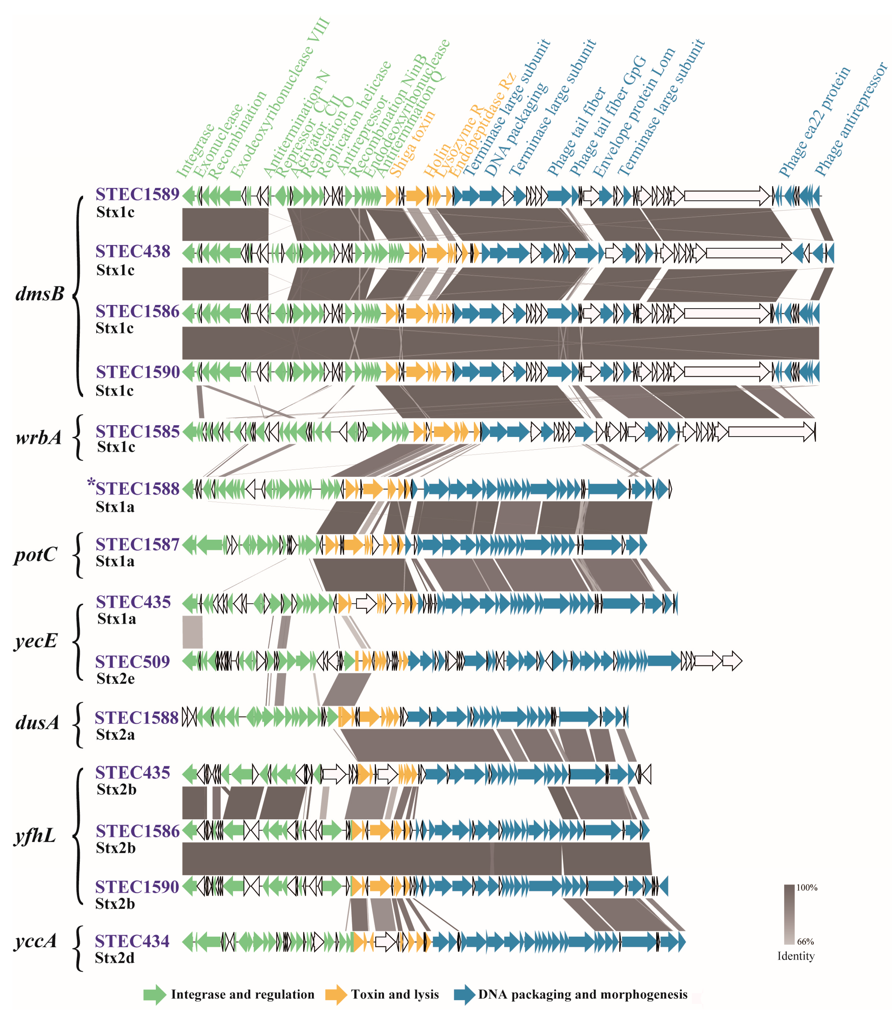
2.3. Genetic Features of Stx Prophages

Fourteen complete sequences of prophages were predicted and extracted from 10 complete STEC genome sequences. The 14 Stx prophages were characterized in terms of insertion site, genetic content, and functional structure (Figure 1). Their sizes ranged from 45,120 bp to 64,251 bp, with predicted CDSs ranging from 61 to 89 (Table S1). Seven different insertion sites were found. Four prophages were integrated into the coding sequence of dmsB (dimethyl sulfoxide reductase subunit B); three were integrated into the yfhL (putative 4Fe-4S cluster-containing protein YfhL); and two were inserted into yecE (DUF72 domain-containing protein YecE). Four prophages were integrated into yccA (Modulator of FtsH protease YccA), wrbA (NAD(P)H: quinone oxidoreductase), potC (spermidine preferential ABC transporter membrane subunit PotC), or dusA (encoding tRNA dihydroxyuridine synthase A), respectively. The insertion site of Stx1a prophage in strain STEC1588 was undetermined (Figure 1).

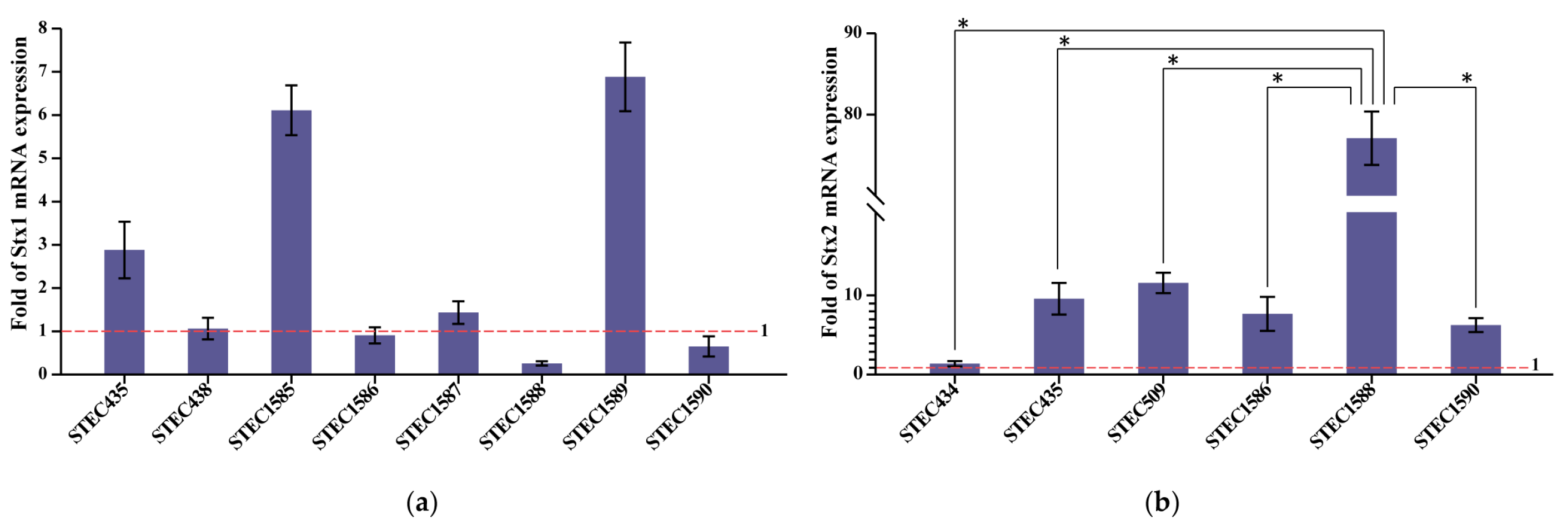
2.4. Variable Stx mRNA Expression Levels

To evaluate the Stx expression at the transcription level, all ten of the STEC strains were induced using mitomycin C. Among the eight Stx1-producing strains, four of them (STEC435, STEC1585, STEC1587, and STEC1589) showed a higher level of Stx1 mRNA transcription after induction (Figure 2a). The Stx2 mRNA transcription of the six Stx2-producing strains increased after induction (Figure 2b). Notably, strain STEC1588, producing Stx2a, showed higher inducibility than the other Stx2 subtype strains (p-value ≤ 0.05).

2.5. Antimicrobial Resistance (AMR) and AMR Genes

Among the ten STEC isolates, strain STEC438 showed resistance to tetracycline and streptomycin, and strain STEC509 showed resistance to tetracycline. The predicted resistance genotype matched well with the phenotypic resistance profiles. A new fosfomycin-resistant gene, fosA7, was identified in three STEC strains (STEC1586, STEC1589, and STEC1590), but the fosfomycin-resistant phenotypes were not tested in this study (Table 2).

2.6. Phylogenetic Relationships of STEC Strains from This Study and Other Sources

To investigate the phylogenetic relationship of the STEC strains between asymptomatic food handlers and reference strains, a wgSNP-based phylogeny tree was constructed. The reference genomes included the “Big 6” non-O157 serogroups (O26, O45, O103, O111, O121, and O145) strains and strains that hold identical serogroups in this study. The analysis showed that the 10 STEC strains from asymptomatic food handlers in this study were generally dispersed; the three strains (STEC1586, STEC1589, and STEC1590) that were isolated from the same location and shared the same serotype and ST type were clustered together with less than 10 SNPs. The other seven strains in this study were distributed separately; among these, STEC1587 and STEC509 clustered with the “Big 6” diarrhea patient-derived strains with 564 and 329 SNPs, respectively. The strain STEC438 clustered with a diarrhea patient-derived strain with 384 SNPs. Two strains (STEC434 and STEC1588) clustered with environment-derived strains, and the SNP distances ranged from 300 to 600. All asymptomatic food handlers-derived STEC strains were separated from HUS strains (Figure 3).

3. Discussion

Many outbreaks and sporadic infections caused by STEC O157:H7/NM have been reported worldwide [25,26,27]. Of note, severe infections caused by non-O157 STEC strains have been increasingly reported in recent years. For example, a large outbreak caused by STEC O104:H4 infected 3816 people and caused 54 deaths in Germany in 2011, affecting other European countries and North America [28]. The incidence of non-O157 STEC infections has risen from 0.19 per 100,000 in 2007 to 0.79 per 100,000 in 2014 in the USA. Non-O157 STEC strains were more common causes of acute diarrhea than O157 strains [29]. In China, an O157:H7 outbreak was reported in Xuzhou in 1999, leading to the hospitalization of 195 HUS patients and 177 deaths [30]. Subsequently, 20-year national surveillance (2001 to 2021) revealed that the isolation rate of STEC O157:H7 in diarrhea patients was 0.07% in Xuzhou City, Jiangsu Province, China [31]. Recently, a foodborne outbreak of diarrhea caused by non-O157 STEC was reported for the first time in China. The STEC O146:H10 strains were isolated from a diarrheal patient and a kitchen worker, respectively [32].
After food safety measures and STEC surveillance system were established in some countries, STEC infection became a notifiable disease. Asymptomatic STEC carriers are legally restricted from working as food handlers. To prevent the spread of infection via food, food handlers are required to carry out routine fecal examinations for a wide variety of infectious pathogens, including STEC [33,34]. The prevalence of STEC in asymptomatic infants and adults has been investigated in several areas. A study in France described STEC carriage in a large cohort, where the estimated rate was 1% among 959 healthy French infants [35]. A cross-sectional analysis in Germany showed a STEC prevalence of 0.5% among 224 asymptomatic children [36]. In Japan, a study showed that 398 out of 472,734 (0.08%) healthy adults were positive for STEC [37]. Since STEC infection is not a mandatory notifiable disease in China, the prevalence of STEC in the population is unclear. In this study, 0.251% of 3987 detected samples from asymptomatic food handlers were positive for STEC. The detection rate for STEC strains was highest in Qinghai (2.041%) compared to Guangdong (0.222%) and Guangxi (0.232%); however, the sample number for Qinghai was relatively small, which warranted further investigations using greater numbers in this region. The prevalence was similar in Guangdong and Guangxi, although the sampling spanned several years. The comparison among different regions should be explained with caution because the prevalence differed in terms of the demography, as well as sampling season, residential areas, and detection methods. It is worth noting that some PCR-positive samples were negative by culture in the study. Several factors could contribute to the failure to isolate STEC from the stx-positive samples, for example, the perturbation of background microflora, the low levels of stx in the samples, and the interference of free Stx phages [38]. The use of different STEC-selective culture media and isolation methods can also have an impact on STEC strain isolation [39]. The O:H serotyping of STEC strains has been used widely to evaluate the potential to cause severe diseases. Even though hundreds of different serotypes have been characterized and new ones continue to be found, the serogroups O26, O45, O103, O111, O121, and O145 (also referred to as the “top six”) have been suggested to be the most related to severe human diseases [40]. A high diversity of serotypes was recognized among the ten strains in our study. The predominant serotype in food handler-derived strains was O112:H8, different from that of diarrhea patient-derived strains in China [41]. The results also differ from a Japanese study, where O26:H11 was the predominant serotype from asymptomatic food handlers-isolated strains [34]. The O112 serogroup has been isolated from beef and caprine samples but not from human samples [42,43]. The serotype O8:H19 in this study had been reported as the most frequent serotype isolated from dairy farm environments [44,45]. The “top 6” non-O157 STEC serogroups were not identified in this study. This implies that asymptomatic carriers are not the reservoirs of predominant serotypes.
Stx2 was reported to be more frequently associated with severe clinical diseases in humans than Stx1 [46]. Stx1a has been linked to human illness; however, STEC that produce subtypes Stx2a, Stx2c, and Stx2d are more often correlated with the development of HC and HUS [47,48]. STEC carrying Stx1c and Stx2b are mainly associated with diarrheal disease [49,50]. In this study, the most common Stx subtype was Stx1c and this was linked to diarrheal disease. Stx1 is the most common type (50.4%) found among healthy adults in Japan, although the exact Stx1 subtypes are unclear [37]. The prevalent Stx subtype among asymptomatic humans remains largely unknown in China. Non-O157 STEC isolated from diarrheal patients in China were dominated by the stx1c subtype [41], which is accordant with our stx subtype from asymptomatic food handlers. Several studies depicted the prevalence and characteristics of STEC strains in retail raw meats in China, showing that stx1c and stx2e were predominant subtypes [51,52]. They could serve as a reservoir for the dissemination of virulence genes through the food chain. We also identified the stx2e subtype, which was a less common subtype in human STEC strains but was linked to edema disease in swine [53]. Some highly pathogenic stx compositions (i.e., stx2c, stx2d) were not present in our strains [41]. The combination of the stx2 and eae genes was also not found in our strains. The stx2 and eae genes together are often found to be associated with hemolytic uremic syndrome and are considered a risk factor for high virulence [54].
The Stx expression is crucial for STEC pathogenesis [55]. We found that the Stx inducibility of the ten strains was diverse. The Stx1-producing strains were non-inducible or less inducible, while all of the Stx2-producing strains were inducible. A study showed that the Stx1-encoding phage is less sensitive to inducing agents than the Stx2-encoding phage and that the level of Stx1 production is lower than that of Stx2 induced by mitomycin C [56]. The expression of Stx1 is regulated by two types of independent promoters. The first is a late phage promoter pR’ that depends on phage induction; this allows the expression and release of the Shiga toxin via the bacteriophage-mediated cell lysis. The other is a special Stx1 promoter, containing a binding site for Fur protein, which makes complexes with iron. Therefore, Fur represses Stx1 expression in the presence of iron, while Stx1 is expressed in the absence of iron. This regulation is completely independent of phage induction, and Stx1 levels of production are similar to those observed under conditions where the Stx1 phage is not induced [57]. However, a different level of Stx production can be observed for Stx2-encoding phages. The Stx2 production is always dependent on phage induction, whereas Stx release is dependent on cell lysis [58]. The strain STEC1588 (O112:H19, stx1a+stx2a) showed a higher Stx2 transcription level than other Stx2 subtype strains, partly indicating that the Stx2a subtype has the potential to cause serious disease.
The Stx phage may play a major role in the development of pathogenic STEC-mediated disease. Although Stx exhibits differences in sequence features, host specificity, and clinical outcomes, the stx genes are all located downstream of the late phage gene promoter. The Stx phage showed a modular construction and sequence heterogeneity, which was in accordance with our study [59]. The 14 Stx prophages showed considerable genomic diversity in terms of genome size and content, structure, and insertion site. Moreover, the prophages with the same insertion site and the same subtype of Stx, the structure and composition are also not entirely identical. It was reported that an STEC strain may carry multiple and different Stx phages [60]. We found that four strains harbored two Stx prophages. The Stx phage integrases seemed to have evolved to recognize specific sites inserted within the bacterial chromosome. Therefore, although one Stx phage integrates preferentially at one specific site, the integrase is able to recognize secondary sites for the integration of another phage genome if the preferred site is occupied or deleted [61]. We found seven different insertion sites in the 14 Stx prophages [53,59], and the dmsB and yfhL were the newly identified insertion sites in this study, indicating that more genes can act as the integration site of Stx phages.
Some studies have indicated that β-lactams, quinolones, trimethoprim, and trimethoprim-sulfamethoxazole can cause bacterial SOS reactions (DNA damage response pathway) and induce Shiga toxin production. However, other studies have suggested that certain antimicrobials, such as tetracycline, azithromycin, fosfomycin, and chloramphenicol, can block the SOS response and Stx production, such that they have been used to treat STEC infection to prevent HUS [62,63]. Among these potential antibiotic classes, resistance to tetracycline was observed in our study. However, all of the isolates were susceptible to azithromycin and chloramphenicol, which might be considered for the treatment of STEC infections when necessary. STEC strains can transfer their resistance to other strains through mobile genetic determinants [64]. Therefore, it is important to monitor AMR in STEC isolates and prevent the overuse of antibiotics.
The whole-genome phylogeny indicated a high diversity of STEC strains isolated from asymptomatic food handlers. Only three strains clustered closely together with less than 10 SNPs distance. When compared with strains from different sources, the strains from asymptomatic food handlers clustered with strains of diarrhea patients and environment-derived strains, but they were genetically distant from the strains of HUS patients. This may suggest that strains from asymptomatic food handlers were less pathogenic and posed a lower potential to cause severe disease in humans.
To our knowledge, this is the first study reporting the prevalence and characteristics of STEC strains isolated from asymptomatic food handlers in China. MLST types, serotypes, stx subtype, prophages, and Stx transcription levels suggest that STEC from asymptomatic carriers in China are highly diverse. The whole-genome phylogeny indicated that the strains from asymptomatic food handlers may pose a low pathogenic potential.

4. Materials and Methods

4.1. Sample Collection and Bacterial Strains

The sampling of asymptomatic food handlers (workers in the industry of food processing and catering companies, and cookers in restaurants) for physical examinations was carried out in three different geographical regions in China. Briefly, 49 anal swabs were collected from Yushu, Qinghai Province in 2013; a total of 1349 anal swabs were collected from Shenzhen, Guangdong Province from 2014 to 2016; and 2589 anal swabs were sampled from Yulin, Guangxi Zhuang Autonomous Region in 2022. Anal swab samples were collected in 15 mL sterile tubes and transported using ice packs to the laboratory at the National Institute for Communicable Disease Control and Prevention, China CDC, for the isolation of STEC. All samples were enriched in EC broth (Land Bridge, Beijing, China), and then enrichments were plated onto MacConkey agar (Oxoid, Hampshire, UK) and CHROMagarTM ECC agar (CHROMagar, Paris, France). The isolates were examined by PCR for the presence of the stx1 and stx2; all stx-positive isolates were confirmed as E. coli using the API-20E system (bioMérieux, Lyon, France), as previously described [65].

4.2. Whole Genome Sequencing (WGS) and Assembling

Genomic DNA was extracted using the Wizard Genomic DNA purification kit (Promega, WI, USA) according to the manufacturer’s instructions. Whole-genome sequencing was performed using two platforms; the 150 bp paired-end short reads were sequenced via the MGISEQ-2000 platform (MGI Tech Co., Ltd., Shenzhen, China). The 10 kb long library was sequenced via the PacBio Sequel platform (Pacific Biosciences, Menlo Park, CA, USA). The low-quality reads (Q-value ≤ 20 for short reads or length ≤ 1000 bp for long reads) and adapter sequences were filtered. The long reads were preliminarily corrected using Canu (v1.5). The assembling was based on the corrected reads using Canu and Falcon (v0.3.0). GATK (v1.6-13) was used to correct the single base error in assembling short reads.

4.3. WGS-Based Molecular Characterization

In silico stx subtyping was conducted by comparing genome assemblies against our in-house stx subtyping database that included all identified stx1 and stx2 subtypes using ABRicate version 0.8.10 with default parameters. SerotypeFinder (https://cge.food.dtu.dk/services/SerotypeFinder/, accessed on 20 March 2023) was used to determine the serotypes. The detection of the virulence genes and antimicrobial resistance genes was performed by comparing assemblies against the E. coli virulence factor database (https://github.com/phac-nml/ecoli_vf, accessed on 20 March 2023) and the Comprehensive Antibiotic Resistance Database (http://arpcard.mcmaster.ca, accessed on 28 March 2023), respectively, using ABRicate with default parameters (coverage ≥ 80% and identity ≥ 80%). Multilocus sequence typing (MLST) of the isolate was conducted through an online tool provided by the Warwick E. coli MLST scheme (https://enterobase.warwick.ac.uk/species/ecoli/allele_st, accessed on 5 April 2023).

4.4. Genomic Characterization of Stx-Converting Prophages

The Stx prophage sequences were extracted from genomes and characterized using the previously described methods [66]. Briefly, the PHAge Search Tool Enhanced Release (PHASTER, http://phaster.ca/, accessed on 2 June 2023) was used to identify the Stx prophages. The genome annotation of the Stx prophages was predicted by the RAST server (http://rast.nmpdr.org/, accessed on 2 June 2023). The gene adjacent to the integrase was defined as the phage insertion site [59]. The Stx prophages were compared and visualized in detail using Easyfig [67].

4.5. RNA Extraction and Relative Quantification of Stx Expression

The STEC overnight cultures in Luria Bertani (LB) broth were inoculated onto fresh LB medium and incubated at 37 °C with shaking until OD600 was about 0.6. Each culture was subdivided into two flasks, and mitomycin C was added to one of the subcultures at a final concentration of 0.5 μg/mL. Both induced and non-induced cultures were continuously grown at 37 °C with shaking for 3 h, and then the total RNA was extracted using RNeasy Mini Kit (Qiagen, Hilden, Germany) [30]. Reverse-transcription quantitative PCR (RT-qPCR) was performed on a Rotor-Gene Q Real-Time PCR system (Qiagen, Hilden, Germany) using the One Step TB Green® PrimeScriptTM RT-PCR kit (TaKaRa, Dalian, China) according to the manufacturer’s instructions. The primer pairs for stx1 (5′-GGAATTTACCTTAGAYTTCTCRAC-3′ and 5′-CCTGTGCCACTATCAATCATC-3′), stx2 (5′-TCCATGACAACGGACAGCAG-3′ and 5′-ACGCCAGATATGATGAAACCAG-3′), and for the housekeeping gene gapA (glyceraldehyde-3-phosphate dehydrogenase A) (5′-TATGACTGGTCCGTCTAAAGACAA-3′ and 5′-GGTTTTCTGAGTAGCGGTAGTAGC-3′) were used in the real-time PCR. The gapA gene was used for a within-sample normalization. The DDCt method was used to determine the relative amount of Stx mRNA expression compared to gapA [68]. The Stx expression levels under mitomycin C-inducing relative to non-induced ones were calculated.

4.6. Antimicrobial Susceptibility Test

The minimal inhibitory concentrations (MICs) were determined via the broth microdilution method using the BD PhoenixTM M50 Automated Microbiology System (BD, Franklin Lakes, NJ, USA). The following 20 antimicrobial agents were tested: amikacin (AMI, 4–64 µg/mL), ampicillin (AMP, 2–32 µg/mL), ampicillin-sulbactam (AMS, 2–32 µg/mL), aztreonam (ATM, 0.25–16 µg/mL), cefotaxime (CTX, 0.25–16 µg/mL), cefoxitin (FOX, 2–64 µg/mL), ceftazidime (CAZ, 0.25–16 µg/mL), chloramphenicol (CHL, 4–32 µg/mL), ciprofloxacin (CIP, 0.015–2 µg/mL), colistin (CT, 0.25–8 µg/mL), ertapenem (ETP, 0.25–8 µg/mL), imipenem (IPM, 0.25–8 µg/mL), meropenem (MEM, 0.125–2 µg/mL), nalidixic (NAL, 4–32 µg/mL), nitrofurantoin (F, 32–256 µg/mL), tetracycline (TET, 1–16 µg/mL), Tigecycline (TIG, 0.25–8), Streptomycin (STR, 4–32), trimethoprim-sulfamethoxazole (SXT, 0.5–8 µg/mL), and ceftazidime-avibactam (CZA, 0.25/4–8/4 µg/mL). The strains were defined as being resistant, intermediate, or susceptible according to the standard reference values (Clinical Laboratory Standards Institute [CLSI], 2022).

4.7. Phylogenomic Analysis

A whole-genome single-nucleotide polymorphism (wgSNP) phylogeny was used to assess the genomic diversity and relatedness of 10 STEC strains obtained in this study as well as 32 strains downloaded from the National Center for Biotechnology Information (NCBI) database. The core SNPs were obtained using Snippy version 4.3.6 (https://github.com/tseemann/snippy, accessed on 5 June 2023) with default parameters. Gubbins v2.3.4 [69] was then used to remove recombination from core SNPs and construct a maximum likelihood tree based on the filtered SNP alignments. The SNPs distances were assessed using snp-dists v0.7.0 (https://github.com/tseemann/snp-dists, accessed on 5 June 2023). The visualization and annotation of the phylogenetic tree were conducted by using an online tool, ChiPlot (https://www.chiplot.online/#Phylogenetic-Tree, accessed on 6 June 2023) [70].

4.8. Statistical Analysis

Statistical analysis was performed using SPSS 26.0. Dunnett’s t-test was carried out to compare Stx2 expression levels. There were statistically significant p-values ≤ 0.05.

4.9. Data Availability

The complete genomes of ten STEC strains are available at GenBank under the accession numbers listed in Figure 3.

Supplementary Materials

The following supporting information can be downloaded at: https://www.mdpi.com/article/10.3390/toxins15110640/s1, Table S1: Characterization of 14 Stx prophages in this study.

Author Contributions

Conceptualization, X.S. and Y.X.; methodology, X.Y.; software, X.S., X.Y. and H.S.; validation, Y.X., X.Y. and X.S.; formal analysis, X.S., X.Y., Q.L. and H.W.; investigation, M.L., Y.J. and Y.W.; resources, M.L., Y.J. and Y.W.; data curation, X.S., H.W., Q.L. and H.S.; writing—original draft preparation, X.S. and X.Y.; writing—review and editing, Y.X. and X.B.; visualization, X.S.; supervision, Y.X.; project administration, Y.X.; funding acquisition, Y.X. All authors have read and agreed to the published version of the manuscript.

Funding

This study was financially supported by the National Natural Science Foundation of China (82072254) and the National Key Research and Development Program of China (2021YFC2301105).

Institutional Review Board Statement

The study was conducted in accordance with the Declaration of Helsinki, and approved by the Institutional Ethics Committee of National Institute for Communicable Disease Control and Prevention (protocol code ICDC-202114, approved on 13 December 2021).

Informed Consent Statement

All of the participants were informed of the nature of the study, and their written consent was obtained.

Data Availability Statement

The data presented in this study are available by contacting the corresponding author upon reasonable request.

Conflicts of Interest

The authors declare no conflict of interest.

References

  1. Smith, J.L.; Fratamico, P.M.; Gunther, N.W.t. Shiga toxin-producing Escherichia coli. Adv. Appl. Microbiol. 2014, 86, 145–197. [Google Scholar]
  2. Jones, G.; Mariani-Kurkdjian, P.; Cointe, A.; Bonacorsi, S.; Lefevre, S.; Weill, F.X.; Le Strat, Y. Sporadic Shiga toxin-producing Escherichia coli-associated pediatric Hemolytic Uremic Syndrome, France, 2012–2021. Emerg. Infect. Dis. 2023, 29, 2054–2064. [Google Scholar] [CrossRef]
  3. Scheutz, F.; Teel, L.D.; Beutin, L.; Pierard, D.; Buvens, G.; Karch, H.; Mellmann, A.; Caprioli, A.; Tozzoli, R.; Morabito, S.; et al. Multicenter evaluation of a sequence-based protocol for subtyping Shiga toxins and standardizing Stx nomenclature. J. Clin. Microbiol. 2012, 50, 2951–2963. [Google Scholar] [CrossRef]
  4. Koutsoumanis, K.; Allende, A.; Alvarez-Ordonez, A.; Bover-Cid, S.; Chemaly, M.; Davies, R.; De Cesare, A.; Herman, L.; Hilbert, F.; Lindqvist, R.; et al. Pathogenicity assessment of Shiga toxin-producing Escherichia coli (STEC) and the public health risk posed by contamination of food with STEC. Efsa J. 2020, 18, e05967. [Google Scholar]
  5. Bai, X.; Fu, S.; Zhang, J.; Fan, R.; Xu, Y.; Sun, H.; He, X.; Xu, J.; Xiong, Y. Identification and pathogenomic analysis of an Escherichia coli strain producing a novel Shiga toxin 2 subtype. Sci. Rep. 2018, 8, 6756. [Google Scholar] [CrossRef]
  6. Lacher, D.W.; Gangiredla, J.; Patel, I.; Elkins, C.A.; Feng, P.C. Use of the Escherichia coli Identification microarray for characterizing the health risks of Shiga toxin-producing Escherichia coli isolated from foods. J. Food Prot. 2016, 79, 1656–1662. [Google Scholar] [CrossRef]
  7. Yang, X.; Bai, X.; Zhang, J.; Sun, H.; Fu, S.; Fan, R.; He, X.; Scheutz, F.; Matussek, A.; Xiong, Y. Escherichia coli strains producing a novel Shiga toxin 2 subtype circulate in China. Int. J. Med. Microbiol. 2020, 310, 151377. [Google Scholar] [CrossRef]
  8. Gill, A.; Dussault, F.; McMahon, T.; Petronella, N.; Wang, X.; Cebelinski, E.; Scheutz, F.; Weedmark, K.; Blais, B.; Carrillo, C. Characterization of atypical Shiga toxin gene sequences and description of Stx2j, a new subtype. J. Clin. Microbiol. 2022, 60, e0222921. [Google Scholar] [CrossRef]
  9. Bai, X.N.; Scheutz, F.; Dahlgren, H.M.; Hedenstrom, I.; Jernberg, C. Characterization of clinical Escherichia coli strains producing a novel Shiga toxin 2 subtype in Sweden and Denmark. Microorganisms 2021, 9, 2374. [Google Scholar] [CrossRef]
  10. Murphy, K.C.; Ritchie, J.M.; Waldor, M.K.; Lobner-Olesen, A.; Marinus, M.G. Dam methyltransferase is required for stable lysogeny of the Shiga toxin (Stx2)-encoding bacteriophage 933W of enterohemorrhagic Escherichia coli O157:H7. J. Bacteriol. 2008, 190, 438–441. [Google Scholar] [CrossRef]
  11. Berger, P.; Kouzel, I.U.; Berger, M.; Haarmann, N.; Dobrindt, U.; Koudelka, G.B.; Mellmann, A. Carriage of Shiga toxin phage profoundly affects Escherichia coli gene expression and carbon source utilization. BMC Genom. 2019, 20, 504. [Google Scholar] [CrossRef]
  12. Bai, X.; Zhang, J.; Ambikan, A.; Jernberg, C.; Ehricht, R.; Scheutz, F.; Xiong, Y.; Matussek, A. Molecular characterization and comparative genomics of clinical hybrid Shiga toxin-producing and enterotoxigenic Escherichia coli (STEC/ETEC) strains in Sweden. Sci. Rep. 2019, 9, 5619. [Google Scholar] [CrossRef]
  13. Hazen, T.H.; Michalski, J.; Luo, Q.; Shetty, A.C.; Daugherty, S.C.; Fleckenstein, J.M.; Rasko, D.A. Comparative genomics and transcriptomics of Escherichia coli isolates carrying virulence factors of both enteropathogenic and enterotoxigenic E. coli. Sci. Rep. 2017, 7, 3513. [Google Scholar] [CrossRef]
  14. Rodriguez-Rubio, L.; Haarmann, N.; Schwidder, M.; Muniesa, M.; Schmidt, H. Bacteriophages of Shiga toxin-producing Escherichia coli and their contribution to pathogenicity. Pathogens 2021, 10, 404. [Google Scholar] [CrossRef]
  15. Kagambega, A.; Martikainen, O.; Siitonen, A.; Traore, A.S.; Barro, N.; Haukka, K. Prevalence of diarrheagenic Escherichia coli virulence genes in the feces of slaughtered cattle, chickens, and pigs in Burkina Faso. Microbiologyopen 2012, 1, 276–284. [Google Scholar] [CrossRef]
  16. Mercer, R.G.; Zheng, J.; Garcia-Hernandez, R.; Ruan, L.; Ganzle, M.G.; McMullen, L.M. Genetic determinants of heat resistance in Escherichia coli. Front. Microbiol. 2015, 6, 932. [Google Scholar] [CrossRef]
  17. Meng, Q.; Bai, X.; Zhao, A.; Lan, R.; Du, H.; Wang, T.; Shi, C.; Yuan, X.; Bai, X.; Ji, S.; et al. Characterization of Shiga toxin-producing Escherichia coli isolated from healthy pigs in China. BMC Microbiol. 2014, 14, 5. [Google Scholar] [CrossRef]
  18. Guh, A.; Phan, Q.; Nelson, R.; Purviance, K.; Milardo, E.; Kinney, S.; Mshar, P.; Kasacek, W.; Cartter, M. Outbreak of Escherichia coli O157 associated with raw milk, Connecticut, 2008. Clin. Infect. Dis. 2010, 51, 1411–1417. [Google Scholar] [CrossRef]
  19. Sethulekshmi, C.; Latha, C.; Anu, C.J. Occurrence and quantification of Shiga toxin-producing Escherichia coli from food matrices. Vet. World 2018, 11, 104–111. [Google Scholar] [CrossRef]
  20. Treacy, J.; Jenkins, C.; Paranthaman, K.; Jorgensen, F.; Mueller-Doblies, D.; Anjum, M.; Kaindama, L.; Hartman, H.; Kirchner, M.; Carson, T.; et al. Outbreak of Shiga toxin-producing Escherichia coli O157:H7 linked to raw drinking milk resolved by rapid application of advanced pathogen characterisation methods, England, August to October 2017. Eurosurveillance 2019, 24, 1800191. [Google Scholar] [CrossRef]
  21. Carlson-Banning, K.M.; Sperandio, V. Catabolite and oxygen regulation of enterohemorrhagic Escherichia coli virulence. mBio 2016, 7, e01852-16. [Google Scholar] [CrossRef] [PubMed]
  22. Kintz, E.; Brainard, J.; Vanderes, M.; Vivancos, R.; Byrne, L.; Butt, S.; Jenkins, C.; Elson, R.; Lake, I.; Hunter, P. Animal and environmental risk factors for sporadic Shiga toxin-producing Escherichia coli (STEC) infection in England: A case control study for O157, O26 and other STEC serotypes. Pathog. Glob. Health 2023, 117, 655–663. [Google Scholar] [CrossRef] [PubMed]
  23. Chen, Y.; Chen, X.; Zheng, S.; Yu, F.; Kong, H.; Yang, Q.; Cui, D.; Chen, N.; Lou, B.; Li, X.; et al. Serotypes, genotypes and antimicrobial resistance patterns of human diarrhoeagenic Escherichia coli isolates circulating in southeastern China. Clin. Microbiol. Infect. 2014, 20, 52–58. [Google Scholar] [CrossRef]
  24. Nuesch-Inderbinen, M.; Treier, A.; Stevens, M.J.A.; Stephan, R. Whole genome sequence-based characterisation of Shiga toxin-producing Escherichia coli isolated from game meat originating from several European countries. Sci. Rep. 2023, 13, 3247. [Google Scholar] [CrossRef]
  25. Riley, L.W.; Remis, R.S.; Helgerson, S.D.; McGee, H.B.; Wells, J.G.; Davis, B.R.; Hebert, R.J.; Olcott, E.S.; Johnson, L.M.; Hargrett, N.T.; et al. Hemorrhagic colitis associated with a rare Escherichia coli serotype. N. Engl. J. Med. 1983, 308, 681–685. [Google Scholar] [CrossRef] [PubMed]
  26. Grant, J.; Wendelboe, A.M.; Wendel, A.; Jepson, B.; Torres, P.; Smelser, C.; Rolfs, R.T. Spinach-associated Escherichia coli O157:H7 outbreak, Utah and New Mexico, 2006. Emerg. Infect. Dis. 2008, 14, 1633–1636. [Google Scholar] [CrossRef] [PubMed]
  27. Michino, H.; Araki, K.; Minami, S.; Takaya, S.; Sakai, N.; Miyazaki, M.; Ono, A.; Yanagawa, H. Massive outbreak of Escherichia coli O157:H7 infection in schoolchildren in Sakai City, Japan, associated with consumption of white radish sprouts. Am. J. Epidemiol. 1999, 150, 787–796. [Google Scholar] [CrossRef]
  28. Frank, C.; Werber, D.; Cramer, J.P.; Askar, M.; Faber, M.; an der Heiden, M.; Bernard, H.; Fruth, A.; Prager, R.; Spode, A.; et al. Epidemic profile of Shiga-toxin-producing Escherichia coli O104:H4 outbreak in Germany. N. Engl. J. Med. 2011, 365, 1771–1780. [Google Scholar] [CrossRef]
  29. Valilis, E.; Ramsey, A.; Sidiq, S.; DuPont, H.L. Non-O157 Shiga toxin-producing Escherichia coli-A poorly appreciated enteric pathogen: Systematic review. Int. J. Infect. Dis. 2018, 76, 82–87. [Google Scholar] [CrossRef]
  30. Xiong, Y.; Wang, P.; Lan, R.; Ye, C.; Wang, H.; Ren, J.; Jing, H.; Wang, Y.; Zhou, Z.; Bai, X.; et al. A novel Escherichia coli O157:H7 clone causing a major hemolytic uremic syndrome outbreak in China. PLoS ONE 2012, 7, e36144. [Google Scholar] [CrossRef]
  31. Zhu, W.; Guo, H.; Xu, J.; Wu, W.; Yi, Y.; Wang, J.; Duan, R.; Tong, J.; Du, Y. Enterohemorrhagic Escherichia coli O157:H7—Xuzhou City, Jiangsu Province, China, 2001–2021. China CDC Wkly. 2023, 5, 311–314. [Google Scholar] [CrossRef] [PubMed]
  32. Huang, H.; Wang, Y.; Wang, C.; Tang, Y.; He, L.; Deng, J.; Pan, X. Complete genome sequences of two Shiga toxin-producing Escherichia coli O146:H10 strains recovered from a foodborne outbreak in China. Microbiol. Resour. Announc. 2021, 10, e0082521. [Google Scholar] [CrossRef] [PubMed]
  33. Balabanova, Y.; Gilsdorf, A.; Buda, S.; Burger, R.; Eckmanns, T.; Gartner, B.; Gross, U.; Haas, W.; Hamouda, O.; Hubner, J.; et al. Communicable diseases prioritized for surveillance and epidemiological research: Results of a standardized prioritization procedure in Germany, 2011. PLoS ONE 2011, 6, e25691. [Google Scholar] [CrossRef] [PubMed]
  34. Baba, H.; Kanamori, H.; Kudo, H.; Kuroki, Y.; Higashi, S.; Oka, K.; Takahashi, M.; Yoshida, M.; Oshima, K.; Aoyagi, T.; et al. Genomic analysis of Shiga toxin-producing Escherichia coli from patients and asymptomatic food handlers in Japan. PLoS ONE 2019, 14, e0225340. [Google Scholar] [CrossRef]
  35. Bizot, E.; Cointe, A.; Bechet, S.; Sobral, E.; Cohen, R.; Mariani-Kurkdjian, P.; Levy, C.; Bonacorsi, S. Shiga toxin-producing Escherichia coli carriage in 959 healthy French infants. Arch. Dis. Child. 2021, 106, 1239–1240. [Google Scholar] [CrossRef] [PubMed]
  36. Harries, M.; Dreesman, J.; Rettenbacher-Riefler, S.; Mertens, E. Faecal carriage of extended-spectrum beta-lactamase-producing Enterobacteriaceae and Shiga toxin-producing Escherichia coli in asymptomatic nursery children in Lower Saxony (Germany), 2014. Epidemiol. Infect. 2016, 144, 3540–3548. [Google Scholar] [CrossRef] [PubMed]
  37. Morita-Ishihara, T.; Iyoda, S.; Iguchi, A.; Ohnishi, M. Secondary Shiga toxin-producing Escherichia coli infection, Japan, 2010-2012. Emerg. Infect. Dis. 2016, 22, 2181–2184. [Google Scholar] [CrossRef]
  38. Quiros, P.; Martinez-Castillo, A.; Muniesa, M. Improving detection of Shiga toxin-producing Escherichia coli by molecular methods by reducing the interference of free Shiga toxin-encoding bacteriophages. Appl. Environ. Microbiol. 2015, 81, 415–421. [Google Scholar] [CrossRef]
  39. Gill, A.; Huszczynski, G.; Gauthier, M.; Blais, B. Evaluation of eight agar media for the isolation of Shiga toxin-producing Escherichia coli. J. Microbiol. Methods 2014, 96, 6–11. [Google Scholar] [CrossRef]
  40. Brooks, J.T.; Sowers, E.G.; Wells, J.G.; Greene, K.D.; Griffin, P.M.; Hoekstra, R.M.; Strockbine, N.A. Non-O157 Shiga toxin-producing Escherichia coli infections in the United States, 1983–2002. J. Infect. Dis. 2005, 192, 1422–1429. [Google Scholar] [CrossRef]
  41. Bai, X.; Hu, B.; Xu, Y.; Sun, H.; Zhao, A.; Ba, P.; Fu, S.; Fan, R.; Jin, Y.; Wang, H.; et al. Molecular and phylogenetic characterization of non-O157 Shiga toxin-producing Escherichia coli strains in China. Front. Cell Infect. Microbiol. 2016, 6, 143. [Google Scholar] [CrossRef] [PubMed]
  42. Mahanti, A.; Samanta, I.; Bandyopadhyay, S.; Joardar, S.N. Molecular characterization and antibiotic susceptibility pattern of caprine Shiga toxin producing-Escherichia coli (STEC) isolates from India. Iran. J. Vet. Res. 2015, 16, 31–35. [Google Scholar] [PubMed]
  43. Franci, T.; Sanso, A.M.; Bustamante, A.V.; Lucchesi, P.M.; Parma, A.E. Genetic characterization of non-O157 verocytotoxigenic Escherichia coli isolated from raw beef products using multiple-locus variable-number tandem repeat analysis. Foodborne Pathog. Dis. 2011, 8, 1019–1023. [Google Scholar] [CrossRef]
  44. Meichtri, L.; Miliwebsky, E.; Gioffre, A.; Chinen, I.; Baschkier, A.; Chillemi, G.; Guth, B.E.; Masana, M.O.; Cataldi, A.; Rodriguez, H.R.; et al. Shiga toxin-producing Escherichia coli in healthy young beef steers from Argentina: Prevalence and virulence properties. Int. J. Food Microbiol. 2004, 96, 189–198. [Google Scholar] [CrossRef] [PubMed]
  45. Polifroni, R.; Etcheverria, A.I.; Sanz, M.E.; Cepeda, R.E.; Kruger, A.; Lucchesi, P.M.; Fernandez, D.; Parma, A.E.; Padola, N.L. Molecular characterization of Shiga toxin-producing Escherichia coli isolated from the environment of a dairy farm. Curr. Microbiol. 2012, 65, 337–343. [Google Scholar] [CrossRef] [PubMed]
  46. Melton-Celsa, A.; Mohawk, K.; Teel, L.; O’Brien, A. Pathogenesis of Shiga-toxin producing Escherichia coli. Curr. Top. Microbiol. Immunol. 2012, 357, 67–103. [Google Scholar] [PubMed]
  47. Baranzoni, G.M.; Fratamico, P.M.; Gangiredla, J.; Patel, I.; Bagi, L.K.; Delannoy, S.; Fach, P.; Boccia, F.; Anastasio, A.; Pepe, T. Characterization of Shiga toxin subtypes and virulence genes in porcine Shiga toxin-producing Escherichia coli. Front. Microbiol. 2016, 7, 574. [Google Scholar] [CrossRef]
  48. Melton-Celsa, A.R. Shiga toxin (Stx) classification, structure, and function. Microbiol. Spectr. 2014, 2, 2–4. [Google Scholar] [CrossRef]
  49. Friedrich, A.W.; Bielaszewska, M.; Zhang, W.L.; Pulz, M.; Kuczius, T.; Ammon, A.; Karch, H. Escherichia coli harboring Shiga toxin 2 gene variants: Frequency and association with clinical symptoms. J. Infect. Dis. 2002, 185, 74–84. [Google Scholar] [CrossRef]
  50. Jelacic, J.K.; Damrow, T.; Chen, G.S.; Jelacic, S.; Bielaszewska, M.; Ciol, M.; Carvalho, H.M.; Melton-Celsa, A.R.; O’Brien, A.D.; Tarr, P.I. Shiga toxin-producing Escherichia coli in Montana: Bacterial genotypes and clinical profiles. J. Infect. Dis. 2003, 188, 719–729. [Google Scholar] [CrossRef]
  51. Bai, X.; Wang, H.; Xin, Y.; Wei, R.; Tang, X.; Zhao, A.; Sun, H.; Zhang, W.; Wang, Y.; Xu, Y.; et al. Prevalence and characteristics of Shiga toxin-producing Escherichia coli isolated from retail raw meats in China. Int. J. Food Microbiol. 2015, 200, 31–38. [Google Scholar] [CrossRef] [PubMed]
  52. Hu, B.; Yang, X.; Liu, Q.; Zhang, Y.; Jiang, D.; Jiao, H.; Yang, Y.; Xiong, Y.; Bai, X.; Hou, P. High prevalence and pathogenic potential of Shiga toxin-producing Escherichia coli strains in raw mutton and beef in Shandong, China. Curr. Res. Food Sci. 2022, 5, 1596–1602. [Google Scholar] [CrossRef] [PubMed]
  53. Yang, X.; Wu, Y.; Liu, Q.; Sun, H.; Luo, M.; Xiong, Y.; Matussek, A.; Hu, B.; Bai, X. Genomic characteristics of Stx2e-producing Escherichia coli strains derived from humans, animals, and meats. Pathogens 2021, 10, 1551. [Google Scholar] [CrossRef] [PubMed]
  54. Ethelberg, S.; Olsen, K.E.; Scheutz, F.; Jensen, C.; Schiellerup, P.; Enberg, J.; Petersen, A.M.; Olesen, B.; Gerner-Smidt, P.; Molbak, K. Virulence factors for hemolytic uremic syndrome, Denmark. Emerg. Infect. Dis. 2004, 10, 842–847. [Google Scholar] [CrossRef] [PubMed]
  55. Joseph, A.; Cointe, A.; Mariani Kurkdjian, P.; Rafat, C.; Hertig, A. Shiga toxin-associated hemolytic uremic syndrome: A narrative review. Toxins 2020, 12, 67. [Google Scholar] [CrossRef]
  56. Ichimura, K.; Shimizu, T.; Matsumoto, A.; Hirai, S.; Yokoyama, E.; Takeuchi, H.; Yahiro, K.; Noda, M. Nitric oxide-enhanced Shiga toxin production was regulated by Fur and RecA in enterohemorrhagic O157. Microbiologyopen 2017, 6, e00461. [Google Scholar] [CrossRef]
  57. Wagner, P.L.; Livny, J.; Neely, M.N.; Acheson, D.W.; Friedman, D.I.; Waldor, M.K. Bacteriophage control of Shiga toxin 1 production and release by Escherichia coli. Mol. Microbiol. 2002, 44, 957–970. [Google Scholar] [CrossRef]
  58. Waldor, M.K.; Friedman, D.I. Phage regulatory circuits and virulence gene expression. Curr. Opin. Microbiol. 2005, 8, 459–465. [Google Scholar] [CrossRef]
  59. Steyert, S.R.; Sahl, J.W.; Fraser, C.M.; Teel, L.D.; Scheutz, F.; Rasko, D.A. Comparative genomics and Stx phage characterization of LEE-negative Shiga toxin-producing Escherichia coli. Front. Cell Infect. Microbiol. 2012, 2, 133. [Google Scholar] [CrossRef]
  60. Nyong, E.C.; Zaia, S.R.; Allue-Guardia, A.; Rodriguez, A.L.; Irion-Byrd, Z.; Koenig, S.S.K.; Feng, P.; Bono, J.L.; Eppinger, M. Pathogenomes of atypical non-Shigatoxigenic Escherichia coli NSF/SF O157:H7/NM: Comprehensive phylogenomic analysis using closed genomes. Front. Microbiol. 2020, 11, 619. [Google Scholar] [CrossRef]
  61. Serra-Moreno, R.; Jofre, J.; Muniesa, M. Insertion site occupancy by Stx2 bacteriophages depends on the locus availability of the host strain chromosome. J. Bacteriol. 2007, 189, 6645–6654. [Google Scholar] [CrossRef] [PubMed]
  62. Oporto, B.; Ocejo, M.; Alkorta, M.; Marimon, J.M.; Montes, M.; Hurtado, A. Zoonotic approach to Shiga toxin-producing Escherichia coli: Integrated analysis of virulence and antimicrobial resistance in ruminants and humans. Epidemiol. Infect. 2019, 147, e164. [Google Scholar] [CrossRef]
  63. Mir, R.A.; Kudva, I.T. Antibiotic-resistant Shiga toxin-producing Escherichia coli: An overview of prevalence and intervention strategies. Zoonoses Public Health 2019, 66, 1–13. [Google Scholar] [CrossRef] [PubMed]
  64. Day, M.; Doumith, M.; Jenkins, C.; Dallman, T.J.; Hopkins, K.L.; Elson, R.; Godbole, G.; Woodford, N. Antimicrobial resistance in Shiga toxin-producing Escherichia coli serogroups O157 and O26 isolated from human cases of diarrhoeal disease in England, 2015. J. Antimicrob. Chemother. 2017, 72, 145–152. [Google Scholar] [CrossRef] [PubMed]
  65. Bai, X.; Zhang, W.; Tang, X.; Xin, Y.; Xu, Y.; Sun, H.; Luo, X.; Pu, J.; Xu, J.; Xiong, Y.; et al. Shiga toxin-producing Escherichia coli in plateau pika (Ochotona curzoniae) on the Qinghai-Tibetan plateau, China. Front. Microbiol. 2016, 7, 375. [Google Scholar] [CrossRef] [PubMed]
  66. Yang, X.; Liu, Q.; Bai, X.; Hu, B.; Jiang, D.; Jiao, H.; Lu, L.; Fan, R.; Hou, P.; Matussek, A.; et al. High prevalence and persistence of Escherichia coli strains producing Shiga toxin subtype 2k in goat herds. Microbiol. Spectr. 2022, 10, e0157122. [Google Scholar] [CrossRef]
  67. Sullivan, M.J.; Petty, N.K.; Beatson, S.A. Easyfig: A genome comparison visualizer. Bioinformatics 2011, 27, 1009–1010. [Google Scholar] [CrossRef]
  68. Livak, K.J.; Schmittgen, T.D. Analysis of relative gene expression data using real-time quantitative PCR and the 2(-Delta Delta C(T)) Method. Methods 2001, 25, 402–408. [Google Scholar] [CrossRef]
  69. Croucher, N.J.; Page, A.J.; Connor, T.R.; Delaney, A.J.; Keane, J.A.; Bentley, S.D.; Parkhill, J.; Harris, S.R. Rapid phylogenetic analysis of large samples of recombinant bacterial whole genome sequences using Gubbins. Nucleic Acids Res. 2015, 43, e15. [Google Scholar] [CrossRef]
  70. Xie, J.; Chen, Y.; Cai, G.; Cai, R.; Hu, Z.; Wang, H. Tree Visualization By One Table (tvBOT): A web application for visualizing, modifying and annotating phylogenetic trees. Nucleic Acids Res. 2023, 51, W587–W592. [Google Scholar] [CrossRef]
Figure 1. Architecture of fourteen prophages from ten STEC isolates. Easyfig plot was used to compare the fourteen prophages. Arrows indicate gene directions. Phage integrase and regulation genes are shown in green; toxin and lysis genes are shown in yellow; DNA packaging and morphogenesis genes are shown in blue; and the genes encoding hypothetic proteins are shown in white. * The insertion site was undetermined.
Toxins 15 00640 g001
Figure 2. Fold of Stx mRNA transcription in induced state relative to non-induced state. Non-induced state was used as calibrator and data are means ± standard deviations from three independent replicates. Fold change greater than one indicates mRNA transcription increased. (a) Fold of Stx1 mRNA transcription; (b) Fold of Stx2 mRNA transcription. * It indicates statistically significant differences.
Toxins 15 00640 g002
Figure 3. Phylogenetic tree based on core-genome single-nucleotide polymorphisms (SNPs) using the maximum likelihood method. The source, serotype, MLST type, stx subtype, eae, and accession numbers of all strains are shown. The red circles represent the strains isolated from this study.
Toxins 15 00640 g003
Table 1. Prevalence of STEC in anal swab samples from food handlers in China.
Sampling Site No. of
Samples
No. of STEC
Isolates
Prevalence of
STEC (%)
Guangdong134930.222
Qinghai4912.041
Guangxi258960.232
Total3987100.251
Table 2. Characteristics of STEC derived from asymptomatic food handlers in this study.
StrainSerotypeSequence Typestx
Subtype
Sampling
Year
Antimicrobial
Resistant Phenotypes
Antimicrobial Resistant Genes
STEC434O43:H2937stx2d2014--
STEC435O91:H1433stx1a+stx2b2013--
STEC438ONT:H2140stx1c2014Streptomycin,
Tetracycline
ant(3)-Ia, tet(A)
STEC509O8:H1921stx2e2016Tetracyclinetet(B)
STEC1585O178:H7278stx1c2022--
STEC1586O112ab:H875stx1c+stx2b2022-fosA7
STEC1587O21:H25971stx1a2022--
STEC1588O112ac:H192038stx1a+stx2a2022--
STEC1589O112ab:H875stx1c2022-fosA7
STEC1590O112ab:H875stx1c+stx2b2022-fosA7
‘-’, susceptible (antimicrobial resistant phenotypes) or absent (antimicrobial resistant genes).
Disclaimer/Publisher’s Note: The statements, opinions and data contained in all publications are solely those of the individual author(s) and contributor(s) and not of MDPI and/or the editor(s). MDPI and/or the editor(s) disclaim responsibility for any injury to people or property resulting from any ideas, methods, instructions or products referred to in the content.

