Next Issue
Volume 13, May
Previous Issue
Volume 13, March


Toxins, Volume 13, Issue 4 (April 2021) – 61 articles

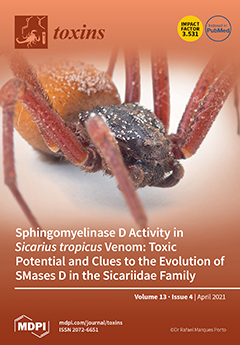
Cover Story (view full-size image): The spider family Sicariidae includes Hexophthalma, Sicarius and Loxosceles genera. Their venoms share a common feature: the presence of Sphingomyelinases D. SMases D are also found in bacteria, ticks and fungi. The bacterial and spider SMases D are functionally similar and considered to have originated from a common ancestor. There are different hypotheses about the evolutionary history of SMases D enzymes, but few studies take into account the similarity or differences in the induction mechanism of effects provoked by SMases D. In this study, Sicarius tropicus and L. laeta venoms were compared regarding the presence and activity of SMases D. Results showed that S. tropicus venom has active SMases D capable of causing cell death and hemolysis dependent on complement system, which, in its induction mechanism, is similar to the one present in bacteria. View this paper
  • Issues are regarded as officially published after their release is announced to the table of contents alert mailing list.
  • You may sign up for e-mail alerts to receive table of contents of newly released issues.
  • PDF is the official format for papers published in both, html and pdf forms. To view the papers in pdf format, click on the "PDF Full-text" link, and use the free Adobe Reader to open them.
Order results
Result details
Section
Select all
Export citation of selected articles as:
16 pages, 6448 KiB  
Article
Effects of Polystyrene Microplastics on Growth and Toxin Production of Alexandrium pacificum
by Chao Liu, Jiangbing Qiu, Zhixuan Tang, Hong Hu, Fanping Meng and Aifeng Li
Toxins 2021, 13(4), 293; https://doi.org/10.3390/toxins13040293 - 20 Apr 2021
Cited by 19 | Viewed by 3396
Abstract
Microplastics (MP) widely distributed in aquatic environments have adverse effects on aquatic organisms. Currently, the impact of MP on toxigenic red tide microalgae is poorly understood. In this study, the strain of Alexandrium pacificum ATHK, typically producing paralytic shellfish toxins (PST), was selected [...] Read more.
Microplastics (MP) widely distributed in aquatic environments have adverse effects on aquatic organisms. Currently, the impact of MP on toxigenic red tide microalgae is poorly understood. In this study, the strain of Alexandrium pacificum ATHK, typically producing paralytic shellfish toxins (PST), was selected as the target. Effects of 1 and 0.1 μm polystyrene MP with three concentration gradients (5 mg L−1, 25 mg L−1 and 100 mg L−1) on the growth, chlorophyll a (Chl a), photosynthetic activity (Fv/Fm) and PST production of ATHK were explored. Results showed that the high concentration (100 mg L−1) of 1 μm and 0.1 μm MP significantly inhibited the growth of ATHK, and the inhibition depended on the size and concentration of MP. Contents of Chl a showed an increase with various degrees after MP exposure in all cases. The photosynthesis indicator Fv/Fm of ATHK was significantly inhibited in the first 11 days, then gradually returned to the level of control group at day 13, and finally was gradually inhibited in the 1 μm MP treatments, and promotion or inhibition to some degree also occurred at different periods after exposure to 0.1 μm MP. Overall, both particle sizes of MP at 5 and 25 mg L−1 had no significant effect on cell toxin quota, and the high concentration 100 mg L−1 significantly promoted the PST biosynthesis on the day 7, 11 and 15. No significant difference occurred in the cell toxin quota and the total toxin content in all treatments at the end of the experiment (day 21). All MP treatments did not change the toxin profiles of ATHK, nor did the relative molar percentage of main PST components. The growth of ATHK, Chl a content, Fv/Fm and toxin production were not affected by MP shading. This is the first report on the effects of MP on the PST-producing microalgae, which will improve the understanding of the adverse impact of MP on the growth and toxin production of A. pacificum. Full article
(This article belongs to the Section Marine and Freshwater Toxins)
Show Figures

Figure 1

21 pages, 23036 KiB  
Review
Predicted Aflatoxin B1 Increase in Europe Due to Climate Change: Actions and Reactions at Global Level
by Marco Camardo Leggieri, Piero Toscano and Paola Battilani
Toxins 2021, 13(4), 292; https://doi.org/10.3390/toxins13040292 - 20 Apr 2021
Cited by 34 | Viewed by 5439
Abstract
Climate change (CC) is predicted to increase the risk of aflatoxin (AF) contamination in maize, as highlighted by a project supported by EFSA in 2009. We performed a comprehensive literature search using the Scopus search engine to extract peer-reviewed studies citing this study. [...] Read more.
Climate change (CC) is predicted to increase the risk of aflatoxin (AF) contamination in maize, as highlighted by a project supported by EFSA in 2009. We performed a comprehensive literature search using the Scopus search engine to extract peer-reviewed studies citing this study. A total of 224 papers were identified after step I filtering (187 + 37), while step II filtering identified 25 of these papers for quantitative analysis. The unselected papers (199) were categorized as “actions” because they provided a sounding board for the expected impact of CC on AFB1 contamination, without adding new data on the topic. The remaining papers were considered as “reactions” of the scientific community because they went a step further in their data and ideas. Interesting statements taken from the “reactions” could be summarized with the following keywords: Chain and multi-actor approach, intersectoral and multidisciplinary, resilience, human and animal health, and global vision. In addition, fields meriting increased research efforts were summarized as the improvement of predictive modeling; extension to different crops and geographic areas; and the impact of CC on fungi and mycotoxin co-occurrence, both in crops and their value chains, up to consumers. Full article
(This article belongs to the Special Issue Mycotoxins in Relation to Climate Change)
Show Figures

Figure 1

12 pages, 2435 KiB  
Article
Vasoactive Effects of Acute Ergot Exposure in Sheep
by Rossalin Yonpiam, Jair Gobbet, Ashok Jadhav, Kaushik Desai, Barry Blakley and Ahmad Al-Dissi
Toxins 2021, 13(4), 291; https://doi.org/10.3390/toxins13040291 - 20 Apr 2021
Cited by 4 | Viewed by 2273
Abstract
Ergotism is a common and increasing problem in Saskatchewan’s livestock. Chronic exposure to low concentrations of ergot alkaloids is known to cause severe arterial vasoconstriction and gangrene through the activation of adrenergic and serotonergic receptors on vascular smooth muscles. The acute vascular effects [...] Read more.
Ergotism is a common and increasing problem in Saskatchewan’s livestock. Chronic exposure to low concentrations of ergot alkaloids is known to cause severe arterial vasoconstriction and gangrene through the activation of adrenergic and serotonergic receptors on vascular smooth muscles. The acute vascular effects of a single oral dose with high-level exposure to ergot alkaloids remain unknown and are examined in this study. This study had two main objectives; the first was to evaluate the role of α1-adrenergic receptors in mediating the acute vasocontractile response after single-dose exposure in sheep. The second was to examine whether terazosin (TE) could abolish the vascular contractile effects of ergot alkaloids. Twelve adult female sheep were randomly placed into control and exposure groups (n = 6/group). Ergot sclerotia were collected and finely ground. The concentrations of six ergot alkaloids (ergocornine, ergocristine, ergocryptine, ergometrine, ergosine, and ergotamine) were determined using HPLC/MS at Prairie Diagnostic Services Inc., (Saskatoon, SK, Canada). Each ewe within the treatment group received a single oral treatment of ground ergot sclerotia at a dose of 600 µg/kg BW (total ergot) while each ewe in the control group received water. Animals were euthanized 12 h after the treatment, and the pedal artery (dorsal metatarsal III artery) from the left hind limb from each animal was carefully dissected and mounted in an isolated tissue bath. The vascular contractile response to phenylephrine (PE) (α1-adrenergic agonist) was compared between the two groups before and after TE (α1-adrenergic antagonist) treatment. Acute exposure to ergot alkaloids resulted in a 38% increase in vascular sensitivity to PE compared to control (Ctl EC50 = 1.74 × 10⁻⁶ M; Exp EC50 = 1.079 × 10⁻⁶ M, p = 0.046). TE treatment resulted in a significant dose-dependent increase in EC50 in both exposure and control groups (p < 0.05 for all treatments). Surprisingly, TE effect was significantly more pronounced in the ergot exposed group compared to the control group at two of the three concentrations of TE (TE 30 nM, p = 0.36; TE 100 nM, p < 0.001; TE 300 nM, p < 0.001). Similar to chronic exposure, acute exposure to ergot alkaloids results in increased vascular sensitivity to PE. TE is a more potent dose-dependent antagonist for the PE contractile response in sheep exposed to ergot compared to the control group. This study may indicate that the dry gangrene seen in sheep, and likely other species, might be related to the activation of α1-adrenergic receptor. This effect may be reversed using TE, especially at early stages of the disease before cell death occurs. This study may also indicate that acute-single dose exposure scenario may be useful in the study of vascular effects of ergot alkaloids. Full article
(This article belongs to the Special Issue Global Impact of Ergot Alkaloids)
Show Figures

Figure 1

15 pages, 4291 KiB  
Article
Short Linear Motifs Characterizing Snake Venom and Mammalian Phospholipases A2
by Caterina Peggion and Fiorella Tonello
Toxins 2021, 13(4), 290; https://doi.org/10.3390/toxins13040290 - 20 Apr 2021
Cited by 7 | Viewed by 2721
Abstract
Snake venom phospholipases A2 (PLA2s) have sequences and structures very similar to those of mammalian group I and II secretory PLA2s, but they possess many toxic properties, ranging from the inhibition of coagulation to the blockage of nerve transmission, and the induction of [...] Read more.
Snake venom phospholipases A2 (PLA2s) have sequences and structures very similar to those of mammalian group I and II secretory PLA2s, but they possess many toxic properties, ranging from the inhibition of coagulation to the blockage of nerve transmission, and the induction of muscle necrosis. The biological properties of these proteins are not only due to their enzymatic activity, but also to protein–protein interactions which are still unidentified. Here, we compare sequence alignments of snake venom and mammalian PLA2s, grouped according to their structure and biological activity, looking for differences that can justify their different behavior. This bioinformatics analysis has evidenced three distinct regions, two central and one C-terminal, having amino acid compositions that distinguish the different categories of PLA2s. In these regions, we identified short linear motifs (SLiMs), peptide modules involved in protein–protein interactions, conserved in mammalian and not in snake venom PLA2s, or vice versa. The different content in the SLiMs of snake venom with respect to mammalian PLA2s may result in the formation of protein membrane complexes having a toxic activity, or in the formation of complexes whose activity cannot be blocked due to the lack of switches in the toxic PLA2s, as the motif recognized by the prolyl isomerase Pin1. Full article
Show Figures

Graphical abstract

13 pages, 534 KiB  
Concept Paper
Protection of Residual Renal Function and Nutritional Treatment: First Step Strategy for Reduction of Uremic Toxins in End-Stage Kidney Disease Patients
by Adamasco Cupisti, Piergiorgio Bolasco, Claudia D’Alessandro, Domenico Giannese, Alice Sabatino and Enrico Fiaccadori
Toxins 2021, 13(4), 289; https://doi.org/10.3390/toxins13040289 - 19 Apr 2021
Cited by 14 | Viewed by 3704
Abstract
The retention of uremic toxins and their pathological effects occurs in the advanced phases of chronic kidney disease (CKD), mainly in stage 5, when the implementation of conventional thrice-weekly hemodialysis is the prevalent and life-saving treatment. However, the start of hemodialysis is associated [...] Read more.
The retention of uremic toxins and their pathological effects occurs in the advanced phases of chronic kidney disease (CKD), mainly in stage 5, when the implementation of conventional thrice-weekly hemodialysis is the prevalent and life-saving treatment. However, the start of hemodialysis is associated with both an acceleration of the loss of residual kidney function (RKF) and the shift to an increased intake of proteins, which are precursors of uremic toxins. In this phase, hemodialysis treatment is the only way to remove toxins from the body, but it can be largely inefficient in the case of high molecular weight and/or protein-bound molecules. Instead, even very low levels of RKF are crucial for uremic toxins excretion, which in most cases are protein-derived waste products generated by the intestinal microbiota. Protection of RKF can be obtained even in patients with end-stage kidney disease (ESKD) by a gradual and soft shift to kidney replacement therapy (KRT), for example by combining a once-a-week hemodialysis program with a low or very low-protein diet on the extra-dialysis days. This approach could represent a tailored strategy aimed at limiting the retention of both inorganic and organic toxins. In this paper, we discuss the combination of upstream (i.e., reduced production) and downstream (i.e., increased removal) strategies to reduce the concentration of uremic toxins in patients with ESKD during the transition phase from pure conservative management to full hemodialysis treatment. Full article
(This article belongs to the Special Issue New Strategies for the Reduction of Uremic Toxins)
Show Figures

Figure 1

14 pages, 1886 KiB  
Article
Cytotoxic Effects and Intracellular Localization of Bin Toxin from Lysinibacillus sphaericus in Human Liver Cancer Cell Line
by Simab Kanwal, Shalini Abeysinghe, Monrudee Srisaisup and Panadda Boonserm
Toxins 2021, 13(4), 288; https://doi.org/10.3390/toxins13040288 - 19 Apr 2021
Cited by 7 | Viewed by 2812
Abstract
Binary toxin (Bin toxin), BinA and BinB, produced by Lysinibacillus sphaericus has been used as a mosquito-control agent due to its high toxicity against the mosquito larvae. The crystal structures of Bin toxin and non-insecticidal but cytotoxic parasporin-2 toxin share some common structural [...] Read more.
Binary toxin (Bin toxin), BinA and BinB, produced by Lysinibacillus sphaericus has been used as a mosquito-control agent due to its high toxicity against the mosquito larvae. The crystal structures of Bin toxin and non-insecticidal but cytotoxic parasporin-2 toxin share some common structural features with those of the aerolysin-like toxin family, thus suggesting a common mechanism of pore formation of these toxins. Here we explored the possible cytotoxicity of Bin proteins (BinA, BinB and BinA + BinB) against Hs68 and HepG2 cell lines. The cytotoxicity of Bin proteins was evaluated using the trypan blue exclusion assay, MTT assay, morphological analysis and LDH efflux assay. The intracellular localization of Bin toxin in HepG2 cells was assessed by confocal laser scanning microscope. HepG2 cells treated with BinA and BinB (50 µg/mL) showed modified cell morphological features and reduced cell viability. Bin toxin showed no toxicity against Hs68 cells. The EC50 values against HepG2 at 24 h were 24 ng/mL for PS2 and 46.56 and 39.72 µg/mL for BinA and BinB, respectively. The induction of apoptosis in treated HepG2 cells was confirmed by upregulation of caspase levels. The results indicated that BinB mediates the translocation of BinA in HepG2 cells and subsequently associates with mitochondria. The study supports the possible development of Bin toxin as either an anticancer agent or a selective delivery vehicle of anticancer agents to target mitochondria of human cancer cells in the future. Full article
(This article belongs to the Section Bacterial Toxins)
Show Figures

Graphical abstract

4 pages, 239 KiB  
Editorial
Staphylococcus aureus Toxins: Promoter or Handicap during Infection?
by Bettina Löffler and Lorena Tuchscherr
Toxins 2021, 13(4), 287; https://doi.org/10.3390/toxins13040287 - 19 Apr 2021
Cited by 4 | Viewed by 4176
Abstract
Staphylococcus aureus is an opportunistic and versatile pathogen that can cause several diseases, which range from acute and destructive, to chronic and difficult-to-treat infections [...] Full article
(This article belongs to the Special Issue Staphylococcus aureus Toxins: Promoter or Handicap during Infection)
16 pages, 3553 KiB  
Article
In Vitro Mechanism Assessment of Zearalenone Removal by Plant-Derived Lactobacillus plantarum BCC 47723
by Saowalak Adunphatcharaphon, Awanwee Petchkongkaew and Wonnop Visessanguan
Toxins 2021, 13(4), 286; https://doi.org/10.3390/toxins13040286 - 19 Apr 2021
Cited by 22 | Viewed by 2601
Abstract
Zearalenone (ZEA) is a harmful secondary fungal metabolite, produced primarily by plant pathogenic fungi mostly belonging to the genus Fusarium. It is involved in reproductive disorders in animals since its structure is similar to the estrogen hormone. This induces precocious pubertal changes, [...] Read more.
Zearalenone (ZEA) is a harmful secondary fungal metabolite, produced primarily by plant pathogenic fungi mostly belonging to the genus Fusarium. It is involved in reproductive disorders in animals since its structure is similar to the estrogen hormone. This induces precocious pubertal changes, fertility problems, and hyper estrogenic disorders. The main objectives of this study were to evaluate the ZEA removal capacity of plant-derived lactic acid bacteria (LAB) and to investigate the possible components and mechanisms involved in the removal of ZEA by physically and chemically treated plant-derived LAB. The bacterial cells were characterized using scanning electron microscopy coupled with energy dispersive X-ray spectroscopy (SEM-EDS), Fourier transform infrared spectroscopy (FTIR), and the analysis of zeta potential, and hydrophobic index. Results revealed that 17 out of 33 plant-derived LAB exhibited ZEA removal from liquid medium. The percentage of removal ranged from 0.5–23% and Lactobacillus plantarum BCC 47723, isolated from wild spider flower pickle (Pag-sian-dorng), exhibited the highest removal. The alteration of proteins on L. plantarum BCC 47723 structure by Sodium dodecyl sulphate (SDS) treatment was positively affected on ZEA removal, whereas that of lipids on ZEA removal was negatively observed. Heat treatment influenced the higher ZEA adsorption. SEM images showed that the morphologies of modified bacterial cells were distinctly deformed and damaged when compared with untreated control. FTIR analysis indicated that the original functional groups, which included amide (C=O, C-N), carboxyl (C=O, C-O, O-H), methylene (C=C), and alcohol (O-H) groups, were not changed after ZEA adsorption. The zeta potential indicated that electrostatic interaction was not involved in the ZEA removal, while hydrophobicity was the main force to interact with ZEA. These findings can conclude that adsorption by hydrophobicity is the main mechanism for ZEA removal of plant-derived L. plantarum BCC 47723. The alteration of bacterial cell structure by heat treatment enhanced the efficiency of L. plantarum BCC 47723 for ZEA reduction. Its activity can be protected by the freeze-drying technique. Hence, plant-derived L. plantarum BCC 47723 can be considered as an organic adsorbent for ZEA reduction in food and feedstuff. Full article
Show Figures

Figure 1

15 pages, 1548 KiB  
Article
Activity of Some Plant and Fungal Metabolites towards Aedes albopictus (Diptera, Culicidae)
by Sonia Ganassi, Marco Masi, Pasqualina Grazioso, Antonio Evidente and Antonio De Cristofaro
Toxins 2021, 13(4), 285; https://doi.org/10.3390/toxins13040285 - 18 Apr 2021
Cited by 2 | Viewed by 2043
Abstract
Aedes albopictus (Skuse) is a widespread mosquito, a vector of important human arboviruses, including Chikungunya, Dengue and Zika. It is an extremely difficult species to control even for the onset of resistances to chemicals insecticides, therefore ecofriendly products are urgently needed. In this [...] Read more.
Aedes albopictus (Skuse) is a widespread mosquito, a vector of important human arboviruses, including Chikungunya, Dengue and Zika. It is an extremely difficult species to control even for the onset of resistances to chemicals insecticides, therefore ecofriendly products are urgently needed. In this study, the activity of Amaryllidaceae alkaloids and some of their semisynthetic derivatives, of 2-methoxy-1,4-naphthoquinone and two analogues, of cyclopaldic acid and epi-epoformin on the survival and development of Ae. albopictus larvae was evaluated. First-instar larval exposure for 24 and 48 h to cyclopaldic acid, resulted in mortality mean per-centage of 82.4 and 96.9 respectively; 1,2-O,O-diacetyllycorine 48h post-treatment caused 84.7% mortality. Larval and pupal duration were proved to decrease significantly when larvae were exposed to cyclopaldic acid, 1,2-O,O-diacetyllycorine and N-methyllycorine iodide. The mean number of third-instar larvae surviving to 2-methyl-1,4-naphthoquinone, 2-hydroxy-1,4-naphthoquinone and 2-methoxy-1,4-naphthoquinone was significantly lower than the number of correspondent control larvae over the time. This study indicated that 1,2-O,O’-diacetyllycorine, N-methyllycorine iodide, cyclopaldic acid and 1,4-naphthoquinone structural derivatives have good potential for developing bioinsecticides for mosquito control programs. The obtained results are of general interest due to the global importance of the seri-ous human diseases such a vector is able to spread. Full article
(This article belongs to the Special Issue Microbial and Plant Phytotoxins)
Show Figures

Graphical abstract

27 pages, 2347 KiB  
Article
Differentiation, Quantification and Identification of Abrin and Abrus precatorius Agglutinin
by Sylvia Worbs, Bettina Kampa, Martin Skiba, Eva-Maria Hansbauer, Daniel Stern, Hervé Volland, François Becher, Stéphanie Simon, Martin B. Dorner and Brigitte G. Dorner
Toxins 2021, 13(4), 284; https://doi.org/10.3390/toxins13040284 - 18 Apr 2021
Cited by 8 | Viewed by 3628
Abstract
Abrin, the toxic lectin from the rosary pea plant Abrus precatorius, has gained considerable interest in the recent past due to its potential malevolent use. However, reliable and easy-to-use assays for the detection and discrimination of abrin from related plant proteins such as [...] Read more.
Abrin, the toxic lectin from the rosary pea plant Abrus precatorius, has gained considerable interest in the recent past due to its potential malevolent use. However, reliable and easy-to-use assays for the detection and discrimination of abrin from related plant proteins such as Abrus precatorius agglutinin or the homologous toxin ricin from Ricinus communis are sparse. To address this gap, a panel of highly specific monoclonal antibodies was generated against abrin and the related Abrus precatorius agglutinin. These antibodies were used to establish two sandwich ELISAs to preferentially detect abrin or A. precatorius agglutinin (limit of detection 22 pg/mL for abrin; 35 pg/mL for A. precatorius agglutinin). Furthermore, an abrin-specific lateral flow assay was developed for rapid on-site detection (limit of detection ~1 ng/mL abrin). Assays were validated for complex food, environmental and clinical matrices illustrating broad applicability in different threat scenarios. Additionally, the antibodies turned out to be suitable for immuno-enrichment strategies in combination with mass spectrometry-based approaches for unambiguous identification. Finally, we were able to demonstrate for the first time how the developed assays can be applied to detect, identify and quantify abrin from a clinical sample derived from an attempted suicide case involving A. precatorius. Full article
(This article belongs to the Special Issue Antibodies for Toxins: From Detection to Therapeutics)
Show Figures

Figure 1

23 pages, 592 KiB  
Review
Aflatoxin in Dairy Cows: Toxicity, Occurrence in Feedstuffs and Milk and Dietary Mitigation Strategies
by Yun Jiang, Ibukun M. Ogunade, Diwakar Vyas and Adegbola T. Adesogan
Toxins 2021, 13(4), 283; https://doi.org/10.3390/toxins13040283 - 17 Apr 2021
Cited by 33 | Viewed by 7176
Abstract
Aflatoxins are poisonous carcinogens produced by fungi, mainly Aspergillus flavus and Aspergillus parasiticus. Aflatoxins can contaminate a variety of livestock feeds and cause enormous economic losses, estimated at between US$52.1 and US$1.68 billion annually for the U.S. corn industry alone. In addition, [...] Read more.
Aflatoxins are poisonous carcinogens produced by fungi, mainly Aspergillus flavus and Aspergillus parasiticus. Aflatoxins can contaminate a variety of livestock feeds and cause enormous economic losses, estimated at between US$52.1 and US$1.68 billion annually for the U.S. corn industry alone. In addition, aflatoxin can be transferred from the diet to the milk of cows as aflatoxin M1 (AFM1), posing a significant human health hazard. In dairy cows, sheep and goats, chronic exposure to dietary aflatoxin can reduce milk production, impair reproduction and liver function, compromise immune function, and increase susceptibility to diseases; hence, strategies to lower aflatoxin contamination of feeds and to prevent or reduce the transfer of the toxin to milk are required for safeguarding animal and human health and improving the safety of dairy products and profitability of the dairy industry. This article provides an overview of the toxicity of aflatoxin to ruminant livestock, its occurrence in livestock feeds, and the effectiveness of different strategies for preventing and mitigating aflatoxin contamination of feeds. Full article
(This article belongs to the Special Issue Removal and Control of Mycotoxins Contamination)
Show Figures

Figure 1

20 pages, 1068 KiB  
Review
Muscle Tone Physiology and Abnormalities
by Jacky Ganguly, Dinkar Kulshreshtha, Mohammed Almotiri and Mandar Jog
Toxins 2021, 13(4), 282; https://doi.org/10.3390/toxins13040282 - 16 Apr 2021
Cited by 42 | Viewed by 13269
Abstract
The simple definition of tone as the resistance to passive stretch is physiologically a complex interlaced network encompassing neural circuits in the brain, spinal cord, and muscle spindle. Disorders of muscle tone can arise from dysfunction in these pathways and manifest as hypertonia [...] Read more.
The simple definition of tone as the resistance to passive stretch is physiologically a complex interlaced network encompassing neural circuits in the brain, spinal cord, and muscle spindle. Disorders of muscle tone can arise from dysfunction in these pathways and manifest as hypertonia or hypotonia. The loss of supraspinal control mechanisms gives rise to hypertonia, resulting in spasticity or rigidity. On the other hand, dystonia and paratonia also manifest as abnormalities of muscle tone, but arise more due to the network dysfunction between the basal ganglia and the thalamo-cerebello-cortical connections. In this review, we have discussed the normal homeostatic mechanisms maintaining tone and the pathophysiology of spasticity and rigidity with its anatomical correlates. Thereafter, we have also highlighted the phenomenon of network dysfunction, cortical disinhibition, and neuroplastic alterations giving rise to dystonia and paratonia. Full article
Show Figures

Figure 1

11 pages, 682 KiB  
Article
Effectiveness of Training and Use of Novasil Binder in Mitigating Aflatoxins in Cow Milk Produced in Smallholder Farms in Urban and Periurban Areas of Kenya
by Gladys Anyango, Irene Kagera, Florence Mutua, Peter Kahenya, Florence Kyallo, Pauline Andang’o, Delia Grace and Johanna F. Lindahl
Toxins 2021, 13(4), 281; https://doi.org/10.3390/toxins13040281 - 15 Apr 2021
Cited by 3 | Viewed by 5114
Abstract
Aflatoxins, which commonly contaminate animal feeds and human food, present a major public health challenge in sub-Saharan Africa. After ingestion by cows, aflatoxin B1 is metabolized to aflatoxin M1 (AFM1), some of which is excreted in milk. This study involved smallholder dairy farms [...] Read more.
Aflatoxins, which commonly contaminate animal feeds and human food, present a major public health challenge in sub-Saharan Africa. After ingestion by cows, aflatoxin B1 is metabolized to aflatoxin M1 (AFM1), some of which is excreted in milk. This study involved smallholder dairy farms in urban and periurban areas of Nairobi and Kisumu, Kenya. The objective was to determine the effectiveness of training and providing farmers with aflatoxin binder (NovaSil®) on AFM1 contamination in raw milk. A baseline survey was undertaken and 30 farmers whose milk had AFM1 levels above 20 ppt were randomly selected for inclusion in the study. Of these, 20 farmers were part of the intervention, and were given training on the usage of the NovaSil® binder, while 10 served as a control group. All farmers were visited biweekly for three months for interviews and milk samples were collected to measure the AFM1 levels. The AFM1 levels were quantified by enzyme linked immunosorbent assay. The NovaSil® binder significantly reduced AFM1 concentrations in the raw milk produced by the farmers in the intervention group over the duration of the study (p < 0.01). The control farms were more likely to have milk with AFM1 levels exceeding the regulatory limit of 50 ppt compared to the intervention farms (p < 0.001) (odds ratio = 6.5). The farmers in the intervention group perceived that there was an improvement in milk yield, and in cow health and appetite. These farmers also felt that the milk they sold, as well as the one they used at home, was safer. In conclusion, the use of binders by dairy farmers can be effective in reducing AFM1 in milk. Further research is needed to understand their effectiveness, especially when used in smallholder settings. Full article
(This article belongs to the Special Issue Occurrence, Toxicity and Mitigation of Aflatoxins)
Show Figures

Figure 1

21 pages, 9115 KiB  
Article
Dispersive Liquid–Liquid Microextraction (DLLME) and LC-MS/MS Analysis for Multi-Mycotoxin in Rice Bran: Method Development, Optimization and Validation
by Sofiyatul Akmal Salim, Rashidah Sukor, Mohd Nazri Ismail and Jinap Selamat
Toxins 2021, 13(4), 280; https://doi.org/10.3390/toxins13040280 - 15 Apr 2021
Cited by 28 | Viewed by 3724
Abstract
Rice bran, a by-product of the rice milling process, has emerged as a functional food and being used in formulation of healthy food and drinks. However, rice bran is often contaminated with numerous mycotoxins. In this study, a method to simultaneous detection of [...] Read more.
Rice bran, a by-product of the rice milling process, has emerged as a functional food and being used in formulation of healthy food and drinks. However, rice bran is often contaminated with numerous mycotoxins. In this study, a method to simultaneous detection of aflatoxins (AFB1, AFB2, AFG1, and AFG2), ochratoxin A (OTA), deoxynivalenol (DON), fumonisins (FB1 and FB2), sterigmatocystin (STG), T-2 toxin, HT-2 toxin, diacetoxyscirpenol (DAS) and zearalenone (ZEA) in rice bran was developed, optimized and validated using dispersive liquid–liquid microextraction (DLLME) and liquid chromatography-tandem mass spectrometry (LC-MS/MS). In DLLME, using a solvent mixture of methanol/water (80:20, v/v) as the dispersive solvent and chloroform as the extraction solvent with the addition of 5% salt improved the extraction recoveries (63–120%). The developed method was further optimized using the response surface methodology (RSM) combined with Box–Behnken Design (BBD). Under the optimized experimental conditions, good linearity was obtained with a correlation coefficient (r2) ≥ 0.990 and a limit of detection (LOD) between 0.5 to 50 ng g−1. The recoveries ranged from 70.2% to 99.4% with an RSD below 1.28%. The proposed method was successfully applied to analyze multi-mycotoxin in 24 rice bran samples. Full article
(This article belongs to the Section Mycotoxins)
Show Figures

Figure 1

15 pages, 1837 KiB  
Article
Intravenous Vipera berus Venom-Specific Fab Fragments and Intramuscular Vipera ammodytes Venom-Specific F(ab’)2 Fragments in Vipera ammodytes-Envenomed Patients
by Tihana Kurtović, Svjetlana Karabuva, Damjan Grenc, Mojca Dobaja Borak, Igor Križaj, Boris Lukšić, Beata Halassy and Miran Brvar
Toxins 2021, 13(4), 279; https://doi.org/10.3390/toxins13040279 - 14 Apr 2021
Cited by 3 | Viewed by 2413
Abstract
Vipera ammodytes (V. ammodytes) is the most venomous European viper. The aim of this study was to compare the clinical efficacy and pharmacokinetic values of intravenous Vipera berus venom-specific (paraspecific) Fab fragments (ViperaTAb) and intramuscular V. ammodytes venom-specific F(ab’)2 fragments [...] Read more.
Vipera ammodytes (V. ammodytes) is the most venomous European viper. The aim of this study was to compare the clinical efficacy and pharmacokinetic values of intravenous Vipera berus venom-specific (paraspecific) Fab fragments (ViperaTAb) and intramuscular V. ammodytes venom-specific F(ab’)2 fragments (European viper venom antiserum, also called “Zagreb” antivenom) in V.ammodytes-envenomed patients. This was a prospective study of V.ammodytes-envenomed patients that were treated intravenously with ViperaTAb or intramuscularly with European viper venom antiserum that was feasible only due to the unique situation of an antivenom shortage. The highest venom concentration, survival, length of hospital stay and adverse reactions did not differ between the groups. Patients treated with intravenous Fab fragments were sicker, with significantly more rhabdomyolysis and neurotoxicity. The kinetics of Fab fragments after one or more intravenous applications matched better with the venom concentration in the early phase of envenomation compared to F(ab’)2 fragments that were given intramuscularly only on admission. F(ab’)2 fragments given intramuscularly had 25-fold longer apparent total body clearance and 14-fold longer elimination half-time compared to Fab fragments given intravenously (2 weeks vs. 24 h, respectively). In V.ammodytes-envenomed patients, the intramuscular use of specific F(ab’)2 fragments resulted in a slow rise of antivenom serum concentration that demanded their early administration but without the need for additional doses for complete resolution of all clinical signs of envenomation. Intravenous use of paraspecific Fab fragments resulted in the immediate rise of antivenom serum concentration that enabled their use according to the clinical progress, but multiple doses might be needed for efficient therapy of thrombocytopenia due to venom recurrence, while the progression of rhabdomyolysis and neurotoxic effects of the venom could not be prevented. Full article
(This article belongs to the Special Issue Toxinology and Pharmacology of Snake Venoms)
Show Figures

Figure 1

8 pages, 761 KiB  
Article
Changes in Muscle Mass after Botulinum Toxin Injection in Children with Spastic Hemiplegic Cerebral Palsy
by Dongwoo Lee, Jaewon Kim, Ja-Young Oh, Mi-Hyang Han, Da-Ye Kim, Ji-Hye Kang and Dae-Hyun Jang
Toxins 2021, 13(4), 278; https://doi.org/10.3390/toxins13040278 - 14 Apr 2021
Cited by 5 | Viewed by 2279
Abstract
We aimed to evaluate muscle mass changes after injection of botulinum toxin (BoNT) in children with spastic hemiplegic cerebral palsy (CP). Children aged between 2 and 12 years who were diagnosed with hemiplegic CP with spastic equinus foot were prospectively recruited and administered [...] Read more.
We aimed to evaluate muscle mass changes after injection of botulinum toxin (BoNT) in children with spastic hemiplegic cerebral palsy (CP). Children aged between 2 and 12 years who were diagnosed with hemiplegic CP with spastic equinus foot were prospectively recruited and administered BoNT in the affected leg. Lean body mass (LBM) of both legs and total limbs was measured by dual-energy X-ray absorptiometry (DXA) preinjection and 4 and 12 weeks after injection. A total of 15 children were enrolled into the study. LBM of both legs and total limbs increased significantly over 12 weeks of growth. The ratio of LBM of the affected leg to total limbs and to the unaffected leg significantly reduced at 4 weeks after injection compared with preinjection but significantly increased at 12 weeks after injection compared with 4 weeks after injection. In conclusion, the muscle mass of the affected leg after BoNT injection in children with hemiplegic spastic CP decreased at 4 weeks after BoNT injection but significantly recovered after 12 weeks after injection. Full article
(This article belongs to the Special Issue Botulinum Toxin Treatment for Spasticity and Pain)
Show Figures

Figure 1

17 pages, 2071 KiB  
Article
Effects of a Low Dose of T-2 Toxin on the Percentage of T and B Lymphocytes and Cytokine Secretion in the Porcine Ileal Wall
by Paweł Wojtacha, Wojciech Trybowski, Piotr Podlasz, Magdalena Żmigrodzka, Józef Tyburski, Magdalena Polak-Śliwińska, Ewa Jakimiuk, Tadeusz Bakuła, Mirosław Baranowski, Krystyna Żuk-Gołaszewska, Łukasz Zielonka and Kazimierz Obremski
Toxins 2021, 13(4), 277; https://doi.org/10.3390/toxins13040277 - 13 Apr 2021
Cited by 8 | Viewed by 2325
Abstract
Plant materials used in the production of pig feed are frequently contaminated with mycotoxins. T-2 toxin is a secondary metabolite of selected Fusarium species, and it can exert a harmful influence on living organisms. Most mycotoxins enter the body via the gastrointestinal tract, [...] Read more.
Plant materials used in the production of pig feed are frequently contaminated with mycotoxins. T-2 toxin is a secondary metabolite of selected Fusarium species, and it can exert a harmful influence on living organisms. Most mycotoxins enter the body via the gastrointestinal tract, and they can modulate the gut-associated lymphoid tissue (GALT) function. However, little is known about the influence of low T-2 toxin doses on GALT. Therefore, the aim of this study was to evaluate the effect of T-2 toxin administered at 50% of the lowest-observed-adverse-effect level (LOAEL) on the percentage of CD2+ T cells, CD4+ T helper cells, CD8+ cytotoxic T cells, CD4+CD8+ double-positive T cells, TCRγδ+ cells, CD5+CD8- B1 cells, and CD21+ B2 cells, and the secretion of proinflammatory (IFN-γ, IL-1β, IL-2, IL-12/23p40, IL-17A), anti-inflammatory, and regulatory (IL-4, IL-10, TGF-β) cytokines in the porcine ileal wall. The results of the study revealed that T-2 toxin disrupts the development of tolerance to food antigens by enhancing the secretion of proinflammatory and regulatory cytokines and decreasing the production of anti-inflammatory TGF-β. T-2 toxin triggered the cellular response, which was manifested by an increase in the percentage of CD8+ T cells and a decrease in the percentage of B2 and Tγδ lymphocytes. Full article
(This article belongs to the Special Issue Effects of Feedborne Mycotoxins on Animal Health)
Show Figures

Figure 1

10 pages, 650 KiB  
Review
Quantitative Modeling of Climate Change Impacts on Mycotoxins in Cereals: A Review
by Cheng Liu and H. J. Van der Fels-Klerx
Toxins 2021, 13(4), 276; https://doi.org/10.3390/toxins13040276 - 12 Apr 2021
Cited by 35 | Viewed by 3915
Abstract
Our climate is projected to change gradually over time. Mycotoxin occurrence in cereal grains is both directly and indirectly related to local weather and to climate changes. Direct routes are via the effects of precipitation, relative humidity, and temperatures on both fungal infection [...] Read more.
Our climate is projected to change gradually over time. Mycotoxin occurrence in cereal grains is both directly and indirectly related to local weather and to climate changes. Direct routes are via the effects of precipitation, relative humidity, and temperatures on both fungal infection of the grain and mycotoxin formation. Indirect routes are via the effects of the wind dispersal of spores, insect attacks, and shifts in cereal grain phenology. This review aimed to investigate available modeling studies for climate change impacts on mycotoxins in cereal grains, and to identify how they can be used to safeguard food safety with future climate change. Using a systematic review approach, in total, 53 relevant papers from the period of 2005–2020 were retrieved. Only six of them focused on quantitative modeling of climate change impacts on mycotoxins, all in pre-harvest cereal grains. Although regional differences exist, the model results generally show an increase in mycotoxins in a changing climate. The models do not give an indication on how to adapt to climate change impacts. If available models were linked with land use and crop models, scenario analyses could be used for analyzing adaptation strategies to avoid high mycotoxin presence in cereal grains and to safeguard the safety of our feed and food. Full article
(This article belongs to the Special Issue Mycotoxins in Relation to Climate Change)
Show Figures

Figure 1

15 pages, 3244 KiB  
Article
Bergamottin and 5-Geranyloxy-7-methoxycoumarin Cooperate in the Cytotoxic Effect of Citrus bergamia (Bergamot) Essential Oil in Human Neuroblastoma SH-SY5Y Cell Line
by Alessandro Maugeri, Giovanni Enrico Lombardo, Laura Musumeci, Caterina Russo, Sebastiano Gangemi, Gioacchino Calapai, Santa Cirmi and Michele Navarra
Toxins 2021, 13(4), 275; https://doi.org/10.3390/toxins13040275 - 10 Apr 2021
Cited by 20 | Viewed by 2841
Abstract
The plant kingdom has always been a treasure trove for valuable bioactive compounds, and Citrus fruits stand out among the others. Bergamottin (BRG) and 5-geranyloxy-7-methoxycoumarin (5-G-7-MOC) are two coumarins found in different Citrus species with well-acknowledged pharmacological properties. Previously, they have been claimed [...] Read more.
The plant kingdom has always been a treasure trove for valuable bioactive compounds, and Citrus fruits stand out among the others. Bergamottin (BRG) and 5-geranyloxy-7-methoxycoumarin (5-G-7-MOC) are two coumarins found in different Citrus species with well-acknowledged pharmacological properties. Previously, they have been claimed to be relevant in the anti-proliferative effects exerted by bergamot essential oil (BEO) in the SH-SY5Y human neuroblastoma cells. This study was designed to verify this assumption and to assess the mechanisms underlying the anti-proliferative effect of both compounds. Our results demonstrate that BRG and 5-G-7-MOC are able to reduce the proliferation of SH-SY5Y cells, inducing apoptosis and increasing cell population in sub-G0/G1 phase. Moreover, we demonstrated the pro-oxidant activity of the two coumarins that increased reactive oxygen species and impaired mitochondrial membrane potential. From a molecular point of view, BRG and 5-G-7-MOC were able to modulate apoptosis related factors at both protein and gene levels. Lastly, we evaluated the synergistic effect of their combination, finding that the highest synergy was observed at a concentration ratio similar to that occurring in the BEO, supporting our initial hypothesis. Taken together, our results deepen the knowledge regarding the effect of BRG and 5-G-7-MOC in SH-SY5Y cells, emphasizing the relevance of their cooperation in achieving this effect. Full article
(This article belongs to the Collection Toxic and Pharmacological Effect of Plant Toxins)
Show Figures

Figure 1

17 pages, 748 KiB  
Review
Contribution of Gut Microbiota-Derived Uremic Toxins to the Cardiovascular System Mineralization
by Iwona Filipska, Agata Winiarska, Monika Knysak and Tomasz Stompór
Toxins 2021, 13(4), 274; https://doi.org/10.3390/toxins13040274 - 10 Apr 2021
Cited by 16 | Viewed by 4264
Abstract
Chronic kidney disease (CKD) affects more than 10% of the world population and leads to excess morbidity and mortality (with cardiovascular disease as a leading cause of death). Vascular calcification (VC) is a phenomenon of disseminated deposition of mineral content within the media [...] Read more.
Chronic kidney disease (CKD) affects more than 10% of the world population and leads to excess morbidity and mortality (with cardiovascular disease as a leading cause of death). Vascular calcification (VC) is a phenomenon of disseminated deposition of mineral content within the media layer of arteries preceded by phenotypic changes in vascular smooth muscle cells (VSMC) and/or accumulation of mineral content within the atherosclerotic lesions. Medial VC results in vascular stiffness and significantly contributes to increased cardio-vascular (CV) morbidity, whereas VC of plaques may rather increase their stability. Mineral and bone disorders of CKD (CKD-MBD) contribute to VC, which is further aggravated by accumulation of uremic toxins. Both CKD-MBD and uremic toxin accumulation affect not only patients with advanced CKD (glomerular filtration rate (GFR) less than 15 mL/min./1.72 m2, end-stage kidney disease) but also those on earlier stages of a disease. The key uremic toxins that contribute to VC, i.e., p-cresyl sulphate (PCS), indoxyl sulphate (IS) and trimethylamine-N-oxide (TMAO) originate from bacterial metabolism of gut microbiota. All mentioned toxins promote VC by several mechanisms, including: Transdifferentiation and apoptosis of VSMC, dysfunction of endothelial cells, oxidative stress, interaction with local renin–angiotensin–aldosterone system or miRNA profile modification. Several attractive methods of gut microbiota manipulations have been proposed in order to modify their metabolism and to limit vascular damage (and VC) triggered by uremic toxins. Unfortunately, to date no such method was demonstrated to be effective at the level of “hard” patient-oriented or even clinically relevant surrogate endpoints. Full article
(This article belongs to the Special Issue Study on the Uremic Toxin Targeting Mechanism)
Show Figures

Figure 1

42 pages, 428 KiB  
Conference Report
Report of the 1st International Electronic Conference on Toxins (IECT2021), 16–31 January 2021
by Jay W. Fox
Toxins 2021, 13(4), 273; https://doi.org/10.3390/toxins13040273 - 09 Apr 2021
Viewed by 3243
Abstract
The 1st International Electronic Conference on Toxins (IECT2021) was successfully held online by https://sciforum [...] Full article
8 pages, 6572 KiB  
Article
Internalization of Clostridium botulinum C2 Toxin Is Regulated by Cathepsin B Released from Lysosomes
by Masahiro Nagahama, Keiko Kobayashi, Sadayuki Ochi and Masaya Takehara
Toxins 2021, 13(4), 272; https://doi.org/10.3390/toxins13040272 - 09 Apr 2021
Cited by 4 | Viewed by 2140
Abstract
Clostridium botulinum C2 toxin is a clostridial binary toxin consisting of actin ADP-ribosyltransferase (C2I) and C2II binding components. Activated C2II (C2IIa) binds to cellular receptors and forms oligomer in membrane rafts. C2IIa oligomer assembles with C2I and contributes to the transport of C2I [...] Read more.
Clostridium botulinum C2 toxin is a clostridial binary toxin consisting of actin ADP-ribosyltransferase (C2I) and C2II binding components. Activated C2II (C2IIa) binds to cellular receptors and forms oligomer in membrane rafts. C2IIa oligomer assembles with C2I and contributes to the transport of C2I into the cytoplasm of host cells. C2IIa induces Ca2+-induced lysosomal exocytosis, extracellular release of the acid sphingomyelinase (ASMase), and membrane invagination and endocytosis through generating ceramides in the membrane by ASMase. Here, we reveal that C2 toxin requires the lysosomal enzyme cathepsin B (CTSB) during endocytosis. Lysosomes are a rich source of proteases, containing cysteine protease CTSB and cathepsin L (CTSL), and aspartyl protease cathepsin D (CTSD). Cysteine protease inhibitor E64 blocked C2 toxin-induced cell rounding, but aspartyl protease inhibitor pepstatin-A did not. E64 inhibited the C2IIa-promoted extracellular ASMase activity, indicating that the protease contributes to the activation of ASMase. C2IIa induced the extracellular release of CTSB and CTSL, but not CTSD. CTSB knockdown by siRNA suppressed C2 toxin-caused cytotoxicity, but not siCTSL. These findings demonstrate that CTSB is important for effective cellular entry of C2 toxin into cells through increasing ASMase activity. Full article
Show Figures

Figure 1

12 pages, 2657 KiB  
Article
Ochratoxin A Induces Oxidative Stress in HepG2 Cells by Impairing the Gene Expression of Antioxidant Enzymes
by Enrique García-Pérez, Dojin Ryu, Chan Lee and Hyun Jung Lee
Toxins 2021, 13(4), 271; https://doi.org/10.3390/toxins13040271 - 09 Apr 2021
Cited by 19 | Viewed by 2754
Abstract
Ochratoxin A (OTA) is a mycotoxin frequently found in raw and processed foods. While it is considered a possible human carcinogen, the mechanism of action remains unclear. OTA has been shown to be hepatotoxic in both in vitro and in vivo models and [...] Read more.
Ochratoxin A (OTA) is a mycotoxin frequently found in raw and processed foods. While it is considered a possible human carcinogen, the mechanism of action remains unclear. OTA has been shown to be hepatotoxic in both in vitro and in vivo models and oxidative stress may be one of the factors contributing to its toxicity. Hence, the effect of OTA on human hepatocellular carcinoma, HepG2 cells, was investigated on oxidative stress parameters. The cytotoxicity of OTA on HepG2 was time- and dose-dependent within a range between 0.1 and 10 µM; while 100 μM of OTA increased the intracellular reactive oxygen species (ROS) in a time-dependent manner. Additionally, the levels of glutathione (GSH) were increased by 9.7% and 11.3% at 10 and 100 nM of OTA, respectively; while OTA at 100 μM depleted GSH by 40.5% after 24 h exposure compared with the control. Finally, the mRNA level of catalase (CAT) was downregulated by 2.33-, 1.92-, and 1.82-fold after cells were treated with 1, 10, and 10 μM OTA for 24 h, respectively; which was linked to a decrease in CAT enzymatic activity. These results suggest that oxidative stress is involved in OTA-mediated toxicity in HepG2 cells. Full article
(This article belongs to the Special Issue New Insights of Ochratoxins)
Show Figures

Figure 1

16 pages, 6347 KiB  
Article
Morphological Analysis Reveals a Compartmentalized Duct in the Venom Apparatus of the Wasp Spider (Argiope bruennichi)
by Henrike Schmidtberg, Björn M. von Reumont, Sarah Lemke, Andreas Vilcinskas and Tim Lüddecke
Toxins 2021, 13(4), 270; https://doi.org/10.3390/toxins13040270 - 09 Apr 2021
Cited by 4 | Viewed by 3961
Abstract
Spiders are one of the most successful groups of venomous animals, but surprisingly few species have been examined in sufficient detail to determine the structure of their venom systems. To learn more about the venom system of the family Araneidae (orb-weavers), we selected [...] Read more.
Spiders are one of the most successful groups of venomous animals, but surprisingly few species have been examined in sufficient detail to determine the structure of their venom systems. To learn more about the venom system of the family Araneidae (orb-weavers), we selected the wasp spider (Argiope bruennichi) and examined the general structure and morphology of the venom apparatus by light microscopy. This revealed morphological features broadly similar to those reported in the small number of other spiders subject to similar investigations. However, detailed evaluation of the venom duct revealed the presence of four structurally distinct compartments. We propose that these subunits facilitate the expression and secretion of venom components, as previously reported for similar substructures in pit vipers and cone snails. Full article
(This article belongs to the Section Animal Venoms)
Show Figures

Graphical abstract

8 pages, 1454 KiB  
Article
Application of Botulinum Neurotoxin Injections in TRAM Flap for Breast Reconstruction: Intramuscular Neural Arborization of the Rectus Abdominis Muscle
by Kyu-Ho Yi, Hyung-Jin Lee, Ji-Hyun Lee, Kyle K. Seo and Hee-Jin Kim
Toxins 2021, 13(4), 269; https://doi.org/10.3390/toxins13040269 - 09 Apr 2021
Cited by 18 | Viewed by 1955
Abstract
Breast reconstruction after mastectomy is commonly performed using transverse rectus abdominis myocutaneous (TRAM) flap. Previous studies have demonstrated that botulinum neurotoxin injections in TRAM flap surgeries lower the risk of necrosis and allow further expansion of arterial cross-sectional diameters. The study was designed [...] Read more.
Breast reconstruction after mastectomy is commonly performed using transverse rectus abdominis myocutaneous (TRAM) flap. Previous studies have demonstrated that botulinum neurotoxin injections in TRAM flap surgeries lower the risk of necrosis and allow further expansion of arterial cross-sectional diameters. The study was designed to determine the ideal injection points for botulinum neurotoxin injection by exploring the arborization patterns of the intramuscular nerves of the rectus abdominis muscle. A modified Sihler’s method was performed on 16 rectus abdominis muscle specimens. Arborization of the intramuscular nerves was determined based on the most prominent point of the xyphoid process to the pubic crest. All 16 rectus abdominis muscle specimens were divided into four muscle bellies by the tendinous portion. The arborized portions of the muscles were located on the 5–15%, 25–35%, 45–55%, and 70–80% sections of the 1st, 2nd, 3rd, and 4th muscle bellies, respectively. The tendinous portion was located at the 15–20%, 35–40%, 55–60%, and 90–100% sections. These results suggest that botulinum neurotoxin injections into the rectus abdominis muscles should be performed in specific sections. Full article
(This article belongs to the Special Issue Botulinum Toxins in Clinical Practice)
Show Figures

Figure 1

12 pages, 25768 KiB  
Article
Characterization of the Exo-Metabolome of the Emergent Phytopathogen Fusarium kuroshium sp. nov., a Causal Agent of Fusarium Dieback
by Angélica Gutiérrez-Sánchez, Javier Plasencia, Juan L. Monribot-Villanueva, José B. Rodríguez-Haas, Jose Abel López-Buenfil, Clemente J. García-Ávila, Eliel Ruiz-May, Diana Sánchez-Rangel and José A. Guerrero-Analco
Toxins 2021, 13(4), 268; https://doi.org/10.3390/toxins13040268 - 09 Apr 2021
Cited by 7 | Viewed by 2688
Abstract
Fusarium kuroshium is the fungal symbiont associated with the ambrosia beetle Euwallacea kuroshio, a plague complex that attacks avocado, among other hosts, causing a disease named Fusarium dieback (FD). However, the contribution of F. kuroshium to the establishment of this disease remains [...] Read more.
Fusarium kuroshium is the fungal symbiont associated with the ambrosia beetle Euwallacea kuroshio, a plague complex that attacks avocado, among other hosts, causing a disease named Fusarium dieback (FD). However, the contribution of F. kuroshium to the establishment of this disease remains unknown. To advance the understanding of F. kuroshium pathogenicity, we profiled its exo-metabolome through metabolomics tools based on accurate mass spectrometry. We found that F. kuroshium can produce several key metabolites with phytotoxicity properties and other compounds with unknown functions. Among the metabolites identified in the fungal exo-metabolome, fusaric acid (FA) was further studied due to its phytotoxicity and relevance as a virulence factor. We tested both FA and organic extracts from F. kuroshium at various dilutions in avocado foliar tissue and found that they caused necrosis and chlorosis, resembling symptoms similar to those observed in FD. This study reports for first-time insights regarding F. kuroshium associated with its virulence, which could lead to the potential development of diagnostic and management tools of FD disease and provides a basis for understanding the interaction of F. kuroshium with its host plants. Full article
(This article belongs to the Special Issue Fusarium and Fusarium Toxins)
Show Figures

Graphical abstract

10 pages, 733 KiB  
Article
Analysis of Multiple Mycotoxins in the Qatari Population and Their Relation to Markers of Oxidative Stress
by Belqes Al-Jaal, Aishah Latiff, Sofia Salama, Huda Mohamed Hussain, Noora Abdulaziz Al-Thani, Noor Al-Naimi, Noof Al-Qasmi, Peter Horvatovich and Morana Jaganjac
Toxins 2021, 13(4), 267; https://doi.org/10.3390/toxins13040267 - 08 Apr 2021
Cited by 4 | Viewed by 2217
Abstract
Mycotoxins are naturally occurring food toxins worldwide that can cause serious health effects. The measurement of mycotoxin biomarkers in biological fluids is needed to assess individuals’ exposure. The aim of this study was to investigate the incidence of mycotoxins in the Qatari population. [...] Read more.
Mycotoxins are naturally occurring food toxins worldwide that can cause serious health effects. The measurement of mycotoxin biomarkers in biological fluids is needed to assess individuals’ exposure. The aim of this study was to investigate the incidence of mycotoxins in the Qatari population. Serum samples from 412 adults and urinary samples from 559 adults were analyzed for the presence of mycotoxin biomarkers. Multimycotoxin approaches have been applied, using liquid chromatography mass spectrometry methods. Samples were further analyzed for the oxidative stress markers and compared with regard to the incidence of mycotoxins. The presence of mycotoxins was identified in 37% of serum samples and in less than 20% of urine samples. It was found that 88% of positive of the samples were positive for only one mycotoxin, while 12% of positive samples had two or more mycotoxins. Trichothecenes and zearalenone metabolites were most commonly detected mycotoxins, followed by aflatoxins, roquefortine C and mycophenolic acid. The presence of mycotoxins was found to positively correlate with oxidative stress markers. The obtained results illustrate the importance of mycotoxin biomonitoring studies in humans and the need to elucidate the underlying mechanisms of mycotoxin-induced toxicity. Full article
(This article belongs to the Special Issue Biomonitoring of Mycotoxins)
Show Figures

Figure 1

24 pages, 3075 KiB  
Article
Innovative and Highly Sensitive Detection of Clostridium perfringens Enterotoxin Based on Receptor Interaction and Monoclonal Antibodies
by Thea Neumann, Maren Krüger, Jasmin Weisemann, Stefan Mahrhold, Daniel Stern, Martin B. Dorner, Cécile Feraudet-Tarisse, Christopher Pöhlmann, Katharina Schulz, Ute Messelhäußer, Dagmar Rimek, Frank Gessler, Thomas Elßner, Stéphanie Simon, Andreas Rummel and Brigitte G. Dorner
Toxins 2021, 13(4), 266; https://doi.org/10.3390/toxins13040266 - 08 Apr 2021
Cited by 4 | Viewed by 2665
Abstract
Clostridium perfringens enterotoxin (CPE) regularly causes food poisoning and antibiotic-associated diarrhea; therefore, reliable toxin detection is crucial. To this aim, we explored stationary and mobile strategies to detect CPE either exclusively by monoclonal antibodies (mAbs) or, alternatively, by toxin-enrichment via the cellular receptor [...] Read more.
Clostridium perfringens enterotoxin (CPE) regularly causes food poisoning and antibiotic-associated diarrhea; therefore, reliable toxin detection is crucial. To this aim, we explored stationary and mobile strategies to detect CPE either exclusively by monoclonal antibodies (mAbs) or, alternatively, by toxin-enrichment via the cellular receptor of CPE, claudin-4, and mAb detection. Among the newly generated mAbs, we identified nine CPE-specific mAbs targeting five distinct epitopes, among them mAbs recognizing CPE bound to claudin-4 or neutralizing CPE activity in vitro. In surface plasmon resonance experiments, all mAbs and claudin-4 revealed excellent affinities towards CPE, ranging from 0.05 to 2.3 nM. Integrated into sandwich enzyme-linked immunosorbent assays (ELISAs), the most sensitive mAb/mAb and claudin-4/mAb combinations achieved similar detection limits of 0.3 pg/mL and 1.0 pg/mL, respectively, specifically detecting recombinant CPE from spiked feces and native CPE from 30 different C. perfringens culture supernatants. The implementation of mAb- and receptor-based ELISAs into a mobile detection platform enabled the fast detection of CPE, which will be helpful in clinical laboratories to diagnose diarrhea of assumed bacterial origin. In conclusion, we successfully employed an endogenous receptor and novel high affinity mAbs for highly sensitive and specific CPE-detection. These tools will be useful for both basic and applied research. Full article
(This article belongs to the Special Issue Antibodies for Toxins: From Detection to Therapeutics)
Show Figures

Figure 1

19 pages, 1478 KiB  
Article
Degradation of Multiple Peptides by Microcystin-Degrader Paucibacter toxinivorans (2C20)
by Allan A. Santos, Sylvia Soldatou, Valeria Freitas de Magalhães, Sandra M. F. O. Azevedo, Dolores Camacho-Muñoz, Linda A. Lawton and Christine Edwards
Toxins 2021, 13(4), 265; https://doi.org/10.3390/toxins13040265 - 08 Apr 2021
Cited by 9 | Viewed by 4606
Abstract
Since conventional drinking water treatments applied in different countries are inefficient at eliminating potentially toxic cyanobacterial peptides, a number of bacteria have been studied as an alternative to biological filters for the removal of microcystins (MCs). Here, we evaluated the degradation of not [...] Read more.
Since conventional drinking water treatments applied in different countries are inefficient at eliminating potentially toxic cyanobacterial peptides, a number of bacteria have been studied as an alternative to biological filters for the removal of microcystins (MCs). Here, we evaluated the degradation of not only MCs variants (-LR/DM-LR/-RR/-LF/-YR), but also non-MCs peptides (anabaenopeptins A/B, aerucyclamides A/D) by Paucibactertoxinivorans over 7 days. We also evaluated the degradation rate of MC-LR in a peptide mix, with all peptides tested, and in the presence of M. aeruginosa crude extract. Furthermore, biodegradation was assessed for non-cyanobacterial peptides with different chemical structures, such as cyclosporin A, (Glu1)-fibrinopeptide-B, leucine-enkephalin, and oxytocin. When cyanopeptides were individually added, P. toxinivorans degraded them (99%) over 7 days, except for MC-LR and -RR, which decreased by about 85 and 90%, respectively. The degradation rate of MC-LR decreased in the peptide mix compared to an individual compound, however, in the presence of the Microcystis extract, it was degraded considerably faster (3 days). It was noted that biodegradation rates decreased in the mix for all MCs while non-MCs peptides were immediately degraded. UPLC–QTOF–MS/MS allowed us to identify two linear biodegradation products for MC-LR and MC-YR, and one for MC-LF. Furthermore, P. toxinivorans demonstrated complete degradation of non-cyanobacterial peptides, with the exception of oxytocin, where around 50% remained after 7 days. Thus, although P. toxinivorans was previously identified as a MC-degrader, it also degrades a wide range of peptides under a range of conditions, which could be optimized as a potential biological tool for water treatment. Full article
(This article belongs to the Special Issue Cyanobacterial Toxins: Their Occurrence, Detection and Removal)
Show Figures

Graphical abstract

10 pages, 913 KiB  
Article
Developing a Consistent, Reproducible Botulinum Toxin Type A Dosing Method for Upper Limb Tremor by Kinematic Analysis
by Olivia Samotus, Jack Lee and Mandar Jog
Toxins 2021, 13(4), 264; https://doi.org/10.3390/toxins13040264 - 08 Apr 2021
Cited by 5 | Viewed by 1998
Abstract
Botulinum toxin type A (BoNT-A) injection patterns customized to each patient’s unique tremor characteristics produce better efficacy and lower adverse effects compared to the fixed-muscle-fixed-dose approach for Essential Tremor (ET) and Parkinson’s disease (PD) tremor therapy. This article outlined how a kinematic-based dosing [...] Read more.
Botulinum toxin type A (BoNT-A) injection patterns customized to each patient’s unique tremor characteristics produce better efficacy and lower adverse effects compared to the fixed-muscle-fixed-dose approach for Essential Tremor (ET) and Parkinson’s disease (PD) tremor therapy. This article outlined how a kinematic-based dosing method to standardize and customize BoNT-A injections for tremors was developed. Seven ET and eight PD participants with significant tremor reduction and minimal perceived weakness using optimized BoNT-A injections determined by clinical and kinematic guidance were retrospectively selected to develop the kinematic-based dosing method. BoNT-A dosages allocated per joint were paired to baseline tremor amplitudes per joint. The final kinematic-based dosing method was prospectively utilized to validate BoNT-A injection pattern selection without clinical/visual assessments in 31 ET and 47 PD participants with debilitating arm tremors (totaling 122 unique tremor patterns). Whole-arm kinematic tremor analysis was performed at baseline and 6-weeks post-injection. Correlation and linear regression analyses between baseline tremor amplitudes and the change in tremor amplitude 6-weeks post-injection, with BoNT-A dosages per joint, were performed. Injection patterns determined using clinical assessment and interpretation of kinematics produced significant associations between baseline tremor amplitudes and optimized BoNT-A dosages in all joints. The change in elbow tremor was only significantly associated with the elbow total dose as the change in the wrist and shoulder tremor amplitudes were not significantly associated with the wrist and shoulder dosages from the selected 15 ET and PD participants. Using the kinematic-based dosing method, significant associations between baseline tremor amplitudes and the change (6-weeks post-first treatment) in tremor at each joint with BoNT-A dosages for all joints was observed in all 78 ET and PD participants. The kinematic-based dosing method provided consistency in dose selection and subsequent tremor reduction and can be used to standardize tremor assessments for whole-arm tremor treatment planning. Full article
Show Figures

Figure 1

Previous Issue
Next Issue
Back to TopTop