Next Issue
Volume 14, January
Previous Issue
Volume 13, November


Toxins, Volume 13, Issue 12 (December 2021) – 82 articles

Cover Story (view full-size image): 
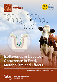
Soybeans are a common ingredient of animal feed. They contain isoflavones, which are known to act as phytoestrogens in animals. Isoflavones were described to have beneficial effects on farm animals, but there are also reports of negative outcomes after the consumption of these phytoestrogens. This review summarizes the current knowledge of the metabolization of isoflavones (including the influence of the microbiome, phase I and phase II metabolism), as well as the distribution of isoflavones and their metabolites in tissues. Published studies on effects of isoflavones in livestock species are reviewed. Furthermore, occurrence data of isoflavones in feed and their co-occurrence with the mycotoxin zearalenone are provided and are supplemented with our own survey data. View this paper.
  • Issues are regarded as officially published after their release is announced to the table of contents alert mailing list.
  • You may sign up for e-mail alerts to receive table of contents of newly released issues.
  • PDF is the official format for papers published in both, html and pdf forms. To view the papers in pdf format, click on the "PDF Full-text" link, and use the free Adobe Reader to open them.
Order results
Result details
Section
Select all
Export citation of selected articles as:
24 pages, 43533 KiB  
Article
rDromaserpin: A Novel Anti-Hemostatic Serpin, from the Salivary Glands of the Hard Tick Hyalomma dromedarii
by Hajer Aounallah, Melissa Regina Fessel, Mauricio Barbugiani Goldfeder, Eneas Carvalho, Chaima Bensaoud, Ana Marisa Chudzinski-Tavassi, Ali Bouattour, Youmna M’ghirbi and Fernanda Faria
Toxins 2021, 13(12), 913; https://doi.org/10.3390/toxins13120913 - 20 Dec 2021
Cited by 6 | Viewed by 3339
Abstract
Hemostatic disorders are caused either by platelet-related dysfunctions, defective blood coagulation, or by a combination of both, leading to an increased susceptibility to cardiovascular diseases (CVD) and other related illnesses. The unique specificity of anticoagulants from hematophagous arthropods, such as ticks, suggests that [...] Read more.
Hemostatic disorders are caused either by platelet-related dysfunctions, defective blood coagulation, or by a combination of both, leading to an increased susceptibility to cardiovascular diseases (CVD) and other related illnesses. The unique specificity of anticoagulants from hematophagous arthropods, such as ticks, suggests that tick saliva holds great promise for discovering new treatments for these life-threatening diseases. In this study, we combined in silico and in vitro analyses to characterize the first recombinant serpin, herein called Dromaserpin, from the sialotranscriptome of the Hyalomma dromedarii tick. Our in silico data described Dromaserpin as a secreted protein of ~43 kDa with high similarities to previously characterized inhibitory serpins. The recombinant protein (rDromaserpin) was obtained as a well-structured monomer, which was tested using global blood coagulation and platelet aggregation assays. With this approach, we confirmed rDromaserpin anticoagulant activity as it significantly delayed plasma clotting in activated partial thromboplastin time and thrombin time assays. The profiling of proteolytic activity shows its capacity to inhibit thrombin in the micromolar range (0.2 to 1 μM) and in the presence of heparin this inhibition was clearly increased. It was also able to inhibit Kallikrein, FXIa and slightly FXIIa, with no significant effect on other factors. In addition, the rDromaserpin inhibited thrombin-induced platelet aggregation. Taken together, our data suggest that rDromaserpin deserves to be further investigated as a potential candidate for developing therapeutic compounds targeting disorders related to blood clotting and/or platelet aggregation. Full article
Show Figures

Figure 1

10 pages, 2569 KiB  
Article
PKCζ-Mitogen-Activated Protein Kinase Signaling Mediates Crotalphine-Induced Antinociception
by Bárbara G. de Freitas, Natália G. Hösch, Leandro M. Pereira, Tereza C. Barbosa, Gisele Picolo, Yara Cury and Vanessa O. Zambelli
Toxins 2021, 13(12), 912; https://doi.org/10.3390/toxins13120912 - 20 Dec 2021
Cited by 4 | Viewed by 2360
Abstract
Crotalphine (CRP) is a structural analogue to a peptide that was first identified in the crude venom from the South American rattlesnake Crotalus durissus terrificus. This peptide induces a potent and long-lasting antinociceptive effect that is mediated by the activation of peripheral [...] Read more.
Crotalphine (CRP) is a structural analogue to a peptide that was first identified in the crude venom from the South American rattlesnake Crotalus durissus terrificus. This peptide induces a potent and long-lasting antinociceptive effect that is mediated by the activation of peripheral opioid receptors. The opioid receptor activation regulates a variety of intracellular signaling, including the mitogen-activated protein kinase (MAPK) pathway. Using primary cultures of sensory neurons, it was demonstrated that crotalphine increases the level of activated ERK1/2 and JNK-MAPKs and this increase is dependent on the activation of protein kinase Cζ (PKCζ). However, whether PKCζ-MAPK signaling is critical for crotalphine-induced antinociception is unknown. Here, we biochemically demonstrated that the systemic crotalphine activates ERK1/2 and JNK and decreases the phosphorylation of p38 in the lumbar spinal cord. The in vivo pharmacological inhibition of spinal ERK1/2 and JNK, but not of p38, blocks the antinociceptive effect of crotalphine. Of interest, the administration of a PKCζ pseudosubstrate (PKCζ inhibitor) prevents crotalphine-induced ERK activation in the spinal cord, followed by the abolishment of crotalphine-induced analgesia. Together, our results demonstrate that the PKCζ-ERK signaling pathway is involved in crotalphine-induced analgesia. Our study opens a perspective for the PKCζ-MAPK axis as a target for pain control. Full article
Show Figures

Graphical abstract

9 pages, 249 KiB  
Article
Effectiveness and Safety of Intradetrusor OnabotulinumtoxinA Injection for Neurogenic Detrusor Overactivity and Overactive Bladder Patients in Taiwan—A Phase IV Prospective, Interventional, Multiple-Center Study (Restore Study)
by Chung-Cheng Wang, Eric Chieh-Lung Chou, Yao-Chi Chuang, Chih-Chieh Lin, Yu-Chao Hsu, Chun-Hou Liao and Hann-Chorng Kuo
Toxins 2021, 13(12), 911; https://doi.org/10.3390/toxins13120911 - 20 Dec 2021
Cited by 4 | Viewed by 2634
Abstract
We conducted a phase IV, pre/post multi-center study to evaluate the efficacy and safety of intradetrusor onabotulinumtoxinA injection in patients with neurogenic detrusor overactivity (NDO, n = 119) or overactive bladder (OAB, n = 215). Patients received either 200U (i.e., NDO) and 100U [...] Read more.
We conducted a phase IV, pre/post multi-center study to evaluate the efficacy and safety of intradetrusor onabotulinumtoxinA injection in patients with neurogenic detrusor overactivity (NDO, n = 119) or overactive bladder (OAB, n = 215). Patients received either 200U (i.e., NDO) and 100U (i.e., OAB) of onabotulinumtoxinA injection into the bladder, respectively. The primary endpoint for all patients was the change in the PPBC questionnaire score at week 4 and week 12 post-treatment compared with baseline. The secondary endpoints were the changes in subjective measures (i.e., questionnaires: NBSS for patients with NDO and OABSS for those with OAB) at week 4 and week 12 post-treatment compared with baseline. Adverse events included symptomatic UTI, de novo AUR, gross hematuria and PVR > 350mL were recorded. The results showed that compared with baseline, PPBC (3.4 versus 2.4 and 2.1, p < 0.001) and NBSS (35.4 versus 20.4 and 18.1, p < 0.001) were significantly improved at 4 weeks and 12 weeks in NDO patients. In addition, compared with baseline, PPBC (3.5 versus 2.3 and 2.0, p < 0.001) and OABSS (9.1 versus 6.2 and 5.7, p < 0.001) were significantly improved at 4 weeks and 12 weeks in OAB patients. Eight (6.7%) had symptomatic UTI and 5 (4.2%) had de novo AUR in NDO patients. Twenty (9.3%) had symptomatic UTI but no de novo AUR in OAB patients. In conclusion, we found that intradetrusor onabotulinumtoxinA injections were safe and improved subjective measures related to NDO or OAB in our cohort. Full article
8 pages, 1539 KiB  
Communication
Aurintricarboxylic Acid Decreases RNA Toxicity in a C. elegans Model of Repeat Expansions
by Maya Braun, Shachar Shoshani, Anna Mellul-Shtern and Yuval Tabach
Toxins 2021, 13(12), 910; https://doi.org/10.3390/toxins13120910 - 20 Dec 2021
Cited by 2 | Viewed by 2932
Abstract
Pathologic expansions of DNA nucleotide tandem repeats may generate toxic RNA that triggers disease phenotypes. RNA toxicity is the hallmark of multiple expansion repeat disorders, including myotonic dystrophy type 1 (DM1). To date, there are no available disease-modifying therapies for DM1. Our aim [...] Read more.
Pathologic expansions of DNA nucleotide tandem repeats may generate toxic RNA that triggers disease phenotypes. RNA toxicity is the hallmark of multiple expansion repeat disorders, including myotonic dystrophy type 1 (DM1). To date, there are no available disease-modifying therapies for DM1. Our aim was to use drug repositioning to ameliorate the phenotype of affected individuals in a nematode model of DM1. As the RNA interference pathway plays a key role in mediating RNA toxicity, we investigated the effect of aurintricarboxylic acid. We demonstrated that by perturbing the RNA interference machinery using aurintricarboxylic acid, we could annihilate the RNA toxicity and ameliorate the phenotype. As our approach targets a universal disease mechanism, it is potentially relevant for more expansion repeat disorders. Full article
(This article belongs to the Special Issue Mechanisms of and Solutions for Diseases Caused by Toxic RNA)
Show Figures

Figure 1

13 pages, 1910 KiB  
Article
Indoxyl Sulfate Contributes to mTORC1-Induced Renal Fibrosis via The OAT/NADPH Oxidase/ROS Pathway
by Takehiro Nakano, Hiroshi Watanabe, Tadashi Imafuku, Kai Tokumaru, Issei Fujita, Nanaka Arimura, Hitoshi Maeda, Motoko Tanaka, Kazutaka Matsushita, Masafumi Fukagawa and Toru Maruyama
Toxins 2021, 13(12), 909; https://doi.org/10.3390/toxins13120909 - 18 Dec 2021
Cited by 18 | Viewed by 3691
Abstract
Activation of mTORC1 (mechanistic target of rapamycin complex 1) in renal tissue has been reported in chronic kidney disease (CKD)-induced renal fibrosis. However, the molecular mechanisms responsible for activating mTORC1 in CKD pathology are not well understood. The purpose of this study was [...] Read more.
Activation of mTORC1 (mechanistic target of rapamycin complex 1) in renal tissue has been reported in chronic kidney disease (CKD)-induced renal fibrosis. However, the molecular mechanisms responsible for activating mTORC1 in CKD pathology are not well understood. The purpose of this study was to identify the uremic toxin involved in mTORC1-induced renal fibrosis. Among the seven protein-bound uremic toxins, only indoxyl sulfate (IS) caused significant activation of mTORC1 in human kidney 2 cells (HK-2 cells). This IS-induced mTORC1 activation was inhibited in the presence of an organic anion transporter inhibitor, a NADPH oxidase inhibitor, and an antioxidant. IS also induced epithelial–mesenchymal transition of tubular epithelial cells (HK-2 cells), differentiation of fibroblasts into myofibroblasts (NRK-49F cells), and inflammatory response of macrophages (THP-1 cells), which are associated with renal fibrosis, and these effects were inhibited in the presence of rapamycin (mTORC1 inhibitor). In in vivo experiments, IS overload was found to activate mTORC1 in the mouse kidney. The administration of AST-120 or rapamycin targeted to IS or mTORC1 ameliorated renal fibrosis in Adenine-induced CKD mice. The findings reported herein indicate that IS activates mTORC1, which then contributes to renal fibrosis. Therapeutic interventions targeting IS and mTORC1 could be effective against renal fibrosis in CKD. Full article
(This article belongs to the Special Issue Contribution of Uremic Toxins to Chronic Kidney Disease Progression)
Show Figures

Figure 1

13 pages, 2659 KiB  
Article
Effect of Lonomia obliqua Venom on Human Neutrophils
by João Alfredo Moraes, Genilson Rodrigues, Daniel Guimarães-Bastos, Vany Nascimento-Silva, Erik Svensjö, Mariana Renovato-Martins, Markus Berger, Jorge Guimarães and Christina Barja-Fidalgo
Toxins 2021, 13(12), 908; https://doi.org/10.3390/toxins13120908 - 18 Dec 2021
Cited by 1 | Viewed by 2079
Abstract
The significant incidence of deforestation in South America culminates in the contact of humans with typical forests species. Among these species, one may highlight Lonomia obliqua caterpillar, which, when touched by humans, can poison them through their bristles. Therefore, better acknowledging the mechanisms [...] Read more.
The significant incidence of deforestation in South America culminates in the contact of humans with typical forests species. Among these species, one may highlight Lonomia obliqua caterpillar, which, when touched by humans, can poison them through their bristles. Therefore, better acknowledging the mechanisms involved in envenomation caused by Lonomia obliqua caterpillar bristle extract (LOCBE) may contribute to further treatments. Recently, we demonstrated that LOCBE induces a pro-inflammatory profile in endothelial cells; thus, we decided to investigate the effects of LOCBE on human polymorphonuclear neutrophils (PMN), which are the first leukocytes that migrate to the inflammatory focus. Our results showed that treatment with LOCBE induced PMN chemotaxis together with alterations in actin cytoskeleton and focal adhesion kinase (FAK) activation, favoring migration. Concurrently, LOCBE induced PMN adhesion to matrix proteins, such as collagen IV, fibronectin, and fibrinogen. Moreover, we observed that LOCBE attenuated PMN apoptosis and increased reactive oxygen species (ROS) production together with nuclear factor kB (NF-κB) activation—a redox-sensitive transcription factor—as well as interleukin (IL)-1β and IL-8 release. We call attention to the ROS-dependent effect of LOCBE on increased cell migration once an antioxidant treatment reverted it. In summary, we report that LOCBE activates PMN, inducing pro-inflammatory responses modulated by ROS. Full article
(This article belongs to the Special Issue Toxinologic and Pharmacological Investigation of Venomous Arthropods)
Show Figures

Graphical abstract

11 pages, 2031 KiB  
Article
Development of Generic Immuno-Magnetic Bead-Based Enzyme-Linked Immunoassay for Ustiloxins in Rice Coupled with Enrichment
by Yi Huang, Xiaoqian Tang, Lu Zheng, Junbin Huang, Qi Zhang and Hao Liu
Toxins 2021, 13(12), 907; https://doi.org/10.3390/toxins13120907 - 17 Dec 2021
Cited by 7 | Viewed by 2388
Abstract
Ustiloxins are a group of mycotoxins produced by rice false smut pathogen. Previous studies have shown that the false smut balls contain six types of ustiloxins, and these toxins are toxic to living organisms. Thus, immunoassay for on-site monitoring of ustiloxins in rice [...] Read more.
Ustiloxins are a group of mycotoxins produced by rice false smut pathogen. Previous studies have shown that the false smut balls contain six types of ustiloxins, and these toxins are toxic to living organisms. Thus, immunoassay for on-site monitoring of ustiloxins in rice is urgently required. The current immunoassays are only for detecting single ustiloxin, and they cannot meet the demand for synchronous and rapid detection of the group toxins. Therefore, this study designed and synthesized a generic antigen with ustiloxin G as material based on the common structure of the mycotoxins. Ustiloxin G was conjugated to two carrier proteins including bovine serum albumin (BSA) and ovalbvmin (OVA) by carbon diimide method. The mice were immunized with ustiloxin-G-BSA to generate the antibody serum, which was further purified to obtain the generic antibody against ustiloxins. The conjugated ustiloxin G-OVA and generic antibodies were used for establishing the enzyme-linked immunosorbent assay (ELISA) for ustiloxin detection and optimizing experiment conditions. The characterization of the antibody showed that the semi-inhibitory concentrations (IC50) of ustiloxin A, B, and G were 0.53, 0.34, and 0.06 µg/mL, respectively, and that their corresponding cross-reactivities were 11.9%, 18.4%, and 100%, respectively. To increase ELISA detection efficiency, generic antibody was combined with magnetic beads to obtain sensitive and class-specific immune-magnetic beads. Based on these immuno-magnetic beads, a high-efficiency enzyme-linked immunoassay method was developed for ustiloxin detection, whose sensitivity to ustiloxin A, B, and G was improved to 0.15 µg/mL, 0.14 µg/mL, and 0.04 µg/mL, respectively. The method accuracy was evaluated by spiking ustiloxin G as standard, and the spiked samples were tested by the immune-magnetic bead-based ELISA. The result showed the ustiloxin G recoveries ranged from 101.9% to 116.4% and were accepted by a standard HPLC method, indicating that our developed method would be promising for on-site monitoring of ustiloxins in rice. Full article
(This article belongs to the Special Issue Research on Pathogenic Fungi and Mycotoxins in China)
Show Figures

Figure 1

18 pages, 3089 KiB  
Article
Bitis arietans Snake Venom and Kn-Ba, a Snake Venom Serine Protease, Induce the Production of Inflammatory Mediators in THP-1 Macrophages
by Ângela Alice Amadeu Megale, Fabio Carlos Magnoli, Felipe Raimondi Guidolin, Kemily Stephanie Godoi, Fernanda Calheta Vieira Portaro and Wilmar Dias-da-Silva
Toxins 2021, 13(12), 906; https://doi.org/10.3390/toxins13120906 - 16 Dec 2021
Cited by 5 | Viewed by 2462
Abstract
Bitis arietans is a snake of medical importance found throughout sub-Saharan Africa and in savannas and pastures of Morocco and western Arabia. The effects of its venom are characterized by local and systemic alterations, such as inflammation and cardiovascular and hemostatic disturbances, which [...] Read more.
Bitis arietans is a snake of medical importance found throughout sub-Saharan Africa and in savannas and pastures of Morocco and western Arabia. The effects of its venom are characterized by local and systemic alterations, such as inflammation and cardiovascular and hemostatic disturbances, which can lead to victims’ death or permanent disability. To better characterize the inflammatory process induced by this snake’s venom, the participation of eicosanoids and PAF (platelet- activating factor) in this response were demonstrated in a previous study. In addition, edema and early increased vascular permeability followed by an accumulation of polymorphonuclear (PMN) cells in the peritoneal cavity were accompanied by the production of the eicosanoids LTB4, LTC4, TXB2, and PGE2, and local and systemic production of IL-6 and MCP-1. In this context, the present study focused on the identification of inflammatory mediators produced by human macrophages derived from THP-1 cells in response to Bitis arietans venom (BaV), and Kn-Ba, a serine protease purified from this venom. Here, we show that Kn-Ba, and even the less intensive BaV, induced the production of the cytokine TNF and the chemokines RANTES and IL-8. Only Kn-Ba was able to induce the production of IL-6, MCP-1, and IP-10, whereas PGE2 was produced only in response to BaV. Finally, the release of IL-1β in culture supernatants suggests the activation of the inflammasomes by the venom of Bitis arietans and by Kn-Ba, which will be investigated in more detail in future studies. Full article
Show Figures

Figure 1

39 pages, 1840 KiB  
Review
Unknown Extracellular and Bioactive Metabolites of the Genus Alexandrium: A Review of Overlooked Toxins
by Marc Long, Bernd Krock, Justine Castrec and Urban Tillmann
Toxins 2021, 13(12), 905; https://doi.org/10.3390/toxins13120905 - 16 Dec 2021
Cited by 19 | Viewed by 4012
Abstract
Various species of Alexandrium can produce a number of bioactive compounds, e.g., paralytic shellfish toxins (PSTs), spirolides, gymnodimines, goniodomins, and also uncharacterised bioactive extracellular compounds (BECs). The latter metabolites are released into the environment and affect a large range of organisms (from protists [...] Read more.
Various species of Alexandrium can produce a number of bioactive compounds, e.g., paralytic shellfish toxins (PSTs), spirolides, gymnodimines, goniodomins, and also uncharacterised bioactive extracellular compounds (BECs). The latter metabolites are released into the environment and affect a large range of organisms (from protists to fishes and mammalian cell lines). These compounds mediate allelochemical interactions, have anti-grazing and anti-parasitic activities, and have a potentially strong structuring role for the dynamic of Alexandrium blooms. In many studies evaluating the effects of Alexandrium on marine organisms, only the classical toxins were reported and the involvement of BECs was not considered. A lack of information on the presence/absence of BECs in experimental strains is likely the cause of contrasting results in the literature that render impossible a distinction between PSTs and BECs effects. We review the knowledge on Alexandrium BEC, (i.e., producing species, target cells, physiological effects, detection methods and molecular candidates). Overall, we highlight the need to identify the nature of Alexandrium BECs and urge further research on the chemical interactions according to their ecological importance in the planktonic chemical warfare and due to their potential collateral damage to a wide range of organisms. Full article
(This article belongs to the Special Issue Bioactivity and Chemical Ecological Interactions of Marine Toxins)
Show Figures

Figure 1

16 pages, 5676 KiB  
Article
Pharmacological Investigation of CC-LAAO, an L-Amino Acid Oxidase from Cerastes cerastes Snake Venom
by Zaineb Abdelkafi-Koubaa, Ines ELBini-Dhouib, Soumaya Souid, Jed Jebali, Raoudha Doghri, Najet Srairi-Abid, Khadija Essafi-Benkhadir, Olivier Micheau and Naziha Marrakchi
Toxins 2021, 13(12), 904; https://doi.org/10.3390/toxins13120904 - 16 Dec 2021
Cited by 6 | Viewed by 2464
Abstract
Snake venom proteins, which are responsible for deadly snakebite envenomation, induce severe injuries including neurotoxicity, myotoxicity, cardiotoxicity, hemorrhage, and the disruption of blood homeostasis. Yet, many snake-venom proteins have been developed as potential drugs for treating human diseases due to their pharmacological effects. [...] Read more.
Snake venom proteins, which are responsible for deadly snakebite envenomation, induce severe injuries including neurotoxicity, myotoxicity, cardiotoxicity, hemorrhage, and the disruption of blood homeostasis. Yet, many snake-venom proteins have been developed as potential drugs for treating human diseases due to their pharmacological effects. In this study, we evaluated the use of, an L-amino acid oxidase isolated from Cerastes cerastes snake venom CC-LAAO, as a potential anti-glioblastoma drug, by investigating its in vivo and in vitro pharmacological effects. Our results showed that acute exposure to CC-LAAO at 1 and 2.5 µg/mL does not induce significant toxicity on vital organs, as indicated by the murine blood parameters including aspartate transaminase (AST), alanine transaminase (ALT), lactate dehydrogenase (LDH) activities, and creatinine levels. The histopathological examination demonstrated that only at high concentrations did CC-LAAO induce inflammation and necrosis in several organs of the test subjects. Interestingly, when tested on human glioblastoma U87 cells, CC-LAAO induced a dose-dependent apoptotic effect through the H2O2 generated during the enzymatic reaction. Taken altogether, our data indicated that low concentration of CC-LAAO may be safe and may have potential in the development of anti-glioblastoma agents. Full article
Show Figures

Figure 1

20 pages, 3726 KiB  
Article
Domoic Acid and Pseudo-nitzschia spp. Connected to Coastal Upwelling along Coastal Inhambane Province, Mozambique: A New Area of Concern
by Holly Kelchner, Katie E. Reeve-Arnold, Kathryn M. Schreiner, Sibel Bargu, Kim G. Roques and Reagan M. Errera
Toxins 2021, 13(12), 903; https://doi.org/10.3390/toxins13120903 - 15 Dec 2021
Cited by 3 | Viewed by 3013
Abstract
Harmful algal blooms (HABs) are increasing globally in frequency, persistence, and geographic extent, posing a threat to ecosystem and human health. To date, no occurrences of marine phycotoxins have been recorded in Mozambique, which may be due to absence of a monitoring program [...] Read more.
Harmful algal blooms (HABs) are increasing globally in frequency, persistence, and geographic extent, posing a threat to ecosystem and human health. To date, no occurrences of marine phycotoxins have been recorded in Mozambique, which may be due to absence of a monitoring program and general awareness of potential threats. This study is the first documentation of neurotoxin, domoic acid (DA), produced by the diatom Pseudo-nitzschia along the east coast of Africa. Coastal Inhambane Province is a biodiversity hotspot where year-round Rhincodon typus (whale shark) sightings are among the highest globally and support an emerging ecotourism industry. Links between primary productivity and biodiversity in this area have not previously been considered or reported. During a pilot study, from January 2017 to April 2018, DA was identified year-round, peaking during Austral winter. During an intense study between May and August 2018, our research focused on identifying environmental factors influencing coastal productivity and DA concentration. Phytoplankton assemblage was diatom-dominated, with high abundances of Pseudo-nitzschia spp. Data suggest the system was influenced by nutrient pulses resulting from coastal upwelling. Continued and comprehensive monitoring along southern Mozambique would provide critical information to assess ecosystem and human health threats from marine toxins under challenges posed by global change. Full article
Show Figures

Figure 1

13 pages, 4605 KiB  
Article
Zearalenone Exposure Triggered Cecal Physical Barrier Injury through the TGF-β1/Smads Signaling Pathway in Weaned Piglets
by Pengfei Zhang, Changwei Jing, Ming Liang, Shuzhen Jiang, Libo Huang, Ning Jiao, Yang Li and Weiren Yang
Toxins 2021, 13(12), 902; https://doi.org/10.3390/toxins13120902 - 15 Dec 2021
Cited by 12 | Viewed by 2447
Abstract
This study aims to investigate the effects of exposure to different dosages of zearalenone (ZEA) on cecal physical barrier functions and its mechanisms based on the TGF-β1/Smads signaling pathway in weaned piglets. Thirty-two weaned piglets were allotted to four groups and fed a [...] Read more.
This study aims to investigate the effects of exposure to different dosages of zearalenone (ZEA) on cecal physical barrier functions and its mechanisms based on the TGF-β1/Smads signaling pathway in weaned piglets. Thirty-two weaned piglets were allotted to four groups and fed a basal diet supplemented with ZEA at 0, 0.15, 1.5, and 3.0 mg/kg, respectively. The results showed that 1.5 and 3.0 mg/kg ZEA damaged cecum morphology and microvilli, and changed distribution and shape of M cells. Moreover, 1.5 and 3.0 mg/kg ZEA decreased numbers of goblet cells, the expressions of TFF3 and tight junction proteins, and inhibited the TGF-β1/Smads signaling pathway. Interestingly, the 0.15 mg/kg ZEA had no significant effect on cecal physical barrier functions but decreased the expressions of Smad3, p-Smad3 and Smad7. Our study suggests that high-dose ZEA exposure impairs cecal physical barrier functions through inhibiting the TGF-β1/Smads signaling pathway, but low-dose ZEA had no significant effect on cecum morphology and integrity through inhibiting the expression of smad7. These findings provide a scientific basis for helping people explore how to reduce the toxicity of ZEA in feeds. Full article
Show Figures

Figure 1

24 pages, 2479 KiB  
Review
The Cytotoxic Necrotizing Factors (CNFs)—A Family of Rho GTPase-Activating Bacterial Exotoxins
by Paweena Chaoprasid and Petra Dersch
Toxins 2021, 13(12), 901; https://doi.org/10.3390/toxins13120901 - 15 Dec 2021
Cited by 6 | Viewed by 3070
Abstract
The cytotoxic necrotizing factors (CNFs) are a family of Rho GTPase-activating single-chain exotoxins that are produced by several Gram-negative pathogenic bacteria. Due to the pleiotropic activities of the targeted Rho GTPases, the CNFs trigger multiple signaling pathways and host cell processes with diverse [...] Read more.
The cytotoxic necrotizing factors (CNFs) are a family of Rho GTPase-activating single-chain exotoxins that are produced by several Gram-negative pathogenic bacteria. Due to the pleiotropic activities of the targeted Rho GTPases, the CNFs trigger multiple signaling pathways and host cell processes with diverse functional consequences. They influence cytokinesis, tissue integrity, cell barriers, and cell death, as well as the induction of inflammatory and immune cell responses. This has an enormous influence on host–pathogen interactions and the severity of the infection. The present review provides a comprehensive insight into our current knowledge of the modular structure, cell entry mechanisms, and the mode of action of this class of toxins, and describes their influence on the cell, tissue/organ, and systems levels. In addition to their toxic functions, possibilities for their use as drug delivery tool and for therapeutic applications against important illnesses, including nervous system diseases and cancer, have also been identified and are discussed. Full article
Show Figures

Figure 1

16 pages, 2928 KiB  
Article
Latitudinal Variation in the Toxicity and Sexual Compatibility of Alexandrium catenella Strains from Southern Chile
by Camilo Rodríguez-Villegas, Patricio A. Díaz, Pilar Riobó, Araceli E. Rossignoli, Francisco Rodríguez, Patricia Loures, Ángela M. Baldrich, Daniel Varela, Alondra Sandoval-Sanhueza and Rosa I. Figueroa
Toxins 2021, 13(12), 900; https://doi.org/10.3390/toxins13120900 - 15 Dec 2021
Cited by 2 | Viewed by 2502
Abstract
The bloom-forming toxic dinoflagellate Alexandrium catenella was first detected in southern Chile (39.5–55° S) 50 years ago and is responsible for most of the area’s cases of paralytic shellfish poisoning (PSP). Given the complex life history of A. catenella, which includes benthic [...] Read more.
The bloom-forming toxic dinoflagellate Alexandrium catenella was first detected in southern Chile (39.5–55° S) 50 years ago and is responsible for most of the area’s cases of paralytic shellfish poisoning (PSP). Given the complex life history of A. catenella, which includes benthic sexual cysts, in this study, we examined the potential link between latitude, toxicity, and sexual compatibility. Nine clones isolated from Chilean Patagonia were used in self- and out-crosses in all possible combinations (n = 45). The effect of latitude on toxicity, reproductive success indexes, and cyst production was also determined. Using the toxin profiles for all strains, consisting of C1, C2, GTX4, GTX1, GTX3, and NeoSTX, a latitudinal gradient was determined for their proportions (%) and content per cell (pg cell−1), with the more toxic strains occurring in the north (−40.6° S). Reproductive success also showed a latitudinal tendency and was lower in the north. None of the self-crosses yielded resting cysts. Rather, the production of resting cysts was highest in pairings of clones separated by distances of 1000–1650 km. Our results contribute to a better understanding of PSP outbreaks in the region and demonstrate the importance of resting cysts in fueling new toxic events. They also provide additional evidence that the introduction of strains from neighboring regions is a cause for concern. Full article
Show Figures

Figure 1

11 pages, 238 KiB  
Review
Botulinum Toxin Therapy in Writer’s Cramp and Musician’s Dystonia
by Elina Zakin and David M. Simpson
Toxins 2021, 13(12), 899; https://doi.org/10.3390/toxins13120899 - 14 Dec 2021
Cited by 8 | Viewed by 3046
Abstract
Task-specific focal dystonia is characterized by muscle contraction(s) during a specific task, resulting in abnormal postures or movements. Specifically, writer’s cramp involves the upper extremity during the act of writing. Musician’s dystonia has a highly variable presentation, and thus makes therapeutic options more [...] Read more.
Task-specific focal dystonia is characterized by muscle contraction(s) during a specific task, resulting in abnormal postures or movements. Specifically, writer’s cramp involves the upper extremity during the act of writing. Musician’s dystonia has a highly variable presentation, and thus makes therapeutic options more limited. Treatments include oral pharmacologic agents, neuromodulation, surgery and, most often, botulinum toxin (BoNT) injection. Selection of target muscles for toxin injection continues to be an area of active research for these task-specific movements. We present a review of the literature selected from a predefined search of the MEDLINE and ClinicalTrials.gov databases. We include six controlled studies of botulinum toxin for the management of writer’s cramp and focal task-specific dystonia (FTSD), including musician’s dystonia. Overall, 139 patients were included across all studies, with 99 individuals injected for writer’s cramp and the remaining 40 individuals with FTSD. The age range of all patients was 18–80 years old. We included studies that utilized only the BoNT-A serotype. These studies utilized various severity scales to quantify response to toxin injection, with ratings of instrument or pen control included as subjective ratings. Of the included 139 patients in this review, pooled data for toxin response show that 73% of patients who received the drug demonstrated improvement. Specific techniques for muscle localization and targeting were difficult to study as variable methods were employed. This remains an area of ongoing exploration. Full article
(This article belongs to the Special Issue Botulinum Toxin in the Movement Disorders Clinic: State of the Art)
12 pages, 2984 KiB  
Article
Trichosanthis Semen Suppresses Lipopolysaccharide-Induced Neuroinflammation by Regulating the NF-κB Signaling Pathway and HO-1 Expression in Microglia
by Seungmin Lee, In Gyoung Ju, Yujin Choi, Sangsu Park and Myung Sook Oh
Toxins 2021, 13(12), 898; https://doi.org/10.3390/toxins13120898 - 14 Dec 2021
Cited by 6 | Viewed by 2510
Abstract
Neuroinflammation, which is mediated by microglia that release various inflammatory cytokines, is a typical feature of neurodegenerative diseases (NDDs), such as Alzheimer’s disease and Parkinson’s disease. Hence, alleviating neuroinflammation by downregulating pro-inflammatory action, and upregulating anti-inflammatory action of microglia is an efficient therapeutic [...] Read more.
Neuroinflammation, which is mediated by microglia that release various inflammatory cytokines, is a typical feature of neurodegenerative diseases (NDDs), such as Alzheimer’s disease and Parkinson’s disease. Hence, alleviating neuroinflammation by downregulating pro-inflammatory action, and upregulating anti-inflammatory action of microglia is an efficient therapeutic target for NDDs. In this study, we evaluated whether trichosanthis semen (TS), a dried ripe seed of Trichosanthes kirilowii Maximowicz, reduces lipopolysaccharide (LPS)-induced neuroinflammation by regulating microglial responses in vitro and in vivo. Our results presented that TS reduced the release of pro-inflammatory mediators, such as nitric oxide (NO), inducible NO synthase, tumor necrosis factor-α, interleukin-1β, and interleukin-6 via inhibition of the nuclear factor kappa B (NF-κB) signaling pathway in LPS-treated BV2 microglial cells. Moreover, TS induced anti-inflammatory mediators, such as interleukin-10, found in inflammatory zone 1, and chitinase 3-like 3 by the upregulation of heme oxygenase 1 (HO-1). We further confirmed that TS administration suppressed microglial activation, but enhanced HO-1 expression in LPS-injected mice. These results suggest that TS has anti-neuroinflammatory effects via inhibition of NF-κB signaling through the activation of HO-1, and that TS may be a therapeutical candidate for NDDs treatment. Full article
(This article belongs to the Special Issue The Frontiers of Toxin in Pharmacology)
Show Figures

Figure 1

13 pages, 1777 KiB  
Review
Tackling the Threat of Cancer Due to Pathobionts Producing Colibactin: Is Mesalamine the Magic Bullet?
by Min Tang-Fichaux, Priscilla Branchu, Jean-Philippe Nougayrède and Eric Oswald
Toxins 2021, 13(12), 897; https://doi.org/10.3390/toxins13120897 - 14 Dec 2021
Cited by 6 | Viewed by 3992
Abstract
Colibactin is a genotoxin produced primarily by Escherichia coli harboring the genomic pks island (pks+ E. coli). Pks+ E. coli cause host cell DNA damage, leading to chromosomal instability and gene mutations. The signature of colibactin-induced mutations [...] Read more.
Colibactin is a genotoxin produced primarily by Escherichia coli harboring the genomic pks island (pks+ E. coli). Pks+ E. coli cause host cell DNA damage, leading to chromosomal instability and gene mutations. The signature of colibactin-induced mutations has been described and found in human colorectal cancer (CRC) genomes. An inflamed intestinal environment drives the expansion of pks+ E. coli and promotes tumorigenesis. Mesalamine (i.e., 5-aminosalycilic acid), an effective anti-inflammatory drug, is an inhibitor of the bacterial polyphosphate kinase (PPK). This drug not only inhibits the production of intestinal inflammatory mediators and the proliferation of CRC cells, but also limits the abundance of E. coli in the gut microbiota and diminishes the production of colibactin. Here, we describe the link between intestinal inflammation and colorectal cancer induced by pks+ E. coli. We discuss the potential mechanisms of the pleiotropic role of mesalamine in treating both inflammatory bowel diseases and reducing the risk of CRC due to pks+ E. coli. Full article
Show Figures

Figure 1

19 pages, 37821 KiB  
Article
Keeping Lagocephalus sceleratus off the Table: Sources of Variation in the Quantity of TTX, TTX Analogues, and Risk of Tetrodotoxication
by Georgios Christidis, Manolis Mandalakis, Thekla I. Anastasiou, George Tserpes, Panagiota Peristeraki and Stylianos Somarakis
Toxins 2021, 13(12), 896; https://doi.org/10.3390/toxins13120896 - 13 Dec 2021
Cited by 13 | Viewed by 2958
Abstract
The invasion of the tetrodotoxin (TTX)-bearing silver-cheeked toadfish and potential poisoning due to its consumption (tetrodotoxication) threatens public safety in the Mediterranean Sea. In this study, TTX and TTX analogues of Lagocephalus sceleratus (Gmelin, 1789) were measured using liquid chromatography tandem mass spectrometry [...] Read more.
The invasion of the tetrodotoxin (TTX)-bearing silver-cheeked toadfish and potential poisoning due to its consumption (tetrodotoxication) threatens public safety in the Mediterranean Sea. In this study, TTX and TTX analogues of Lagocephalus sceleratus (Gmelin, 1789) were measured using liquid chromatography tandem mass spectrometry (LC-MS/MS) in fish collected off the island of Crete (Southern Mediterranean). We tested the synergistic effect of a suite of factors potentially affecting toxins’ levels and tetrodotoxication risk using general and generalized linear models, respectively. The type of tissue, geographic origin (Cretan Sea, Libyan Sea), sex, and fish maturity stage were significant predictors of toxin concentrations. Mean TTX was higher in gonads and lower in muscles, higher in the Libyan Sea and in female fish, and lower in juvenile (virgin) fish. The concentration of TTX was also significantly and positively correlated with the concentration of several TTX analogues (4-epiTTX, 4,9-anhydroTTX, 11-deoxyTTX, 5,11/6,11-dideoxyTTX, 5,6,11-trideoxyTTX, 11-norTTX-6-ol). The analysis showed that fish originating from the Libyan Sea had significantly higher probability to cause tetrodotoxication in case of consumption. The variability explained by the models developed in this study was relatively low, indicating that toxin levels are hard to predict and the consumption of L. sceleratus should therefore be avoided. Full article
(This article belongs to the Special Issue Monitoring of Marine Biotoxins)
Show Figures

Figure 1

13 pages, 7283 KiB  
Article
Ecophysiology of Fusarium chaquense a Novel Type A Trichothecene Producer Species Isolated from Natural Grasses
by Maria J. Nichea, Eugenia Cendoya, Miriam Haidukowski, Adriana M. Torres and María L. Ramirez
Toxins 2021, 13(12), 895; https://doi.org/10.3390/toxins13120895 - 13 Dec 2021
Viewed by 2089
Abstract
Fusarium chaquense, a recently formally described novel species, has been identified as an T-2 toxin (T-2), HT-2 toxin (HT-2) and other toxins producer in natural grasses (Poaceae) from Argentina. The major objective of this study was to describe the effect of water [...] Read more.
Fusarium chaquense, a recently formally described novel species, has been identified as an T-2 toxin (T-2), HT-2 toxin (HT-2) and other toxins producer in natural grasses (Poaceae) from Argentina. The major objective of this study was to describe the effect of water activity (aW, 0.995, 0.98, 0.95, 0.93 and 0.91), temperature (15, 25 and 30 °C) and incubation time (5, 15 and 25 days) on growth and to evaluate the production of T-2, HT-2 toxins and beauvericin (BEA) by two F. chaquense strains in a grass-based media. The results showed a wide range of conditions for F. chaquense growth and mycotoxin production. Both strains had a maximum growth rate at the highest aW (0.995) and 25 °C. Regarding mycotoxin production, more T-2 than the other analysed mycotoxins were produced by the two strains. T-2 production was favoured at 0.995 aW and 30 °C, while HT-2 production at 0.98–0.95 aW and 15 °C. The maximum levels of BEA were produced at 0.995 aW and 25–30 °C. Two-dimensional profiles of aW by temperature interactions were obtained from these data in order to identify areas where conditions indicate a significant risk of mycotoxins accumulation on grass. For its versatility on growth and mycotoxin production in a wide range of aW and temperatures, F. chaquense would have an adaptive advantage over other Fusarium species, and this would explain its high frequency of isolation in natural grasses grown up in the Chaco wetlands. Full article
(This article belongs to the Special Issue Fusarium and Fusarium Toxins)
Show Figures

Figure 1

13 pages, 12768 KiB  
Article
Temporal Patterns of Bacterial and Viral Communities during Algae Blooms of a Reservoir in Macau
by Dini Hu, John P. Giesy, Min Guo, Wai Kin Ung, Yijun Kong, Kai Meng Mok and Simon Ming-Yuen Lee
Toxins 2021, 13(12), 894; https://doi.org/10.3390/toxins13120894 - 13 Dec 2021
Cited by 2 | Viewed by 2802
Abstract
Compositions of microbial communities associated with blooms of algae in a storage reservoir in Macau, China were investigated between 2013 and 2016. Algae were enumerated by visible light microscopy. Profiles of organisms in water were examined by 16S rRNA sequences and viral metagenomics, [...] Read more.
Compositions of microbial communities associated with blooms of algae in a storage reservoir in Macau, China were investigated between 2013 and 2016. Algae were enumerated by visible light microscopy. Profiles of organisms in water were examined by 16S rRNA sequences and viral metagenomics, based on next generation sequencing. Results of 16S rRNA sequencing indicated that majority of the identified organisms were bacteria closely related to Proteobacteria, Cyanobacteria, Verrucomicrobia, Bacteroidetes, and Actinobacteria. Metagenomics sequences demonstrated that the dominant virus was Phycodnavirus, accounting for 70% of the total population. Patterns of relative numbers of bacteria in the microbial community and their temporal changes were determined through alpha diversity indices, principal coordinates analysis (PCoA), relative abundance, and visualized by Venn diagrams. Ways in which the bacterial and viral communities are influenced by various water-related variables were elucidated based on redundancy analysis (RDA). Relationships of the relative numbers of bacteria with trophic status in a reservoir used for drinking water in Macau, provided insight into associations of Phycodnavirus and Proteobacteria with changes in blooms of algae. Full article
(This article belongs to the Special Issue Ecology and Toxicology of Cyanobacteria and Cyanotoxins)
Show Figures

Figure 1

14 pages, 5418 KiB  
Article
Platelet Endothelial Cell Adhesion Molecule 1 (CD31) Is Essential for Clostridium perfringens Beta-Toxin Mediated Cytotoxicity in Human Endothelial and Monocytic Cells
by Basma Tarek, Julia Bruggisser, Filippo Cattalani and Horst Posthaus
Toxins 2021, 13(12), 893; https://doi.org/10.3390/toxins13120893 - 13 Dec 2021
Cited by 4 | Viewed by 4642
Abstract
Beta toxin (CPB) is a small hemolysin beta pore-forming toxin (β-PFT) produced by Clostridium perfringens type C. It plays a central role in the pathogenesis of necro-hemorrhagic enteritis in young animals and humans via targeting intestinal endothelial cells. We recently identified the membrane [...] Read more.
Beta toxin (CPB) is a small hemolysin beta pore-forming toxin (β-PFT) produced by Clostridium perfringens type C. It plays a central role in the pathogenesis of necro-hemorrhagic enteritis in young animals and humans via targeting intestinal endothelial cells. We recently identified the membrane protein CD31 (PECAM-1) as the receptor for CPB on mouse endothelial cells. We now assess the role of CD31 in CPB cytotoxicity against human endothelial and monocytic cells using a CRISPR/Cas9 gene knockout and an antibody blocking approach. CD31 knockout human endothelial and monocytic cells were resistant to CPB and CPB oligomers only formed in CD31-expressing cells. CD31 knockout endothelial and monocytic cells could be selectively enriched out of a polyclonal cell population by exposing them to CPB. Moreover, antibody mediated blocking of the extracellular Ig6 domain of CD31 abolished CPB cytotoxicity and oligomer formation in endothelial and monocytic cells. In conclusion, this study confirms the role of CD31 as a receptor of CPB on human endothelial and monocytic cells. Specific interaction with the CD31 molecule can thus explain the cell type specificity of CPB observed in vitro and corresponds to in vivo observations in naturally diseased animals. Full article
Show Figures

Graphical abstract

13 pages, 1406 KiB  
Article
Evidence of the Involvement of a Cyclase Gene in the Biosynthesis of Ochratoxin A in Aspergillus carbonarius
by Massimo Ferrara, Antonia Gallo, Carla Cervini, Lucia Gambacorta, Michele Solfrizzo, Scott E. Baker and Giancarlo Perrone
Toxins 2021, 13(12), 892; https://doi.org/10.3390/toxins13120892 - 13 Dec 2021
Cited by 13 | Viewed by 3323
Abstract
Ochratoxin A (OTA) is a well-known mycotoxin with wide distribution in food and feed. Fungal genome sequencing has great utility for identifying secondary metabolites gene clusters for known and novel compounds. A comparative analysis of the OTA-biosynthetic cluster in A. steynii, A. westerdijkiae, [...] Read more.
Ochratoxin A (OTA) is a well-known mycotoxin with wide distribution in food and feed. Fungal genome sequencing has great utility for identifying secondary metabolites gene clusters for known and novel compounds. A comparative analysis of the OTA-biosynthetic cluster in A. steynii, A. westerdijkiae, A. niger, A. carbonarius, and P. nordicum has revealed a high synteny in OTA cluster organization in five structural genes (otaA, otaB, ota, otaR1, and otaD). Moreover, a recent detailed comparative genome analysis of Aspergilli OTA producers led to the identification of a cyclase gene, otaY, located in the OTA cluster between the otaA and otaB genes, encoding for a predicted protein with high similarity to SnoaLs domain. These proteins have been shown to catalyze ring closure steps in the biosynthesis of polyketide antibiotics produced in Streptomyces. In the present study, we demonstrated an upregulation of the cyclase gene in A. carbonarius under OTA permissive conditions, consistent with the expression trends of the other OTA cluster genes and their role in OTA biosynthesis by complete gene deletion. Our results pointed out the involvement of a cyclase gene in OTA biosynthetic pathway for the first time. They represent a step forward in the understanding of the molecular basis of OTA biosynthesis in A. carbonarius. Full article
(This article belongs to the Special Issue Evolution and Diversity of Mycotoxin Gene Clusters)
Show Figures

Figure 1

15 pages, 3764 KiB  
Article
The Antagonistic Effect of Glutamine on Zearalenone-Induced Apoptosis via PI3K/Akt Signaling Pathway in IPEC-J2 Cells
by Tianhu Wang, Jingjing Wang, Tong Zhang, Aixin Gu, Jianping Li and Anshan Shan
Toxins 2021, 13(12), 891; https://doi.org/10.3390/toxins13120891 - 12 Dec 2021
Cited by 14 | Viewed by 2827
Abstract
Zearalenone (ZEN) is a non-steroidal estrogen mycotoxin produced by Fusarium fungi, which inevitably exists in human and animal food or feed. Previous studies indicated that apoptosis seems to be a key determinant of ZEN-induced toxicity. This experiment aimed to investigate the protective effects [...] Read more.
Zearalenone (ZEN) is a non-steroidal estrogen mycotoxin produced by Fusarium fungi, which inevitably exists in human and animal food or feed. Previous studies indicated that apoptosis seems to be a key determinant of ZEN-induced toxicity. This experiment aimed to investigate the protective effects of Glutamine (Gln) on ZEN-induced cytotoxicity in IPEC-J2 cells. The experimental results showed that Gln was able to alleviate the decline of cell viability and reduce the production of reactive oxygen species and calcium (Ca2+) induced by ZEN. Meanwhile, the mRNA expression of antioxidant enzymes such as glutathione reductase, glutathione peroxidase, and catalase was up-regulated after Gln addition. Subsequently, Gln supplementation resulted in the nuclear fission and Bad-fluorescence distribution of apoptotic cells were weakened, and the mRNA expression and protein expression of pro-apoptotic genes and apoptotic rates were significantly reduced. Moreover, ZEN reduced the phosphorylation Akt, decreased the expression of Bcl-2, and increased the expression of Bax. Gln alleviated the above changes induced by ZEN and the antagonistic effects of Gln were disturbed by PI3K inhibitor (LY294002). To conclude, this study revealed that Gln exhibited significant protective effects on ZEN-induced apoptosis, and this effect may be attributed to the PI3K/Akt signaling pathway. Full article
(This article belongs to the Special Issue Novel Strategies for Biodegradation and Detoxification of Mycotoxins)
Show Figures

Graphical abstract

10 pages, 276 KiB  
Article
Prevalence of Mycotoxins and Endotoxins in Total Mixed Rations and Different Types of Ensiled Forages for Dairy Cows in Lithuania
by Gintarė Vaičiulienė, Bronius Bakutis, Jurgita Jovaišienė, Rimvydas Falkauskas, Gediminas Gerulis, Sigita Kerzienė and Violeta Baliukonienė
Toxins 2021, 13(12), 890; https://doi.org/10.3390/toxins13120890 - 12 Dec 2021
Cited by 8 | Viewed by 2648
Abstract
In this study, 119 samples of total mixed rations and different types of ensiled forage (maize and grass silage, and haylage) collected in 2019–2020 from dairy farms in Lithuania were analyzed to evaluate the quantitative occurrence of mycotoxins and endotoxins. Samples were analyzed [...] Read more.
In this study, 119 samples of total mixed rations and different types of ensiled forage (maize and grass silage, and haylage) collected in 2019–2020 from dairy farms in Lithuania were analyzed to evaluate the quantitative occurrence of mycotoxins and endotoxins. Samples were analyzed using high-performance liquid chromatography (HPLC) with a fluorescent (FLD) and an ultraviolet detector (UV) of mycotoxins and a detection assay based on the ELISA technology for endotoxins. The study included toxins regulated within the European Union (aflatoxin B1 (AFB1), zearalenone (ZEA), deoxynivalenol (DON) and T-2 toxin) and nonregulated toxins (endotoxins). Mycotoxin analysis showed that 49.58% of the samples out of 119 were positive for AFB1, 52.11% for ZEA and DON, 55.47% for T-2 toxin and 84.04% for endotoxins. In the contaminated samples, the highest mean values of AFB1 and T-2 toxin were determined in the grass silage samples, while ZEA and DON–were determined in the maize silage samples. Maize silage samples had the highest ZEA and DON concentrations, exceeding the EU maximum permissible concentration limits. In the haylage samples, AFB1 mycotoxin exceeded the maximum concentration limits. The highest mean value of endotoxins was determined in the total mixed rations samples. This is the first study to provide information about the concentrations of mycotoxins and endotoxins in total mixed rations and different types of ensiled forages for dairy cows in Lithuania. Full article
14 pages, 2078 KiB  
Article
Triceps Surae Muscle Characteristics in Spastic Hemiparetic Stroke Survivors Treated with Botulinum Toxin Type A: Clinical Implications from Ultrasonographic Evaluation
by Marco Battaglia, Lucia Cosenza, Lorenza Scotti, Michele Bertoni, Marco Polverelli, Alberto Loro, Andrea Santamato and Alessio Baricich
Toxins 2021, 13(12), 889; https://doi.org/10.3390/toxins13120889 - 12 Dec 2021
Cited by 2 | Viewed by 2777
Abstract
Equinovarus foot is one of the most commonly spasticity related conditions in stroke survivors, leading to an impaired gait and poor functional performances. Notably, spastic muscles undergo a dynamic evolution following typical pathophysiological patterns. Botulinum Neurotoxin Type A (BoNT-A) is the gold standard [...] Read more.
Equinovarus foot is one of the most commonly spasticity related conditions in stroke survivors, leading to an impaired gait and poor functional performances. Notably, spastic muscles undergo a dynamic evolution following typical pathophysiological patterns. Botulinum Neurotoxin Type A (BoNT-A) is the gold standard for focal spasticity treatment, and ultrasound (US) imaging is widely recommended to guide injections and monitor muscle evolution. The role of BoNT-A in influencing muscle fibroadipose degeneration is still unclear. In this study, we analyzed medial gastrocnemius (MG) and soleus (SOL) US characteristics (cross-sectional area, muscle thickness, pennation angle, and mean echo intensity) in 53 patients. MG and SOL alterations, compared to the unaffected side, depend on the spasticity only and not on the BoNT-A treatment. In functionally preserved patients (functional ambulation classification, FAC > 3; modified Ashworth scale, MAS ≤ 2), the ultrasonographic changes of MG compared to ipsilateral SOL observed in the paretic limb alone seems to be due to histological, anatomical, pathophysiological, and biomechanical differences between the two muscles. In subjects with poor walking capability and more severe spasticity, such ipsilateral difference was found in both calves. In conclusion, BoNT-A does not seem to influence muscle degeneration. Similar muscles undergo different evolution depending on the grade of walking deficit and spasticity. Full article
Show Figures

Figure 1

17 pages, 3483 KiB  
Article
Solution Structures of Bacillus anthracis Protective Antigen Proteins Using Small Angle Neutron Scattering and Protective Antigen 63 Ion Channel Formation Kinetics
by Ariel Michelman-Ribeiro, Kenneth A. Rubinson, Vitalii Silin and John J. Kasianowicz
Toxins 2021, 13(12), 888; https://doi.org/10.3390/toxins13120888 - 11 Dec 2021
Viewed by 2665
Abstract
We are studying the structures of bacterial toxins that form ion channels and enable macromolecule transport across membranes. For example, the crystal structure of the Staphylococcus aureus α-hemolysin (α-HL) channel in its functional state was confirmed using neutron reflectometry (NR) with the protein [...] Read more.
We are studying the structures of bacterial toxins that form ion channels and enable macromolecule transport across membranes. For example, the crystal structure of the Staphylococcus aureus α-hemolysin (α-HL) channel in its functional state was confirmed using neutron reflectometry (NR) with the protein reconstituted in membranes tethered to a solid support. This method, which provides sub-nanometer structural information, could also test putative structures of the Bacillus anthracis protective antigen 63 (PA63) channel, locate where B. anthracis lethal factor and edema factor toxins (LF and EF, respectively) bind to it, and determine how certain small molecules can inhibit the interaction of LF and EF with the channel. We report here the solution structures of channel-forming PA63 and its precursor PA83 (which does not form channels) obtained with small angle neutron scattering. At near neutral pH, PA83 is a monomer and PA63 a heptamer. The latter is compared to two cryo-electron microscopy structures. We also show that although the α-HL and PA63 channels have similar structural features, unlike α-HL, PA63 channel formation in lipid bilayer membranes ceases within minutes of protein addition, which currently precludes the use of NR for elucidating the interactions between PA63, LF, EF, and potential therapeutic agents. Full article
Show Figures

Graphical abstract

16 pages, 2397 KiB  
Article
Pain Reduction in Adults with Limb Spasticity Following Treatment with IncobotulinumtoxinA: A Pooled Analysis
by Jörg Wissel, Alexandre Camões-Barbosa, Georg Comes, Michael Althaus, Astrid Scheschonka and David M. Simpson
Toxins 2021, 13(12), 887; https://doi.org/10.3390/toxins13120887 - 11 Dec 2021
Cited by 8 | Viewed by 3221
Abstract
Some studies have shown that incobotulinumtoxinA reduces spasticity-associated pain, but further evidence is needed. This exploratory analysis pooled pain-relief data from six Phase 2 or 3 studies of incobotulinumtoxinA (four placebo-controlled studies) for treating upper limb spasticity in adults. Spasticity-associated pain was assessed [...] Read more.
Some studies have shown that incobotulinumtoxinA reduces spasticity-associated pain, but further evidence is needed. This exploratory analysis pooled pain-relief data from six Phase 2 or 3 studies of incobotulinumtoxinA (four placebo-controlled studies) for treating upper limb spasticity in adults. Spasticity-associated pain was assessed at baseline and 4 weeks post incobotulinumtoxinA injection using the disability assessment scale (DAS) for pain. Only data for patients with pain at baseline were analysed. Overall, 544 (incobotulinumtoxinA, N = 415; placebo, N = 129) of 937 patients (58.1%) experienced pain at baseline. At Week 4, a significantly greater proportion of incobotulinumtoxinA- (52.1%) than placebo-treated patients (28.7%; Chi-square p < 0.0001) showed a response (≥1-point improvement in DAS pain score). In logistic regression analysis, incobotulinumtoxinA-treated patients were 2.6 times more likely to achieve this endpoint than placebo-treated patients. A significant difference between incobotulinumtoxinA and placebo was observed regardless of baseline pain severity. Additionally, 27.1% of incobotulinumtoxinA- versus 12.4% of placebo-treated patients reported complete pain relief at Week 4 (p = 0.0006). Pain relief increased with multiple injection cycles. To achieve patient-centred care, pain relief may be considered a treatment goal in adults with spasticity-associated pain regardless of pain severity. This study contributes to understanding the benefits of incobotulinumtoxinA in treating limb spasticity-associated pain. Full article
(This article belongs to the Special Issue Botulinum Toxin Treatment for Spasticity and Pain)
Show Figures

Figure 1

12 pages, 2006 KiB  
Article
Cytotoxicity of an Innovative Pressurised Cyclic Solid–Liquid (PCSL) Extract from Artemisia annua
by Rosanna Culurciello, Andrea Bosso, Giovanni Di Fabio, Armando Zarrelli, Angela Arciello, Francesca Carella, Leonardo Leonardi, Laura Pazzaglia, Gionata De Vico and Elio Pizzo
Toxins 2021, 13(12), 886; https://doi.org/10.3390/toxins13120886 - 11 Dec 2021
Cited by 2 | Viewed by 2325
Abstract
Therapeutic treatments with Artemisia annua have a long-established tradition in various diseases due to its antibacterial, antioxidant, antiviral, anti-malaria and anti-cancer effects. However, in relation to the latter, virtually all reports focused on toxic effects of A. annua extracts were obtained mostly through [...] Read more.
Therapeutic treatments with Artemisia annua have a long-established tradition in various diseases due to its antibacterial, antioxidant, antiviral, anti-malaria and anti-cancer effects. However, in relation to the latter, virtually all reports focused on toxic effects of A. annua extracts were obtained mostly through conventional maceration methods. In the present study, an innovative extraction procedure from A. annua, based on pressurised cyclic solid–liquid (PCSL) extraction, resulted in the production of a new phytocomplex with enhanced anti-cancer properties. This extraction procedure generated a pressure gradient due to compressions and following decompressions, allowing to directly perform the extraction without any maceration. The toxic effects of A. annua PCSL extract were tested on different cells, including three cancer cell lines. The results of this study clearly indicate that the exposure of human, murine and canine cancer cells to serial dilutions of PCSL extract resulted in higher toxicity and stronger propensity to induce apoptosis than that detected by subjecting the same cells to Artemisia extracts obtained through canonical extraction by maceration. Collected data suggest that PCSL extract of A. annua could be a promising and economic new therapeutic tool to treat human and animal tumours. Full article
Show Figures

Figure 1

20 pages, 2039 KiB  
Article
Comprehensive Analysis and Biological Characterization of Venom Components from Solitary Scoliid Wasp Campsomeriella annulata annulata
by Carlos Alberto-Silva, Fernanda Calheta Vieira Portaro, Roberto Tadashi Kodama, Halyne Queiroz Pantaleão, Hidetoshi Inagaki, Ken-ichi Nihei and Katsuhiro Konno
Toxins 2021, 13(12), 885; https://doi.org/10.3390/toxins13120885 - 10 Dec 2021
Cited by 5 | Viewed by 2396
Abstract
Venoms of solitary wasps are utilized for prey capture (insects and spiders), paralyzing them with a stinger injection to be offered as food for their larvae. Thus, the identification and characterization of the components of solitary wasp venoms can have biotechnological application. In [...] Read more.
Venoms of solitary wasps are utilized for prey capture (insects and spiders), paralyzing them with a stinger injection to be offered as food for their larvae. Thus, the identification and characterization of the components of solitary wasp venoms can have biotechnological application. In the present study, the venom components profile of a solitary scoliid wasp, Campsomeriella annulata annulata, was investigated through a comprehensive analysis using LC-MS and -MS/MS. Online mass fingerprinting revealed that the venom extract contains 138 components, and MS/MS analysis identified 44 complete sequences of the peptide components. The peptides are broadly divided into two classes: bradykinin-related peptides, and linear α-helical peptides. Among the components of the first class, the two main peptides, α-campsomerin (PRLRRLTGLSPLR) and β-campsomerin (PRLRRLTGLSPLRAP), had their biological activities evaluated. Both peptides had no effects on metallopeptidases [human neprilysin (NEP) and angiotensin-converting enzyme (ACE)] and acetylcholinesterase (AChE), and had no cytotoxic effects. Studies with PC12 neuronal cells showed that only α-campsomerin was able to enhance cell viability, while β-campsomerin had no effect. It is noteworthy that the only difference between the primary structures from these peptides is the presence of the AP extension at the C-terminus of β-campsomerin, compared to α-campsomerin. Among the linear α-helical peptides, annulatin (ISEALKSIIVG-NH2) was evaluated for its biological activities. Annulatin showed histamine releasing activity from mast cells and low hemolytic activity, but no antimicrobial activities against all microbes tested were observed. Thus, in addition to providing unprecedented information on the whole components, the three peptides selected for the study suggest that molecules present in solitary scoliid wasp venoms may have interesting biological activities. Full article
(This article belongs to the Special Issue Toxinologic and Pharmacological Investigation of Venomous Arthropods)
Show Figures

Figure 1

11 pages, 883 KiB  
Article
Patterns of Diversity of Fusarium Fungi Contaminating Soybean Grains
by Maciej Żelechowski, Tomasz Molcan, Katarzyna Bilska, Kamil Myszczyński, Jacek Olszewski, Krzysztof Karpiesiuk, Joanna Wyrębek and Tomasz Kulik
Toxins 2021, 13(12), 884; https://doi.org/10.3390/toxins13120884 - 10 Dec 2021
Cited by 5 | Viewed by 2657
Abstract
Soybean is an important, high protein source of food and feed. However, like other agricultural grains, soybean may pose a risk to human and animal health due to contamination of the grains with toxigenic Fusaria and associated mycotoxins. In this study, we investigated [...] Read more.
Soybean is an important, high protein source of food and feed. However, like other agricultural grains, soybean may pose a risk to human and animal health due to contamination of the grains with toxigenic Fusaria and associated mycotoxins. In this study, we investigated the diversity of Fusaria on a panel of 104 field isolates obtained from soybean grains during the growing seasons in 2017–2020. The results of species-specific PCR analyses showed that Fusarium avenaceum was the most common (n = 40) species associated with soybean grains in Poland, followed by F. equiseti (n = 22) and F. sporotrichioides (11 isolates). A set of isolates, which was not determined based on PCR analyses, was whole genome sequenced. Multiple sequence analyses using tef-1α, top1, rpb1, rpb2, tub2, pgk, cam and lsu genes showed that most of them belonged to Equiseti clade. Three cryptic species from this clade: F. clavum, F. flagelliforme and FIESC 31 (lacking Latin binomial) were found on soybean for the first time. This is the first report demonstrating the prevalence of Fusaria on soybean grains in Poland. Full article
(This article belongs to the Section Mycotoxins)
Show Figures

Figure 1

Previous Issue
Next Issue
Back to TopTop