Review

Browning of Adipocytes: A Potential Therapeutic Approach to Obesity

1
Endocrinology and Care of Diabetes Unit—Azienda Ospedaliero-Universitaria S. Orsola Malpighi, Alma Mater Studiorum University of Bologna, 40126 Bologna, Italy
2
IRCCS—Azienda Ospedaliero-Universitaria S. Orsola Malpighi, Alma Mater Studiorum University of Bologna, 40126 Bologna, Italy
3
PRISM Lab, Department of Biomedical and Neuromotor Sciences, Alma Mater Studiorum University of Bologna, 40126 Bologna, Italy
4
International Center for the Assessment of Nutritional Status and the Development of Dietary Intervention Strategies (ICANS-DIS), Department of Food, Environmental and Nutritional Sciences (DeFENS), University of Milan, 20133 Milan, Italy
*
Authors to whom correspondence should be addressed.
Nutrients 2023, 15(9), 2229; https://doi.org/10.3390/nu15092229
Submission received: 6 April 2023 / Revised: 1 May 2023 / Accepted: 4 May 2023 / Published: 8 May 2023
(This article belongs to the Special Issue Modulation by Dietary Supplements in Obesity)

Abstract

:
The increasing prevalence of overweight and obesity suggests that current strategies based on diet, exercise, and pharmacological knowledge are not sufficient to tackle this epidemic. Obesity results from a high caloric intake and energy storage, the latter by white adipose tissue (WAT), and when neither are counterbalanced by an equally high energy expenditure. As a matter of fact, current research is focused on developing new strategies to increase energy expenditure. Against this background, brown adipose tissue (BAT), whose importance has recently been re-evaluated via the use of modern positron emission techniques (PET), is receiving a great deal of attention from research institutions worldwide, as its main function is to dissipate energy in the form of heat via a process called thermogenesis. A substantial reduction in BAT occurs during normal growth in humans and hence it is not easily exploitable. In recent years, scientific research has made great strides and investigated strategies that focus on expanding BAT and activating the existing BAT. The present review summarizes current knowledge about the various molecules that can be used to promote white-to-brown adipose tissue conversion and energy expenditure in order to assess the potential role of thermogenic nutraceuticals. This includes tools that could represent, in the future, a valid weapon against the obesity epidemic.

1. Introduction

Obesity is becoming one of the leading causes of death in the world, representing an increasingly alarming health emergency [1]. Obesity is recognized as a chronic disease caused by genetic, environmental, psychological and social factors; it is also considered a condition that increases the probability of developing a wide range of non-transmissible comorbidities, such as cardiovascular and metabolic disorders including type 2 diabetes and numerous forms of cancer [2]. One of the greatest challenges of this millennium is to halt the rapid increase in obesity worldwide, which should be considered a form of malnutrition on par with undernutrition, if not more critical. In order to achieve this goal, dietary prescriptions and physical activity are not enough. It is necessary to reform food systems, from the agricultural sector to large-scale distribution, ensure the availability and accessibility of healthy foods, invest in the promotion of educational programs, provide consumers with tools for making informed choices and encourage the population to engage in more physical activity. These are only some of the projects that would ideally be implemented soon, but they require significant political and economic commitment [3]. The scientific community is focusing on searching for alternative therapeutic strategies that accompany the refinement of modern technologies. The identification of BAT with its thermogenic potential may represent a valid way to tackle obesity, both by increasing energy expenditure and by modulating numerous metabolic targets [4,5,6]. The aim of this review is to address this hot topic by exploring the most recent studies in the literature and to examine a wide range of new bioactive dietary compounds that can expand and activate BAT, and to simultaneously promote the browning process in humans.

2. Adipose Tissue: Typologies, Role, Physiology

According to their antagonistic functions, two types of adipose tissue can be distinguished: the white adipose tissue (WAT), which stores excess energy, and the brown adipose tissue (BAT), which is specialized in dissipating energy via the production of heat. WAT represents the main form of storage of excess energy derived from food. The excess energy is stored as triglycerides and when necessary, is released in the form of three fatty acids and a glycerol. This feature guarantees the survival of an organism even during long fasting periods. In addition to its primary function as an energy reserve, WAT also has an important mechanical and insulating function, and protects the organs against trauma and cold. No less important is the endocrine function of WAT, which is now defined as an endocrine organ, being a source of the production of hormones and biologically active substances, including adipokines. It serves as a central node of inter-organ metabolic communication and a regulator of reproduction and satiety [1].
Similar to WAT, BAT synthesizes and secretes “batokines” such as fibroblast growth factors (FGFs), including FGF21, neuregulin 4, vascular endothelial growth factor (VEGF), and cytokines, such as interleukin 6 (IL-6). Given the relatively small amount of BAT present in humans, the endocrine potential of batokines is relatively unknown, but it is clear that factors secreted from BAT exert paracrine and autocrine functions. While the relative BAT mass in humans and rodents is small compared to other adipose depots, its relative contribution to metabolic health may be higher [7].

3. Distribution of Adipose Tissue

WAT can be broadly classified by location, largely defined as either subcutaneous (located under the skin) or visceral/omental (located intra-abdominally, adjacent to internal organs). In most lean, healthy individuals, WAT is confined to defined depots, but in certain conditions, such as obesity and lipodystrophy, WAT mass can increase ectopically in areas that may influence an individual’s susceptibility to comorbidities such as diabetes and atherosclerosis [8]. Such ectopic WAT areas are mostly located within the visceral cavity, and include intrahepatic, epicardial (between the heart and the pericardium), perivascular (surrounding major blood vessels), mesenteric fat (contiguous with digestive organs in the viscera), omental fat (an apron of fat that stretches over the intestines, liver, and stomach), and retroperitoneal fat (surrounding the kidneys). Visceral fat is highly metabolically active and is constantly releasing free fatty acids (FFA) into the portal circulation. As such, visceral fat content contributes to various features of the metabolic syndrome, such as hyperinsulinemia, systemic inflammation, dyslipidaemia, and atherosclerosis [9].
BAT is localized to distinct anatomical regions that have been well-characterized in rodents, helping them to survive cold temperatures [10]. While originally believed to be a depot exclusive to hibernating and small mammals, and present to some degree in human infants, adult humans have recently been shown to have functional and inducible levels of BAT. Thanks to an innovative technique called fluorodeoxyglucose positron emission tomography (FDG PET), combined with X-ray computed tomography, it has also been possible to identify metabolically active areas of BAT in adult humans that have total fat stores of between 1% and 2%, and that are localized primarily in the cervical, supraclavicular, interscapular, axillary, paravertebral, mediastinal, and upper abdominal regions [11]. Differently localized vessels in the organism present different proportions between WAT and BAT. This variability may be in conjunction with the different predominant functions of perivascular tissue according to localization. Large central vessels, for example, such as the aorta and its main ramifications, are mostly surrounded by brown adipose tissue, thus playing a key part in maintaining the central temperature within normal ranges. Peripherally increased perivascular adipose tissue, on the other hand, has been associated with increased insulin resistance [8]. Considering this, BAT has aroused great interest in scientific research, with the hope of exploiting its unique characteristics in the treatment of obesity-associated comorbidities.

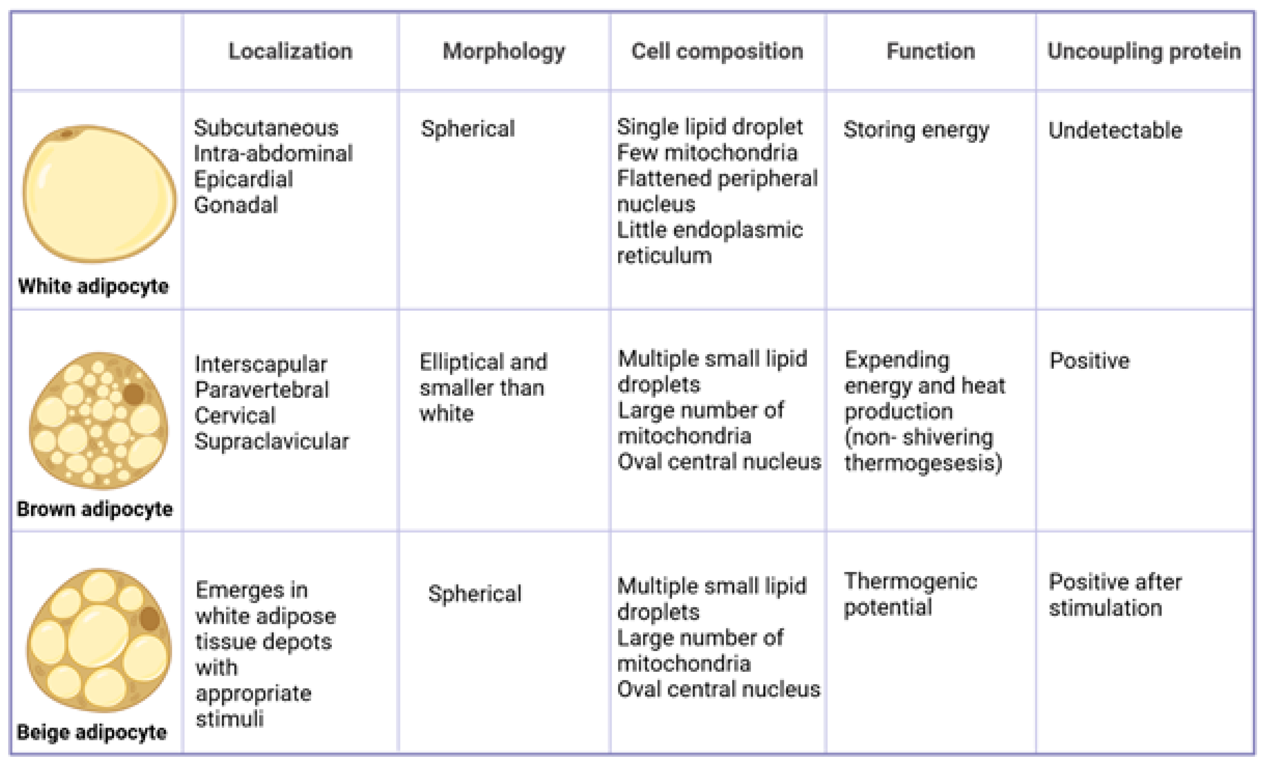
4. Differences between White and Brown Adipocytes

The biology, the origins of BAT and the differences between WAT and BAT have been deeply reviewed in recent years. The most important differences are as follows: white adipocytes are spherical and unilocular cells (50–100 microns in diameter) with more than 90% of their volume represented by a single lipid droplet and a modest number of mitochondria. The spherical shape provides the adipocyte with a good way of accumulating volume in the smallest space and exporting energy molecules without the excessive anatomical breakdown of the tissue. White adipocytes derive from mesenchymal stem cells (MSCs), the precursors of adipocytes, but also cartilage, bone, and muscle cells. MSCs are directed towards the adipocyte line, in case of high energy intake. WAT has a great capacity for expansion, including hypertrophy and hyperplasia, in order to store large amounts of energy [12].
Brown adipocyte is defined as a multilocular cell, as it is composed of multiple, small lipid droplets and is rich in mitochondria with dense ridges, which give the tissue its brown colour, together with dense vascularisation. Mitochondria store energy as a proton gradient across the inner mitochondrial membrane, and this energy is used to synthesize adenosine triphosphate (ATP); when protons move along the gradient without producing ATP, the stored energy is dissipated as heat. This is due to the presence, exclusively in the brown adipocyte, of a specific protein: “the uncoupling protein 1 (UCP1)” in the inner mitochondrial membrane, which immunohistochemically is the defining protein marker of BAT. In contrast with WAT, the origin of brown adipocytes is related to the origin of skeletal muscle: both derive from specific cells of the dermomyotome, a portion of the mesoderm, that express myogenic factor 5 (Myf5) [13]. Another distinguishing feature of BAT is the innervation by the sympathetic nervous system, which is extremely reduced in WAT [14,15].

5. Conversion of White Adipocytes into Brown-like Adipocytes

Adipose tissue is an extremely dynamic organ, capable of transforming in response to environmental or dietary stimuli. One of the possible ways to increase the presence of functional UCP1-rich cells in the adipose tissue is the conversion of white (pre)adipocytes into brown-like fat cells. This phenomenon is known as the “browning” of adipocytes or as the reversible and transdifferentiation of the adipose organ [16]. The browning process of WAT has become a key area of interest in research due to its role in fat burning; it is thus a potential useful strategy for treating obesity. The result is the appearance of dispersed masses of brown-like adipocytes in WAT, termed as beige or brite adipocytes. These cells possess similar characteristics to brown adipocytes, such as the multilocularised accumulation of lipids, a high mitochondrial content and an elevated UCP1 expression, along with factors that stimulate the transcriptional activity of thermogenic proteins. Nevertheless, brite adipocytes also display a distinct gene expression pattern that differs from both white and brown adipocyte profiles [15,16]. The morphology, location, origin and function of white, brown and beige adipocytes are reported in Figure 1.
At present, the mechanisms known to induce the browning of WAT in humans are cold exposure and β-adrenergic receptor stimulation. However, both these approaches are difficult to implement in human beings due to the non-specific nature of β-adrenergic agonists and the inability of humans to tolerate cold conditions for prolonged periods. Researchers in recent years have asked if it is possible to induce beige adipocyte differentiation without cold exposure and adrenergic stimulation. Recent studies would seem to offer answers: certain key genes, such as Proline Rich Domain Containing Protein 16 (PRDM16) [17] and Early B-cell Factor 2 (EBF2) [18], which appear to play a dominant role in programming brown adipocytes, have been identified [19]. The activation of these genes initiates a signal transduction cascade that culminates in the overexpression of UCP1 and other thermogenic proteins. Numerous studies have confirmed that, endogenously, the mechanisms responsible for classic BAT recruitment and WAT browning are identical, whereas exogenous molecules may selectively activate BAT thermogenesis or recruit brite adipocytes [20].

6. Thermogenesis and Thermogenin

In physiology, thermogenesis refers to a specific metabolic process that results in the production of heat by the body, particularly in adipose and muscle tissue. Heat is produced by the transformation of chemical energy via oxidative or catabolic processes. Thermogenesis is responsible for maintaining a constant internal temperature to cope with changing external climatic conditions and to ensure thermoneutrality: the ideal internal temperature for the body’s proper functioning [17]. Thermogenesis depends on both endogenous factors, genetic in nature, and exogenous factors, and therefore varies from individual to individual. An important exogenous factor affecting thermogenesis is caused by a cold environmental temperature, a metabolic process that leads to the production of heat by the body depending on how cold the environment is. Cold-induced thermogenesis (TIF) is divided into two types: ‘non-shivering thermogenesis’ and ‘shivering thermogenesis’. Non-shivering thermogenesis represents an increase in heat production by the body when exposed to cold, but is not associated with muscular contraction. It is caused by the increased activity of the sympathetic nervous system, which innervates BAT to a greater extent, and skeletal muscle. An alteration in BAT leads, in fact, to a high intolerance to cold in mammals. By contrast, ‘shivering thermogenesis’ requires the rhythmic isometric contraction of skeletal muscle, and it plays a less important role than ‘non-shivering’ thermogenesis; it takes over after it only [18,19]. BAT has an extraordinary ability to stimulate thermogenesis more than any other district of the body, thus increasing the total energy expenditure [20]. BAT heat production takes place in the mitochondria of brown adipocytes via the process of mitochondrial uncoupling, which involves the transmembrane protein UCP1 or thermogenin. It is responsible for ‘non-shivering thermogenesis’ and promotes the utilization of fatty acids that are not associated with ATP production but instead with heat production, reducing the efficiency of cellular respiration. The UCP1 is activated by fatty acids and is inhibited by nucleotides. Its thermogenic function is to dissipate the proton gradient generated by the respiratory chain and increase the permeability of the mitochondrial matrix, allowing proton dispersion [21]. This mechanism, used to produce heat, is a mammalian physiological response to low temperatures or excess nutrients from high-calorie diets. When sensory neurons are activated by cold, they transmit the stimulus to the brain, which in turn responds by increasing the activity of afferent nerves and releases noradrenaline. This neurotransmitter induces thermogenesis and lipid oxidation. UCP1-induced thermogenesis during exposure to cold is under the control of the hypothalamus, which integrates peripheral signals from skin thermoreceptors [22].

7. BAT-Secreted Factors with Potential Direct and/or Indirect Cardioprotective Effects

Current knowledge suggests the general beneficial effect of BAT activation regarding a reduction in the risk of cardiovascular disease (CVD) [23]. BAT secretes several molecules, which are collectively termed batokines. These batokines may alter the metabolism via autocrine, paracrine, and endocrine mechanisms, thus modifying BAT itself or acting remotely on other organs. Some BAT-secreted factors have potential direct and/or indirect cardioprotective effects via the modulation of the systemic metabolism. Summarizing, BAT generates heat via non-shivering thermogenesis, a process that helps to maintain body temperature and contributes to the overall energy expenditure. An increased energy expenditure may help to prevent excess weight gain and the development of obesity, which is a risk factor for CVD [24]. BAT consumes fatty acids as a primary fuel source for thermogenesis. By increasing the uptake and oxidation of fatty acids, BAT can help reduce circulating levels of triglycerides and lower the risk of atherosclerosis [25], a key factor in CVD. BAT also contributes to glucose homeostasis by taking up and metabolizing glucose. Improved glucose metabolism can help maintain insulin sensitivity and reduce the risk of developing type 2 diabetes, another CVD risk factor [26]. BAT secretes various hormones and cytokines, such as FGF21 and IL-6, which can have beneficial effects on metabolism, inflammation, and cardiovascular health. Some studies have suggested that BAT activation may help modulate blood pressure by promoting the release of nitric oxide, a vasodilator that helps relax blood vessels and lower blood pressure [27].

8. Browning Strategies: Cold, Physical Activity, and Adrenergic Agonists

Nowadays, it is known that UCP1 activation can be mediated by several factors: cold exposure, physical activity, fasting, nutraceutical foods, amino acids such as tyrosine (noradrenaline precursor), triiodothyronine (FT3), thyroxine (FT4), molecules that stimulate β-adrenergic receptors, and drugs that inhibit noradrenaline reuptake [6,28,29] (Table 1). The mechanism of action of the different browning strategies are summarized in Table 2.
Cold exposure (<20 °C) generates the production of noradrenergic signals that move from the periphery to the hypothalamus, via the orthosympathetic nerves that reach the BAT and release catecholamines such as noradrenaline [30]. Norepinephrine acts on the β-adrenergic receptors present on the cell membrane of brown adipocytes. These receptors activate adenylate cyclase, which catalyses the conversion of ATP to cyclic AMP (cAMP). cAMP in turn activates protein kinase A (PKA). Such a protein induces peroxisome proliferation, which activates the peroxisome proliferator-activated receptor gamma coactivator 1-alpha (PGC1α) receptor, which is the master regulator of the transcriptional cascade of UCP1 and other thermogenic genes [31]. To date, cold exposure has been proven to be the most effective strategy regarding BAT activation. Past studies on mouse models have shown that long-term cold exposure leads to an increase in adipocytes expressing UCP1, in both brown and beige adipose deposits, which can increase non-chill thermogenesis [19]. Studies on humans have shown that even a less prolonged exposure to the cold (17 °C for 2–6 h a day) is still capable of expanding BAT and increasing its functionality: the increase in BAT perfusion is directly associated with an increased total energy expenditure; and the increase in glucose uptake is due to the increased gene expression of the GLUT4 transporter in BAT. These effects of cold exposure on BAT are severely attenuated in individuals with excessive WAT deposition and the presence of insulin resistance [32,33]. Promising results are also emerging from the use of BAT activation strategies that exploit the modulation of adrenergic activity in the sympathetic nervous system. The stimulation of β-adrenergic receptors by adrenergic agonists [34], but also the combination of cold exposure with the infusion of sympathomimetics (isoprenaline and ephedrine) [35], can induce an increase in the levels of catecholamines released by the sympathetic nervous system and amplify thermogenesis. These results need further verification in order to clarify the modulation pathways [36]. Similar to cold exposure, exercise also promotes the upregulation of the activation markers of BAT (PGC1α and UCP1) and the stimulation of endocrine activators (cardiac natriuretic peptides, irisin and FGF21). Recent findings [37,38] show that exercise can have both an acute effect on BAT-releasing catecholamines via the activation of UCP1 in BAT and induce lipogenesis in WAT. The chronic effect of exercise on BAT concerns the discovery of specific myokines produced by skeletal muscle and released into the bloodstream during exercise. These myokines, identified in early 2014, are as follows: irisin, β-aminoisobutyric acid (BAIBA) and FGF21; these would appear to govern white adipocyte browning, independent of sympathetic nervous system stimulation [39,40].
Table 2. Mechanism of action of the different browning strategies: cold exposure, physical activity, adrenergic agonists and myokines (irisin, BAIBA and FGF21).
InterventionMechanism
of Action
Bat ActivationExperimental ModelRef.
COLD EXPOSURE yes
UCP1 activation human[28,29,31]
↑ production of noradrenergic signals human[30]
↑ PGC1α mouse
human
[31]
↑ UCP1 in BAT human[19]
↑ BAT and ↑ its functionality human[32,33]
ADRENERGIC
AGONISTS
yes
stimulate β-adrenergic receptors human[34]
↑ levels of catecholamines and amplify thermogenesis mouse[35]
PHYSICAL
ACTIVITY
yes
UCP1 activation human[37,38]
↑ PGC1α human[37,38]
releases catecholamines human[6,28]
activates lipogenesis in WAT human[6,28]
MYOKINES
Irisingoverns white adipocyte browning human
mouse
[29,30]
β-aminoisobutyric acid (BAIBA)governs white adipocyte browning human
mouse
[29,30]
Fibroblast growth factor 21 (FGF21)governs white adipocyte browning human
mouse
[29,30]
Abbreviations: UCP1: uncoupling protein 1, PGC1α: peroxisome proliferator-activated receptor gamma coactivator 1-alpha, BAT: brown adipose tissue, WAT: white adipose tissue, ↑: increase.

9. Thermogenic Nutraceuticals

Current evidence, both on animal and human models, indicates that several diet components may have beneficial effects on obesity by affecting BAT and energy metabolism, including polyunsaturated fatty acids, capsaicin and capsinoids, catechins, curcumin, resveratrol and berberine, oleuropein, anthocyanins, quercetin, gingerol, shogaol, 6-paradol, thiacremonone, cinnamaldehyde and menthol. The molecular mechanisms involved in WAT browning and the BAT activation mediated by thermogenic nutraceuticals are represented in Figure 2 and summarized in Table 3.

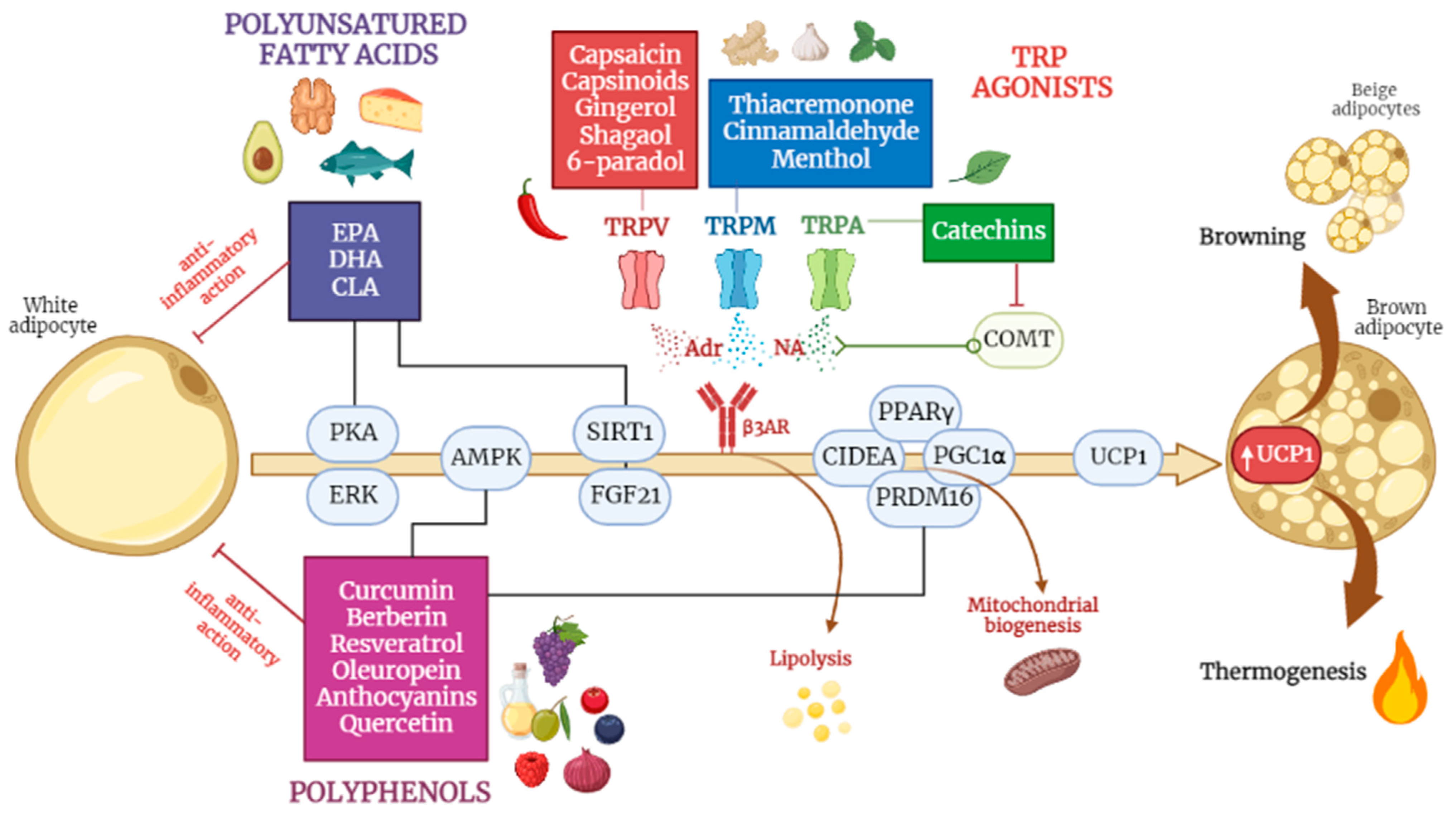
9.1. Polyunsatured Fatty Acids

Fish oil is rich in the polyunsaturated fatty acids (PUFA) eicosapentaenoic acid (EPA) and docosahexaenoic acid (DHA), which stimulate thermogenesis in BAT [41,42,43]. In the early 2000s, several studies in mouse models compared the effects of a diet enriched with n-3 PUFAs and a diet rich in n-6 PUFAs in order to study their impact on BAT. It turned out that fats from the n-3 series were much more effective in activating BAT than the n-6 series [44]. Therefore, subsequent research focused on the effects of n-3 PUFAs, especially EPA, and confirmed its anti-obesogenic effect, revealing that it can promote the browning of WAT. Rats fed with a supplementation of fish oil at a 20% EPA for 4 weeks showed an increased mRNA level of UCP1 in WAT [45]. Another study showed that supplementation with 27.5% fish oil, administered for one month, stimulates SNS-mediated mitochondrial and thermogenic activity in rats [46]. A diet enriched with a mixture of EPA and DHA increases the expression of UCP1 in BAT and reduces adipose accumulation via the induction of marked, non-shivering thermogenesis [47,48]. Further implications emerged from a study conducted in 2016, which revealed that a high-fat diet (HFD), enriched with fish oil (12% of total lipids) and administered to mice for 8 weeks, modulates BAT activation via epigenetic regulation [49]. EPA promotes both the adipogenesis of mature brown adipocytes [50] and the differentiation of pre-adipocytes into beige adipocytes, within WAT stores, particularly in the inguinal WAT [48]. It is important to note that the conversion of mature white adipocytes to beige adipocytes was achieved via supplementation with EPA in human adipocyte cultures [51] and that the same effect was not observed in rodents [43].

9.2. Capsaicin and Capsinoids

Capsaicin is an alkaloid found in chilli peppers that is mainly concentrated in the seeds of the fruit and is the main component responsible for the spicy flavour. Capsaicin has a role in preventing the obesogenic effects of diet, due to its ability to increase energy expenditure by activating BAT [52]. Capsaicin implements BAT function via several signalling pathways, primarily via the activation of Transient Receptor Potential Vanilloid 1 channels (TRPV1), which stimulate the central nervous system to produce catecholamines involved in thermogenesis. Capsaicin seems to regulate the epigenetic expression of the transcription factors involved in WAT browning [53,54]. Important evidence has also emerged from studies on humans: Yoneshiro et al. [55] examined the acute effect of an oral ingestion of a single dose of capsinoids (9 mg) on energy expenditure, in relation to BAT activity in humans; this was measured by 18F-FDG PET, a classical imaging technique used to visualize BAT activation. The ingestion of capsinoids resulted in a 3-fold increase in energy expenditure in the BAT-positive group compared to the BAT-negative group. Furthermore, the chronic intake of capsinoids (9 mg/day in capsule form) for 6 weeks promoted BAT activity and reduced fat mass after cold exposure, even in human subjects with low BAT activity. In 2018, Camps et al. [56] tested the same treatment used in mice in 2016 [57] on humans. They gave a group of normal-weight individuals a dose of capsinoids (12 mg), combined with exposure to cold (14.5 °C). The subjects underwent, before and after exposure, both 18F-FDG PET and indirect calorimetry, in order to assess the total energy expenditure. The results showed that capsinoids increased energy expenditure in BAT-positive participants and, when combined with cold, increased fat oxidation, improved insulin sensitivity and increased HDL-cholesterol [58,59].

9.3. Green Tea Catechins

Green tea consumption is associated with weight loss and the modulation of fat metabolism and energy expenditure [60,61]. Catechin supplementation for 8 weeks reduced the mass of perirenal WAT and increased the expression of mRNA coding for UCP1 in BAT, compared to the control group in rats [62]. Yan and al. [63] showed that the oral administration of catechins (100 mg/kg body weight) for 4 weeks significantly reduced the total fat mass (subcutaneous and visceral) and liver size in rats fed with HFD. No browning of the WAT was observed, but fatty acid oxidation in the BAT increased twofold [63]. Several experiments testing the effects of catechin supplementation on rodents were also conducted on humans. The habitual intake of green tea (>300 mg catechins/day) was found to be as effective as in rats in terms of reducing body weight and preventing weight regain [64]. Similar results were obtained in a study on subjects with metabolic syndrome, where catechin supplementation additionally improved the lipid profile and hypertension [65]. The consumption of 300 mg/day of Epigallocatechin Gallate (EGCG) or 200 mg of caffeine [66] for 3 days in subjects with obesity increased postprandial fat oxidation, but not the total energy expenditure [67]. A 12-week treatment with green tea extracts containing 583 mg catechins resulted in a small but significant reduction in body fat (3–5%) in healthy, normal-weight subjects [68]. A more recent study [69] suggested that catechins also act as inhibitors of catechol-O-methyltransferases (COMT), enzymes that degrade catecholamines. The inhibition of COMTs results in the increased metabolism of catecholamines, including noradrenaline, which, as we have seen, is involved in thermogenesis and fat oxidation processes.

9.4. Curcumin

Curcumin is the most abundant and active element within the turmeric rhizome; it is a widely studied polyphenol with antioxidant and anti-inflammatory properties, also known for its anti-obesity effects [70]. Isolated WAT cells of obese rats, in response to treatment with 20 μM of curcumin for 6–8 days, showed an increase in the thermogenic markers of BAT and in the hormone-sensitive lipase (HSL), which is responsible for triglyceride mobilization and lipolysis in WAT. These results suggest curcumin’s role in improving the lipid profile in an obese condition [71]. The administration of high doses of curcumin (45 mg/kg body weight) causes an increase in energy expenditure via the induction of mitochondrial biogenesis in mice [72]. Experimental evidence on humans is still limited. However, one significant study in the literature stands out; this was conducted in 2019 on 60 participants [73,74], with the aim of assessing the effects of curcumin supplementation on cardiovascular risk factors among overweight adolescents. It was a randomized controlled trial involving 60 girls aged 13–18 years, randomly assigned to the intervention or control group. The intervention group was supplemented with 500 mg of curcumin (in bioavailable form) daily for 10 weeks, while the control group received a placebo. At the same time, they were asked to undergo a mild weight loss or weight maintenance diet, depending on the degree of overweight or obesity. The results revealed that curcumin supplementation induced significant improvements compared to the control group, such as a reduction in the body mass index, a reduction in waist circumference and hip circumference, and reduction in the triglyceride/HDL ratio; meanwhile, it induced an increase in the HDL cholesterol. However, clinical studies on a larger population and with a longer duration are needed to confirm the results of these studies [73,74] and to test the WAT darkening effects observed in rodents also in humans.

9.5. Resveratrol

Resveratrol is a polyphenol naturally contained in grape skins and other vegetables such as red fruits, cocoa and peanuts [75]. The supplementation of 30 mg/kg body weight of resveratrol in mouse models for 8 weeks showed a significant reduction in fat mass, plasma glucose concentrations and total cholesterol, compared to the control group [76]. There was also evidence of increased UCP1 expression in both BAT and skeletal muscle [77]. This is attributed, at least in part, to the ability of resveratrol to activate upstream AMPK, which promotes the production of PGC1α and Sirtuin 1 (SIRT1); these, in turn, promote mitochondrial biogenesis and WAT browning [78]. The effect of resveratrol on the neo-formation of beige adipocytes is thought to be mediated by the phosphorylation of AMPK, as the deletion of this protein in mice abolishes the browning effects of resveratrol [79]. A recent study [80] revealed the additional signalling pathways exerted by resveratrol. The results reported regarding the cell cultures and animal models suggest that high doses of resveratrol (100 and 200 μM) induce epigenetic regulatory mechanisms. Polyphenol can up-regulate the expression of the genes that code for the proteins involved in WAT adipogenesis, such as fatty acid synthase (FAS), the sterol response element-binding protein (SREBP1), lipoprotein lipase (LPL) and hormone-sensitive lipase (HSL) [81]. Overall, resveratrol oversees antiadipogenic and anti-inflammatory effects, which are dependent and independent of BAT activation; however, evidence supporting its thermogenic effects in humans is currently lacking. This is due to the poor bioavailability of polyphenolic compounds [82], so it is difficult to achieve effective doses that are able to stimulate BAT through food alone. Diet alone is not sufficient in order to mimic the efficacy of resveratrol observed in rodents.

9.6. Berberine

Berberine is an alkaloid compound found in the roots, rhizomes and bark of certain plants of the genus Berberis, primarily known for its anti-cancer activity [83]. Zhang et al. [84] proved that the intraperitoneal administration of berberine, administered at 5 mg/kg/day for 4 weeks in obesity-induced mice, played a key role in increasing energy expenditure and the mobilization of lipids. More specifically, the thermogenic effects attributed to berberine concerned the increase in the mitochondrial content and thermogenic markers in BAT (UCP1, PGC1α, Cidea). The increase in BAT was detected via PET-CT, while the increase in the resting energy expenditure was quantified by measuring the oxygen consumption (VO2) and carbon dioxide release (VCO2) before and after treatment. The total expenditure increased by 20%.
An interesting finding, which did not emerge from studies on the other phytochemical compounds, is that this treatment caused a significant reduction in the respiratory quotient, suggesting that berberine causes a shift in cell metabolism, taking energy from the oxidation of fatty acids, rather than from carbohydrates. Furthermore, mice berberine not only stimulates BAT activity but also induces the browning of the inguinal WAT, probably via the phosphorylation of AMPK, which leads to the increased expression of UCP1 and PGC1α [85]. An in vitro study was recently conducted on human preadipocytes, and it was found that the thermogenic effect induced by berberine on WAT depends on the recruitment of the AMPK-PRDM16 axis: the activation of AMPK could lead to DNA demethylation and thus upregulate the expression of PRDM16, which acts as a master regulator of the transdifferentiation of white adipocytes into beige adipocytes; this occurs via the direct modulation of the transcription factors PPARγ and PGC1α [86,87].

9.7. Other Nutraceutical Compounds

Many other dietary polyphenolic compounds have been found, in rodent studies, to influence BAT thermogenesis and WAT browning: oleuropein, anthocyanins, quercetin, and structural analogues of capsaicin (i.e., menthol, cinnamaldehyde, allyl and benzyl isothiocyanates, which are the spicy elements found in mustard and wasabi, and thiacremonone). Some of these compounds have also shown promising results in humans.

9.7.1. Oleuropein

Oleuropein is the main polyphenol contained in olive leaves and fruits, and is responsible for the pungent and bitter taste of raw olives. Olive leaf extract (3 mg of oleuropein, injected intravenously in mice for 7 weeks) increases UCP1 content in BAT by activating SIRT1, PPARγ and PGC1α. In addition, it stimulates the secretion of adrenalin and noradrenalin via the activation of TRP channels [88]. Oleuropein aglycone (the absorbed form of oleuropein) appears to be able to attenuate diet-induced obesity by supporting the expression of thermogenic genes and genes related to mitochondrial biogenesis in the BAT of overfed mice [89], and to promote the browning of adipose tissue via mesenchymal stem cells in humans [90].

9.7.2. Anthocyanins

Anthocyanins are the polyphenols mainly contained in red fruits, grapes, black soy, and red beans. Anthocyanins are known first and foremost for their strong antioxidant power, but they have also been re-evaluated for their thermogenic action in stimulating adrenalin secretion and energy metabolism in humans; meanwhile, in rodents, they exert an anti-obesity action that is related to BAT activation [91,92]. In humans, the intake of 150 mg/day of an extract of Aronia melanocarpa, a particular type of blueberry rich in anthocyanins, procyanidins and other flavonoids, increases the surface body temperature and plasma adrenalin levels, suggesting that it has a stimulating effect on the SNS [93]. Numerous studies have also shown that long-term treatment with cyanidin-3-glucoside, which is found in raspberry and mulberry extract, increases UCP1 expression and mitochondrial biogenesis during the adipogenic differentiation of brown and white preadipocytes [94,95]. Similarly, black soybean peel extract upregulates the expression of thermogenic genes in BAT, induces WAT browning and increases the lipid respiration quotient, thus preventing visceral fat accumulation in mice on a hyperlipidic diet [96].

9.7.3. Quercetin

Quercetin is a flavonoid contained in a wide variety of fruits (apples, grapes, olives, citrus fruits, berries) and vegetables (tomatoes, onions, broccoli, and capers). It has a thermogenic effect in mice and an ability to modulate the gut microbiota in mice. Obese mice fed a HFD supplemented with 1% quercetin for 16 weeks lost weight and had reduced plasma cholesterol levels, compared to mice fed a HFD alone [97]. The improvement in obesity is explained by the ability of quercetin to increase the expression of thermogenic genes in BAT (UCP1, PGC1α. FGF21), as well as and genes coding for β-adrenergic receptors and AMPK; this results in increased non-excitation thermogenesis. The increase in AMPK also suggests that quercetin may predispose these signalling pathways to WAT browning [98]. Other studies point out the flavonoid’s ability to modulate the intestinal microbial composition of mice. Quercetin reduces the ratio of Firmicutes to Bacteroidetes in the microbiota of HFD-fed mice, improving the obesity-related dysbiosis picture. In fact, a eubiotic microbiota allows greater energy extraction from the diet via the increased production of short-chain fatty acids, which have been found in the faeces of mice supplemented with quercetin [97].

9.7.4. Analogues of Capsaicin

Structural analogues of capsaicin include menthol, which is a cyclic monoterpene alcohol obtained from peppermint, the activator of TRPM8 receptors [99,100], cinnamaldehyde, a pungent compound that increases UCP1 expression in human white adipocytes [101] and is found in cinnamon, allyl and benzyl isothiocyanates, which are spicy elements found in mustard, ginger and wasabi (Japanese horseradish), and finally, thiacremonone, a sulphur compound isolated in garlic [102]. The compounds just mentioned can activate TRPV1 and TRPA1 channels (also belonging to the TRP receptor family, but activated by cold, mechanical stimuli and cooling nutritional compounds), which modulate thermoregulation. The combination of sub-effective doses of capsaicin, cinnamaldehyde and menthol induce the “brite” phenotype in the differentiation of the 3T3-L1 cells and subcutaneous white adipose tissue of HFD-fed obese mice [103]. The intervention prevented adipose tissue hypertrophy and weight gain, and enhanced the thermogenic potential, mitochondrial biogenesis, and overall activation of brown adipose tissue. These changes, observed in vitro as well as in vivo, were linked to the increased phosphorylation of kinases, AMPK and ERK. In the liver, this combination treatment enhanced insulin sensitivity, improved the gluconeogenic potential and lipolysis, prevented fatty acid accumulation and enhanced glucose utilization. Finally, it is worth mentioning that gingerol, shogaol and 6-paradol, which are all contained in ginger root, activate TRPV1 channels [104,105,106]. An interesting study in humans looked at a type of ginger native to West Africa (black ginger), from which the extract Kaempferia parviflora is derived. The administration of 100 mg/day of this extract appears to increase energy expenditure in BAT-positive subjects, and the activation of brown adipocytes is detectable via FDG-PET after exposure to cold [107].
Table 3. Mechanism of action of the thermogenic nutraceuticals examined.
Thermogenic NutraceuticalsDoseMechanism of ActionExperimental ModelRef.
EPA DHA simulates thermogenesis in BATmouse
human
[41,42,43,44]
↑ expression of UCP1 in BAT,mouse[47,48]
↓ adipose accumulation via the induction of marked, non-shivering thermogenesis,mouse[36,37]
promotes the adipogenesis of mature brown adipocytesmouse[50]
promotes the differentiation of pre-adipocytes into beige adipocytes, particularly in the inguinal WATmouse[48]
Capsaicin activates TRPV1 channels: implements BAT functionin vitro and pre-clinical studies[53,54]
regulates the epigenetic expression of the transcription factors involved in WAT browningin vitro and pre-clinical studies[53,54]
Capsinoids9 mg/day in capsule form for 6 weekspromotes BAT activity and reduces fat masshuman[55]
12 mg combined with exposure to cold (14.5 °C)↑ energy expenditure and, when combined with cold, ↑ fat oxidation, ↑ insulin sensitivity and ↑ HDL-cholesterolhuman[56,57,58]
Catechinsfor 8 weeks↓ mass of perirenal WAT, ↑ expression of mRNA coding for UCP1 in BATrat[62]
100 mg/kg body weight for 4 weeks↓ total fat mass (subcutaneous and visceral) and liver size, fatty acid oxidation in the BAT increased twofoldrat[63]
>300 mg catechins/day↓ body weight and prevents weight regainhuman[64]
inhibits catechol-O-methyltransferaseshuman[69]
Curcumin20 μM for 6–8 days↑ in thermogenic markers of BAT and in hormone-sensitive lipase (HSL),isolated WAT cells of obese rats[71]
45 mg/kg of body weight↑ energy expenditure via the induction of mitochondrial biogenesismouse[73]
500 mg (in bioavailable form) daily for 10 weeks↓ in body mass index, waist circumference and hip circumference, and triglyceride/HDL ratio, and ↑ HDL cholesterolhuman[73,74]
Resveratrol30 mg/kg of body weight for 8 weeks↓ fat mass, plasma glucose concentrations and total cholesterolmouse[76]
↑ UCP1 expressionmouse[77]
activates upstream AMPK, which promotes the production of PGC1α, and SIRT1, which promotes mitochondrial biogenesis and WAT browningmouse[78]
up-regulates the expression of genes coding for proteins involved in WAT adipogenesis (FAS, SREBP1, LPL and HSL)human[81]
Berberine5 mg/kg/day for 4 weeks↑ energy expenditure and the mobilization of lipidsmouse[84]
stimulates BAT activity, and induces browning of the inguinal WATmouse[85]
Oleuropein3 mg injected intravenously for 7 weeks↑ UCP1 content in BAT by activating SIRT1, PPARγ and PGC1α,
stimulates the secretion of adrenalin and noradrenalin via the activation of TRP channels
mouse[88]
Oleuropein aglycone (the absorbed form of oleuropein) attenuates diet-induced obesity by supporting the expression of thermogenic genes and genes related to mitochondrial biogenesis in the BAT of overfed micemouse[89]
promotes the browning of adipose tissue from mesenchymal stem cells in humansin vitro[90]
Anthocyanins150 mg/day of an extract of Aronia melanocarpa↑ surface body temperature and plasma adrenalin levels, suggesting that it has stimulating effect on the SNShuman[91]
long-term treatment with cyanidin-3-glucoside, such as raspberry and mulberry extract↑ UCP1 expression and mitochondrial biogenesis during adipogenic differentiation of brown and white preadipocytesrat[92,93]
black soybean peel extract↑ the expression of thermogenic genes in BAT, induces WAT browning and increases the lipid respiration quotient, preventing visceral fat accumulation on a hyperlipidic dietmouse[94]
QuercetinHFD supplemented with 1% quercetin for 16 weeksreduces plasma cholesterol levelsmouse[95]
↑ the expression of thermogenic genes in BAT (UCP1, PGC1α, FGF21) and genes coding for β-adrenergic receptors and AMPKin vitro[96]
Menthol activates TRPM8 receptorsin vitro[98]
Cinnamaldehyde ↑ UCP1 expressionin vitro[97]
Gingerol, Shogaol, 6-Paradol activates TRPV1 channelsin vitro
mouse
[102,103,104]
100 mg/day of Kaempferia parviflora↑ energy expenditure in BAT-positive subjects; activates brown adipocyteshuman[107]
BAT: brown adipose tissue; UCP1: uncoupling protein 1; WAT: white adipose tissue; TRPV1: transient receptor potential vanilloid 1; HSL: hormone-sensitive lipase; AMPK: adenosine monophosphate-activated protein kinase; PGC1α: peroxisome proliferator-activated receptor gamma coactivator 1-alpha; SIRT1: Sirtuin 1; FAS: fatty acid synthase; SREBP1: sterol response element-binding protein; LPL: lipoprotein lipase; PPAR: peroxisome proliferator-activated receptor; TRP: transient receptor potential; SNS: sympathetic nervous system; FGF21: fibroblast growth factor 21; TRPM8: transient receptor potential cation channel subfamily M member 8; ↑: increase, ↓ decrease.

10. Conclusions

To face the increasing rate of obesity and the associated metabolic risks, the scientific community is focusing on the search for alternative therapeutic strategies and pharmacological treatments, which currently are rather limited and correlated with numerous side effects. New therapeutic strategies accompany the refinement of modern technologies, such as PET-CT, which has made it possible to re-evaluate the role of adipose tissue. Thanks to these recent imaging techniques, the BAT, with its thermogenic potential, may represent a valid strategy for combating obesity both by increasing energy expenditure and by modulating numerous metabolic targets [108,109,110]. Today, the goal is to find strategies that can expand and activate BAT, and simultaneously promote the browning process of adipose tissue in order to increase energy expenditure. One such strategy is cold exposure, which has long been known as the most effective way of activating BAT and browning WAT. An alternative method is the use of adrenergic agonists that directly stimulate the sympathetic nervous system to activate BAT. Recent findings regarding the role of metabolites produced during exercise, such as irisin, β-aminoisobutyric acid, FGF21 and natriuretic peptides, which could act as BAT activators, are also very promising. In recent years, research has focused on identifying food and nutraceutical components that have similar effects to cold exposure. The mechanisms of action of several nutraceutical factors have been characterized, and innovative methods for BAT activation and WAT darkening, without causing the side effects associated with previous strategies, have been discovered. The most consistent findings concern the role of capsinoids and catechins in green tea. The consistent supplementation of capsinoids in high doses (>10 mg) is effective in promoting thermogenic activation in humans, which has important implications regarding the prevention and therapeutic treatment of obesity. The habitual consumption of green tea (~100 mg/kg body weight of catechins) appears to promote BAT recruitment and induce weight loss, at least in animals. Further studies in humans are needed in order to understand whether the beneficial effects of green tea are due to catechins or to other components. A widely studied dietary compound is fish oil, particularly EPA. Although there is some controversy as to the effective dose, there is evidence that fish oil consumption activates multiple signalling pathways in order to increase BAT and trigger WAT darkening. Several polyphenolic compounds, such as resveratrol, curcumin, oleuropein, quercetin and berberine, have been recognised as thermogenic activators and play an important role in the transdifferentiation of white adipocytes into beige adipocytes. Thermogenic activation, in animals, was evidenced with the supplementation of high doses of polyphenols (>0.1% or 100 mg/kg body weight), which are considered to have super-physiological effects in humans. A major challenge for future experiments will be to overcome the poor bioavailability of polyphenol molecules in food.
To conclude, lifestyle modification interventions remain the first line of intervention in the treatment of obesity. However, considering the difficulties generally experienced by obese patients in terms of following a low-calorie diet and exercising on a regular basis, this therapy can be enhanced via the combination of innovative strategies. In view of these considerations, a personalized dietary program could be complemented by the proper supplementation of thermogenic nutraceuticals, which are molecules capable of increasing energy expenditure by enhancing BAT’s own thermogenesis. Further studies in humans are needed to confirm the effects of the proposed nutritional factors, which could represent, in the future, a promising intervention for the treatment of obesity and associated comorbidities.

Author Contributions

Conceptualization, V.S., C.B. and A.L.; methodology, V.S., C.P., C.B. and A.L.; writing—original draft preparation, V.S.; writing—review and editing, C.P., C.B. and A.L.; supervision, V.S., C.P., C.B. and A.L.; All authors have read and agreed to the published version of the manuscript.

Funding

This research received no external funding.

Institutional Review Board Statement

Not applicable.

Informed Consent Statement

Not applicable.

Data Availability Statement

Not applicable.

Conflicts of Interest

The authors declare no conflict of interest.

Abbreviations

ADRB3β-3 adrenoceptor
AMPAdenosine monophosphate
AMPKadenosine monophosphate-activated protein kinase
ATPadenosine triphosphate
BAIBAβ-aminoisobutyric acid
BATbrown adipose tissue
cAMPcyclic adenosine monophosphate
COMTcatechol-O-methyltransferase
CREBcAMP response element binding protein
CVDcardiovascular disease
DHAdocosahexaenoic acid
DITdiet-induced thermogenesis
EGCGepigallocatechin gallate
EPAeicosapentaenoic acid
ERKextracellular signal-regulated kinase
FASfatty acid synthase
FGF21fibroblast growth factor 21
FT3free triiodothyronine
FT4free thyroxine
GLUTglucose transporter
HFDhigh fat diet
HSLlipase sensitive hormone
IL-6interleukin 6
LPLlipoprotein lipase
MSCsmesenchymal stem cells
Myf5myogenic factor 5
NPYneuropeptide Y
NSTnon-shivering thermogenesis
OAoleuropein
PETpositron emission tomography
PGC1αperoxisome proliferator–activated receptor gamma coactivator 1-alpha
PKAprotein kinase A
PPARperoxisome proliferator-activated receptor
PRDM16proline rich domain-containing protein 16
SIRT1Sirtuin 1
SREBP1sterol response element-binding protein
SNSsympathetic nervous system
TIFcold-induced thermogenesis
TLRtoll-like receptor
TNFαtumour necrosis factor alpha
TRPA1transient receptor potential cation channel subfamily A member 1
TRPM8transient receptor potential cation channel subfamily M member 8
TRPV1transient receptor potential vanilloid 1
UCP1 uncoupling protein 1
VEGFvascular endothelial growth factor
WATwhite adipose tissue

References

  1. Golden, A. Obesity’s Impact. Nurs. Clin. N. Am. 2021, 4, 13–15. [Google Scholar] [CrossRef] [PubMed]
  2. Blüher, M. Obesity: Global epidemiology and pathogenesis. Nat. Rev. Endocrinol. 2019, 5, 176–178. [Google Scholar] [CrossRef] [PubMed]
  3. Safaei, M.; Sundararajan, E.A.; Driss, M.; Boulila, W.; Shapi’I, A. A systematic literature review on obesity: Understanding the causes & consequences of obesity and reviewing various machine learning approaches used to predict obesity. Comput. Biol. Med. 2021, 136, 104754. [Google Scholar]
  4. Gavaldà-Navarro, A.; Villarroya, J.; Cereijo, R.; Giralt, M.; Villarroya, F. The endocrine role of brown adipose tissue: An update on actors and actions. Rev. Endocr. Metab. Disord. 2022, 1, 40–46. [Google Scholar] [CrossRef] [PubMed]
  5. Kuryłowicz, A.; Puzianowska-Kuźnicka, M. Induction of Adipose Tissue Browning as a Strategy to Combat Obesity. Int. J. Mol. Sci. 2020, 21, 6241. [Google Scholar] [CrossRef] [PubMed]
  6. Armani, A.; Feraco, A.; Camajani, E.; Gorini, S.; Lombardo, M.; Caprio, M. Nutraceuticals in Brown Adipose Tissue Activation. Cells 2022, 11, 3996. [Google Scholar] [CrossRef]
  7. Martins, F.F.; Souza-Mello, V.; Aguila, M.B.; Mandarim-De-Lacerda, C.A. Brown adipose tissue as an endocrine organ: Updates on the emerging role of batokines. Horm. Mol. Biol. Clin. Investig. 2022, 10, 22–44. [Google Scholar] [CrossRef]
  8. Negrea, M.O.; Neamtu, B.; Dobrotă, I.; Sofariu, C.R.; Crisan, R.M.; Ciprian, B.I.; Domnariu, C.D.; Teodoru, M. Causative Mechanisms of Childhood and Adolescent Obesity Leading to Adult Cardiometabolic Disease: A Literature Review. Appl. Sci. 2021, 11, 11565. [Google Scholar] [CrossRef]
  9. Chait, A.; den Hartigh, L.J. Adipose Tissue Distribution, Inflammation and Its Metabolic Consequences, Including Diabetes and Cardiovascular Disease. Front. Cardiovasc. Med. 2020, 7, 22. [Google Scholar] [CrossRef]
  10. Zhang, F.; Hao, G.; Shao, M.; Nham, K.; An, Y.; Wang, Q.; Zhu, Y.; Kusminski, C.M.; Hassan, G.; Gupta, R.K.; et al. An Adipose Tissue Atlas: An Image-Guided Identification of Human-like BAT and Beige Depots in Rodents. Cell Metab. 2018, 27, 252–262.e3. [Google Scholar] [CrossRef]
  11. Srivastava, S.; Veech, R.L. Brown and Brite: The Fat Soldiers in the Anti-obesity Fight. Front. Physiol. 2019, 10, 38. [Google Scholar] [CrossRef] [PubMed]
  12. Gesta, S.; Tseng, Y.-H.; Kahn, C.R. Developmental Origin of Fat: Tracking Obesity to Its Source. Cell 2007, 131, 242–256. [Google Scholar] [CrossRef] [PubMed]
  13. Long, J.Z.; Svensson, K.J.; Tsai, L.; Zeng, X.; Roh, H.C.; Kong, X.; Rao, R.R.; Lou, J.; Lokurkar, I.; Baur, W.; et al. A Smooth Muscle-Like Origin for Beige Adipocytes. Cell Metab. 2014, 19, 810–820. [Google Scholar] [CrossRef] [PubMed]
  14. Rosenwald, M.; Wolfrum, C. The origin and definition of brite versus white and classical brown adipocytes. Adipocyte 2013, 3, 4–9. [Google Scholar] [CrossRef] [PubMed]
  15. Bartness, T.J.; Vaughan, C.H.; Song, C.K. Sympathetic and sensory innervation of brown adipose tissue. Int. J. Obes. 2010, 34, S36–S42. [Google Scholar] [CrossRef]
  16. Cinti, S. La Transdifferenziazione Dell’organo Adiposo; Società Italiana dell’Obesità (SIO), Istituto di Morfologia Umana Normale, Università Politecnica delle Marche: Ancona, Italy, 2008. [Google Scholar]
  17. Widmaier, E.; Raff, H.; Strang, T.; Vander, J. Vander’s Human Physiology: The Mechanisms of Body Function; New York McGraw-Hill Education: New York, NY, USA, 2016. [Google Scholar]
  18. Perez, L.; Perez, L.; Nene, Y.; Umpierrez, G. Interventions associated with brown adipose tissue activation and the impact on energy expenditure and weight loss. Front. Endocrinol. 2022, 13, 1037458. [Google Scholar] [CrossRef]
  19. Van Der Lans, A.A.J.J.; Hoeks, J.; Brans, B.; Vijgen, G.H.E.J.; Visser, M.G.W.; Vosselman, M.J.; Hansen, J.; Jörgensen, J.A.; Wu, J.; Mottaghy, F.M.; et al. Cold acclimation recruits human brown fat and increases nonshivering thermogenesis. J. Clin. Investig. 2013, 123, 3395–3403. [Google Scholar] [CrossRef]
  20. Cannon, B.; Nedergaard, J. Metabolic consequences of the presence or absence of the thermogenic capacity of brown adipose tissue in mice (and probably in humans). Int. J. Obes. 2010, 34, S7–S16. [Google Scholar] [CrossRef]
  21. Wankhade, U.D.; Shen, M.; Yadav, H.; Thakali, K.M. Novel Browning Agents, Mechanisms, and Therapeutic Potentials of Brown Adipose Tissue. BioMed Res. Int. 2016, 2016, 65–69. [Google Scholar] [CrossRef]
  22. Morrison, S.F.; Madden, C.J.; Tupone, D. Central Control of Brown Adipose Tissue Thermogenesis. Front. Endocrinol. 2012, 3, 33–39. [Google Scholar] [CrossRef]
  23. Pereira, R.O.; McFarlane, S.I. The Role of Brown Adipose Tissue in Cardiovascular Disease Protection: Current Evidence and Future Directions. Int. J. Clin. Res. Trials 2019, 4, 136. [Google Scholar] [CrossRef] [PubMed]
  24. Tseng, Y.-H.; Cypess, A.M.; Kahn, C.R. Cellular bioenergetics as a target for obesity therapy. Nat. Rev. Drug Discov. 2010, 9, 465–482. [Google Scholar] [CrossRef] [PubMed]
  25. Preusch, M.R.; Baeuerle, M.; Albrecht, C.; Blessing, E.; Bischof, M.; A Katus, H.; Bea, F. GDF-15 protects from macrophage accumulation in a mousemodel of advanced atherosclerosis. Eur. J. Med. Res. 2013, 18, 19. [Google Scholar] [CrossRef] [PubMed]
  26. Stanford, K.; Middelbeek, R.; Townsend, R.; An, D.; Nygaard, E. Brown adipose tissue regulates glucose homeostasis and insulin sensitivity. J. Clin. Investig. 2013, 123, 215–223. [Google Scholar] [CrossRef] [PubMed]
  27. Aldiss, P.; Davies, G.; Woods, R.; Budge, H.; Sacks, H.S.; Symonds, M.E. ‘Browning’ the cardiac and peri-vascular adipose tissues to modulate cardiovascular risk. Int. J. Cardiol. 2016, 228, 265–274. [Google Scholar] [CrossRef] [PubMed]
  28. Sun, L.; Goh, H.J.; Verma, S.; Govindharajulu, P.; Sadananthan, S.A.; Michael, N.; Jadegoud, Y.; Henry, C.J.; Velan, S.S.; Yeo, P.S.; et al. Metabolic effects of brown fat in transitioning from hyperthyroidism to euthyroidism. Eur. J. Endocrinol. 2021, 185, 553–563. [Google Scholar] [CrossRef] [PubMed]
  29. Loeliger, R.C.; Maushart, C.I.; Gashi, G.; Senn, J.R.; Felder, M.; Becker, A.S.; Müller, J.; Balaz, M.; Wolfrum, C.; Burger, I.A.; et al. Relation of diet-induced thermogenesis to brown adipose tissue activity in healthy men. Am. J. Physiol. Metab. 2021, 320, E93–E101. [Google Scholar] [CrossRef]
  30. Van Marken Lichtenbelt, W.D.; Vanhommerig, J.W.; Smulders, N.M.; Drossaerts, J.M.A.F.L.; Kemerink, G.J.; Bouvy, N.D.; Schrauwen, P.; Teule, G.J.J. Cold-Activated Brown Adipose Tissue in Healthy Men. N. Engl. J. Med. 2009, 360, 1500–1508. [Google Scholar] [CrossRef]
  31. Labbé, S.M.; Mouchiroud, M.; Caron, A.; Secco, B.; Freinkman, E.; Lamoureux, G.; Gélinas, Y.; Lecomte, R.; Bossé, Y.; Chimin, P.; et al. mTORC1 is Required for Brown Adipose Tissue Recruitment and Metabolic Adaptation to Cold. Sci. Rep. 2016, 6, 37223. [Google Scholar] [CrossRef]
  32. Singh, R.; Barrios, A.; Dirakvand, G.; Pervin, S. Human Brown Adipose Tissue and Metabolic Health: Potential for Therapeutic Avenues. Cells 2021, 10, 3030. [Google Scholar] [CrossRef]
  33. Orava, J.; Nuutila, P.; Noponen, T.; Parkkola, R.; Viljanen, T.; Enerbäck, S.; Rissanen, A.; Pietiläinen, K.; Virtanen, K.A. Blunted metabolic responses to cold and insulin stimulation in brown adipose tissue of obese humans. Obesity 2013, 21, 2279–2287. [Google Scholar] [CrossRef] [PubMed]
  34. Vosselman, M.J.; van der Lans, A.A.; Brans, B.; Wierts, R.; van Baak, M.A.; Schrauwen, P.; Lichtenbelt, W.D.V.M. Systemic β-Adrenergic Stimulation of Thermogenesis Is Not Accompanied by Brown Adipose Tissue Activity in Humans. Diabetes 2012, 61, 3106–3113. [Google Scholar] [CrossRef] [PubMed]
  35. Chitraju, C.; Fischer, A.; Farese, R.V.; Walther, T.C. Lipid Droplets in Brown Adipose Tissue Are Dispensable for Cold-Induced Thermogenesis. Cell Rep. 2020, 5, 108–134. [Google Scholar] [CrossRef] [PubMed]
  36. Scheele, C.; Wolfrum, C. Brown Adipose Crosstalk in Tissue Plasticity and Human Metabolism. Endocr. Rev. 2019, 41, 53–65. [Google Scholar] [CrossRef]
  37. Sanchez-Delgado, G.; Martinez-Tellez, B.; Olza, J.; Aguilera, C.M.; Gil, Á.; Ruiz, J.R. Role of Exercise in the Activation of Brown Adipose Tissue. Ann. Nutr. Metab. 2015, 67, 21–32. [Google Scholar] [CrossRef]
  38. Ruiz, J.; Tellez, B.; Delgado, G.; Aguilera, C. Regulation of energy balance by brown adipose tissue: At least three potential roles for physical activity. Br. J. Sport Med. 2015, 49, 37–45. [Google Scholar] [CrossRef]
  39. Roberts, L.D.; Boström, P.; O’sullivan, J.F.; Schinzel, R.T.; Lewis, G.D.; Dejam, A.; Lee, Y.-K.; Palma, M.J.; Calhoun, S.; Georgiadi, A.; et al. β-Aminoisobutyric Acid Induces Browning of White Fat and Hepatic β-Oxidation and Is Inversely Correlated with Cardiometabolic Risk Factors. Cell Metab. 2014, 19, 96–108. [Google Scholar] [CrossRef]
  40. Boström, P.; Wu, J.; Jedrychowski, M.P.; Korde, A.; Ye, L.; Lo, J.C.; Rasbach, K.A.; Boström, E.A.; Choi, J.H.; Long, J.Z.; et al. A PGC1-α-dependent myokine that drives brown-fat-like development of white fat and thermogenesis. Nature 2012, 481, 463–468. [Google Scholar] [CrossRef]
  41. Minj, K.; Tsuyoshi, G.; Rina, Y. Fish oil intake induces UCP1 upregulation in brown and white adipose tissue via the sympathetic nervous system. Sci. Rep. 2016, 5, 186–197. [Google Scholar]
  42. Matta, J.A.; Miyares, R.L.; Ahern, G.P.; Brothwell, S.L.C.; Barber, J.L.; Monaghan, D.T.; Jane, D.E.; Gibb, A.J.; Jones, S. TRPV1 is a novel target for omega-3 polyunsaturated fatty acids. J. Physiol. 2007, 578, 397–411. [Google Scholar] [CrossRef]
  43. Ming, Z.; XIaoli, C. Eicosapentaenoic acid promotes thermogenic and fatty acid storage capacity in mouse subcutaneous adipocytes. Biochem. Biophys. Res. Commun. 2014, 4, 1446–1461. [Google Scholar]
  44. Tsuboyama, N.; Mayumi, K.; Hyounjulu, T. Up-Regulation of Liver Uncoupling Protein-2 mRNA by either Fish Oil Feeding or Fibrate Administration in Mice. Biochem. Biophys. Res. Commun. 2002, 3, 879–885. [Google Scholar] [CrossRef] [PubMed]
  45. Takahashi, Y.; Ide, T. Dietary n-3 fatty acids affect mRNA level of brown adipose tissue uncoupling protein 1, and white adipose tissue leptin and glucose transporter 4 in the rat. Br. J. Nutr. 2000, 2, 175–184. [Google Scholar] [CrossRef]
  46. Oudart, H.; Groscolas, R.; Calgari, C.; Nibbelink, M.; Leray, C.; Le Maho, Y.; Malan, A. Brown fat thermogenesis in rats fed high-fat diets enriched with n-3 polyunsaturated fatty acids. Int. J. Obes. 1997, 21, 955–962. [Google Scholar] [CrossRef] [PubMed]
  47. Sadurskis, A.; Dicker, A.; Cannon, B.; Nedergaard, J. Polyunsaturated fatty acids recruit brown adipose tissue: Increased UCP content and NST capacity. Am. J. Physiol. Metab. 1995, 269, E351–E360. [Google Scholar] [CrossRef] [PubMed]
  48. Bargut, T.C.L.; Silva-E-Silva, A.C.A.G.; Souza-Mello, V.; Mandarim-De-Lacerda, C.A.; Aguila, M.B. Mice fed fish oil diet and upregulation of brown adipose tissue thermogenic markers. Eur. J. Nutr. 2016, 55, 159–169. [Google Scholar] [CrossRef]
  49. Eckel, M.; Fleckenstein-Elsen, D.; Dennis, T. Eicosapentaenoic acid and arachidonic acid differentially regulate adipogenesis, acquisition of a brite phenotype and mitochondrial function in primary human adipocytes. Mol. Nutr. Food Res. 2016, 9, 2065–2075. [Google Scholar]
  50. Kim, J.; Okla, M.; Erickson, A.; Carr, T.; Natarajan, S.K.; Chung, S. Eicosapentaenoic Acid Potentiates Brown Thermogenesis through FFAR4-dependent Up-regulation of miR-30b and miR-378. J. Biol. Chem. 2016, 291, 20551–20562. [Google Scholar] [CrossRef]
  51. Laiglesia, L.; Lorente-Cebrián, S.; Prieto-Hontoria, P.; Fernández-Galilea, M.; Ribeiro, S.; Sáinz, N.; Martínez, J.; Moreno-Aliaga, M. Eicosapentaenoic acid promotes mitochondrial biogenesis and beige-like features in subcutaneous adipocytes from overweight subjects. J. Nutr. Biochem. 2016, 37, 76–82. [Google Scholar] [CrossRef]
  52. Ludy, M.-J.; Moore, G.E.; Mattes, R.D. The Effects of Capsaicin and Capsiate on Energy Balance: Critical Review and Meta-analyses of Studies in Humans. Chem. Senses 2011, 37, 103–121. [Google Scholar] [CrossRef]
  53. Panchal, S.K.; Bliss, E.; Brown, L. Capsaicin in Metabolic Syndrome. Nutrients 2018, 10, 630. [Google Scholar] [CrossRef] [PubMed]
  54. Baskaran, P.; Krishnan, V.; Ren, J.; Thyagarajan, B. Capsaicin induces browning of white adipose tissue and counters obesity by activating TRPV1 channel-dependent mechanisms. Br. J. Pharmacol. 2016, 173, 2369–2389. [Google Scholar] [CrossRef] [PubMed]
  55. Yoneshiro, T.; Aita, S.; Matsushita, M.; Kayahara, T.; Kameya, T.; Kawai, Y.; Iwanaga, T.; Saito, M. Recruited brown adipose tissue as an antiobesity agent in humans. J. Clin. Investig. 2013, 123, 3404–3408. [Google Scholar] [CrossRef] [PubMed]
  56. Sun, L.; Camps, S.G.; Goh, H.J.; Govindharajulu, P.; Schaefferkoetter, J.D.; Townsend, D.W.; Verma, S.K.; Velan, S.S.; Sun, L.; Sze, S.K.; et al. Capsinoids activate brown adipose tissue (BAT) with increased energy expenditure associated with subthreshold 18-fluorine fluorodeoxyglucose uptake in BAT-positive humans confirmed by positron emission tomography scan. Am. J. Clin. Nutr. 2018, 107, 62–70. [Google Scholar] [CrossRef]
  57. Ohyama, K.; Nogusa, Y.; Shinoda, K.; Suzuki, K.; Bannai, M.; Kajimura, S. A synergistic anti-obesity effect by a combination of capsinoids and cold temperature through promoting beige adipocyte biogenesis. Diabetes 2016, 1, 1410–1423. [Google Scholar] [CrossRef]
  58. Maliszewska, K.; Kretowski, A. Brown Adipose Tissue and Its Role in Insulin and Glucose Homeostasis. Int. J. Mol. Sci. 2021, 22, 1530. [Google Scholar] [CrossRef]
  59. Schaik, L.; Kettle, C.; Green, R. Both caffeine and Capsicum annuum fruit powder lower blood glucose levels and increase brown adipose tissue temperature in healthy adult males. Front. Physiol. 2022, 13, 20–27. [Google Scholar]
  60. Gosselin, C.; Haman, F. Effects of green tea extracts on non-shivering thermogenesis during mild cold exposure in young men. Br. J. Nutr. 2012, 110, 282–288. [Google Scholar] [CrossRef]
  61. Kurogi, M.; Kawai, Y.; Nagatomo, K.; Tateyama, M.; Kubo, Y.; Saitoh, O. Auto-oxidation Products of Epigallocatechin Gallate Activate TRPA1 and TRPV1 in Sensory Neurons. Chem. Senses 2014, 40, 27–46. [Google Scholar] [CrossRef]
  62. Nomura, S.; Ichinose, T.; Jinde, M.; Kawashima, Y.; Tachiyashiki, K.; Imaizumi, K. Tea catechins enhance the mRNA expression of uncoupling protein 1 in rat brown adipose tissue. J. Nutr. Biochem. 2008, 19, 840–847. [Google Scholar] [CrossRef]
  63. Yan, J.; Zhao, Y.; Zhao, B. Green tea catechins prevent obesity through modulation of peroxisome proliferator-activated receptors. Sci. China Life Sci. 2013, 56, 804–810. [Google Scholar] [CrossRef] [PubMed]
  64. Hursel, R.; Viechtbauer, W.; Dulloo, A.; Tremblay, A.; Tappy, L.; Rumpler, W.; Westerterp-Plantenga, M.S. The effects of catechin rich teas and caffeine on energy expenditure and fat oxidation: A meta-analysis. Obes. Rev. 2011, 12, e573–e581. [Google Scholar] [CrossRef] [PubMed]
  65. Basu, A.; Sanchez, K.; Leyva, M.J.; Wu, M.; Betts, N.M.; E Aston, C.; Lyons, T.J. Green Tea Supplementation Affects Body Weight, Lipids, and Lipid Peroxidation in Obese Subjects with Metabolic Syndrome. J. Am. Coll. Nutr. 2010, 29, 31–40. [Google Scholar] [CrossRef] [PubMed]
  66. Van Schaik, L.; Kettle, C.; Green, R.; Irving, H.R.; Rathner, J.A. Effects of Caffeine on Brown Adipose Tissue Thermogenesis and Metabolic Homeostasis: A Review. Front. Neurosci. 2021, 15, 56–62. [Google Scholar] [CrossRef]
  67. Thielecke, F.; Rahn, G.; Böhnke, J.; Adams, F.; Birkenfeld, A.L.; Jordan, J.; Boschmann, M. Epigallocatechin-3-gallate and postprandial fat oxidation in overweight/obese male volunteers: A pilot study. Eur. J. Clin. Nutr. 2010, 64, 704–713. [Google Scholar] [CrossRef] [PubMed]
  68. Yoneshiro, T.; Matsushita, M.; Hibi, M.; Tone, H.; Takeshita, M.; Yasunaga, K.; Katsuragi, Y.; Kameya, T.; Sugie, H.; Saito, M. Tea catechin and caffeine activate brown adipose tissue and increase cold-induced thermogenic capacity in humans. Am. J. Clin. Nutr. 2017, 105, 873–881. [Google Scholar] [CrossRef]
  69. Akhtar, J.; Yar, M.S.; Grover, G.; Nath, R. Neurological and psychiatric management using COMT inhibitors: A review. Bioorganic Chem. 2019, 94, 103418. [Google Scholar] [CrossRef]
  70. Di Pierro, F.; Bressan, A.; Ranaldi, D.; Rapacioli, G.; Giacomelli, L.; Bertuccioli, A. Potential role of bioavailable curcumin in weight loss and omental adipose tissue decrease: Preliminary data of a randomized, controlled trial in overweight people with metabolic syndrome. Preliminary study. Eur. Rev. Med. Pharmacol. Sci. 2015, 19, 4195–4202. [Google Scholar]
  71. Kim, S.W.; Choi, J.H.; Mukherjee, R.; Hwang, K.-C.; Yun, J.W. Proteomic identification of fat-browning markers in cultured white adipocytes treated with curcumin. Mol. Cell. Biochem. 2016, 415, 51–66. [Google Scholar] [CrossRef]
  72. Nishikawa, S.; Kamiya, M.; Aoyama, H.; Nomura, M. Highly Dispersible and Bioavailable Curcumin but not Native Curcumin Induces Brown-Like Adipocyte Formation in Mice. Mol. Nutr. Food. Res. 2018, 5, 700–731. [Google Scholar]
  73. Nishikawa, S.; Kamiya, M.; Aoyama, H.; Yoshimura, K.; Miyata, R.; Kumazawa, S.; Tsuda, T. Co-Administration of Curcumin and Artepillin C Induces Development of Brown-Like Adipocytes in Association with Local Norepinephrine Production by Alternatively Activated Macrophages in Mice. J. Nutr. Sci. Vitaminol. 2019, 65, 328–334. [Google Scholar] [CrossRef] [PubMed]
  74. Saraf-Bank, S.; Ahmadi, A.; Paknahad, Z.; Maracy, M.; Nourian, M. Effects of curcumin on cardiovascular risk factors in obese and overweight adolescent girls: A randomized clinical trial. Sao Paulo Med. J. 2019, 137, 414–422. [Google Scholar] [CrossRef] [PubMed]
  75. Glinjak, S.; Aebisher, D.; Bartusik, B. Health benefits of resveratrol administration. Acta Biochim. Pol. 2019, 1, 13–21. [Google Scholar]
  76. Andrade, J.M.O.; Frade, A.C.M.; Guimarães, J.B.; Freitas, K.M.; Lopes, M.T.P.; Guimaraes, A.; De Paula, A.M.B.; Coimbra, C.C.; Santos, S.H.S. Resveratrol increases brown adipose tissue thermogenesis markers by increasing SIRT1 and energy expenditure and decreasing fat accumulation in adipose tissue of mice fed a standard diet. Eur. J. Nutr. 2014, 53, 1503–1510. [Google Scholar] [CrossRef] [PubMed]
  77. Alberdi, G.; Rodríguez, V.M.; Miranda, J.; Macarulla, M.T.; Churruca, I.; Portillo, M.P. Thermogenesis is involved in the body-fat lowering effects of resveratrol in rats. Food Chem. 2013, 141, 1530–1535. [Google Scholar] [CrossRef]
  78. Wang, S.; Liang, X.; Yang, Q.; Fu, X.; Rogers, C.J.; Zhu, M.; Rodgers, B.D.; Jiang, Q.; Dodson, M.V.; Du, M. Resveratrol induces brown-like adipocyte formation in white fat through activation of AMP-activated protein kinase (AMPK) α1. Int. J. Obes. 2015, 39, 967–976. [Google Scholar] [CrossRef]
  79. Um, J.-H.; Park, S.-J.; Kang, H.; Yang, S.; Foretz, M.; McBurney, M.W.; Kim, M.K.; Viollet, B.; Chung, J.H. AMP-Activated Protein Kinase–Deficient Mice Are Resistant to the Metabolic Effects of Resveratrol. Diabetes 2010, 59, 554–563. [Google Scholar] [CrossRef]
  80. Milton-Laskíbar, I.; Gómez-Zorita, S.; Arias, N.; Romo-Miguel, N.; González, M.; Fernández-Quintela, A.; Portillo, M.P. Effects of resveratrol and its derivative pterostilbene on brown adipose tissue thermogenic activation and on white adipose tissue browning process. J. Physiol. Biochem. 2020, 76, 269–278. [Google Scholar] [CrossRef]
  81. Scarano, F.; Gliozzi, M.; Zito, M.C.; Guarnieri, L.; Carresi, C.; Macrì, R.; Nucera, S.; Scicchitano, M.; Bosco, F.; Ruga, S.; et al. Potential of Nutraceutical Supplementation in the Modulation of White and Brown Fat Tissues in Obesity-Associated Disorders: Role of Inflammatory Signalling. Int. J. Mol. Sci. 2021, 22, 3351. [Google Scholar] [CrossRef]
  82. Manach, C.; Williamson, G.; Morand, C.; Scalbert, A.; Rémésy, C. Bioavailability and bioefficacy of polyphenols in humans. I. Review of 97 bioavailability studies. Am. J. Clin. Nutr. 2005, 81, 230S–242S. [Google Scholar] [CrossRef]
  83. Pinto-Garcia, L.; Efferth, T.; Torres, A.; Hoheisel, J.D.; Youns, M. Berberine Inhibits Cell Growth and Mediates Caspase-Independent Cell Death in Human Pancreatic Cancer Cells. Planta Medica 2010, 76, 1155–1161. [Google Scholar] [CrossRef] [PubMed]
  84. Zhang, Z.; Zhang, H.; Li, B.; Meng, X.; Wang, J.; Zhang, Y.; Yao, S.; Ma, Q.; Jin, L.; Yang, J.; et al. Berberine activates thermogenesis in white and brown adipose tissue. Nat. Commun. 2014, 5, 5493. [Google Scholar] [CrossRef] [PubMed]
  85. van Dam, A.D.; Kooijman, S.; Schilperoort, M.; Rensen, P.C.; Boon, M.R. Regulation of brown fat by AMP-activated protein kinase. Trends Mol. Med. 2015, 21, 571–579. [Google Scholar] [CrossRef] [PubMed]
  86. Wu, L.; Xia, M.; Duan, Y.; Zhang, L.; Jiang, H.; Hu, X.; Yan, H.; Zhang, Y.; Gu, Y.; Shi, H.; et al. Berberine promotes the recruitment and activation of brown adipose tissue in mice and humans. Cell Death Dis. 2019, 10, 468. [Google Scholar] [CrossRef] [PubMed]
  87. Jiang, N.; Yang, M.; Han, Y.; Zhao, H.; Sun, L. PRDM16 Regulating Adipocyte Transformation and Thermogenesis: A Promising Therapeutic Target for Obesity and Diabetes. Front. Pharmacol. 2022, 13, 25–47. [Google Scholar] [CrossRef]
  88. Oi-Kano, Y.; Iwasaki, Y.; Nakamura, T.; Watanabe, T.; Goto, T.; Kawada, T.; Watanabe, K.; Iwai, K. Oleuropein aglycone enhances UCP1 expression in brown adipose tissue in high-fat-diet-induced obese rats by activating β-adrenergic signaling. J. Nutr. Biochem. 2017, 40, 209–218. [Google Scholar] [CrossRef]
  89. Oi-Kano, Y.; Kawada, T.; Watanabe, T.; Koyama, F. Oleuropein, a phenolic compound in extra virgin olive oil, increases uncoupling protein 1 content in brown adipose tissue and enhances noradrenaline and adrenaline secretions in rats. J Nutr Sci Vitaminol 2018, 5, 363–370. [Google Scholar] [CrossRef]
  90. Palmeri, R.; Monteleone, J.I.; Spagna, G.; Restuccia, C.; Raffaele, M.; Vanella, L.; Volti, G.L.; Barbagallo, I. Olive Leaf Extract from Sicilian Cultivar Reduced Lipid Accumulation by Inducing Thermogenic Pathway during Adipogenesis. Front. Pharmacol. 2016, 7, 1–10. [Google Scholar] [CrossRef]
  91. Pei, L.; Wan, T.; Wang, S.; Ye, M.; Qiu, Y.; Jiang, R.; Pang, N.; Huang, Y.; Zhou, Y.; Jiang, X.; et al. Cyanidin-3-O-β-glucoside regulates the activation and the secretion of adipokines from brown adipose tissue and alleviates diet induced fatty liver. Biomed. Pharmacother. 2018, 105, 625–632. [Google Scholar] [CrossRef]
  92. Chen, K.; Kortesniemi, M.K.; Linderborg, K.M.; Yang, B. Anthocyanins as Promising Molecules Affecting Energy Homeostasis, Inflammation, and Gut Microbiota in Type 2 Diabetes with Special Reference to Impact of Acylation. J. Agric. Food Chem. 2022, 71, 1002–1017. [Google Scholar] [CrossRef]
  93. Zhu, Y.; Zhang, J.; Wei, Y.; Hao, J. The polyphenol-rich extract from chokeberry (Aronia melanocarpa L.) modulates gut microbiota and improves lipid metabolism in diet-induced obese rats. Nutr. Metab. 2020, 17, 50–54. [Google Scholar] [CrossRef] [PubMed]
  94. Matsukawa, T.; Villareal, M.O.; Motojima, H.; Isoda, H. Increasing cAMP levels of preadipocytes by cyanidin-3-glucoside treatment induces the formation of beige phenotypes in 3T3-L1 adipocytes. J. Nutr. Biochem. 2017, 40, 77–85. [Google Scholar] [CrossRef] [PubMed]
  95. Sheng, Y.; Liu, J.; Zheng, S.; Liang, F.; Luo, Y.; Huang, K.; Xu, W.; He, X. Mulberry leaves ameliorate obesity through enhancing brown adipose tissue activity and modulating gut microbiota. Food Funct. 2019, 10, 4771–4781. [Google Scholar] [CrossRef] [PubMed]
  96. Lee, M.; My, L. The Effects of C3G and D3G Anthocyanin-Rich Black Soybean on Energy Metabolism in Beige-like Adipocytes. J. Agric Food Chem. 2020, 43, 12011–12018. [Google Scholar] [CrossRef]
  97. Pei, Y.; Otieno, D.; Gu, I.; Lee, S.-O.; Parks, J.S.; Schimmel, K.; Kang, H.W. Effect of quercetin on nonshivering thermogenesis of brown adipose tissue in high-fat diet-induced obese mice. J. Nutr. Biochem. 2020, 88, 108532. [Google Scholar] [CrossRef]
  98. Gil Lee, S.; Parks, J.S.; Kang, H.W. Quercetin, a functional compound of onion peel, remodels white adipocytes to brown-like adipocytes. J. Nutr. Biochem. 2017, 42, 62–71. [Google Scholar]
  99. Typolt, O.; Filingeri, D. Evidence for the involvement of peripheral cold-sensitive TRPM8 channels in human cutaneous hygrosensation. Am. J. Physiol. Integr. Comp. Physiol. 2020, 318, R579–R589. [Google Scholar] [CrossRef]
  100. Vizin, R.C.L.; Motzko-Soares, A.C.P.; Armentano, G.M.; Ishikawa, D.T.; Cruz-Neto, A.P.; Carrettiero, D.C.; Almeida, M.C.; Motzko-Soares, A.C.P. Short-term menthol treatment promotes persistent thermogenesis without induction of compensatory food consumption in Wistar rats: Implications for obesity control. J. Appl. Physiol. 2018, 124, 672–683. [Google Scholar] [CrossRef]
  101. Jiang, J.; Emont, M.; Jun, H.; Qiao, X.; Liao, J.; Kim, D.-I.; Wu, J. Cinnamaldehyde induces fat cell-autonomous thermogenesis and metabolic reprogramming. Metabolism 2017, 77, 58–64. [Google Scholar] [CrossRef]
  102. Young, Y.; Eun, J.; Kima, D. Thiacremonone, a sulfur compound isolated from garlic, attenuates lipid accumulation partially mediated via AMPK activation in 3T3-L1 adipocytes. J. Nutr. Biochem. 2012, 12, 1552–1558. [Google Scholar]
  103. Kaur, J.; Singh, D.P.; Kumar, V.; Kaur, S.; Bhunia, R.K.; Kondepudi, K.K.; Kuhad, A.; Bishnoi, M. Transient Receptor Potential (TRP) based polypharmacological combination stimulates energy expending phenotype to reverse HFD-induced obesity in mice. Life Sci. 2023, 12, 1704. [Google Scholar] [CrossRef] [PubMed]
  104. Deng, X.; Zhang, S.; Wu, J.; Sun, X. Promotion of Mitochondrial Biogenesis via Activation of AMPK-PGC1α Signaling Pathway by Ginger (Zingiber officinale Roscoe) Extract, and Its Major Active Component 6-Gingerol. J. Food Sci. 2019, 84, 2101–2111. [Google Scholar] [CrossRef] [PubMed]
  105. Konstantinidi, M.; Koutelidakis, A.E. Functional Foods and Bioactive Compounds: A Review of Its Possible Role on Weight Management and Obesity’s Metabolic Consequences. Medicines 2019, 6, 94. [Google Scholar] [CrossRef] [PubMed]
  106. Hattori, H.; Yamauchi, K.; Onwona-Agyeman, S.; Mitsunaga, T. Identification of vanilloid compounds in grains of paradise and their effects on sympathetic nerve activity. J. Sci. Food Agric. 2018, 98, 4742–4748. [Google Scholar] [CrossRef]
  107. Matsushita, M.; Yoneshiro, T.; Aita, S.; Kamiya, T.; Kusaba, N.; Yamaguchi, K.; Takagaki, K.; Kameya, T.; Sugie, H.; Saito, M. Kaempferia parviflora Extract Increases Whole-Body Energy Expenditure in Humans: Roles of Brown Adipose Tissue. J. Nutr. Sci. Vitaminol. 2015, 61, 79–83. [Google Scholar] [CrossRef] [PubMed]
  108. Saito, M.; Okamatsu-Ogura, Y.; Matsushita, M.; Watanabe, K. Human brown adipose tissue: Regulation and anti-obesity potential. Endocr. J. 2014, 2, 15–27. [Google Scholar] [CrossRef]
  109. Poekes, L.; Lanthier, N.; Leclercq, I.A. Brown adipose tissue: A potential target in the fight against obesity and the metabolic syndrome. Clin. Sci. 2015, 129, 933–949. [Google Scholar] [CrossRef]
  110. Wang, X.; Xu, M.; Li, Y. Adipose Tissue Aging and Metabolic Disorder, and the Impact of Nutritional Interventions. Nutrients 2022, 14, 3134. [Google Scholar] [CrossRef]
Figure 1. Overview of the main characteristics of white, brown, and beige adipocytes.
Nutrients 15 02229 g001
Figure 2. Molecular mechanisms involved in WAT browning and BAT activation mediated by thermogenic nutraceuticals. Each group of nutraceuticals, through different pathways, induces the expression of thermogenic genes (PPARγ, PRDM16, PGC1α, SIRT-1, CIDEA) leading to mitochondrial biogenesis and to the increase in UCP1 in white and brown adipocytes, which is responsible for the browning of WAT and enhancement of thermogenesis in BAT. TRP agonists, via the stimulation of adrenergic receptors, not only are crucial for the initiation of thermogenic pathways and beige differentiation, but also promote lipolysis. Polyunsaturated fatty acids and polyphenols, in addition, play an anti-inflammatory role in WAT.
Nutrients 15 02229 g002
Table 1. Factors responsible for activation of UCP1.
UCP1 activationCold Exposure [6,28,29]
Physical Activity
Fasting
Nutraceutical Foods
Amino Acids such as Tyrosine (Noradrenaline precursor)
Triiodothyronine (Ft3), Thyroxine (Ft4)
Molecules that stimulate β-adrenergic receptors and drugs that inhibit noradrenaline reuptake
Disclaimer/Publisher’s Note: The statements, opinions and data contained in all publications are solely those of the individual author(s) and contributor(s) and not of MDPI and/or the editor(s). MDPI and/or the editor(s) disclaim responsibility for any injury to people or property resulting from any ideas, methods, instructions or products referred to in the content.

