
Polyunsaturated Fatty Acids (PUFAs): Sources, Digestion, Absorption, Application and Their Potential Adjunctive Effects on Visual Fatigue


Abstract
:1. Introduction
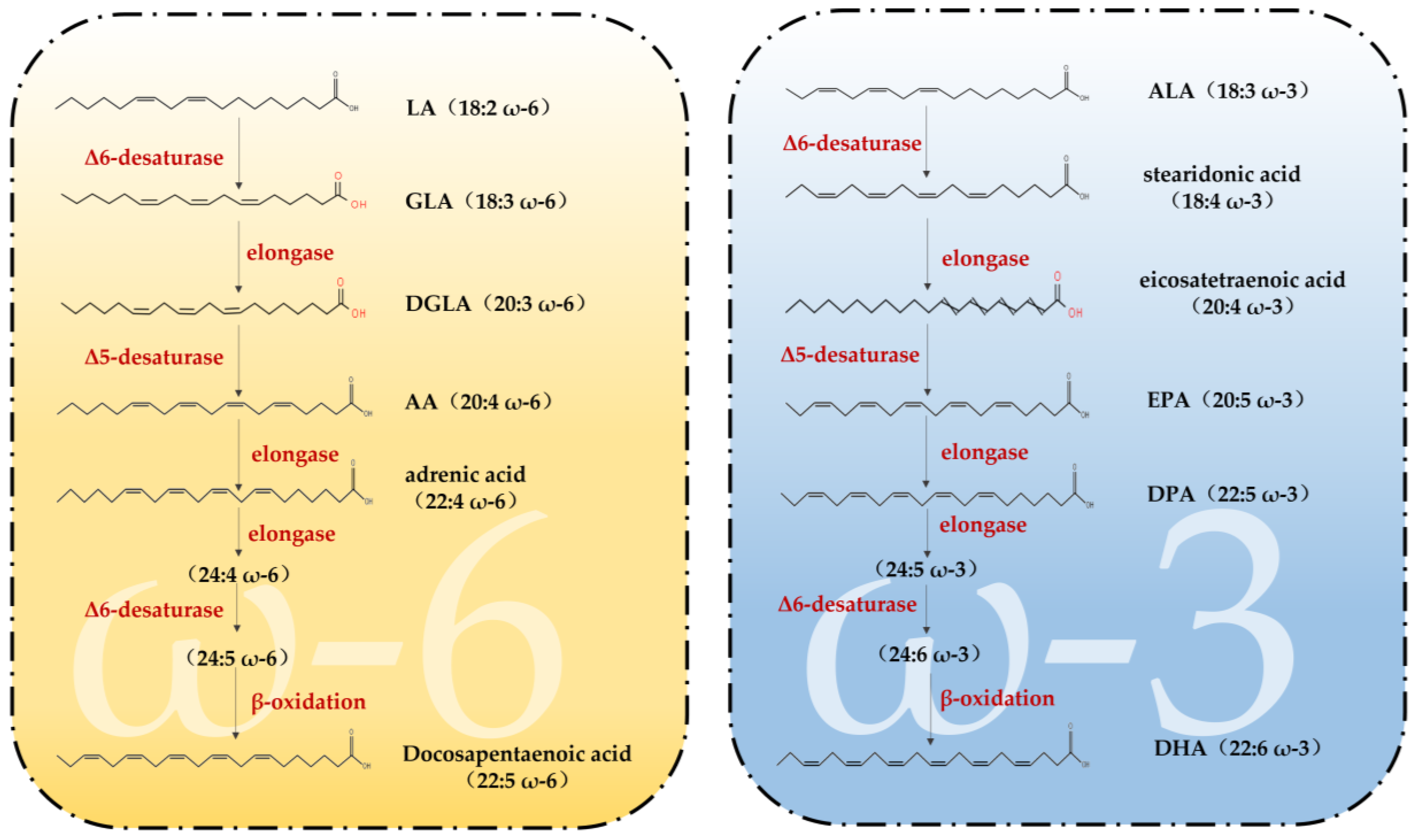
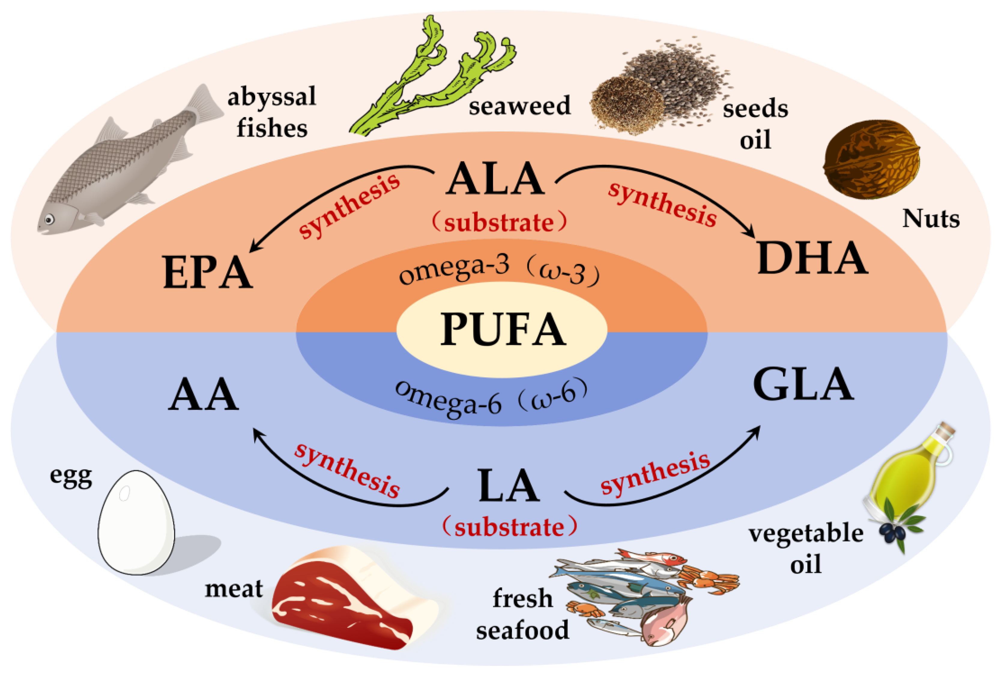
2. Sources of PUFAs
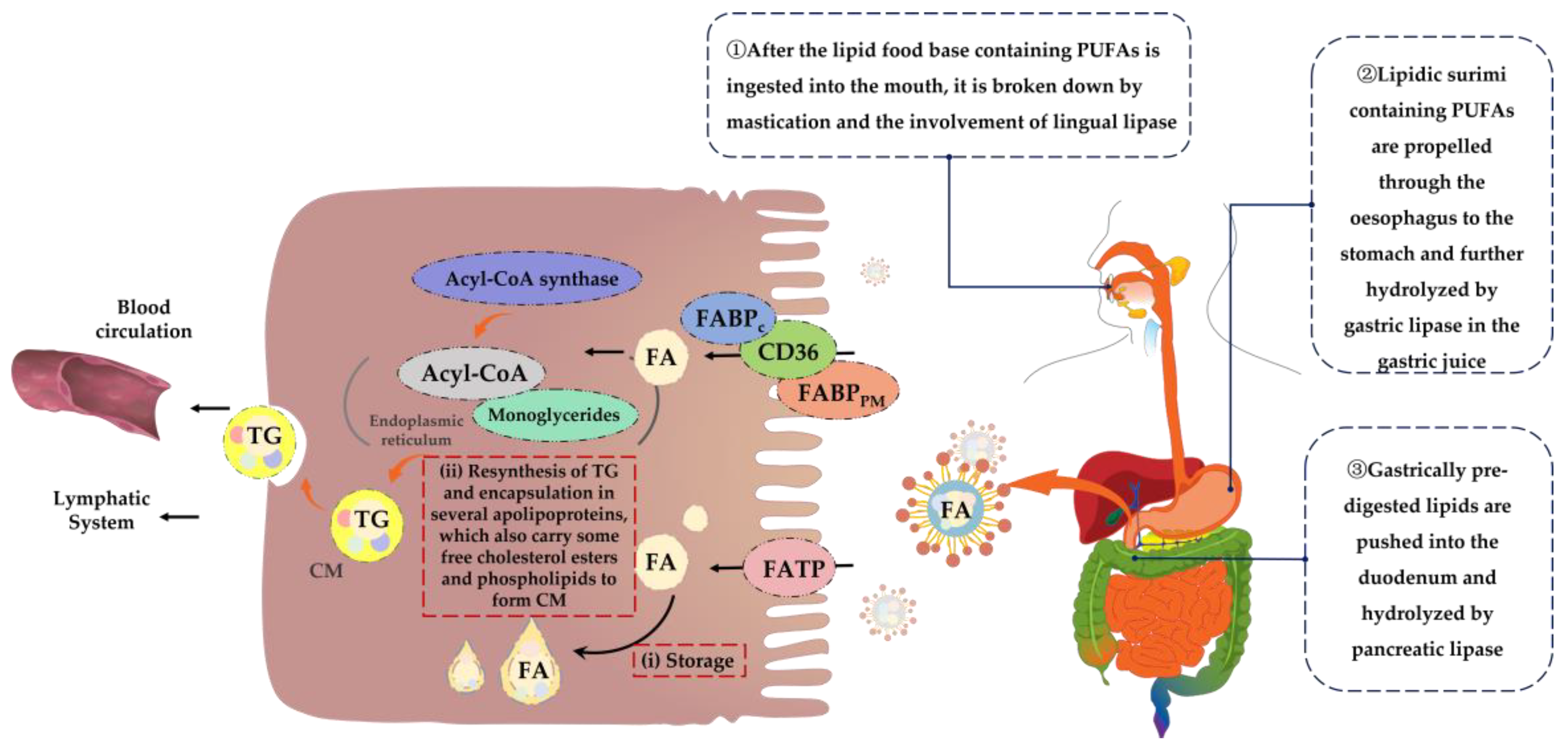
3. Mechanisms of Digestion and Absorption of PUFAs in the Organism
4. Application Safety of PUFAs
5. Study on the Mechanism of PUFAs to Assist in the Relief of Visual Fatigue
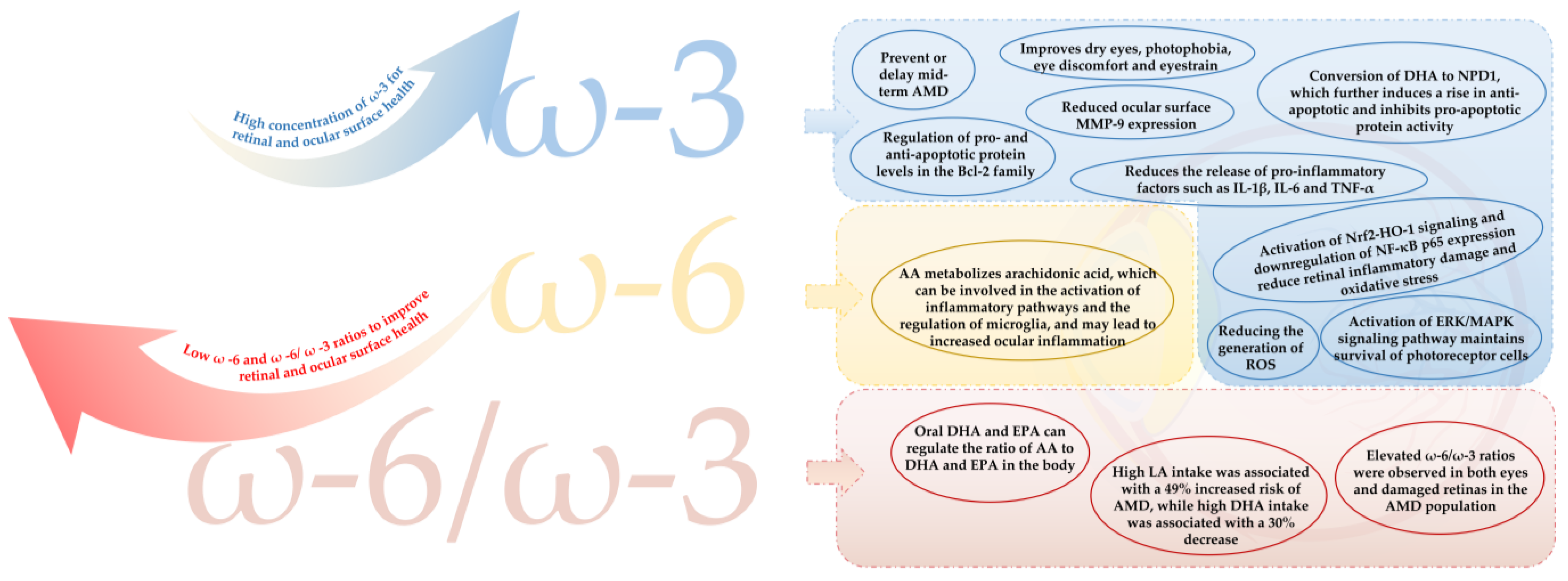
5.1. PUFAs Improve Damaged Ocular Surfaces to Act as a Visual Fatigue Reliever
5.2. PUFAs Improve Damaged Eye Fundus to Exert Visual Fatigue Relief
6. Conclusions
Author Contributions
Funding
Institutional Review Board Statement
Informed Consent Statement
Data Availability Statement
Acknowledgments
Conflicts of Interest
References
- Vilela, M.A.P.; Pellanda, L.C.; Fassa, A.G.; Castagno, V.D. Prevalence of asthenopia in children: A systematic review with meta-analysis. J. Pediatr. 2015, 91, 320–325. [Google Scholar] [CrossRef] [PubMed] [Green Version]
- Kaur, K.; Gurnani, B.; Nayak, S.; Deori, N.; Kaur, S.; Jethani, J.; Singh, D.; Agarkar, S.; Hussaindeen, J.R.; Sukhija, J.; et al. Digital Eye Strain- A Comprehensive Review. Ophthalmol. Ther. 2022, 11, 1655–1680. [Google Scholar] [CrossRef] [PubMed]
- Abdi, S.; Rydberg, A. Asthenopia in Schoolchildren, Orthoptic and Ophthalmological Findings and Treatment. Doc. Ophthalmol. 2005, 111, 65–72. [Google Scholar] [CrossRef] [PubMed]
- Sheppard, A.L.; Wolffsohn, J.S. Digital eye strain: Prevalence, measurement and amelioration. BMJ Open Ophthalmol. 2018, 3, e000146. [Google Scholar] [CrossRef] [Green Version]
- American Academy of Ophthalmology; Tripathy, K.; Chandrasekaran, P.R. Computer Vision Syndrome (Digital Eye Strain). Available online: https://eyewiki.aao.org/Computer_Vision_Syndrome_(Digital_Eye_Strain) (accessed on 20 September 2021).
- Lem, D.W.; Gierhart, D.L.; Davey, P.G. Can Nutrition Play a Role in Ameliorating Digital Eye Strain? Nutrients 2022, 14, 4005. [Google Scholar] [CrossRef]
- Balsam, A. Effect of digital device use during COVID-19 on digital eye strain. Clin. Exp. Optom. 2021, 104, 698–704. [Google Scholar]
- Zheng, F.; Hou, F.; Chen, R.; Mei, J.; Huang, P.; Chen, B.; Wang, Y. Investigation of the Relationship Between Subjective Symptoms of Visual Fatigue and Visual Functions. Front. Neurosci. 2021, 15, 686740. [Google Scholar] [CrossRef]
- Portello, J.K.; Rosenfield, M.; Bababekova, Y.; Estrada, J.M.; Leon, A. Computer-related visual symptoms in office workers. Ophthalmic Physiol. Opt. 2012, 32, 375–382. [Google Scholar] [CrossRef]
- Reddy, S.C.; Low, C.K.; Lim, Y.P.; Low, L.L.; Mardina, F.; Nursaleha, M.P. Computer vision syndrome: A study of knowledge and practices in university students. Nepal. J. Ophthalmol. 2013, 5, 161–168. [Google Scholar] [CrossRef]
- Duan, H.; Song, W.; Guo, J.; Yan, W. Taurine: A Source and Application for the Relief of Visual Fatigue. Nutrients 2023, 15, 1843. [Google Scholar] [CrossRef]
- Ayaki, M.; Kuze, M.; Kondo, M.; Tsubota, K.; Negishi, K. Association between Retinal Nerve Fiber Layer Thickness and Eye Fatigue. BioMed Res. Int. 2019, 2019, 3014567. [Google Scholar] [CrossRef] [PubMed]
- Fan, B.; Zhang, C.; Chi, J.; Liang, Y.; Bao, X.; Cong, Y.; Yu, B.; Li, X.; Li, G.-Y. The Molecular Mechanism of Retina Light Injury Focusing on Damage from Short Wavelength Light. Oxidative Med. Cell. Longev. 2022, 2022, 8482149. [Google Scholar] [CrossRef] [PubMed]
- Castelli, V.; Paladini, A.; D’angelo, M.; Allegretti, M.; Mantelli, F.; Brandolini, L.; Cocchiaro, P.; Cimini, A.; Varrassi, G. Taurine and oxidative stress in retinal health and disease. CNS Neurosci. Ther. 2021, 27, 403–412. [Google Scholar] [CrossRef] [PubMed]
- Yang, S.; Zhou, J.; Li, D. Functions and Diseases of the Retinal Pigment Epithelium. Front. Pharmacol. 2021, 12, 727870. [Google Scholar] [CrossRef] [PubMed]
- Upadhyay, M.; Milliner, C.; Bell, B.A.; Bonilha, V.L. Oxidative stress in the retina and retinal pigment epithelium (RPE): Role of aging, and DJ-1. Redox Biol. 2020, 37, 101623. [Google Scholar] [CrossRef] [PubMed]
- Kurlak, L.O.; Stephenson, T.J. Plausible explanations for effects of long chain polyunsaturated fatty acids (LCPUFA) on neonates. Arch. Dis. Child.-Fetal Neonatal Ed. 1999, 80, F148–F154. [Google Scholar] [CrossRef] [Green Version]
- Subramaniam, M.D.; Iyer, M.; Nair, A.P.; Venkatesan, D.; Mathavan, S.; Eruppakotte, N.; Kizhakkillach, S.; Chandran, M.K.; Roy, A.; Gopalakrishnan, A.V.; et al. Oxidative stress and mitochondrial transfer: A new dimension towards ocular diseases. Genes Dis. 2020, 9, 610–637. [Google Scholar] [CrossRef]
- Ruiz-Pastor, M.J.; Kutsyr, O.; Lax, P.; Cuenca, N. Decrease in DHA and other fatty acids correlates with photoreceptor degeneration in retinitis pigmentosa. Exp. Eye Res. 2021, 209, 108667. [Google Scholar] [CrossRef]
- Enoki, M.; Shinto, S.; Matsuoka, Y.; Otsuka, A.; Kaidzu, S.; Tanito, M.; Shibata, T.; Uchida, K.; Ohira, A.; Yamato, M.; et al. Lipid radicals cause light-induced retinal degeneration. Chem. Commun. 2017, 53, 10922–10925. [Google Scholar] [CrossRef]
- Suzumura, A.; Terao, R.; Kaneko, H. Protective Effects and Molecular Signaling of n-3 Fatty Acids on Oxidative Stress and Inflammation in Retinal. Antioxidants 2020, 9, 920. [Google Scholar] [CrossRef]
- McCusker, M.M.; Durrani, K.; Payette, M.J.; Suchecki, J. An eye on nutrition: The role of vitamins, essential fatty acids, and antioxidants in age-related macular degeneration, dry eye syndrome, and cataract. Clin. Dermatol. 2016, 34, 276–285. [Google Scholar] [CrossRef]
- Britten-Jones, A.C.; Craig, J.P.; Downie, L.E. Omega-3 polyunsaturated fatty acids and corneal nerve health: Current evidence and future directions. Ocul. Surf. 2023, 27, 1–12. [Google Scholar] [CrossRef]
- Saccà, S.C.; Cutolo, C.A.; Ferrari, D.; Corazza, P.; Traverso, C.E. The Eye, Oxidative Damage and Polyunsaturated Fatty Acids. Nutrients 2018, 10, 668. [Google Scholar] [CrossRef] [Green Version]
- Saccà, S.C.; Roszkowska, A.M.; Izzotti, A. Environmental light and endogenous antioxidants as the main determinants of non-cancer ocular diseases. Mutat. Res. Mol. Mech. Mutagen. 2013, 752, 153–171. [Google Scholar] [CrossRef]
- Lee, J.; Lee, H.K.; Kim, C.Y.; Hong, Y.J.; Choe, C.M.; You, T.W.; Seong, G.J. Purified high-dose anthocyanoside oligomer administration improves nocturnal vision and clinical symptoms in myopia subjects. Br. J. Nutr. 2005, 93, 895–899. [Google Scholar] [CrossRef] [Green Version]
- Tao, Y.; He, M.; Yang, Q.; Ma, Z.; Qu, Y.; Chen, W.; Peng, G.; Teng, D. Systemic taurine treatment provides neuroprotection against retinal photoreceptor degeneration and visual function impairments. Drug Des. Dev. Ther. 2019, 13, 2689–2702. [Google Scholar] [CrossRef] [Green Version]
- Kawabata, F.; Tsuji, T. Effects of dietary supplementation with a combination of fish oil, bilberry extract, and lutein on subjective symptoms of asthenopia in humans. Biomed. Res. 2011, 32, 387–393. [Google Scholar] [CrossRef] [PubMed] [Green Version]
- Giannaccare, G.; Pellegrini, M.; Senni, C.; Bernabei, F.; Scorcia, V.; Cicero, A.F.G. Clinical Applications of Astaxanthin in the Treatment of Ocular Diseases: Emerging Insights. Mar. Drugs 2020, 18, 239. [Google Scholar] [CrossRef] [PubMed]
- Park, C.Y.; Gu, N.; Lim, C.-Y.; Oh, J.-H.; Chang, M.; Kim, M.; Rhee, M.-Y. The effect of Vaccinium uliginosum extract on tablet computer-induced asthenopia: Randomized placebo-controlled study. BMC Complement. Altern. Med. 2016, 16, 296. [Google Scholar] [CrossRef] [PubMed] [Green Version]
- Hao, D.; Yan, W. Progress in the research of raw materials and their efficacious components for the function of relieving visual fatigue. J. Food Industry Sci. 2023, 44, 420–427. [Google Scholar]
- SanGiovanni, J.P.; Chew, E.Y. The role of omega-3 long-chain polyunsaturated fatty acids in health and disease of the retina. Prog. Retin. Eye Res. 2005, 24, 87–138. [Google Scholar] [CrossRef] [PubMed]
- Saini, R.K.; Keum, Y.-S. Omega-3 and omega-6 polyunsaturated fatty acids: Dietary sources, metabolism, and significance—A review. Life Sci. 2018, 203, 255–267. [Google Scholar] [CrossRef] [PubMed]
- Lee, J.M.; Lee, H.; Kang, S.; Park, W.J. Fatty Acid Desaturases, Polyunsaturated Fatty Acid Regulation, and Biotechnological Advances. Nutrients 2016, 8, 23. [Google Scholar] [CrossRef] [Green Version]
- Zárate, R.; el Jaber-Vazdekis, N.; Tejera, N.; Pérez, J.A.; Rodríguez, C. Significance of long chain polyunsaturated fatty acids in human health. Clin. Transl. Med. 2017, 6, 25. [Google Scholar] [CrossRef] [Green Version]
- Shahidi, F.; Ambigaipalan, P. Omega-3 Polyunsaturated Fatty Acids and Their Health Benefits. Annu. Rev. Food Sci. Technol. 2018, 9, 345–381. [Google Scholar] [CrossRef] [PubMed]
- Gecgel, U.; Gumus, T.; Tasan, M.; Daglioglu, O.; Arici, M. Determination of fatty acid composition of γ-irradiated hazelnuts, walnuts, almonds, and pistachios. Radiat. Phys. Chem. 2011, 80, 578–581. [Google Scholar] [CrossRef]
- Zamany, A.J.; Samadi, G.R.; Kim, D.H.; Keum, Y.-S.; Saini, R.K. Comparative Study of Tocopherol Contents and Fatty Acids Composition in Twenty Almond Cultivars of Afghanistan. J. Am. Oil Chem. Soc. 2017, 94, 805–817. [Google Scholar] [CrossRef]
- Nogales-Bueno, J.; Baca-Bocanegra, B.; Hernández-Hierro, J.M.; Garcia, R.; Barroso, J.M.; Heredia, F.J.; Rato, A.E. Assessment of Total Fat and Fatty Acids in Walnuts Using Near-Infrared Hyperspectral Imaging. Front. Plant Sci. 2021, 12, 729880. [Google Scholar] [CrossRef]
- Amaral, J.S.; Casal, S.; Citová, I.; Santos, A.; Seabra, R.M.; Oliveira, B.P. Characterization of several hazelnut (Corylus avellana L.) cultivars based in chemical, fatty acid and sterol composition. Eur. Food Res. Technol. 2006, 222, 274–280. [Google Scholar] [CrossRef] [Green Version]
- Kosker, A.R.; Regenstein, J.M.; Özogul, F. Proximate composition and fatty acid profiles of common pufferfish species in the Mediterranean Sea. Int. J. Food Sci. Technol. 2021, 56, 874–884. [Google Scholar] [CrossRef]
- Han, C.; Dong, S.; Li, L.; Gao, Q.; Zhou, Y. Assessment of phospholipid fatty acid profiles for discrimination of salmonids cultured in freshwater and seawater. Food Control 2020, 120, 107493. [Google Scholar] [CrossRef]
- Joensen, H.; Grahl-Nielsen, O. Distinction among North Atlantic cod Gadus morhua stocks by tissue fatty acid profiles. J. Fish Biol. 2014, 84, 1904–1925. [Google Scholar] [CrossRef] [PubMed]
- Rizzo, G.; Baroni, L.; Lombardo, M. Promising Sources of Plant-Derived Polyunsaturated Fatty Acids: A Narrative Review. Int. J. Environ. Res. Public Health 2023, 20, 1683. [Google Scholar] [CrossRef]
- Khursheed, T.; Fatima, T.; Qadri, T.; Rafiq, A.; Malik, A.; Naseer, B.; Hussain, S.Z. Biochemical, nutraceutical and phytochemical characterization of chia and basil seeds: A comparative study. Int. J. Food Prop. 2022, 26, 1–13. [Google Scholar] [CrossRef]
- Kiczorowska, B.; Samolińska, W.; Andrejko, D.; Kiczorowski, P.; Antoszkiewicz, Z.; Zając, M.; Winiarska-Mieczan, A.; Bąkowski, M. Comparative analysis of selected bioactive components (fatty acids, tocopherols, xanthophyll, lycopene, phenols) and basic nutrients in raw and thermally processed camelina, sunflower, and flax seeds (Camelina sativa L. Crantz, Helianthus L., and Linum L.). J. Food Sci. Technol. 2019, 56, 4296–4310. [Google Scholar] [CrossRef] [Green Version]
- Khotimchenko, S.V.; Vaskovsky, V.E.; Titlyanova, T.V. Fatty Acids of Marine Algae from the Pacific Coast of North California. Bot. Mar. 2002, 45, 17–22. [Google Scholar] [CrossRef]
- Yu, Y.; Wang, G.; Sun, Y.; Ge, C.; Liao, G. Changes in physicochemical parameters, free fatty acid profile and water-soluble compounds of Yunnan dry-cured beef during processing. J. Food Process. Preserv. 2020, 44, e14380. [Google Scholar] [CrossRef]
- Nudda, A.; McGuire, M.K.; Battacone, G.; Manca, M.G.; Boe, R.; Pulina, G. Documentation of Fatty Acid Profiles in Lamb Meat and Lamb-Based Infant Foods. J. Food Sci. 2011, 76, H43–H47. [Google Scholar] [CrossRef]
- Aquino-Bolaños, E.N.; Mapel-Velazco, L.; Martín-del-Campo, S.T.; Chávez-Servia, J.L.; Martínez, A.J.; Verdalet-Guzmán, I. Fatty acids profile of oil from nine varieties of Macadamia nut. Int. J. Food Prop. 2016, 20, 1262–1269. [Google Scholar] [CrossRef] [Green Version]
- Jacobsen, C.; Warncke, S.A.; Hansen, S.H.; Sørensen, A.-D.M. Fish Liver Discards as a Source of Long-Chain Omega-3 Polyunsaturated Fatty Acids. Foods 2022, 11, 905. [Google Scholar] [CrossRef]
- Minami, I.; Nakamura, Y.; Todoriki, S.; Murata, Y. Effect of γ Irradiation on the Fatty Acid Composition of Soybean and Soybean Oil. Biosci. Biotechnol. Biochem. 2012, 76, 900–905. [Google Scholar] [CrossRef] [Green Version]
- Suri, K.; Singh, B.; Kaur, A.; Yadav, M.P.; Singh, N. Influence of microwave roasting on chemical composition, oxidative stability and fatty acid composition of flaxseed (Linum usitatissimum L.) oil. Food Chem. 2020, 326, 126974. [Google Scholar] [CrossRef]
- Hamosh, M.; Scow, R.O. Lingual Lipase and Its Role in the Digestion of Dietary Lipid. J. Clin. Investig. 1973, 52, 88–95. [Google Scholar] [CrossRef]
- Brignot, H.; Feron, G. Oral lipolysis and its association with diet and the perception and digestion of lipids: A systematic literature review. Arch. Oral Biol. 2019, 108, 104550. [Google Scholar] [CrossRef]
- Dias, C.B.; Zhu, X.; Thompson, A.K.; Singh, H.; Garg, M.L. Effect of the food form and structure on lipid digestion and postprandial lipaemic response. Food Funct. 2018, 10, 112–124. [Google Scholar] [CrossRef] [PubMed]
- Maldonado-Valderrama, J.; Wilde, P.; Macierzanka, A.; Mackie, A. The role of bile salts in digestion. Adv. Colloid Interface Sci. 2011, 165, 36–46. [Google Scholar] [CrossRef] [PubMed]
- Dawson, P.A.; Karpen, S.J. Intestinal transport and metabolism of bile acids. J. Lipid Res. 2015, 56, 1085–1099. [Google Scholar] [CrossRef] [PubMed] [Green Version]
- Schonewille, M.; de Boer, J.F.; Groen, A.K. Bile salts in control of lipid metabolism. Curr. Opin. Infect. Dis. 2016, 27, 295–301. [Google Scholar] [CrossRef] [PubMed]
- Kennelly, J.P.; van der Veen, J.N.; Nelson, R.C.; Leonard, K.-A.; Havinga, R.; Buteau, J.; Kuipers, F.; Jacobs, R.L. Intestinal de novo phosphatidylcholine synthesis is required for dietary lipid absorption and metabolic homeostasis. J. Lipid Res. 2018, 59, 1695–1708. [Google Scholar] [CrossRef] [PubMed] [Green Version]
- Guo, Q.; Ye, A.; Bellissimo, N.; Singh, H.; Rousseau, D. Modulating fat digestion through food structure design. Prog. Lipid Res. 2017, 68, 109–118. [Google Scholar] [CrossRef] [PubMed]
- Qin, D.; Yang, X.; Gao, S.; Yao, J.; McClements, D.J. Influence of dietary fibers on lipid digestion: Comparison of single-stage and multiple-stage gastrointestinal models. Food Hydrocoll. 2017, 69, 382–392. [Google Scholar] [CrossRef]
- Goncalves, A.; Roi, S.; Nowicki, M.; Dhaussy, A.; Huertas, A.; Amiot, M.J.; Reboul, E. Fat-soluble vitamin intestinal absorption: Absorption sites in the intestine and interactions for absorption. Food Chem. 2015, 172, 155–160. [Google Scholar] [CrossRef]
- Hamilton, J.A. Fatty acid transport: Difficult or easy? J. Lipid Res. 1998, 39, 467–481. [Google Scholar] [CrossRef] [PubMed]
- Hamilton, J.A. Fast flip-flop of cholesterol and fatty acids in membranes: Implications for membrane transport proteins. Curr. Opin. Lipidol. 2003, 14, 263–271. [Google Scholar] [CrossRef] [PubMed]
- Ehehalt, R.; Füllekrug, J.; Pohl, J.; Ring, A.; Herrmann, T.; Stremmel, W. Translocation of long chain fatty acids across the plasma membrane—Lipid rafts and fatty acid transport proteins. Mol. Cell. Biochem. 2006, 284, 135–140. [Google Scholar] [CrossRef] [PubMed]
- Hajri, T.; Abumrad, N.A. FATTY ACID TRANSPORT ACROSS MEMBRANES: Relevance to Nutrition and Metabolic Pathology. Annu. Rev. Nutr. 2002, 22, 383–415. [Google Scholar] [CrossRef]
- Stremmel, W.; Staffer, S.; Wannhoff, A.; Pathil, A. The overall fatty acid absorption controlled by basolateral chylomicron excretion under regulation of p-JNK1. Biochim. Biophys. Acta (BBA)-Mol. Cell Biol. Lipids 2017, 1862, 917–928. [Google Scholar] [CrossRef]
- Storch, J.; McDermott, L. Structural and functional analysis of fatty acid-binding proteins. J. Lipid Res. 2009, 50, S126–S131. [Google Scholar] [CrossRef] [Green Version]
- Carey, M.C.; Small, D.M.; Bliss, C.M. Lipid Digestion and Absorption. Annu. Rev. Physiol. 1983, 45, 651–677. [Google Scholar] [CrossRef]
- Watkins, J.B. Lipid Digestion and Absorption. Pediatrics 1985, 75, 151–156. [Google Scholar] [CrossRef]
- Mansbach, C.M., II; Gorelick, F. Development and physiological regulation of intestinal lipid absorption. II. Dietary lipid absorption, complex lipid synthesis, and the intracellular packaging and secretion of chylomicrons. Am. J. Physiol. Gastrointest. Liver Physiol. 2007, 293, G645–G650. [Google Scholar] [CrossRef]
- Zhang, Y.; Zhang, T.; Liang, Y.; Jiang, L.; Sui, X. Dietary Bioactive Lipids: A Review on Absorption, Metabolism, and Health Properties. J. Agric. Food Chem. 2021, 69, 8929–8943. [Google Scholar] [CrossRef] [PubMed]
- Xu, E.; Chen, C.; Fu, J.; Zhu, L.; Shu, J.; Jin, M.; Wang, Y.; Zong, X. Dietary fatty acids in gut health: Absorption, metabolism and function. Anim. Nutr. 2021, 7, 1337–1344. [Google Scholar] [CrossRef] [PubMed]
- Fredman, G.; Tabas, I. Boosting Inflammation Resolution in Atherosclerosis: The Next Frontier for Therapy. Am. J. Pathol. 2017, 187, 1211–1221. [Google Scholar] [CrossRef] [PubMed] [Green Version]
- Serhan, C.N.; Savill, J. Resolution of inflammation: The beginning programs the end. Nat. Immunol. 2005, 6, 1191–1197. [Google Scholar] [CrossRef]
- Del Gobbo, L.C.; Imamura, F.; Aslibekyan, S.; Marklund, M.; Virtanen, J.K.; Wennberg, M.; Yakoob, M.Y.; Chiuve, S.E.; Cruz, L.D.; Frazier-Wood, A.C.; et al. ω-3 Polyunsaturated Fatty Acid Biomarkers and Coronary Heart Disease. JAMA Intern. Med. 2016, 176, 1155–1166. [Google Scholar] [CrossRef] [Green Version]
- Bork, C.S.; Lundbye-Christensen, S.; Venø, S.K.; Lasota, A.N.; Tjønneland, A.; Schmidt, E.B.; Overvad, K. Intake of marine and plant-derived n-3 fatty acids and development of atherosclerotic cardiovascular disease in the Danish Diet, Cancer and Health cohort. Eur. J. Nutr. 2023, 62, 1389–1401. [Google Scholar] [CrossRef]
- Dietary Reference Intakes for Energy, Carbohydrate, Fiber, Fat, Fatty Acids, Cholesterol, Protein, and Amino Acids (2005) Chapter: 8 Dietary Fats: Total Fat and Fatty Acids. Available online: https://nap.nationalacademies.org/read/10490/chapter/10?term=DHA#464-472 (accessed on 29 May 2023).
- Dietary Reference Values for Nutrients Summary Report. Available online: https://www.efsa.europa.eu/sites/default/files/2017_09_DRVs_summary_report.pdf (accessed on 29 May 2023).
- Nutrient Reference Values for Australia and New Zealand Including Recommended Dietary Intakes. Available online: https://www.nhmrc.gov.au/about-us/publications/nutrient-reference-values-australia-and-new-zealand-including-recommended-dietary-intakes (accessed on 29 May 2023).
- Dietary Reference Nutrient Intakes for Chinese Residents Part 1: Macronutrients. Available online: http://www.nhc.gov.cn/wjw/yingyang/201710/fdade20feb8144ba921b412944ffb779/files/0fa10dfb812a48b483d931972df1ccb8.pdf (accessed on 29 May 2023).
- Dietary Reference Intakes for Japanese. 2015. Available online: https://www.mhlw.go.jp/file/06-Seisakujouhou-10900000-Kenkoukyoku/Full_DRIs2015.pdf (accessed on 29 May 2023).
- Dietary Reference Intakes Definitions. Available online: https://www.canada.ca/content/dam/hc-sc/migration/hc-sc/fn-an/alt_formats/hpfb-dgpsa/pdf/nutrition/dri_tables-eng.pdf (accessed on 29 May 2023).
- Cruzat, A.; Qazi, Y.; Hamrah, P. In Vivo Confocal Microscopy of Corneal Nerves in Health and Disease. Ocul. Surf. 2016, 15, 15–47. [Google Scholar] [CrossRef] [Green Version]
- Aslan, M. Polyunsaturated Fatty Acid and Sphingolipid Measurements by Tandem Mass Spectrometry. Mini-Reviews Org. Chem. 2020, 18, 3–10. [Google Scholar] [CrossRef]
- Britten-Jones, A.C.; Craig, J.P.; Anderson, A.J.; Downie, L.E. Association between systemic omega-3 polyunsaturated fatty acid levels, and corneal nerve structure and function. Eye 2022, 1–8. [Google Scholar] [CrossRef]
- Aragona, P.; Bucolo, C.; Spinella, R.; Giuffrida, S.; Ferreri, G. Systemic omega-6 essential fatty acid treatment and pge1 tear content in Sjögren’s syndrome patients. Ophthalmol. Vis. Sci. 2005, 46, 4474–4479. [Google Scholar] [CrossRef] [PubMed] [Green Version]
- Macrì, A.; Giuffrida, S.; Amico, V.; Iester, M.; Traverso, C.E. Effect of linoleic acid and gamma-linolenic acid on tear production, tear clearance and on the ocular surface after photorefractive keratectomy. Graefe’s Arch. Clin. Exp. Ophthalmol. 2003, 241, 561–566. [Google Scholar] [CrossRef] [PubMed]
- Miljanović, B.; Trivedi, K.A.; Dana, M.R.; Gilbard, J.P.; Buring, J.E.; Schaumberg, D.A. Relation between dietary n − 3 and n − 6 fatty acids and clinically diagnosed dry eye syndrome in women. Am. J. Clin. Nutr. 2005, 82, 887–893. [Google Scholar] [CrossRef] [Green Version]
- Rashid, S.; Jin, Y.; Ecoiffier, T.; Barabino, S.; Schaumberg, D.A.; Dana, M.R. Topical Omega-3 and Omega-6 Fatty Acids for Treatment of Dry Eye. Arch. Ophthalmol. 2008, 126, 219–225. [Google Scholar] [CrossRef] [Green Version]
- Park, J.; Yoo, Y.-S.; Shin, E.; Han, G.; Shin, K.; Lim, D.H.; Chung, T.-Y. Effects of the re-esterified triglyceride (rTG) form of omega-3 supplements on dry eye following cataract surgery. Br. J. Ophthalmol. 2020, 105, 1504–1509. [Google Scholar] [CrossRef]
- Kangari, H.; Eftekhari, M.H.; Sardari, S.; Hashemi, H.; Salamzadeh, J.; Ghassemi-Broumand, M.; Khabazkhoob, M. Short-term Consumption of Oral Omega-3 and Dry Eye Syndrome. Ophthalmology 2013, 120, 2191–2196. [Google Scholar] [CrossRef]
- Li, D.-Q.; Lokeshwar, B.L.; Solomon, A.; Monroy, D.; Ji, Z.; Pflugfelder, S.C. Regulation of MMP-9 Production by Human Corneal Epithelial Cells. Exp. Eye Res. 2001, 73, 449–459. [Google Scholar] [CrossRef]
- Epitropoulos, A.; Donnenfeld, E.D.; Shah, Z.A.; Holland, E.J.; Gross, M.; Faulkner, W.J.; Matossian, C.; Lane, S.S.; Toyos, M.; Bucci, F.A.; et al. Effect of Oral Re-esterified Omega-3 Nutritional Supplementation on Dry Eyes. Cornea 2016, 35, 1185–1191. [Google Scholar] [CrossRef] [PubMed]
- Schnebelen, C.; Pasquis, B.; Salinas-Navarro, M.; Joffre, C.; Creuzot-Garcher, C.P.; Vidal-Sanz, M.; Bron, A.M.; Bretillon, L.; Acar, N. A dietary combination of omega-3 and omega-6 polyunsaturated fatty acids is more efficient than single supplementations in the prevention of retinal damage induced by elevation of intraocular pressure in rats. Graefe’s Arch. Clin. Exp. Ophthalmol. 2009, 247, 1191–1203. [Google Scholar] [CrossRef]
- Brignole-Baudouin, F.; Baudouin, C.; Aragona, P.; Rolando, M.; Labetoulle, M.; Pisella, P.J.; Barabino, S.; Siou-Mermet, R.; Creuzot-Garcher, C. A multicentre, double-masked, randomized, controlled trial assessing the effect of oral supplementation of omega-3 and omega-6 fatty acids on a conjunctival inflammatory marker in dry eye patients. Acta Ophthalmol. 2011, 89, e591–e597. [Google Scholar] [CrossRef]
- Layé, S.; Nadjar, A.; Joffre, C.; Bazinet, R.P. Anti-Inflammatory Effects of Omega-3 Fatty Acids in the Brain: Physiological Mechanisms and Relevance to Pharmacology. Pharmacol. Rev. 2017, 70, 12–38. [Google Scholar] [CrossRef] [PubMed] [Green Version]
- German, O.L.; Monaco, S.; Agnolazza, D.L.; Rotstein, N.P.; Politi, L.E. Retinoid X receptor activation is essential for docosahexaenoic acid protection of retina photoreceptors. J. Lipid Res. 2013, 54, 2236–2246. [Google Scholar] [CrossRef] [PubMed] [Green Version]
- Rotstein, N.P.; Politi, L.E.; German, O.L.; Girotti, R. Protective Effect of Docosahexaenoic Acid on Oxidative Stress-Induced Apoptosis of Retina Photoreceptors. Investig. Opthalmology Vis. Sci. 2003, 44, 2252–2259. [Google Scholar] [CrossRef] [PubMed] [Green Version]
- Bazan, N.G.; Calandria, J.M.; Gordon, W.C. Docosahexaenoic acid and its derivative neuroprotectin D1 display neuroprotective properties in the retina, brain and central nervous system. Nestlé Nutr. Inst. Workshop Ser. 2013, 77, 121–131. [Google Scholar]
- Leyrolle, Q.; Layé, S.; Nadjar, A. Direct and indirect effects of lipids on microglia function. Neurosci. Lett. 2019, 708, 134348. [Google Scholar] [CrossRef]
- Gorica, E.; Calderone, V. Arachidonic Acid Derivatives and Neuroinflammation. CNS Neurol. Disord.-Drug Targets (Former. Curr. Drug Targets-CNS Neurol. Disord.) 2021, 21, 118–129. [Google Scholar] [CrossRef]
- Vidal, E.; Jun, B.; Gordon, W.C.; Maire, M.-A.; Martine, L.; Grégoire, S.; Khoury, S.; Cabaret, S.; Berdeaux, O.; Acar, N.; et al. Bioavailability and spatial distribution of fatty acids in the rat retina after dietary omega-3 supplementation. J. Lipid Res. 2020, 61, 1733–1746. [Google Scholar] [CrossRef]
- Deng, Q.; Wang, Y.; Wang, C.; Ji, B.; Cong, R.; Zhao, L.; Chen, P.; Zang, X.; Lu, F.; Han, F.; et al. Dietary supplementation with omega-3 polyunsaturated fatty acid-rich oils protects against visible-light-induced retinal damage in vivo. Food Funct. 2018, 9, 2469–2479. [Google Scholar] [CrossRef]
- Deng, Q.; Huang, F.; Huang, Q.; Song, Y.; Yang, J. Alleviating Eye Fatigue Evaluation of Flaxseed Oil Soft Capsule. J. Food Res. Dev. 2011, 32, 118–122. [Google Scholar]
- Tu, C.-F.; Lee, C.-H.; Chen, H.-N.; Tsao, L.-Y.; Chen, J.-Y.; Hsiao, C.-C. Effects of fish oil-containing lipid emulsions on retinopathy of prematurity in very low birth weight infants. Pediatr. Neonatol. 2020, 61, 224–230. [Google Scholar] [CrossRef]
- Meydani, S.N.; Endres, S.; Woods, M.M.; Goldin, B.R.; Soo, C.; Morrill-Labrode, A.; Dinarello, C.A.; Gorbach, S.L. Oral (n-3) fatty acid supplementation suppresses cytokine production and lymphocyte proliferation: Comparison between young and Older Women. J. Nutr. 1991, 121, 547–555. [Google Scholar] [CrossRef]
- Sadeghi, S.; Wallace, F.; Calder, P.C. Dietary lipids modify the cytokine response to bacterial lipopolysaccharide in mice. Immunology 1999, 96, 404–410. [Google Scholar] [CrossRef] [PubMed]
- Wallace, F.A.; Miles, E.A.; Calder, P.C. Activation state alters the effect of dietary fatty acids on pro-inflammatory mediator production by murine macrophages. Cytokine 2000, 12, 1374–1379. [Google Scholar] [CrossRef] [PubMed]
- Kim, M.J.; Kim, D.H.; Kwak, H.S.; Yu, I.-S.; Um, M.Y. Protective Effect of Chrysanthemum boreale Flower Extracts against A2E-Induced Retinal Damage in ARPE-19 Cell. Antioxidants 2022, 11, 669. [Google Scholar] [CrossRef] [PubMed]
- Ren, J.; Ren, A.; Deng, X.; Huang, Z.; Jiang, Z.; Li, Z.; Gong, Y. Long-Chain Polyunsaturated Fatty Acids and Their Metabolites Regulate Inflammation in Age-Related Macular Degeneration. J. Inflamm. Res. 2022, 15, 865–880. [Google Scholar] [CrossRef]
- Wu, J.; Cho, E.; Giovannucci, E.L.; Rosner, B.A.; Sastry, S.M.; Willett, W.C.; Schaumberg, D.A. Dietary Intakes of Eicosapentaenoic Acid and Docosahexaenoic Acid and Risk of Age-Related Macular Degeneration. Ophthalmology 2017, 124, 634–643. [Google Scholar] [CrossRef] [Green Version]
- Wei, Y.; Meng, Y.; Li, N.; Wang, Q.; Chen, L. The effects of low-ratio n-6/n-3 PUFA on biomarkers of inflammation: A systematic review and meta-analysis. Food Funct. 2021, 12, 30–40. [Google Scholar] [CrossRef]
- Fu, Z.; Yan, W.; Chen, C.T.; Nilsson, A.K.; Bull, E.; Allen, W.; Yang, J.; Ko, M.; SanGiovanni, J.P.; Akula, J.D.; et al. Omega-3/Omega-6 Long-Chain Fatty Acid Imbalance in Phase I Retinopathy of Prematurity. Nutrients 2022, 14, 1333. [Google Scholar] [CrossRef]
- Lister, T. Nutritional, Alternative, and Complementary Therapies for Age-related Macular Degeneration. Integr. Med. (Encinitas Calif.) 2019, 18, 30–36. [Google Scholar]
- Carneiro, Â.; Andrade, J.P. Nutritional and Lifestyle Interventions for Age-Related Macular Degeneration: A Review. Oxidative Med. Cell. Longev. 2017, 2017, 6469138. [Google Scholar]
- Simopoulos, A.P. An Increase in the Omega-6/Omega-3 Fatty Acid Ratio Increases the Risk for Obesity. Nutrients 2016, 8, 128. [Google Scholar] [CrossRef] [PubMed] [Green Version]
- Georgakopoulos, C.D.; Makri, O.E.; Pagoulatos, D.; Vasilakis, P.; Peristeropoulou, P.; Kouli, V.; Eliopoulou, M.I.; Psachoulia, C. Effect of Omega-3 Fatty Acids Dietary Supplementation on Ocular Surface and Tear Film in Diabetic Patients with Dry Eye. J. Am. Coll. Nutr. 2016, 36, 38–43. [Google Scholar] [CrossRef] [PubMed]
- Wang, Y.E.; Tseng, V.L.; Yu, F.; Caprioli, J.; Coleman, A.L. Association of Dietary Fatty Acid Intake with Glaucoma in the United States. JAMA Ophthalmol 2018, 136, 141–147. [Google Scholar] [CrossRef] [PubMed] [Green Version]


| Category | Name | Total PUFAs | ω-3 PUFAs | ω-6 PUFAs | Ref. | |||
|---|---|---|---|---|---|---|---|---|
| ALA (18:3) | EPA (20:5) | DHA (22:6) | LA (18:2) | AA (20:4) | ||||
| Nuts | Almond | 9.93~26.66 | 0.07~0.29 | / | / | 8.17~26.66 | / | [37,38] |
| Walnut | 72.66~77.48 | 12.61~16.92 | / | / | 58.64~63.74 | / | [37,39] | |
| Hazelnut | 7.30~11.48 | 0.08~0.12 | / | / | 7.20~11.25 | / | [37,40] | |
| Fish | Blowfish | 30.20~41.80 | 0.10~0.40 | 1.30~6.90 | 14.00~26.70 | 1.40~10.60 | 7.00~12.20 | [41] |
| salmonids | 49.18~49.28 | 0.62~1.62 | 3.86~6.37 | 27.65~28.88 | 9.41~11.81 | 2.12~2.30 | [42] | |
| Cod | 59.00~62.00 | 0.18~0.32 | 0.07~0.09 | 30.00~31.00 | 0.50~0.70 | 2.40~6.00 | [43] | |
| Seed | Chia seed | 24.77~28.37 | 17.8~19.55 | / | / | 5.25~5.84 | / | [44,45] |
| Flaxseed | 70.29~71.65 | 55.72~57.93 | / | 0.11~0.14 | 13.58~14.68 | / | [46] | |
| Sunflower | 58.90~61.80 | 0.06~0.11 | / | / | 58.83~61.68 | <0.01 | [46] | |
| Seaweed | Red algae | / | 0.20~0.50 | 27.80~45.40 | / | 0.60~1.60 | 5.30~23.40 | [47] |
| Brown algae | / | 2.10~9.70 | 3.10~13.20 | / | 3.70~9.90 | 7.90~18.60 | [47] | |
| Green algae | / | 0.50~21.90 | 1.00~1.40 | / | 2.30~28.60 | 0.30~1.20 | [47] | |
| Meat | Beef | 9.48~12.52 | 0.13~0.25 | 2.87~3.71 | / | 0.80~1.84 | 0.06~0.24 | [48] |
| Lamb | 11.01~1.60 | 1.25~1.35 | 0.60~0.70 | 0.59~0.69 | / | 1.87~2.19 | [49] | |
| Oil | Argan oil | 1.90~3.90 | 0.18~0.30 | / | / | 1.60~3.20 | / | [50] |
| Fish oil | / | 1.04~1.37 | 6.21~8.13 | 12.80~13.30 | 1.32~1.67 | 0.85~0.95 | [51] | |
| Soybean oil | / | 5.50~6.62 | / | / | 50.9~57.10 | / | [44,52] | |
| Flaxseed oil | 67.40~68.27 | 53.40~54.33 | / | / | 13.94~14.20 | / | [44,53] | |
| Country/Institution | Adult Male | Adult Women | Ref. | ||
|---|---|---|---|---|---|
| ω-3, g/d (ALA) | ω-6, g/d (LA) | ω-3, g/d (ALA) | ω-6, g/d (LA) | ||
| United States (NIH) | 1.6 | 14–17 | 1.1 | 11–12 | [79] |
| Europe (EFSA) | 0.5% E | 4.0% E | 0.5% E | 4.0% E | [80] |
| Australia and New Zealand (NHMRC) | 1.6 | 14–17 | 1.1–1.3 | 11–13 | [81] |
| China (NHC) | 0.6% E | 4.0% E | 0.6% E | 4.0% E | [82] |
| Japan (MHLW) | (2.0–2.4) a | (8–11) a | (1.1–1.3) a | (7–9) a | [83] |
| Canada (Health Canada) | 1.3 | 13 | 0.8 | 8 | [84] |
| Subjects/Animals | Subjects | Periodicity | Results | Conclusions/Potential Mechanisms | Ref. |
|---|---|---|---|---|---|
| People with visual fatigue | EPA: 162 mg/d/ DHA: 783 mg/d and lingonberry extract and lutein | 4 w | Omega-3-rich dietary supplements can safely improve the subjective symptoms of eye strain and mental fatigue in humans. | Dietary supplements rich in omega-3s can help alleviate the onset of visual fatigue. | [28] |
| Dry eye mice | ALA or LA | 5~10 d | Increased corneal fluorescein staining; increased corneal IL-1 β and TNF-α expression; and conjunctival IL-1 β, TNF-β, IFN-β, IL-2, IL-6 and IL-10. The ALA group, on the other hand, showed significantly reduced corneal fluorescein staining, corneal IL-1 and TNF-α expression and a significant reduction in conjunctival TNF-α associated. | ALA helps to relieve dry eyes and eye discomfort, and it reduces the expression of inflammatory factors on the ocular surface, thus relieving eye strain symptoms such as dry eyes. | [91] |
| Dry eye people | EPA: 1680 mg/d/ DHA: 560 mg/d | 2 m | Two months of omega-3 supplementation significantly improved the subjective symptom scores of OSDI and DEQ in the dry eye population, significantly inhibiting the increase in levels of the ocular surface pro-inflammatory factor MMP-9. | Omega-3 may have alleviated the onset of dry eye by reducing ocular surface inflammation. | [92] |
| Dry eye people | EPA: 360 mg/d/ DHA: 240 mg/d | 1 m | One month after the ω-3 intervention, the dry eye population showed significant improvement in TBUT, OSDI and Schirmer scores than the placebo group. The intervention and placebo groups showed significant differences in TBUT with changes of 71%, and 3.3%, dry eye symptoms changes of 26% and 4% and Schirmer score changes of 22.3% and 5.1%, respectively. | Omega-3 is effective at reducing the rate of tear evaporation; improving dry eyes and eye discomfort and effectively increasing tear production. | [93] |
| Dry eye people | EPA: 1680 mg/d/ DHA: 560 mg/d | 12 w | At week 6, the ω-3 intervention group showed a significant reduction in tear osmolarity. At 12 weeks, both ω-3 index levels and TBUT were significant at 12 weeks, both ω-3 index levels and TBUT significantly improved and had reduced ocular surface MMP-9 expression. | ω-3 was associated with statistically significant improvements in tear osmolality, ω-3 index levels, TBUT, MMP-9 and OSDI symptom scores. | [95] |
| Rats with high eye pressure | Omega-3 PUFAs group: EPA + DHA ω-6 PUFAs group: GLA ω-3 + ω-6 PUFAs group: EPA + DHA + GLA | 6 m | Significant activation of glial cells was observed in the eyes of control rats with high IOP, while animals in the ω-3 + ω-6 PUFAs group diet showed significant improvement. | The results of the study showed that the effects of the combination of ω-3 + ω-6 PUFAs were more effective than single supplementation in preventing retinal cell structure and glial cell activation induced by elevated IOP in rats. | [96] |
| Dry eye people | EPA: 427.5 mg/d/ DHA: 285 mg/d and other vitamins and zinc | 3 m | ω-3 significantly reduced the percentage of HLA-DR-positive cells. | Supplementation with omega-3 may reduce the expression of HLA-DR conjunctival inflammatory markers and may help improve dry eye and ocular discomfort. | [97] |
| Rabbit with retinal damage | Algae oil (contains 33.34% DHA) | 3 w | The intervention group significantly reduced the levels of IL-1β, TNF-α, IL-8 and COX-2 in the retina, downregulated NF-κB expression and significantly attenuated light-induced retinal apoptosis and neovascularization. | Diets containing omega-3 PUFA may protect against visible light-induced retinal damage. The mechanism may be related to reducing the expression of inflammatory factors in the damaged retina. | [105] |
| People with visual fatigue | Flaxseed oil capsules | 30 d | After 30 d, the test food group showed a significant reduction in visual fatigue and a 70.3% improvement in the persistence of binocular vision, with a mean improvement of 11.2 ± 7.3% and an overall effective rate of 51%. | It is shown that omega-3 rich flaxseed oil softgels are effective in relieving human visual fatigue. | [106] |
| AMD crowd | EPA- and DHA-rich diet | 3 m | Higher intakes of EPA and DHA prevented or delayed the onset of moderate AMD, but there appeared to be only a trend towards improvement for late-stage ADM. The results of the study were not available but not with significant differences. | Higher intake of EPA and DHA may prevent or delay the onset of moderate AMD | [113] |
Disclaimer/Publisher’s Note: The statements, opinions and data contained in all publications are solely those of the individual author(s) and contributor(s) and not of MDPI and/or the editor(s). MDPI and/or the editor(s) disclaim responsibility for any injury to people or property resulting from any ideas, methods, instructions or products referred to in the content. |
© 2023 by the authors. Licensee MDPI, Basel, Switzerland. This article is an open access article distributed under the terms and conditions of the Creative Commons Attribution (CC BY) license (https://creativecommons.org/licenses/by/4.0/).
Share and Cite
Duan, H.; Song, W.; Zhao, J.; Yan, W. Polyunsaturated Fatty Acids (PUFAs): Sources, Digestion, Absorption, Application and Their Potential Adjunctive Effects on Visual Fatigue. Nutrients 2023, 15, 2633. https://doi.org/10.3390/nu15112633
Duan H, Song W, Zhao J, Yan W. Polyunsaturated Fatty Acids (PUFAs): Sources, Digestion, Absorption, Application and Their Potential Adjunctive Effects on Visual Fatigue. Nutrients. 2023; 15(11):2633. https://doi.org/10.3390/nu15112633
Chicago/Turabian StyleDuan, Hao, Wei Song, Jian Zhao, and Wenjie Yan. 2023. "Polyunsaturated Fatty Acids (PUFAs): Sources, Digestion, Absorption, Application and Their Potential Adjunctive Effects on Visual Fatigue" Nutrients 15, no. 11: 2633. https://doi.org/10.3390/nu15112633