
The Role of Vitamin D in Stroke Prevention and the Effects of Its Supplementation for Post-Stroke Rehabilitation: A Narrative Review


Abstract
:1. Introduction
2. Methods
3. Role of Vitamin D on Brain
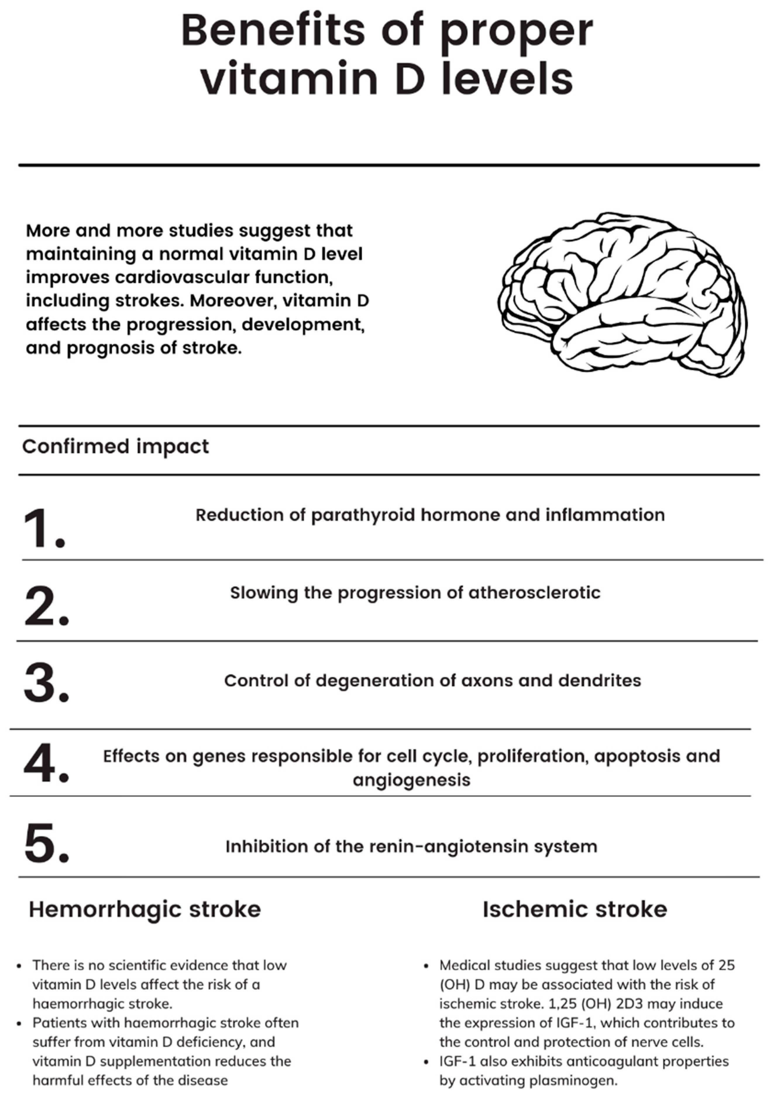
4. Vitamin D and Stroke Incidence
5. Gut–Brain Axis
6. The Effect of Vitamin D Supplementation on Post-Stroke Rehabilitation
7. Discussion
8. Conclusions
Author Contributions
Funding
Conflicts of Interest
References
- Deluca, H.F. History of the discovery of vitamin D and its active metabolites. BoneKEy Rep. 2014, 3, 479. [Google Scholar] [CrossRef] [PubMed] [Green Version]
- Muscogiuri, G.; Barrea, L.; Scannapieco, M.; Di Somma, C.; Scacchi, M.; Aimaretti, G.; Savastano, S.; Colao, A.; Marzullo, P. The lullaby of the sun: The role of vitamin D in sleep disturbance. Sleep Med. 2019, 54, 262–265. [Google Scholar] [CrossRef] [PubMed]
- Suvarna, V.; Mohan, V. Vitamin D and its role in coronavirus disease 2019 (COVID-19). J. Diabetol. 2020, 11, 71–80. [Google Scholar] [CrossRef]
- Rafeeq, H.; Ahmad, S.; Tareen, M.B.K.; Shahzad, K.; Bashir, A.; Jabeen, R.; Tariq, S.; Shehzadi, I. Biochemistry of Fat Soluble Vitamins, Sources, Biochemical Functions and Toxicity. J. Life Sci. 2020, 5, 188–196. [Google Scholar] [CrossRef]
- Holick, M.F. Vitamin D deficiency. N. Engl. J. Med. 2007, 357, 266–281. [Google Scholar] [CrossRef]
- Melamed, M.L.; Michos, E.D.; Post, W.; Astor, B. 25-hydroxyvitamin D levels and the risk of mortality in the general population. Arch. Intern. Med. 2008, 168, 1629–1637. [Google Scholar] [CrossRef]
- Ashouri, R.; Fangman, M.; Brielmaier, J.; Fields, Z.A.; Campo, N.; Doré, S. Nutritional Supplementation of Naturally Occurring Vitamin D to Improve Hemorrhagic Stroke Outcomes. Front. Neurol. 2021, 12, 670245. [Google Scholar] [CrossRef]
- Holick, M.F. Vitamin D: Important for prevention of osteoporosis, cardiovascular heart disease, type 1 diabetes, autoimmune diseases, and some cancers. South. Med. J. 2005, 98, 1024–1027. [Google Scholar] [CrossRef] [Green Version]
- Brøndum-Jacobsen, P.; Benn, M.; Jensen, G.B.; Nordestgaard, B.G. 25-hydroxyvitamin d levels and risk of ischemic heart disease, myocardial infarction, and early death: Population-based study and meta-analyses of 18 and 17 studies. Arterioscler. Thromb. Vasc. Biol. 2012, 32, 2794–2802. [Google Scholar] [CrossRef] [Green Version]
- Moan, J.; Dahlback, A.; Ma, L.; Juzeniene, A. Influenza, solar radiation and vitamin D. Derm.-Endocrinol. 2009, 1, 307–309. [Google Scholar] [CrossRef] [Green Version]
- Laaksi, I.; Ruohola, J.P.; Mattila, V.; Auvinen, A.; Ylikomi, T.; Pihlajamäki, H. Vitamin D supplementation for the prevention of acute respiratory tract infection: A randomized, double-blinded trial among young Finnish men. J. Infect. Dis. 2010, 202, 809–814. [Google Scholar] [CrossRef] [PubMed] [Green Version]
- Tripkovic, L.; Lambert, H.; Hart, K.; Smith, C.P.; Bucca, G.; Penson, S.; Chope, G.; Hyppönen, E.; Berry, J.; Vieth, R.; et al. Comparison of vitamin D2 and vitamin D3 supplementation in raising serum 25-hydroxyvitamin D status: A systematic review and meta-analysis. Am. J. Clin. Nutr. 2012, 95, 1357–1364. [Google Scholar] [CrossRef] [PubMed] [Green Version]
- Aspell, N.; Lawlor, B.; O’Sullivan, M. Is there a role for vitamin D in supporting cognitive function as we age? Proc. Nutr. Soc. 2018, 77, 124–134. [Google Scholar] [CrossRef] [PubMed]
- Holick, M.F. Sunlight and vitamin D for bone health and prevention of autoimmune diseases, cancers, and cardiovascular disease. Am. J. Clin. Nutr. 2004, 80, 1678S–1688S. [Google Scholar] [CrossRef] [Green Version]
- Fraikin, G.Y. Signaling Mechanisms Regulating Diverse Plant Cell Responses to UVB Radiation. Biochemistry 2018, 83, 787–794. [Google Scholar] [CrossRef]
- Nair, R.; Maseeh, A. Vitamin D: The “sunshine” vitamin. J. Pharmacol. Pharmacother. 2012, 3, 118–126. [Google Scholar] [CrossRef]
- Schuster, I. Cytochromes P450 are essential players in the vitamin D signaling system. Biochim. Biophys. Acta (BBA)-Proteins Proteom. 2011, 1814, 186–199. [Google Scholar] [CrossRef]
- Institute of Medicine (US) Committee to Review Dietary Reference Intakes for Vitamin D and Calcium. The national academies collection: Reports funded by national institutes of health. In Dietary Reference Intakes for Calcium and Vitamin D; Ross, A.C., Taylor, C.L., Yaktine, A.L., Del Valle, H.B., Eds.; National Academies Press: Cambridge, MA, USA; National Academy of Sciences: Washington, DC, USA, 2011. [Google Scholar]
- Holick, M.F. Vitamin D: A D-Lightful health perspective. Nutr. Rev. 2008, 66, S182–S194. [Google Scholar] [CrossRef]
- Holick, M.F. Resurrection of vitamin D deficiency and rickets. J. Clin. Investig. 2006, 116, 2062–2072. [Google Scholar] [CrossRef] [Green Version]
- Holick, M.F. The Vitamin D Deficiency Pandemic: A Forgotten Hormone Important for Health. Public Health Rev. 2010, 32, 267–283. [Google Scholar] [CrossRef] [Green Version]
- Siniscalchi, A.; Lochner, P.; Anticoli, S.; Chirchiglia, D.; De Sarro, G.; Gallelli, L. What is the Current Role for Vitamin D and the Risk of Stroke? Curr. Neurovasc. Res. 2019, 16, 178–183. [Google Scholar] [CrossRef] [PubMed]
- Yarlagadda, K.; Ma, N.; Doré, S. Vitamin D and Stroke: Effects on Incidence, Severity, and Outcome and the Potential Benefits of Supplementation. Front. Neurol. 2020, 11, 384. [Google Scholar] [CrossRef]
- Won, S.; Sayeed, I.; Peterson, B.L.; Wali, B.; Kahn, J.S.; Stein, D.G. Vitamin D prevents hypoxia/reoxygenation-induced blood-brain barrier disruption via vitamin D receptor-mediated NF-kB signaling pathways. PLoS ONE 2015, 10, e0122821. [Google Scholar] [CrossRef] [PubMed] [Green Version]
- Afzal, S.; Nordestgaard, B.G. Vitamin D, Hypertension, and Ischemic Stroke in 116 655 Individuals from the General Population: A Genetic Study. Hypertension 2017, 70, 499–507. [Google Scholar] [CrossRef] [PubMed]
- Sheerah, H.A.; Eshak, E.S.; Cui, R.; Imano, H.; Iso, H.; Tamakoshi, A. Relationship Between Dietary Vitamin D and Deaths From Stroke and Coronary Heart Disease: The Japan Collaborative Cohort Study. Stroke 2018, 49, 454–457. [Google Scholar] [CrossRef]
- Gepner, A.D.; Ramamurthy, R.; Krueger, D.C.; Korcarz, C.E.; Binkley, N.; Stein, J.H. A prospective randomized controlled trial of the effects of vitamin D supplementation on cardiovascular disease risk. PLoS ONE 2012, 7, e36617. [Google Scholar] [CrossRef] [PubMed] [Green Version]
- Momosaki, R.; Abo, M.; Urashima, M. Vitamin D Supplementation and Post-Stroke Rehabilitation: A Randomized, Dou-ble-Blind, Placebo-Controlled Trial. Nutrients 2019, 11, 1295. [Google Scholar] [CrossRef] [Green Version]
- Tague, S.E.; Smith, P.G. Vitamin D receptor and enzyme expression in dorsal root ganglia of adult female rats: Modulation by ovarian hormones. J. Chem. Neuroanat. 2011, 41, 1–12. [Google Scholar] [CrossRef] [Green Version]
- Anderson, P.H.; May, B.K.; Morris, H.A. Vitamin D metabolism: New concepts and clinical implications. Clin. Biochem. Rev. 2003, 24, 13–26. [Google Scholar]
- Siotto, M.; Santoro, M.; Aprile, I. Vitamin D and Rehabilitation after Stroke: Status of Art. Appl. Sci. 2020, 10, 1973. [Google Scholar] [CrossRef] [Green Version]
- Wikvall, K. Cytochrome P450 enzymes in the bioactivation of vitamin D to its hormonal form (review). Int. J. Mol. Med. 2001, 7, 201–209. [Google Scholar] [CrossRef] [PubMed]
- Prosser, D.E.; Jones, G. Enzymes involved in the activation and inactivation of vitamin D. Trends Biochem. Sci. 2004, 29, 664–673. [Google Scholar] [CrossRef] [PubMed]
- Eelen, G.; Verlinden, L.; van Camp, M.; van Hummelen, P.; Marchal, K.; de Moor, B.; Mathieu, C.; Carmeliet, G.; Bouillon, R.; Verstuyf, A. The effects of 1alpha,25-dihydroxyvitamin D3 on the expression of DNA replication genes. J. Bone Miner. Res. 2004, 19, 133–146. [Google Scholar] [CrossRef] [PubMed]
- Peterlik, M.; Cross, H.S. Vitamin D and calcium insufficiency-related chronic diseases: Molecular and cellular pathophysiology. Eur. J. Clin. Nutr. 2009, 63, 1377–1386. [Google Scholar] [CrossRef]
- Eyles, D.W.; Smith, S.; Kinobe, R.; Hewison, M.; McGrath, J.J. Distribution of the vitamin D receptor and 1 alpha-hydroxylase in human brain. J. Chem. Neuroanat. 2005, 29, 21–30. [Google Scholar] [CrossRef]
- Zelzer, S.; Meinitzer, A.; Herrmann, M.; Goessler, W.; Enko, D. A Novel Method for the Determination of Vitamin D Metabolites Assessed at the Blood-Cerebrospinal Fluid Barrier. Biomolecules 2021, 11, 1288. [Google Scholar] [CrossRef]
- DeLuca, G.C.; Kimball, S.M.; Kolasinski, J.; Ramagopalan, S.V.; Ebers, G.C. Review: The role of vitamin D in nervous system health and disease. Neuropathol. Appl. Neurobiol. 2013, 39, 458–484. [Google Scholar] [CrossRef]
- Kalueff, A.V.; Minasyan, A.; Keisala, T.; Kuuslahti, M.; Miettinen, S.; Tuohimaa, P. The vitamin D neuroendocrine system as a target for novel neurotropic drugs. CNS Neurol. Disord.-Drug Targets 2006, 5, 363–371. [Google Scholar] [CrossRef]
- Bivona, G.; Agnello, L.; Ciaccio, M. The immunological implication of the new vitamin D metabolism. Cent. Eur. J. Immunol. 2018, 43, 331–334. [Google Scholar] [CrossRef]
- Landel, V.; Stephan, D.; Cui, X.; Eyles, D.; Feron, F. Differential expression of vitamin D-associated enzymes and receptors in brain cell subtypes. J. Steroid. Biochem. Mol. Biol. 2018, 177, 129–134. [Google Scholar] [CrossRef]
- Eyles, D.; Brown, J.; Mackay-Sim, A.; McGrath, J.; Feron, F. Vitamin D3 and brain development. Neuroscience 2003, 118, 641–653. [Google Scholar] [CrossRef]
- Marini, F.; Bartoccini, E.; Cascianelli, G.; Voccoli, V.; Baviglia, M.G.; Magni, M.V.; Garcia-Gil, M.; Albi, E. Effect of 1alpha,25-dihydroxyvitamin D3 in embryonic hippocampal cells. Hippocampus 2010, 20, 696–705. [Google Scholar] [CrossRef] [PubMed]
- O’Loan, J.; Eyles, D.W.; Kesby, J.; Ko, P.; McGrath, J.J.; Burne, T.H. Vitamin D deficiency during various stages of pregnancy in the rat; its impact on development and behaviour in adult offspring. Psychoneuroendocrinology 2007, 32, 227–234. [Google Scholar] [CrossRef] [PubMed]
- Whitehouse, A.J.; Holt, B.J.; Serralha, M.; Holt, P.G.; Kusel, M.M.; Hart, P.H. Maternal serum vitamin D levels during pregnancy and offspring neurocognitive development. Pediatrics 2012, 129, 485–493. [Google Scholar] [CrossRef] [PubMed]
- Lucas, R.M.; Ponsonby, A.L.; Pasco, J.A.; Morley, R. Future health implications of prenatal and early-life vitamin D status. Nutr. Rev. 2008, 66, 710–720. [Google Scholar] [CrossRef] [PubMed]
- McGrath, J.; Welham, J.; Pemberton, M. Month of birth, hemisphere of birth and schizophrenia. Br. J. Psychiatry 1995, 167, 783–785. [Google Scholar] [CrossRef] [Green Version]
- Maddock, J.; Berry, D.J.; Geoffroy, M.C.; Power, C.; Hyppönen, E. Vitamin D and common mental disorders in mid-life: Cross-sectional and prospective findings. Clin. Nutr. 2013, 32, 758–764. [Google Scholar] [CrossRef]
- Armstrong, D.J.; Meenagh, G.K.; Bickle, I.; Lee, A.S.; Curran, E.S.; Finch, M.B. Vitamin D deficiency is associated with anxiety and depression in fibromyalgia. Clin. Rheumatol. 2007, 26, 551–554. [Google Scholar] [CrossRef]
- Annweiler, C.; Bartha, R.; Karras, S.N.; Gautier, J.; Roche, F.; Beauchet, O. Vitamin D and white matter abnormalities in older adults: A quantitative volumetric analysis of brain MRI. Exp. Gerontol. 2015, 63, 41–47. [Google Scholar] [CrossRef]
- van Schoor, N.M.; Comijs, H.C.; Llewellyn, D.J.; Lips, P. Cross-sectional and longitudinal associations between serum 25-hydroxyvitamin D and cognitive functioning. Int. Psychogeriatr. 2016, 28, 759–768. [Google Scholar] [CrossRef]
- Wilson, V.K.; Houston, D.K.; Kilpatrick, L.; Lovato, J.; Yaffe, K.; Cauley, J.A.; Harris, T.B.; Simonsick, E.M.; Ayonayon, H.N.; Kritchevsky, S.B.; et al. Relationship between 25-hydroxyvitamin D and cognitive function in older adults: The Health, Aging and Body Composition Study. J. Am. Geriatr. Soc. 2014, 62, 636–641. [Google Scholar] [CrossRef] [PubMed] [Green Version]
- Croll, P.H.; Boelens, M.; Vernooij, M.W.; van de Rest, O.; Zillikens, M.C.; Ikram, M.A.; Voortman, T. Associations of vitamin D deficiency with MRI markers of brain health in a community sample. Clin. Nutr. 2021, 40, 72–78. [Google Scholar] [CrossRef] [PubMed]
- Buell, J.S.; Dawson-Hughes, B. Vitamin D and neurocognitive dysfunction: Preventing “D”ecline? Mol. Asp. Med. 2008, 29, 415–422. [Google Scholar] [CrossRef] [PubMed] [Green Version]
- Brown, J.; Bianco, J.I.; McGrath, J.J.; Eyles, D.W. 1,25-dihydroxyvitamin D3 induces nerve growth factor, promotes neurite outgrowth and inhibits mitosis in embryonic rat hippocampal neurons. Neurosci. Lett. 2003, 343, 139–143. [Google Scholar] [CrossRef]
- Garcion, E.; Wion-Barbot, N.; Montero-Menei, C.N.; Berger, F.; Wion, D. New clues about vitamin D functions in the nervous system. Trends Endocrinol. Metab. 2002, 13, 100–105. [Google Scholar] [CrossRef]
- Wang, T.J.; Pencina, M.J.; Booth, S.L.; Jacques, P.F.; Ingelsson, E.; Lanier, K.; Benjamin, E.J.; D’Agostino, R.B.; Wolf, M.; Vasan, R.S. Vitamin D deficiency and risk of cardiovascular disease. Circulation 2008, 117, 503–511. [Google Scholar] [CrossRef] [Green Version]
- Talebi, A.; Amirabadizadeh, A.; Nakhaee, S.; Ahmadi, Z.; Mousavi-Mirzaei, S.M. Cerebrovascular disease: How serum phosphorus, vitamin D, and uric acid levels contribute to the ischemic stroke. BMC Neurol. 2020, 20, 116. [Google Scholar] [CrossRef]
- Perna, L.; Schöttker, B.; Holleczek, B.; Brenner, H. Serum 25-hydroxyvitamin D and incidence of fatal and nonfatal cardio-vascular events: A prospective study with repeated measurements. J. Clin. Endocrinol. Metab. 2013, 98, 4908–4915. [Google Scholar] [CrossRef] [Green Version]
- Michos, E.D.; Reis, J.P.; Post, W.S.; Lutsey, P.L.; Gottesman, R.F.; Mosley, T.H.; Sharrett, A.R.; Melamed, M.L. 25-Hydroxyvitamin D deficiency is associated with fatal stroke among whites but not blacks: The NHANES-III linked mor-tality files. Nutrition 2012, 28, 367–371. [Google Scholar] [CrossRef] [Green Version]
- Nibbelink, K.A.; Tishkoff, D.X.; Hershey, S.D.; Rahman, A.; Simpson, R.U. 1,25(OH)2-vitamin D3 actions on cell proliferation, size, gene expression, and receptor localization, in the HL-1 cardiac myocyte. J. Steroid Biochem. Mol. Biol. 2007, 103, 533–537. [Google Scholar] [CrossRef] [Green Version]
- Tarcin, O.; Yavuz, D.G.; Ozben, B.; Telli, A.; Ogunc, A.V.; Yuksel, M.; Toprak, A.; Yazici, D.; Sancak, S.; Deyneli, O.; et al. Effect of vitamin D deficiency and replacement on endothelial function in asymptomatic subjects. J. Clin. Endocrinol. Metab. 2009, 94, 4023–4030. [Google Scholar] [CrossRef] [PubMed] [Green Version]
- Dusso, A.S.; Brown, A.J.; Slatopolsky, E. Vitamin D. Am. J. Physiol. Ren. Physiol. 2005, 289, F8–F28. [Google Scholar] [CrossRef] [PubMed]
- Nagpal, S.; Na, S.; Rathnachalam, R. Noncalcemic actions of vitamin D receptor ligands. Endocr. Rev. 2005, 26, 662–687. [Google Scholar] [CrossRef] [PubMed]
- Masuda, S.; Jones, G. Promise of vitamin D analogues in the treatment of hyperproliferative conditions. Mol. Cancer Ther. 2006, 5, 797–808. [Google Scholar] [CrossRef] [PubMed] [Green Version]
- Li, Y.C.; Kong, J.; Wei, M.; Chen, Z.F.; Liu, S.Q.; Cao, L.P. 1,25-Dihydroxyvitamin D(3) is a negative endocrine regulator of the renin-angiotensin system. J. Clin. Investig. 2002, 110, 229–238. [Google Scholar] [CrossRef]
- Khundmiri, S.J.; Murray, R.D.; Lederer, E. PTH and Vitamin D. Compr. Physiol. 2016, 6, 561–601. [Google Scholar] [CrossRef]
- Yin, K.; You, Y.; Swier, V.; Tang, L.; Radwan, M.M.; Pandya, A.N.; Agrawal, D.K. Vitamin D Protects Against Atherosclerosis via Regulation of Cholesterol Efflux and Macrophage Polarization in Hypercholesterolemic Swine. Arterioscler. Thromb. Vasc. Biol. 2015, 35, 2432–2442. [Google Scholar] [CrossRef] [Green Version]
- Koyama, T.; Shibakura, M.; Ohsawa, M.; Kamiyama, R.; Hirosawa, S. Anticoagulant effects of 1alpha,25-dihydroxyvitamin D3 on human myelogenous leukemia cells and monocytes. Blood 1998, 92, 160–167. [Google Scholar] [CrossRef]
- Zhang, R.; Li, B.; Gao, X.; Tian, R.; Pan, Y.; Jiang, Y.; Gu, H.; Wang, Y.; Wang, Y.; Liu, G. Serum 25-hydroxyvitamin D and the risk of cardiovascular disease: Dose-response meta-analysis of prospective studies. J. Clin. Nutr. 2017, 105, 810–819. [Google Scholar] [CrossRef] [Green Version]
- Leung, R.Y.H.; Han, Y.; Sing, C.-W.; Cheung, B.M.; Wong, I.C.; Tan, K.C.; Kung, A.W.C.; Cheung, C.L. Serum 25-hydroxyvitamin D and the risk of stroke in Hong Kong Chinese. Thromb. Haemost. 2017, 117, 158–163. [Google Scholar] [CrossRef] [Green Version]
- Zhou, R.; Wang, M.; Huang, H.; Li, W.; Hu, Y.; Wu, T. Lower Vitamin D Status Is Associated with an Increased Risk of Ischemic Stroke: A Systematic Review and Meta-Analysis. Nutrients 2018, 10, 277. [Google Scholar] [CrossRef] [PubMed] [Green Version]
- Barbarawi, M.; Kheiri, B.; Zayed, Y.; Barbarawi, O.; Dhillon, H.; Swaid, B.; Yelangi, A.; Sundus, S.; Bachuwa, G.; Alkotob, M.L.; et al. Vitamin D Supplementation and Cardiovascular Disease Risks in More Than 83 000 Individuals in 21 Randomized Clinical Trials: A Meta-analysis. JAMA Cardiol. 2019, 4, 765–776. [Google Scholar] [CrossRef] [PubMed]
- Su, C.; Jin, B.; Xia, H.; Zhao, K. Association between Vitamin D and Risk of Stroke: A PRISMA-Compliant Systematic Review and Meta-Analysis. Eur. Neurol. 2021, 84, 399–408. [Google Scholar] [CrossRef]
- Yalbuzdag, S.A.; Sarifakioglu, B.; Afsar, S.I.; Celik, C.; Can, A.; Yegin, T.; Senturk, B.; Guzelant, A.Y. Is 25(OH)D Associated with Cognitive Impairment and Functional Improvement in Stroke? A Retrospective Clinical Study. J. Stroke Cerebrovasc. Dis. 2015, 24, 1479–1486. [Google Scholar] [CrossRef] [PubMed]
- Kilkkinen, A.; Knekt, P.; Aro, A.; Rissanen, H.; Marniemi, J.; Heliövaara, M.; Impivaara, O.; Reunanen, A. Vitamin D status and the risk of cardiovascular disease death. Am. J. Epidemiol. 2009, 170, 1032–1039. [Google Scholar] [CrossRef]
- Makariou, S.E.; Michel, P.; Tzoufi, M.S.; Challa, A.; Milionis, H.J. Vitamin D and stroke: Promise for prevention and better outcome. Curr. Vasc. Pharmacol. 2014, 12, 117–124. [Google Scholar] [CrossRef]
- Asif, A.; Farooq, N. Vitamin D toxicity. In StatPearls; StatPearls Publishing LLC: Treasure Island, FL, USA, 2022. [Google Scholar]
- Miao, H.; Zhu, H.; Luan, X.; Huang, G.; Chen, M.; Yuan, Z.; Wang, Z. Risk Factors of Vitamin D Deficiency in Chinese Is-chemic Stroke Patients: A Cross-Sectional Study. Front. Aging Neurosci. 2020, 12, 613498. [Google Scholar] [CrossRef]
- Tu, W.J.; Zhao, S.J.; Xu, D.J.; Chen, H. Serum 25-hydroxyvitamin D predicts the short-term outcomes of Chinese patients with acute ischaemic stroke. Clin. Sci. 2014, 126, 339–346. [Google Scholar] [CrossRef] [Green Version]
- Wajda, J.; Świat, M.; Owczarek, A.J.; Brzozowska, A.; Olszanecka-Glinianowicz, M.; Chudek, J. Severity of Vitamin D Deficiency Predicts Mortality in Ischemic Stroke Patients. Dis. Markers 2019, 2019, 3652894. [Google Scholar] [CrossRef]
- Wei, Z.N.; Kuang, J.G. Vitamin D deficiency in relation to the poor functional outcomes in nondiabetic patients with ischemic stroke. Biosci. Rep. 2018, 38, BSR20171509. [Google Scholar] [CrossRef] [Green Version]
- Suthar, O.P.; Mathur, S.; Gupta, V.; Agarwal, H.; Mathur, A.; Singh, P.; Sharma, S.L. Study of Correlation of Serum Vitamin D Levels with Arterial Stiffness and Cardiovascular Morbidity in Elderly Individuals of Western Rajasthan. J. Assoc. Physicians India 2018, 66, 18–21. [Google Scholar]
- Feng, C.; Tang, N.; Huang, H.; Zhang, G.; Qi, X.; Shi, F. 25-Hydroxy vitamin D level is associated with total MRI burden of cerebral small vessel disease in ischemic stroke patients. Int. J. Neurosci. 2019, 129, 49–54. [Google Scholar] [CrossRef] [PubMed]
- Michos, E.D.; Melamed, M.L. Vitamin D and cardiovascular disease risk. Curr. Opin. Clin. Nutr. Metab. Care 2008, 11, 7–12. [Google Scholar] [CrossRef] [PubMed]
- Kashefiolasl, S.; Leisegang, M.S.; Helfinger, V.; Schürmann, C.; Pflüger-Müller, B.; Randriamboavonjy, V.; Vasconez, A.E.; Carmeliet, G.; Badenhoop, K.; Hintereder, G.; et al. Vitamin D-A New Perspective in Treatment of Cerebral Vasospasm. Neurosurgery 2021, 88, 674–685. [Google Scholar] [CrossRef] [PubMed]
- Turetsky, A.; Goddeau, R.P., Jr.; Henninger, N. Low Serum Vitamin D Is Independently Associated with Larger Lesion Volumes after Ischemic Stroke. J. Stroke Cerebrovasc. Dis. 2015, 24, 1555–1563. [Google Scholar] [CrossRef]
- Nie, Z.; Ji, X.C.; Wang, J.; Zhang, H.X. Serum levels of 25-hydroxyvitamin D predicts infarct volume and mortality in ischemic stroke patients. J. Neuroimmunol. 2017, 313, 41–45. [Google Scholar] [CrossRef]
- Huang, H.; Zheng, T.; Wang, S.; Wei, L.; Wang, Q.; Sun, Z. Serum 25-hydroxyvitamin D predicts early recurrent stroke in ischemic stroke patients. Nutr. Metab. Cardiovasc. Dis. 2016, 26, 908–914. [Google Scholar] [CrossRef]
- Lasek-Bal, A.; Jedrzejowska-Szypulka, H.; Student, S.; Warsz-Wianecka, A.; Zareba, K.; Puz, P.; Bal, W.; Pawletko, K.; Lewin-Kowalik, J. The importance of selected markers of inflammation and blood-brain barrier damage for short-term ischemic stroke prognosis. J. Physiol. Pharmacol. 2019, 70, 209–217. [Google Scholar] [CrossRef]
- Di Napoli, M.; Singh, P. Is plasma fibrinogen useful in evaluating ischemic stroke patients? Why, how, and when. Stroke 2009, 40, 1549–1552. [Google Scholar] [CrossRef] [Green Version]
- Rasyid, A.; Harris, S.; Kurniawan, M.; Mesiano, T.; Hidayat, R. Fibrinogen and LDL Influence on Blood Viscosity and Out-come of Acute Ischemic Stroke Patients in Indonesia. Ann. Neurosci. 2019, 26, 30–34. [Google Scholar] [CrossRef]
- Tao, L.; ShiChuan, W.; DeTai, Z.; Lihua, H. Evaluation of lipoprotein-associated phospholipase A2, serum amyloid A, and fibrinogen as diagnostic biomarkers for patients with acute cerebral infarction. J. Clin. Lab. Anal. 2020, 34, e23084. [Google Scholar] [CrossRef] [PubMed]
- Xiao, M.; Xiao, Z.J.; Yang, B.; Lan, Z.; Fang, F. Blood-Brain Barrier: More Contributor to Disruption of Central Nervous System Homeostasis Than Victim in Neurological Disorders. Front. Neurosci. 2020, 14, 764. [Google Scholar] [CrossRef] [PubMed]
- Belcher, J.D.; Chen, C.; Nguyen, J.; Milbauer, L.; Abdulla, F.; Alayash, A.I.; Smith, A.; Nath, K.A.; Hebbel, R.P.; Vercellotti, G.M. Heme triggers TLR4 signaling leading to endothelial cell activation and vaso-occlusion in murine sickle cell disease. Blood 2014, 123, 377–390. [Google Scholar] [CrossRef] [PubMed] [Green Version]
- Judd, S.E.; Morgan, C.J.; Panwar, B.; Howard, V.J.; Wadley, V.G.; Jenny, N.S.; Kissela, B.M.; Gutiérrez, O.M. Vitamin D de-ficiency and incident stroke risk in community-living black and white adults. Int. J. Stroke 2016, 11, 93–102. [Google Scholar] [CrossRef] [PubMed]
- D’Hellencourt, C.L.; Montero-Menei, C.N.; Bernard, R.; Couez, D. Vitamin D3 inhibits proinflammatory cytokines and nitric oxide production by the EOC13 microglial cell line. J. Neurosci. Res. 2003, 71, 575–582. [Google Scholar] [CrossRef]
- Zielińska-Nowak, E.; Cichon, N.; Saluk-Bijak, J.; Bijak, M.; Miller, E. Nutritional Supplements and Neuroprotective Diets and Their Potential Clinical Significance in Post-Stroke Rehabilitation. Nutrients 2021, 13, 2704. [Google Scholar] [CrossRef]
- Yang, F.Z.; Jehu, D.A.M.; Ouyang, H.; Lam, F.M.H.; Pang, M.Y.C. The impact of stroke on bone properties and muscle-bone relationship: A systematic review and meta-analysis. Osteoporos. Int. 2020, 31, 211–224. [Google Scholar] [CrossRef]
- Carda, S.; Cisari, C.; Invernizzi, M.; Bevilacqua, M. Osteoporosis after stroke: A review of the causes and potential treatments. Cerebrovasc. Dis. 2009, 28, 191–200. [Google Scholar] [CrossRef]
- Montiel-Castro, A.J.; González-Cervantes, R.M.; Bravo-Ruiseco, G.; Pacheco-López, G. The microbiota-gut-brain axis: Neurobehavioral correlates, health and sociality. Front. Integr. Neurosci. 2013, 7, 70. [Google Scholar] [CrossRef] [Green Version]
- Srikantha, P.; Mohajeri, M.H. The Possible Role of the Microbiota-Gut-Brain-Axis in Autism Spectrum Disorder. Int. J. Mol. Sci. 2019, 20, 2115. [Google Scholar] [CrossRef] [Green Version]
- Levy, M.; Kolodziejczyk, A.A.; Thaiss, C.A.; Elinav, E. Dysbiosis and the immune system. Nat. Rev. Immunol. 2017, 17, 219–232. [Google Scholar] [CrossRef] [PubMed]
- Battaglini, D.; Pimentel-Coelho, P.M.; Robba, C.; dos Santos, C.C.; Cruz, F.F.; Pelosi, P.; Rocco, P.R.M. Gut Microbiota in Acute Ischemic Stroke: From Pathophysiology to Therapeutic Implications. Front. Neurol. 2020, 11, 598. [Google Scholar] [CrossRef] [PubMed]
- Yamashiro, K.; Kurita, N.; Urabe, T.; Hattori, N. Role of the Gut Microbiota in Stroke Pathogenesis and Potential Therapeutic Implications. Ann. Nutr. Metab. 2021, 77 (Suppl. 2), 36–44. [Google Scholar] [CrossRef] [PubMed]
- Singh, P.; Rawat, A.; Alwakeel, M.; Sharif, E.; Al Khodor, S. The potential role of vitamin D supplementation as a gut mi-crobiota modifier in healthy individuals. Sci. Rep. 2020, 10, 21641. [Google Scholar] [CrossRef] [PubMed]
- Charoenngam, N.; Shirvani, A.; Kalajian, T.A.; Song, A.; Holick, M.F. The Effect of Various Doses of Oral Vitamin D(3) Supplementation on Gut Microbiota in Healthy Adults: A Randomized, Double-blinded, Dose-response Study. Anticancer Res. 2020, 40, 551–556. [Google Scholar] [CrossRef]
- Kanhere, M.; He, J.; Chassaing, B.; Ziegler, T.R.; Alvarez, J.A.; Ivie, E.A.; Hao, L.; Hanfelt, J.; Gewirtz, A.T.; Tangpricha, V. Bolus Weekly Vitamin D3 Supplementation Impacts Gut and Airway Microbiota in Adults With Cystic Fibrosis: A Dou-ble-Blind, Randomized, Placebo-Controlled Clinical Trial. J. Clin. Endocrinol. Metab. 2018, 103, 564–574. [Google Scholar] [CrossRef] [Green Version]
- Cantarel, B.L.; Waubant, E.; Chehoud, C.; Kuczynski, J.; DeSantis, T.Z.; Warrington, J.; Venkatesan, A.; Fraser, C.M.; Mowry, E.M. Gut Microbiota in Multiple Sclerosis. J. Investig. Med. 2015, 63, 729. [Google Scholar] [CrossRef]
- Amadi, C.N.; Orish, C.N.; Frazzoli, C.; Orisakwe, O.E. Dietary interventions for autism spectrum disorder: An updated systematic review of human studies. Psychiatriki 2022, in press. [Google Scholar] [CrossRef]
- Ogbu, D.; Xia, E.; Sun, J. Gut instincts: Vitamin D/vitamin D receptor and microbiome in neurodevelopment disorders. Open Biol. 2020, 10, 200063. [Google Scholar] [CrossRef]
- Hussein, H.M.; Elyamany, M.F.; Rashed, L.A.; Sallam, N.A. Vitamin D mitigates diabetes-associated metabolic and cognitive dysfunction by modulating gut microbiota and colonic cannabinoid receptor 1. Eur. J. Pharm. Sci. 2022, 170, 106105. [Google Scholar] [CrossRef]
- Akimbekov, N.S.; Digel, I.; Sherelkhan, D.K.; Lutfor, A.B.; Razzaque, M.S. Vitamin D and the Host-Gut Microbiome: A Brief Overview. Acta Histochem. Cytochem. 2020, 53, 33–42. [Google Scholar] [CrossRef] [PubMed]
- Krutzik, S.R.; Hewison, M.; Liu, P.T.; Robles, J.A.; Stenger, S.; Adams, J.S.; Modlin, R.L. IL-15 links TLR2/1-induced mac-rophage differentiation to the vitamin D-dependent antimicrobial pathway. J. Immunol. 2008, 181, 7115–7120. [Google Scholar] [CrossRef] [PubMed]
- Kempker, J.A.; Han, J.E.; Tangpricha, V.; Ziegler, T.R.; Martin, G.S. Vitamin D and sepsis: An emerging relationship. Derm.-Endocrinol. 2012, 4, 101–108. [Google Scholar] [CrossRef] [PubMed]
- Zhang, Y.G.; Wu, S.; Sun, J. Vitamin D, Vitamin D Receptor, and Tissue Barriers. Tissue Barriers 2013, 1, e23118. [Google Scholar] [CrossRef]
- Gupta, A.; Prabhakar, S.; Modi, M.; Bhadada, S.K.; Kalaivani, M.; Lal, V.; Khurana, D. Effect of Vitamin D and calcium supplementation on ischaemic stroke outcome: A randomised controlled open-label trial. Int. J. Clin. Pract. 2016, 70, 764–770. [Google Scholar] [CrossRef]
- Sari, A.; Durmus, B.; Karaman, C.A.; Ogut, E.; Aktas, I. A randomized, double-blind study to assess if vitamin D treatment affects the outcomes of rehabilitation and balance in hemiplegic patients. J. Phys. Ther. Sci. 2018, 30, 874–878. [Google Scholar] [CrossRef] [Green Version]
- Narasimhan, S.; Balasubramanian, P. Role of Vitamin D in the Outcome of Ischemic Stroke—A Randomized Controlled Trial. J. Clin. Diagn. Res. 2017, 11, CC6–CC10. [Google Scholar] [CrossRef]
- Torrisi, M.; Bonanno, L.; Formica, C.; Arcadi, F.A.; Cardile, D.; Cimino, V.; Bramanti, P.; Morini, E. The role of rehabilitation and vitamin D supplementation on motor and psychological outcomes in poststroke patients. Medicine 2021, 100, e27747. [Google Scholar] [CrossRef]
- Karasu, A.U.; Karataş, G.K. Effect of vitamin D supplementation on lower extremity motor function and ambulation in stroke patients. Turk. J. Med. Sci. 2021, 51, 1413–1419. [Google Scholar] [CrossRef]
- Schilling, S. Epidemic vitamin D deficiency among patients in an elderly care rehabilitation facility. Dtsch. Arztebl. Int. 2012, 109, 33–38. [Google Scholar] [CrossRef]
- Neo, J.J.; Kong, K.H. Prevalence of Vitamin D Deficiency in Elderly Patients Admitted to an Inpatient Rehabilitation Unit in Tropical Singapore. Rehabil. Res. Pract. 2016, 2016, 9689760. [Google Scholar] [CrossRef] [PubMed] [Green Version]
- Bakradze, E.; McCullough, L.; Staff, I.; Nouh, A. Vitamin D deficiency correlates with stroke severity on presentation in in-tracerebral hemorrhage. Stroke 2016, 47, ATP375. [Google Scholar] [CrossRef]
- Türkanoğlu Özçelik, A.; Öner, T.; Can Demirdöğen, B.; Bek, V.S.; Demirkaya, S.; Adalı, O. Genetic polymorphisms of vit-amin D3 metabolizing CYP24A1 and CYP2R1 enzymes in Turkish patients with ischemic stroke. Neurol. Res. 2018, 40, 364–371. [Google Scholar] [CrossRef] [PubMed]


| Study, Year, Reference | Types of Criteria | |||||
|---|---|---|---|---|---|---|
| Type of Stroke | Testing Serum Vitamin D Levels before the Study | Age | How to Evaluate Vitamin D | Exclusions | Other | |
| Narasimhan et al., 2017 [119]. | Ischemic, middle cerebral artery ischemia | Serum 25-hydroxy vitamin D level 21–29 ng/mL and Serum 25-hydroxy vitamin D level ≤ 20 ng/mL | 50–80 years | Measurement of serum 25-hydroxyvitamin D by electrochemiluminescence binding assay. Interpretation of the result according to the US Endocrine Society | Hemorrhagic stroke, large MCA stroke, lacunar stroke, thrombolytic therapy, very poor general condition, multiple organ failure, normal vitamin D levels (serum 25-hydroxyvitamin D concentration ≥ 30 ng/mL) | Diagnosis of stroke, clinical evaluative, CT, MRI |
| Momosaki et al., 2019 [28]. | Cerebral infarction, intracerebral hemorrhage or subarachnoid hemorrhage | Serum 25-hydroxyvitamin D level was not included as an inclusion criterion | At least 20 years | Irrelevant | History of stones in the urinary tract, vitamin D3 or activated vitamin D supplementation before stroke, osteoporosis, bone structure, dysphagia, or other disorder that would make it difficult to take an oral vitamin D supplement, inability to participate in the study in the opinion of the attending physiologist | First stroke in life, admission to the convalescent rehabilitation unit after acute stroke treatment, deemed by the attending physiologist to require 8 weeks of inpatient rehabilitation |
| Torrisi et al., 2021 [120]. | Ischemic/hemorrhage stroke | Irrelevant, Serum 25-hydroxyvitamin D levels, were tested, but this was not a criterion | Irrelevant | Irrelevant | Mini Mental State Examination < 15, psychiatric conditions/treatment with antidepressants, patients already on vitamin D supplementation, also in combination with calcium, multivitamins or other medications, and conditions that do not allow for a neurorehabilitation program | Stroke that occurred between 30 and 60 days before, eligibility for rehabilitation treatment |
| Karasu et al., 2021 [121]. | Ischemic/hemorrhage stroke | Serum 25-hydroxyvitamin D (25(OH)D) levels measured in ng/mL | Irrelevant | Irrelevant | No pre-rehabilitation measurement of vitamin D levels, chronic kidney, liver, or lung disease that may affect vitamin D levels, current steroid treatment, previous orthopedic problems known to affect lower extremity function | First stroke in life, diagnosis of stroke, clinical evaluative, CT, MRI, inpatient stroke rehabilitation treatment May 2018–February 2020 |
| Sari et al., 2018 [118]. | Ischemic stroke | Measured during winter, <30 ng/mL | Irrelevant | Measurement of 25 (OH) vitamin D3 using the RIA CT kit (BioSource Europe SA, Nivelles, Belgium) by radioimmunoassay | End-stage disease (cancer) or disease other than stroke that may affect balance or mobility (e.g., multiple sclerosis, Parkinson’s disease, or pelvic and lower extremity surgery), limit sun exposure (e.g., acquired vitiligo and psoriasis), or affect vitamin D levels (e.g., chronic renal failure and celiac disease) | Current hemiplegia after stroke, Hospitalization for neurological rehabilitation (hemiplegia) between September 2014 and March 2015, no cerebrovascular disease, at least a 2-month gap that has passed since the last stroke |
| Gupta et al., 2016 [117]. | Ischemic stroke | 25(OH)D concentration < 75 nmol/L | Age ≥ 35 years | Serum 25(OH)D concentration measured by chemiluminescence | Previously taken vitamin D and calcium supplementation, thrombolysis performed, kidney and liver dysfunction | MRS (Modified Rankin Score) before stroke < 2, diagnosis of stroke, CT, MRI, |
| Study, Year, Reference | Type of Study | Duration of the Study | Amount and Method of Administration of Vitamin D | Scales, Tests | Results | Conclusions |
|---|---|---|---|---|---|---|
| Narasimhan et al., 2017 [119]. | Randomized, controlled, unblinded | 3 months | Single dose of 6 lac IU cholecalciferol, intramuscular injection IIM) | Scandinavian stroke scale (SSS)—stroke severity assessment | The differences in SSS from admission and after three months in group A—study (6.39 ± 4.56) and group B—control (2.50 ± 2.20) were statistically analyzed and found to be highly significant (p < 0.001) | After three months, there was a significant improvement in stroke outcome in patients who received vitamin D |
| Momosaki et al., 2019 [28]. | Randomized, multicenter, double-blind, placebo-controlled trial | 8 weeks | Vitamin D3, 400 IU, 5 times daily (2000 IU vit. D3 per day), oral tablets | Barthel Index score, Brunnstrom stage (arm, hand, and leg on the affected side), hand grip strength (bilaterally), and calf circumference (bilaterally) | The mean (±standard deviation) increase in Barthel Index score was 19.0 ± 14.8 in the supplementation group and 19.5 ± 13.1 in the placebo group (p = 0.88). The effectiveness of the Barthel Index was 0.32 ± 0.31 in the supplementation group and 0.28 ± 0.21 in the placebo group (p = 0.38). There were no differences between groups in other secondary endpoints | Oral vitamin D3 supplementation does not improve rehabilitation outcomes after acute stroke. |
| Torrisi et al., 2021 [120]. | Randomized, double-blind, parallel, monocentric, clinical | 3 months | 2000 IU/day cholecalciferol, oral | Montgomery Aasberg Depression Rating Scale (MADR), Functional Independent Measures (FIM) | In the vitamin D group, we highlighted significant differences between T0 and T1 in calcium (p < 0.001), vitamin D (p < 0.001), MADR (p = 0.001), and FIM (p < 0.001). In the health control group, we found a significant difference in calcium (p = 0.003), vitamin D (p < 0.001), MADR (p = 0.006), overall sense of self-efficacy (p = 0.009), and in FIM (p < 0.001) | Beneficial effects on improved mood and function are mainly due to neurorehabilitation rather than vitamin D supplementation |
| Karasu et al., 2021 [121]. | Retrospective | 3 months | Weekly vitamin D supplementation (50,000 IU) for 4–12 weeks, orally, total vitamin D intake ranged from 200,000 to 600,000 IU | Brunnstrom recovery stage (lower extremity), (BRS), functional ambulation classification (FAC) | At the end of rehabilitation, the change in FAC and Brunnstrom scores was higher in patients receiving vitamin D supplementation (p = 0.005 p = 0.018). The change in FAC and Brunnstrom scores in patients undergoing rehabilitation for the first time and/or within the first 3 months after stroke was higher in the group receiving vitamin D supplementation compared with the group not receiving vitamin D (p < 0.05) | Vitamin D supplementation may increase the effectiveness of rehabilitation therapy in patients during the first 3 months after stroke |
| Sari et al., 2018 [118]. | Randomized, double-blind, placebo-controlled | 3 months | 300,000 IU Vitamin D, 2 mL fluid, intramuscular injection (IM) | Brunnstrom recovery staging (BRS), functional ambulation scale (FAS), modified Barthel index (MBI) scores, Berg balance scale (BBS) | By the end of the third month, The Berg balance scale results and modified Barthel index scores significantly differed between the two groups, whereas Brunnstrom recovery staging and functional ambulation scale test results did not. | Vitamin D administration increased activity levels and accelerated recovery, but did not significantly affect movement or motor recovery |
| Gupta et al., 2016 [117]. | Randomized, controlled, open-label | 6 months | Single intramuscular injection of 600,000 IU cholecalciferol, oral cholecalciferol 60,000 IU once a month with one gram elemental calcium daily | Modified Rankin scale (mRS) | Serum 25(OH)D levels increased by 47.3 (25.0–69.5) nmol/L in the vitamin D and calcium supplementation group (p < 0.001) and by 0.8 (−7.5–8.8) nmol/L in the group receiving usual care without supplementation (p = 0.86) up to 6 months. Patients who received usual care were 2–3 times more likely to be vitamin D deficient/deficient at 6 months compared with those who received vitamin D and calcium supplementation | After 6 months 11 patients (52.4%)—mRS score between 0 and 2 in the vitamin D plus calcium-supplemented arm and 10 patients (43.5%) had a good outcome in the usual care arm, Adjusted OR 1.9, 95% CI 0.6–6.4; p = 0.31, Risk difference 4.7% |
Publisher’s Note: MDPI stays neutral with regard to jurisdictional claims in published maps and institutional affiliations. |
© 2022 by the authors. Licensee MDPI, Basel, Switzerland. This article is an open access article distributed under the terms and conditions of the Creative Commons Attribution (CC BY) license (https://creativecommons.org/licenses/by/4.0/).
Share and Cite
Marek, K.; Cichoń, N.; Saluk-Bijak, J.; Bijak, M.; Miller, E. The Role of Vitamin D in Stroke Prevention and the Effects of Its Supplementation for Post-Stroke Rehabilitation: A Narrative Review. Nutrients 2022, 14, 2761. https://doi.org/10.3390/nu14132761
Marek K, Cichoń N, Saluk-Bijak J, Bijak M, Miller E. The Role of Vitamin D in Stroke Prevention and the Effects of Its Supplementation for Post-Stroke Rehabilitation: A Narrative Review. Nutrients. 2022; 14(13):2761. https://doi.org/10.3390/nu14132761
Chicago/Turabian StyleMarek, Klaudia, Natalia Cichoń, Joanna Saluk-Bijak, Michał Bijak, and Elżbieta Miller. 2022. "The Role of Vitamin D in Stroke Prevention and the Effects of Its Supplementation for Post-Stroke Rehabilitation: A Narrative Review" Nutrients 14, no. 13: 2761. https://doi.org/10.3390/nu14132761