Article

Chemical Composition and Toxicological Evaluation of Landfill Leachate from Białystok, Poland

by
Agata Jabłońska-Trypuć
1,*,
Elżbieta Wołejko
1,
Urszula Wydro
1,
Jacek Leszczyński
1,
Mariola Wasil
1 and
Anna Kiełtyka-Dadasiewicz
2
1
Faculty of Civil Engineering and Environmental Sciences, Bialystok University of Technology, 15-351 Bialystok, Poland
2
Department of Plant Production Technology and Commodity, University of Life Sciences in Lublin, 20-950 Lublin, Poland
*
Author to whom correspondence should be addressed.
Sustainability 2023, 15(23), 16497; https://doi.org/10.3390/su152316497
Submission received: 11 November 2023 / Revised: 29 November 2023 / Accepted: 30 November 2023 / Published: 1 December 2023
(This article belongs to the Special Issue Safe Disposal of Solid Waste in Landfill)

Abstract

:
Leachates from landfills constitute a very complex environmental matrix with potentially toxic properties for both the environment and humans. Due to this fact, toxicological tests were carried out on landfill leachate (LL) obtained from the local landfill in Hryniewicze (Poland). The analyses included physicochemical studies of leachates and their impact on soil and plants, and studies conducted in bacterial models and human cell models. The results obtained indicate high contents of metals and organic matter, especially dangerous PAHs, in the tested leachate. This results in the influence of LL on changes in the content of assimilation pigments and oxidative stress observed in plants grown in soil fed with leachates. The effect of leachate on the growth of Sporosarcina pasteurii, Staphylococcus aureus, Lactobacillus rhamnosus, Saccharomyces boulardii and Candida albicans varied depending on the strain and LL dose. A particularly significant increase in proliferation after exposure to LL was noted for S. aureus. In studies conducted on human cancer cell lines representing three types of glioblastomas and one type of colorectal adenocarcinoma, a particularly significant increase in the viability of cells treated with LL was noted for the DLD-1 cell line. The results obtained, especially the stimulation of the growth of cancer cells and an increase in the number of pathogenic bacteria, indicate the potential toxic properties of the tested leachates. This is confirmed by the high level of oxidative stress in plants. The results indicate the need for continuous monitoring of waste landfills and leachates generated there.

1. Introduction

Waste landfills are facilities built for depositing mainly municipal waste. They must meet certain conditions, such as proper location, and technical and operational parameters. In Poland, despite attempts to reduce the number of landfills, they are still one of the most common methods of waste disposal [1,2]. Some of the waste is recycled, some is subject to thermal transformation and composting, but nearly 40% of waste is still stored in a traditional way. This method poses the greatest threat to the natural environment and humans [3].
A landfill is a kind of biochemical reactor in which continuous chemical reactions, and biochemical and microbiological transformations take place. The changes taking place are influenced by atmospheric factors and the presence of microorganisms [4,5]. A landfill is a highly heterogeneous environment, which results from the fact that different transformations occur simultaneously in different areas and different stages of decomposition processes coexist [5,6].
According to EU Council Directive 1999/31/EC, landfill leachate (LL) is described as “any liquid percolating through landfilled waste and emitted from/or contained in a landfill”. Leachates are generated by direct infiltration of rainwater through the landfill body, but also as a result of changes taking place in the landfill itself [7]. The composition of leachates depends on many factors, including the composition of the stored waste, the method of its storage, the density of the stored waste, the age of the landfill and the rate of formation of new layers. The technology of landfill construction and the method of its operation are also important. The composition of leachates includes organic components (organic nitrogen, volatile fatty acids, alkanes, alcohols, aldehydes, terpenes, and aromatic hydrocarbons), inorganic components (ammonium nitrogen, nitrates III and V, sulphates, bicarbonates, chlorides, sodium, and potassium) and heavy metals (arsenic, mercury, iron, cadmium, copper, nickel, and lead). Key indicators in monitoring leachate from landfills are pH, specific electrolytic conductivity, total organic carbon, the sum of polycyclic aromatic hydrocarbons and the content of individual heavy metals, such as copper, zinc, lead, cadmium, chromium (VI) and mercury [8,9].
When considering the potential toxicity of landfill leachates, it should be taken into account that they are mixtures of many substances, which may act synergistically or even additively with each other. However, the complexity of these mixtures makes it impossible to determine which substance is toxic in the leachate. It should also not be forgotten that, in addition to penetrating into soils and groundwater, leachates may be emitted in the form of bioaerosols and biogas. Bioaerosols contain microbiological factors that are potentially dangerous and infectious to humans, while biogas is produced as a result of uncontrolled biochemical transformations deposited in a landfill [10,11].
Research on the impact of waste landfills on human health is often associated with significant methodological limitations. One such limitation is the difficulty in estimating the level of population exposure to chemicals found in leachates. In our previous experiment, we also examined the potential toxicity of leachate from a landfill near Bialystok, but we focused on different research models and conducted the experiment in a different way. In the above-mentioned manuscript, we demonstrated stimulation of the growth of bacterial cells exposed to a wide range of LL concentrations, while in the case of human cells we obtained a toxic effect for healthy cells and a stimulating effect for the growth of cancer cells [12].
The aim of this work is to examine the impact of leachate from a local municipal waste landfill on various elements of the ecosystem, including humans. For this purpose, the chemical composition of leachates, their physicochemical properties, their influence on soil properties and plant metabolism, and the effect of leachates on selected bacterial strains and selected cell lines were analyzed. The novelty of this research study is the application of a wide range of bioassays, including plant metabolism analysis and microbial bioassays, and in vitro human cell culture, as a biological model for toxicological studies. This approach will allow for the complex estimation of LL influence on different ecosystem elements, including soil, plant, human and environmental microbiomes and the human organism.

2. Materials and Methods

2.1. LL Sample Collecting

Samples of landfill leachate were collected in December 2022 from a solid waste landfill located in the northeastern part of Poland (Hryniewicze) and prepared according to our previous studies [12]. The main physicochemical properties of the LL are summarized in the Table 1.

2.2. Pot Experiment Design and Plant Sampling

In order to determine the effect of LL on plants, a pot experiment was prepared according to the method described by Wydro et al. [13]. The properties of the soil on which the experiment was carried out are summarized in Table 1. Nitrogen doses were calculated according to the following data: a single dose of 85 kg/ha means 50% of the N dose, 50 LL, while 170 kg/ha means 100% of the N dose, 100 LL. Seeds of a lawn grass mixture containing Lollium perenne, Festuca rubra and Poa pratensis were sown before LL application. The prepared pots with plants were watered with distilled water until the humidity reached 60–70% of the maximum water-holding capacity. The pot experiment was conducted for 6 weeks, after which plant samples were collected to determine the physiological parameters. In addition, soil filtrates were prepared according to the procedure described by Ming et al. and Wydro et al. [13,14].

2.3. Soil Filtrates Physicochemical Characteristics

Physicochemical properties of soil filtrates, such as pH, total organic carbon (TOC), heavy metals (Ni, Cr, Mn, Fe, Cd, Cu, Zn and Pb) content, electrical conductivity (EC), inorganic ions (PO43−, SO42−, Cl, NO3) content and 16 PAHs content were analyzed according to [12].

2.4. Assimilation Dyes Content in Plants

1 g of fresh plant sample was ground in a mortar with the addition of calcium carbonate and 20 mL of 90% methanol. The obtained suspension was filtered through filter paper. The absorbance in the plant extract was determined at the following wavelengths: 470 nm, 665 nm, 652 nm to determine the content of chlorophyll a (Chla), chlorophyll b (Chlb) and carotenoids (carot). After adding 50% HCl to the extract, the absorbance was determined at a wavelength of 654 nm to determine the content of total pheophytins (Phe(a + b)). The dye contents were determined from Equations (1)–(4) and expressed as mg/g of fresh weight (mg/g fw). Moreover, the ratio of Chla/Chlb was calculated.
Chla = ((16.82 × A665−9.28 × A652) × V)/(m × 1000)
Chlb = (36.82 × A652−16.54 × A665) × V)/(m × 1000)
Carot = ((1000 × A470−1.91 × Chla−95.15 × Chlb) × V)/(m × 1000)
Phe(a + b) = (23.1 × A654 × V)/(m × 1000)
where:
  • A665, A652, A470, A654—absorbance at wavelengths 665 nm, 652 nm, 470 nm, 654 nm, respectively;
  • Chla—chlorophyll a; Chlb—chlorophyll b; carot—carotenoids; Phe(a + b)—total pheophytins;
  • M—sample weight (g);
  • V—volume of extract (mL).

2.5. Analysis of Oxidative Stress Parameters in Plants

In the plant samples, the activity of catalase (CAT), NADH-dependent substances, thiobarbituric acid reactive substances (TBARS), and superoxide dismutase (SOD), and the content of protein thiol groups (–SH), were analyzed according to Łozowicka et al. 2021 [15]. Each sample was analyzed three times.

2.6. Microbial Cell Viability and Cytotoxicity in Human Cells

The following strains were used to test the viability of microorganisms after treatment with LL: Staphylococcus aureus (ATCC 6538), Candida albicans (ATCC 10231), Sporosarcina pasteurii (ATCC 6453), Lactobacillus rhamnosus (ATCC 53103), and Saccharomyces boulardii (ATCC MYA-796). The tested microorganisms were cultured at 37 °C in Mueller Hinton II Broth medium. After 24 h, the cultures were diluted in fresh MH II Broth to obtain 106 CFU/mL inoculum. The details have been described previously in [12]. Antimicrobial activity after LL application was analyzed according to the methodology described previously by Liang et al. and Jahn et al. [16,17]. Each sample was analyzed three times.
The cytotoxic effect of leachate was examined in three different glioblastoma cell lines (LN-229, U-118MG, A-172) and one colorectal adenocarcinoma cell line (DLD-1), which were obtained from the American Type Culture Collection (ATCC, Manassas, VA, USA). Cells were maintained in DMEM (Gibco, San Diego, CA, USA) supplemented with 10% FBS (Gibco, San Diego, CA, USA), penicillin (100 U/mL), and streptomycin (100 μg/mL) at 37 °C in a humidified atmosphere of 5% CO2 in air. Leachate cytotoxicity was measured according to Carmichael et al. using an MTT cytotoxicity assay test [18].

2.7. Estimation of Chronic Daily Intake (CDI) and Cancer Risk (CR)

Chronic daily intake (CDI) and cancer risk (CR) were calculated for estimating the carcinogenic risk for selected LL components. CDI and CR factors were calculated according to [19,20,21,22].

2.8. Data Analysis

The effects of leachate and soil filtrate application on the studied parameters were determined using analysis of variance (ANOVA). When significant differences were detected depending on the level of the experimental factor (LL dose), the differences between the means were evaluated by Tukey’s test at p < 0.05. For cytotoxicity and viability tests, the differences between the control and treated cells were established using Dunnett’s test at p < 0.05, p < 0.01 and 0.001. Relations between soil filtrate properties and plant physiological characteristics were identified by Pearson correlation analysis, and correlations were significant at p < 0.05.

3. Results

3.1. Physicochemical Properties of Soil Filtrates

Prepared soil filtrates from various experimental variants contain various components (ions, heavy metals, PAHs, etc.) in forms that can potentially be taken up by plants together with soil water (Table 2). The sum of PAHs in the filtrate in the control soil was 1006.78 ng/L, while in the filtrate where the soil was treated with N-50% and N-100% it was 1350.53 ng/L and 1688.29 ng/L, respectively, which was approximately 34% and 67% more compared to the control. Among all analyzed PAHs where N-100% was applied, the highest amounts were observed for benzo[a]anthracene, chrysene, benzo[b]fluoranthene, and fluoranthene, at 356.67 ng/L, 284.82 ng/L, 239.51 ng/L and 209.12 ng/L, respectively. In the filtrate from the control soil, the highest amount of benzo[k]fluoranthene was observed—225.6 ng/L—while the highest amount of phenanthrene—286.75 ng/L—was observed in the filtrate treated with N-50%. Among all the analyzed metals in the filtrate, the highest values were observed for Fe, around 2.3 μg/L. However, among the tested ions in the filtrate, the highest values were obtained for Cl and NO3 treated with N-50% and N-100% leachate.

3.2. Assimilation Dyes Content in Plants

The doses of leachate used in the tests influenced the content of chlorophyll a and b and their proportion in grass leaves. The amount of chlorophyll a was at a similar level and amounted to approximately 0.16 mg/g FM (Figure 1), while the content of chlorophyll b in the aboveground parts of grass was statistically significantly higher where N-100% was used. The total chlorophyll content in grasses was from 0.273 mg/g FM (N-50%) to 0.365 mg/g FM (N-100%). The calculated chlorophyll a/b ratio indicates the stress that the plants may have experienced after the leachate was applied. The application of N-100% significantly reduced the a/b ratio and its value was 0.72 mg/g FM. In turn, after application of N-50%, the a/b ratio increased to 1.6 (Figure 1). Analysis of the carotene content in grass leaves indicates an increase in their content with the leachate dose applied. The highest amounts of carotenes were observed where N-100% was used. The content of pheophytin a + b was the highest in the control and the lowest—0.15 mg/g FM—where the leachate dose of N-50% was used.

3.3. Oxidative Stress Parameters in Plants

Figure 2 shows the parameters of oxidative stress in plants after the application of leachate (N-50% and N-100%). Catalase activity ranged from 0.74 U/mg protein to 1.39 U/mg protein. Treatment with leachate at a dose of N-100% increased the activity of this enzyme by approximately 87% compared to control samples. The activity of NADH-dependent peroxidase in control samples and samples treated with leachate at a dose of N-50% were at a similar level of 0.12 U/mg protein and 0.15 U/mg protein, respectively. However, in plants treated with the highest dose of leachate, a significant statistical increase in NADH-dependent peroxidase by approximately 133% was observed compared to the control. The content of –SH groups ranged from approximately 68 nM/mg protein to approximately 78 nM/mg protein (Figure 2). A slight decrease in the number of -SH groups was observed after the application of N-50% and N-100% leachate, approximately 8% and 16%, respectively, compared to the control. The TBARS content in plants increased with the increase in the leachate dose and at the highest dose it was at the level of 1.27 nM/mg protein (Figure 2). Compared to control samples, an increase in TBARS content was observed by approximately 29% for N-50% and 84% for N-100%. SOD activity in the tested samples ranged from 18.6 U/mg protein to 40.7 U/mg protein. After applying the leachate to the soil, a significant increase in SOD activity was observed compared to control samples by approximately 32% for N-50% and 118% for N-100%.
Table 3 shows the Pearson correlation coefficients for the physiological parameters of plants and the properties of soil filtrates. The obtained results indicate a positive significant correlation between Cd and parameters such as TBARS, SOD and -SH groups (0.79; 0.87 and 0.76, respectively), and a negative one for Chla (−0.89). Moreover, a positive significant correlation was detected between Pb and -SH groups (0.68), and a negative one between Pb and Chla (−0.91). In the case of TBARS and SOD, a positive significant correlation with Cl concentration was observed (0.89 and 0.98, respectively). The content of 16 tested PAHs correlated positively with CAT (0.89) and SH groups (0.65) and negatively with Chla (−0.85).

3.4. Microbial Cell Viability

Figure 3 shows the effect of leachate on the growth of S. pasteurii, Staphylococcus aureus, Lactobacillus rhamnosus, Saccharomyces boulardii and Candida albicans. Their viability was measured using the colorimetric method with an MTT reagent. The results obtained for C. albicans show that the stimulating effect after using the leachate was observed at concentrations ranging from 0.8% to 12.5%. However, a statistically significant inhibitory effect on the growth of C. albicans of approximately 22% was observed only at the highest concentration of 50% as compared to the control. Regarding the effect of leachate on the viability of S. pasteurii, it was observed that treatment with leachate at concentrations of 0.8%, 1.6%, 3.10%, 6.30%, 12.5%, 25% and 50% showed a slight decrease in the growth of S. pasteurii of 2% to 7% compared to the control. In turn, S. aureus leachate treatment resulted in an increase in cell viability of 2% to 5% at concentrations of 0.8%, 1.6%, 3.10%, and 6.30% compared to the control, while a decrease in relative cell viability was observed at the highest leachate concentrations used (from 12.5% to 50%). The highest inhibitory effect on S. aureus was observed after using the leachate at a concentration of 50% and was approximately 51%.
In turn, treatment with the leachate of probiotic bacteria L. rhamnosus and S. boulardii had a stimulating effect on the relative viability in the lowest concentrations of 0.8%, 1.6%, 3.10%, and 6.30%. The highest relative cell viability was observed in S. boulardii at a concentration of 0.8% and amounted to 193%. However, in L. rhamnosus, the highest inhibitory effect on relative viability was observed after applying the highest concentration of 50% and amounted to 76% compared to untreated cells. However, in the case of S. boulardii, treatment with leachate at a concentration of 50% resulted in a slight growth decrease of approximately 4% compared to control cells.

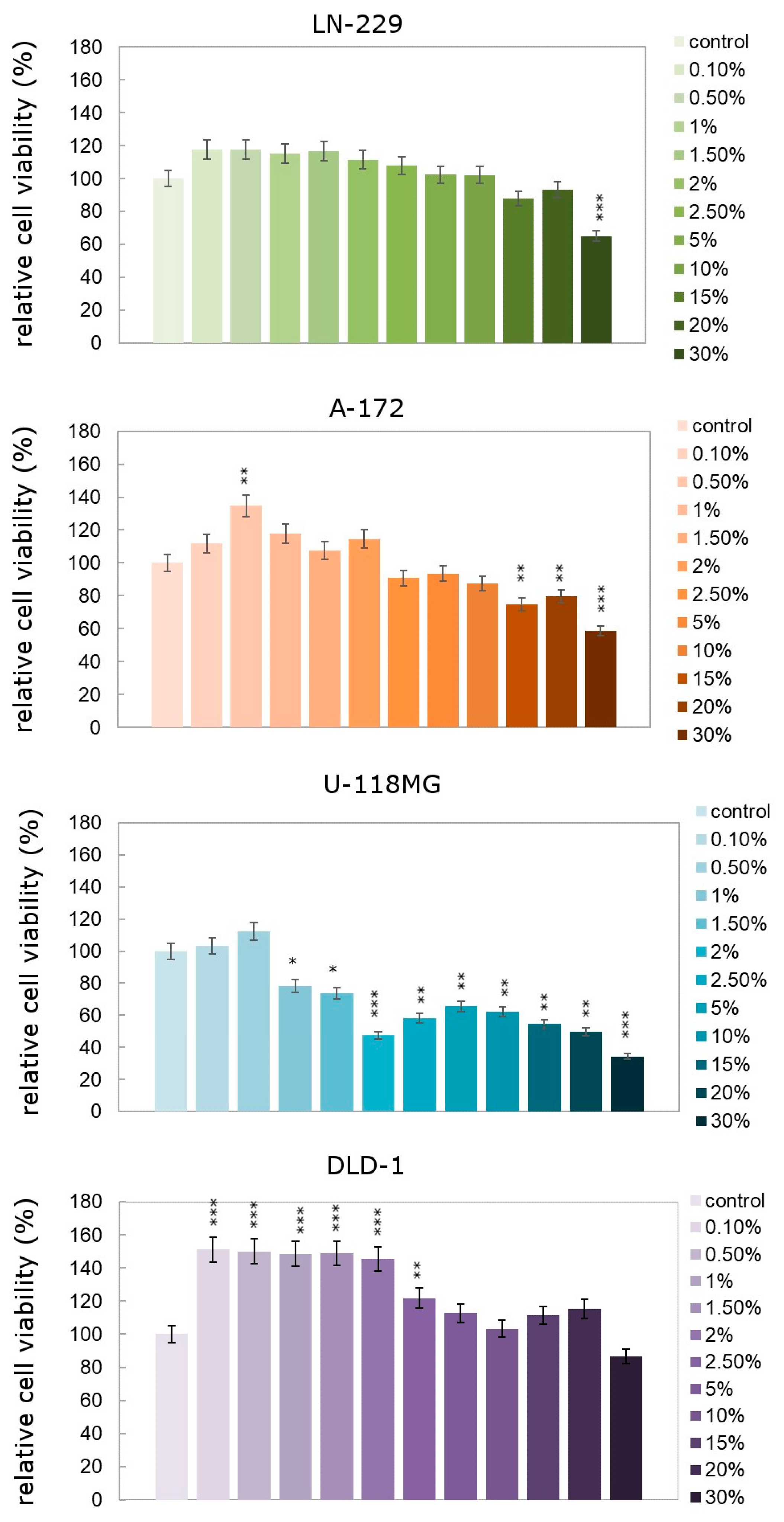
3.5. Human Cell Viability

In the case of human cells, the vast majority of them observed an increase in cell proliferation under the influence of the analyzed leachates (Figure 4). All cell lines used as models in this experiment were cancer lines. Both in the case of the DLD-1 colorectal adenocarcinoma line and in the case of all glioblastoma lines, increases in proliferation were observed, with the exception of the U118-MG line, which was the only one that responded by inhibiting cell proliferation as a result of exposure to leachates. However, it should be noted that even in this U118-MG cell line, an increase in the level of cell proliferation above the control level was noted under the influence of the two lowest analyzed leachate concentrations—0.1% and 0.5%. Statistical analysis indicates particularly high increases in proliferation under the influence of LL in the DLD-1 cell line. Virtually no concentration tested dropped below the control level, but particularly high levels of proliferation were observed in the concentration range of 0.1% to 2%. This seems obvious considering that it is a gastrointestinal cancer, which is the most exposed to any xenobiotics/contaminants entering the body through food.

3.6. Chronic Daily Intake (CDI) and Cancer Risk (CR) for Selected LL Components

LL contains many potentially dangerous substances such as heavy metals and PAHs. CR was estimated for selected particularly hazardous metals and PAHs (Table 4). It was found that the tested leachates have carcinogenic properties and pose a danger to human health. Among the tested compounds, Cd and Cr appear to exceed values indicating high toxicity. The remaining analyzed compounds do not exceed the standards adopted for toxicological safety.

4. Discussion

Leachates from municipal waste landfills are characterized by diverse chemical composition, which is one of the factors that significantly complicate their chemical analysis and testing of their toxic properties. The composition of leachates changes over time and depends on the type of waste stored and the method of landfill operation. Data from the literature indicate that in most cases leachates contain high concentrations of nitrogen, the content of which, like other organic compounds, changes during the operation of the landfill [23,24].
Due to the complexity of the environmental matrix constituted by leachates, the methodology for examining them should include various scopes of research, both physicochemical and biological. The results obtained during physicochemical analyses indicate a relatively high content of PAHs, especially fluorene and phenanthrene, as well as BOD and COD. The high content of organic matter, total nitrogen and nitrates in the leachate indicates a high potential danger to the natural environment as a result of possible penetration of the leachate into, for example, groundwater. These results are consistent with data from the literature and show some analogies to our earlier work [12]. In a study by Jabłońska-Trypuć et al., the authors also analyzed leachate from the landfill in Hryniewicze, but both experiments differ in the dates of sampling and the biological models used. In our previous experiment we focused mainly on different bacterial strains and human cell lines representing healthy cells and cancerous cells. For the first time in the LL study, here we used a comprehensive approach covering a wide range of biological tests. We started analyzing the impact of LLs on the environment by examining their impact on plants. The key aspects turned out to be the content of assimilation dyes and oxidative stress parameters.
In plants, chlorophyll a and chlorophyll b are the main photosynthetic pigments that are an important element of proper functioning. In our studies, the addition of leachate to the soil had a significant impact on the content of chlorophyll a and b, carotenoids and pheophytin in the aboveground parts of the grass mixture. In most cases, the chlorophyll a to b ratio was relatively low (less than 1) (Figure 1). The reason for this may be the addition of leachate, as well as the properties of the urban soil on which the lawn grass grew, which could have been an additional stress factor. This may be explained by the particularly similar chlorophyll a to b content in plants collected from control pots. As noted by Hänsch and Mendel [25], the proper functioning of photosynthetic pigments may be disturbed by heavy metals contained in urban soil, which contribute to physiological and anatomical changes in plants. In our own research, an imbalanced ratio of chlorophyll a to b and an increase in carotene content could be observed at the highest dose of leachate used. Prasad observed a similar trend in his research [26], where he indicated the negative impact of metals on the inhibition of chlorophyll production and thus on the biochemical processes occurring in plants, i.e., respiration, photosynthesis, and transpiration.
Pheophytin is a functional component of the reaction center of photosystem II, where it participates in electron transfer from P-680. The increase in the pheophytin content in plants may be an expression of chlorophyll destruction. Among the assimilation pigments, chlorophyll a is more susceptible to pheophytinization than chlorophyll b [27]. Based on the results obtained, it can be seen that treating plants with doses of N-50% and N-100% of the leachate does not affect the increase in the sum of pheophytins a and b in the leaves, and even causes their value to decrease by approximately 11% compared to the control. Moreover, a positive correlation between the sum of the pheophytins and the content of chlorophyll a was observed, which may indicate the mutual relationship between these pigments during environmental stress.
Reactive oxygen species (ROS) in low concentrations are needed by organisms because they function as signaling molecules, participate in programmed cell death, participate in tissue development, and regulate gene expression. Antioxidant systems protect the body against excessive accumulation of ROS, including the enzymatic and nonenzymatic systems [28]. The literature sources show that the enzymatic activity of peroxidase is considered a very important indicator of oxidative stress in plants. In response to stress factors in the plant’s environment, there is usually an increase in the activity of this enzyme in its tissues [29], which was also observed in our own research. As Huang et al. pointed out [30], peroxidase is one of the enzymes that participate in maintaining the balance between ROS production and their removal. However, catalase activity is related to the removal of hydrogen peroxide, which is a reactive oxygen species [28]. In our own research, the formation of H2O2 in plant tissues may be a defensive reaction to the dose of leachate that was applied in the experiment. According to Møller et al. [31], H2O2 transported by aquaporins in the cell membrane will not only cause oxidative damage but will also participate in the regulation of cell signaling. However, as shown by Kärkönen and Kuchitsu [32], an increase in the H2O2 content in cells may be the result of cell differentiation and the formation of a cell wall. Moreover, it is closely related to and interacts with hormones that regulate development processes in plants and the aging of plant cells. SOD is the main intracellular antioxidant that is involved in protecting the cell against ROS [33]. When it comes to plants, we can distinguish three isoforms of SOD, such as the Mn-SOD isoform found in the mitochondria; the chloroplastic Fe-SOD isoform; and the chloroplastic and cytosolic Cu/Zn-SOD isoform [34]. According to the literature, nutrient deficiencies and the presence of metals in the soil may increase SOD activity and also cause the appearance of new isoforms [33,35]. In our own research, an increase in SOD activity was observed with the applied dose of leachate, which suggests that the metals (Pb and Cd) contained in these leachates could induce the activity of this enzyme and counteract stress in plants. Moreover, a positive correlation between SO42− and Cl ions is also observed. Oxidation of the -SH groups of proteins indicates that the plant is under oxidative stress because this metabolite can act as a detoxifier and antioxidant [36]. In our study, a decrease in -SH groups was observed, which may indicate that the plants had started to activate their defense system. Zagorchev et al. [37] report that plant thiols are involved in the response of plants to many stress factors, and their quantities in plants, redox status, and regulation are important criteria for understanding the interconnections between various biochemical pathways and plant tolerance to stress.
The formation of TBARS in plants indicates the generation of free radicals, and this indicator can be used as a marker in biological systems [38]. TBARS analysis in plants showed that after the use of leachate, the amount of this indicator in plant leaves increased statistically significantly, which indicates a high level of damage to cell membranes, especially in plants where the highest dose of leachate was applied. According to Turan [39], changes occurring in the properties of cell membranes, such as an increase in their permeability to H+ ions and other elements, indicate a significant level of membrane damage and an increase in lipid peroxidation.
Among the bioassays selected by researchers to analyze LL toxicity are often microbial bioassays. Their most important advantages are speed of execution, repeatability of results and good quality-to-cost ratio. The most common tests are the test using Vibrio fisherii, based on the bioluminescent properties of this bacterium, and the test using the Salmonella strain. In our research, we chose a wide spectrum of bacterial strains and one species of fungus. There were significant differences between the tested strains in the intensity of the proliferative response to the tested leachates. This is consistent with data from the literature indicating differences in sensitivity to chemical substances contained in leachates and other environmental pollutants [40,41,42]. In our work, we selected so many different strains that the results of toxicological analysis based on microbial bioassays were as reliable as possible. Different strains of bacteria and species of fungi represent taxonomically, phylogenetically, and structurally a wide range of microorganism diversity in the environment and, at the same time, their different sensitivities to xenobiotics [43,44]. Similarly to Zawierucha et al., we noticed an evidently large amount of metal ions, especially such as lead [45]. Heavy metal ions are highly toxic to living organisms, and even trace amounts may cause an effect due to their accumulation in living cells. Bacterial cells are more resistant to the presence of many pollutants, including heavy metals. Therefore, we observed a smaller effect of the leachate in them than in the case of human cells. The results obtained are similar to those that we presented previously [12]. TU was calculated according to Kalka J., and it amounted to 6.19 for Lactobacillus rhamnosus and 17.85 for Saccharomyces boulardii, which indicates that the studied LL is toxic for L. rhamnosus because the TU value is below 10 [46].
In the literature on toxicological analyses of landfill leachates and other xenobiotics, some in vitro biological models based on selected human cell lines have been described. These lines are primarily HepG2, MCF-7, lymphocytes, CHO, MVLN, and NIH/3T3 [47]. Typically, cell lines for toxicity testing are selected due to the route of exposure to the tested substances. Toxic compounds, including those found in landfills and in leachates generated there, may be absorbed through ingestion (e.g., of contaminated water), inhalation, or through the skin. Each of the considered routes poses a significant threat to human health [48]. However, it should be remembered that potentially toxic substances/mixtures reach tissues further away from the site of direct contact and generate various forms of damage, stimulating, among others, the process of carcinogenesis. That is why we decided to analyze, among others, glioma cells and colorectal adenocarcinoma. Orescanin et al. also used the colorectal adenocarcinoma line—Caco-2—as a model, with which they demonstrated extremely high cytotoxicity of untreated sludge [49]. We also observed an obvious stimulation of cancer cell proliferation as a result of exposure to LL, which was similar to an increase observed in our previous study [12]. This may be due to an increase in the level of oxidative stress in these cells, which usually translates into increased tumor growth. In turn, very high concentrations of LL caused the death of cancer cells, most likely by necrosis.
Taking everything together, it can be assumed that in such a complex environmental matrix as landfill leachates, various reactions undoubtedly occur between its components, which translates into the effect observed in plant tissues, bacterial and fungal cells, as well as in human cells.

5. Conclusions

Regardless of the biological models used, the toxic effect of LL was confirmed in our studies. Any release of such leachates into the environment without prior appropriate treatment is potentially extremely dangerous. Therefore, both landfills and leachates generated there should be constantly monitored to exclude the possibility of their entering ground or surface waters. Research such as ours is extremely valuable from the point of view of the need to implement new methods and technologies for leachate treatment and landfill control to prevent possible environmental contamination. Such studies indicate the need to develop toxicological analyses in order to implement the fastest and most effective research methods possible in the future.
The area where the waste landfill is located, where the samples were taken, is not an industrialized area. It is a typical municipal waste landfill, and the areas of the Podlaskie Voivodeship are agricultural rather than industrial areas. Therefore, taking into account the results obtained indicating the toxicity of the tested leachates, we conclude that even the smallest landfills, in areas where particularly toxic waste is not generated, should not be underestimated. According to Zuo et al., if the CR index is lower than 1 × 10⁻⁴, it is an acceptable value and does not indicate a high level of risk [50]. Our results indicate that only heavy metals such as Cd, Pb and Cr may pose a danger to humans (Table 4). The analyzed PAHs did not exceed acceptable standards; therefore, we conclude that the tested leachates do not generally pose an unacceptable health risk to the average exposure population. The exceptions are some heavy metals. When comparing our results with the data obtained by Kalka J. [46], attention should be paid to the origin of the tested leachates. Differences in the composition and toxicity of leachates may result from different geographical locations, and also from different levels of industrialization of a given region. Although our results, including the analysis of CDI and CR indicators, do not indicate significant toxicity of the tested leachates, we believe that they should be constantly and carefully monitored. Concentrations of 25% and higher turned out to be toxic in the bacterial model, while even very low concentrations in the range of 0.1–1.5% stimulated the growth of cancer cells, which clearly indicates their potential carcinogenic properties.

Author Contributions

Conceptualization, A.J.-T., E.W. and U.W.; methodology, A.J.-T., E.W., J.L. and U.W.; software, E.W. and U.W.; validation, A.J.-T., M.W. and A.K.-D.; formal analysis, J.L.; investigation, A.J.-T., E.W. and U.W.; resources, A.J.-T., E.W., J.L. and U.W.; data curation, M.W. and A.K.-D.; writing—original draft preparation, A.J.-T., E.W. and U.W.; writing—review and editing, A.J.-T., E.W., M.W., A.K.-D. and U.W.; visualization, E.W. and U.W.; supervision, A.J.-T.; project administration, A.J.-T.; funding acquisition, A.J.-T. All authors have read and agreed to the published version of the manuscript.

Funding

This work was financially supported by the Ministry of Education and Science, Poland, under the research project number WZ/WB-IIŚ/6/2022.

Institutional Review Board Statement

Not applicable.

Informed Consent Statement

Not applicable.

Data Availability Statement

The data are available on request due to restrictions, e.g., privacy or ethical.

Conflicts of Interest

The authors declare no conflict of interest.

References

  1. Duan, Z.; Scheutz, C.; Kjeldsen, P. Trace Gas Emissions from Municipal Solid Waste Landfills: A Review. Waste Manag. 2021, 119, 39–62. [Google Scholar] [CrossRef] [PubMed]
  2. Ren, Y.; Zhang, Z.; Huang, M. A Review on Settlement Models of Municipal Solid Waste Landfills. Waste Manag. 2022, 149, 79–95. [Google Scholar] [CrossRef] [PubMed]
  3. Juarez, M.B.; Mondelli, G.; Giacheti, H.L. Correction to: An Overview of In Situ Testing and Geophysical Methods to Investigate Municipal Solid Waste Landfills. Environ. Sci. Pollut. Res. 2023, 30, 24779–24789. [Google Scholar] [CrossRef] [PubMed]
  4. Annapareddy, V.S.R.; Pain, A.; Sufian, A.; Godas, S.; Scheuermann, A. Influence of Heterogeneity and Elevated Temperatures on the Seismic Translational Stability of Engineered Landfills. Waste Manag. 2023, 158, 1–12. [Google Scholar] [CrossRef] [PubMed]
  5. Hussain, S.; Aneggi, E.; Comuzzi, C.; Baderna, D.; Zuccaccia, D.; Trovarelli, A.; Goi, D. Abatement of the Ecotoxicological Risk of Landfill Leachate by Heterogeneous Fenton-like Oxidation. Environ. Sci. Pollut. Res. 2023, 30, 21025–21032. [Google Scholar] [CrossRef] [PubMed]
  6. Hussain, S.; Aneggi, E.; Trovarelli, A.; Goi, D. Removal of Organics from Landfill Leachate by Heterogeneous Fenton-like Oxidation over Copper-Based Catalyst. Catalysts 2022, 12, 338. [Google Scholar] [CrossRef]
  7. Directive, L. Council Directive 1999/31/EC of 26 April 1999 on the Landfill of Waste. Off. J. Eur. Commun. 1999, 182, 26. [Google Scholar]
  8. Wdowczyk, A.; Szymańska-Pulikowska, A. Differences in the Composition of Leachate from Active and Non-Operational Municipal Waste Landfills in Poland. Water 2020, 12, 3129. [Google Scholar] [CrossRef]
  9. Abdel-Shafy, H.I.; Ibrahim, A.M.; Al-Sulaiman, A.M.; Okasha, R.A. Landfill Leachate: Sources, Nature, Organic Composition, and Treatment: An Environmental Overview. Ain Shams Eng. J. 2023, 15, 102293. [Google Scholar] [CrossRef]
  10. Nair, A.T. Bioaerosols in the Landfill Environment: An Overview of Microbial Diversity and Potential Health Hazards. Aerobiologia 2021, 37, 185–203. [Google Scholar] [CrossRef]
  11. Akpeimeh, G.F.; Fletcher, L.A.; Evans, B.E. Exposure to Bioaerosols at Open Dumpsites: A Case Study of Bioaerosols Exposure from Activities at Olusosun Open Dumpsite, Lagos Nigeria. Waste Manag. 2019, 89, 37–47. [Google Scholar] [CrossRef]
  12. Jabłońska-Trypuć, A.; Wydro, U.; Wołejko, E.; Pietryczuk, A.; Cudowski, A.; Leszczyński, J.; Rodziewicz, J.; Janczukowicz, W.; Butarewicz, A. Potential Toxicity of Leachate from the Municipal Landfill in View of the Possibility of Their Migration to the Environment through Infiltration into Groundwater. Environ. Geochem. Health 2021, 43, 3683–3698. [Google Scholar] [CrossRef] [PubMed]
  13. Wydro, U.; Wołejko, E.; Sokołowska, G.; Leszczyński, J.; Jabłońska-Trypuć, A. Investigating Landfill Leachate Influence on Soil Microbial Biodiversity and Its Cytotoxicity. Water 2022, 14, 3634. [Google Scholar] [CrossRef]
  14. Ming, L.; Li, L.; Zhang, Y.; Lin, W.; Wang, G.; Zhang, S.; Guo, P. Effects of Dissolved Organic Matter on the Desorption of Cd in Freeze–Thaw Treated Cd-Contaminated Soils. Chem. Ecol. 2014, 30, 76–86. [Google Scholar] [CrossRef]
  15. Łozowicka, B.; Wołejko, E.; Kaczyński, P.; Konecki, R.; Iwaniuk, P.; Drągowski, W.; Łozowicki, J.; Tujtebajeva, G.; Wydro, U.; Jablońska-Trypuć, A. Effect of Microorganism on Behaviour of Two Commonly Used Herbicides in Wheat/Soil System. Appl. Soil Ecol. 2021, 162, 103879. [Google Scholar] [CrossRef]
  16. Liang, H.; Xing, Y.; Chen, J.; Zhang, D.; Guo, S.; Wang, C. Antimicrobial activities of endophytic fungi isolated from Ophiopogon japonicus (Liliaceae). BMC Complement. Altern. Med. 2012, 12, 238. [Google Scholar] [CrossRef]
  17. Jahn, B.; Martin, E.; Stueben, A.; Bhakdi, S. Susceptibility testing of Candida albicans and Aspergillus species by a simple microtiter menadione-augmented 3-(4,5-dimethyl-2-thiazolyl)-2,5-diphenyl-2H-tetrazolium bromide assay. J. Clin. Microbiol. 1995, 33, 661–667. [Google Scholar] [CrossRef] [PubMed]
  18. Carmichael, J.; DeGraff, W.G.; Gazdar, A.F.; Minna, J.D.; Mitchell, J.B. Evaluation of a tetrazolium-based semiautomated colorimetric assay: Assessment of chemosensitivity testing. Cancer Res. 1987, 47, 936–942. [Google Scholar]
  19. Parveen, N.; Goel, S. Trihalomethane Cancer Risk Assessment for Private and Shared Residences: Addressing the Differences in Inhalation Exposure. Toxics 2023, 11, 295. [Google Scholar] [CrossRef]
  20. Dokubo, A.; Igwe, F.U. Assessment of Polycyclic Aromatic Hydrocarbons (PAHs) in Commonly Consumed Shellfish from Kula, Rivers State, Nigeria. Environ. Manag. Sustain. Dev. 2019, 8, 58. [Google Scholar] [CrossRef]
  21. Oni, A.A.; Babalola, S.O.; Adeleye, A.D.; Olagunju, T.E.; Amama, I.A.; Omole, E.O.; Adegboye, E.A.; Ohore, O.G. Non-carcinogenic andcarcinogenic health risks associated with heavy metals and polycyclic aromatic hydrocarbons in well-water samples from an automobile junk market in Ibadan, SW-Nigeria. Heliyon 2022, 8, e10688. [Google Scholar] [CrossRef]
  22. Gerba, C.P. Risk Assessment. Available online: https://web.iitd.ac.in/~arunku/files/CEL899_Y13/Gerba%20Risk%20Assessment.pdf (accessed on 10 November 2023).
  23. Chiang, L.-C.; Chang, J.-E.; Wen, T.-C. Indirect Oxidation Effect in Electrochemical Oxidation Treatment of Landfill Leachate. Water Res. 1995, 29, 671–678. [Google Scholar] [CrossRef]
  24. Kang, K.-H.; Shin, H.S.; Park, H. Characterization of Humic Substances Present in Landfill Leachates with Different Landfill Ages and Its Implications. Water Res. 2002, 36, 4023–4032. [Google Scholar] [CrossRef] [PubMed]
  25. Hänsch, R.; Mendel, R.R. Physiological Functions of Mineral Micronutrients (Cu, Zn, Mn, Fe, Ni, Mo, B, Cl). Curr. Opin. Plant Biol. 2009, 12, 259–266. [Google Scholar] [CrossRef] [PubMed]
  26. Prasad, M.N.V.; Strzałka, K. Impact of Heavy Metals on Photosynthesis. In Heavy Metal Stress in Plants: From Molecules to Ecosystems; Prasad, M.N.V., Hagemeyer, J., Eds.; Springer: Berlin/Heidelberg, Germany, 1999; pp. 117–138. ISBN 978-3-662-07745-0. [Google Scholar]
  27. Zalewska, M.; Tukaj, Z. Biochemiczne i fizjologiczne aspekty rozkładu barwników chlorofilowych. Postępy Biochem. 2019, 65, 128–134. [Google Scholar] [CrossRef] [PubMed]
  28. Fryzova, R.; Pohanka, M.; Martinkova, P.; Cihlarova, H.; Brtnicky, M.; Hladky, J.; Kynicky, J. Oxidative Stress and Heavy Metals in Plants. In Reviews of Environmental Contamination and Toxicology; de Voogt, P., Ed.; Springer International Publishing: Cham, Switzerland, 2018; Volume 245, pp. 129–156. ISBN 978-3-319-75037-8. [Google Scholar]
  29. Stręk, M.; Telesiński, A. The effect of calcium peroxide on the activity of some oxidoreductases in soil contaminated with creosote. Chem. Environ. Biotechnol. 2016, 19, 7–11. [Google Scholar] [CrossRef]
  30. Huang, H.; Ullah, F.; Zhou, D.-X.; Yi, M.; Zhao, Y. Mechanisms of ROS Regulation of Plant Development and Stress Responses. Front. Plant Sci. 2019, 10, 800. [Google Scholar] [CrossRef]
  31. Møller, I.M.; Jensen, P.E.; Hansson, A. Oxidative Modifications to Cellular Components in Plants. Annu. Rev. Plant Biol. 2007, 58, 459–481. [Google Scholar] [CrossRef]
  32. Kärkönen, A.; Kuchitsu, K. Reactive Oxygen Species in Cell Wall Metabolism and Development in Plants. Phytochemistry 2015, 112, 22–32. [Google Scholar] [CrossRef]
  33. Miller, A.-F. Superoxide Dismutases. In Encyclopedia of Biophysics; Roberts, G.C.K., Ed.; Springer: Berlin/Heidelberg, Germany, 2013; pp. 2517–2522. ISBN 978-3-642-16712-6. [Google Scholar]
  34. Stephenie, S.; Chang, Y.P.; Gnanasekaran, A.; Esa, N.M.; Gnanaraj, C. An Insight on Superoxide Dismutase (SOD) from Plants for Mammalian Health Enhancement. J. Funct. Foods 2020, 68, 103917. [Google Scholar] [CrossRef]
  35. Huseynova, I.M.; Aliyeva, D.R.; Aliyev, J.A. Subcellular Localization and Responses of Superoxide Dismutase Isoforms in Local Wheat Varieties Subjected to Continuous Soil Drought. Plant Physiol. Biochem. PPB 2014, 81, 54–60. [Google Scholar] [CrossRef] [PubMed]
  36. Chen, H.; Zhang, Q.; Zhang, Z. Comparative Transcriptome Combined with Metabolomic and Physiological Analyses Revealed ROS-Mediated Redox Signaling Affecting Rice Growth and Cellular Iron Homeostasis under Varying pH Conditions. Plant Soil 2019, 434, 343–361. [Google Scholar] [CrossRef]
  37. Zagorchev, L.; Seal, C.E.; Kranner, I.; Odjakova, M. A Central Role for Thiols in Plant Tolerance to Abiotic Stress. Int. J. Mol. Sci. 2013, 14, 7405–7432. [Google Scholar] [CrossRef] [PubMed]
  38. Srivastava, M.; Ma, L.Q.; Singh, N.; Singh, S. Antioxidant Responses of Hyper-Accumulator and Sensitive Fern Species to Arsenic. J. Exp. Bot. 2005, 56, 1335–1342. [Google Scholar] [CrossRef]
  39. Turan, V. Confident Performance of Chitosan and Pistachio Shell Biochar on Reducing Ni Bioavailability in Soil and Plant plus Improved the Soil Enzymatic Activities, Antioxidant Defense System and Nutritional Quality of Lettuce. Ecotoxicol. Environ. Saf. 2019, 183, 109594. [Google Scholar] [CrossRef]
  40. Takigami, H.; Matsui, S.; Matsuda, T.; Shimizu, Y. The Bacillus Subtilis Rec-Assay: A Powerful Tool for the Detection of Genotoxic Substances in the Water Environment. Prospect for Assessing Potential Impact of Pollutants from Stabilized Wastes. Waste Manag. 2002, 22, 209–213. [Google Scholar] [CrossRef]
  41. Chekabab, S.M.; Paquin-Veillette, J.; Dozois, C.M.; Harel, J. The Ecological Habitat and Transmission of Escherichia coli O157:H7. FEMS Microbiol. Lett. 2013, 341, 1–12. [Google Scholar] [CrossRef]
  42. van Elsas, J.D.; Semenov, A.V.; Costa, R.; Trevors, J.T. Survival of Escherichia coli in the Environment: Fundamental and Public Health Aspects. ISME J. 2011, 5, 173–183. [Google Scholar] [CrossRef]
  43. Wadhia, K.; Thompson, K.C. Low-Cost Ecotoxicity Testing of Environmental Samples Using Microbiotests for Potential Implementation of the Water Framework Directive. TrAC Trends Anal. Chem. 2007, 26, 300–307. [Google Scholar] [CrossRef]
  44. Wadhia, K.; Dando, T.; Thompson, K.C. Intra-Laboratory Evaluation of Microbial Assay for Risk Assessment (MARA) for Potential Application in the Implementation of the Water Framework Directive (WFD). J. Environ. Monit. 2007, 9, 953–958. [Google Scholar] [CrossRef] [PubMed]
  45. Zawierucha, I.; Kozlowski, C.; Malina, G. Removal of Toxic Metal Ions from Landfill Leachate by Complementary Sorption and Transport across Polymer Inclusion Membranes. Waste Manag. 2013, 33, 2129–2136. [Google Scholar] [CrossRef]
  46. Kalka, J. Landfill leachate toxicity removal in combined treatment with municipal wastewater. Sci. World J. 2012, 2012, 202897. [Google Scholar] [CrossRef]
  47. Baderna, D.; Caloni, F.; Benfenati, E. Investigating Landfill Leachate Toxicity In Vitro: A Review of Cell Models and Endpoints. Environ. Int. 2019, 122, 21–30. [Google Scholar] [CrossRef] [PubMed]
  48. Rehman, K.; Fatima, F.; Waheed, I.; Akash, M.S.H. Prevalence of Exposure of Heavy Metals and Their Impact on Health Consequences. J. Cell. Biochem. 2018, 119, 157–184. [Google Scholar] [CrossRef] [PubMed]
  49. Orescanin, V.; Durgo, K.; Mikelic, I.L.; Halkijevic, I.; Kuspilic, M. Toxicity Assessment of Untreated/Treated Electroplating Sludge Using Human and Plant Bioassay. J. Environ. Sci. Health Part A Tox. Hazard. Subst. Environ. Eng. 2018, 53, 925–930. [Google Scholar] [CrossRef] [PubMed]
  50. Zuo, T.T.; Jin, H.Y.; Zhang, L.; Liu, Y.L.; Nie, J.; Chen, B.L.; Fang, C.F.; Xue, J.; Bi, X.Y.; Zhou, L.; et al. Innovative health risk assessment of heavy metals in Chinese herbal medicines based on extensive data. Pharmacol Res. 2020, 159, 104987. [Google Scholar] [CrossRef]
Figure 1. Content of Chla, Chb, carot, Phe(a + b) and Chla/Chlb in plants after application of LL in doses of C (control), N-50% and N-100%. The same letters above bars mean nonsignificant differences between treatments assessed by the Tukey test at p < 0.05.
Sustainability 15 16497 g001
Figure 2. The main parameters of oxidative stress (CAT, NADPH-dependent peroxidase, SOD, TBARS and -SH groups) in plants after application of LL in doses of C (control), N-50% and N-100%. The same letters above bars mean nonsignificant differences between treatments assessed by the Tukey test at p < 0.05.
Sustainability 15 16497 g002
Figure 3. Relative viability of microbial cells (S. pasteurii, S. aureus, C. albicans, L. rhamnosus and S. boulardii) treated with LL in different concentrations. Each value presented as a column with bars represents the mean with the standard deviation (±SD). * p < 0.05, ** p < 0.01 and *** p < 0.001 describe significant effects between treatments and control assessed by Dunnett’s test.
Sustainability 15 16497 g003
Figure 4. Relative viability of cells exposed to grading concentrations of landfill leachate. Each value presented as a column with bars represents the mean with the standard deviation (±SD). * p < 0.05, ** p < 0.01 and *** p < 0.001 describe significant effects between treatments and control assessed by Dunnett’s test.
Sustainability 15 16497 g004
Table 1. Physicochemical parameters of landfill leachate (LL) and soil before starting the experiment.
ParameterMatriceParameterMatrice
LLSoilLLSoil
pH7.858.70BOD 1 (mg/L)61-
TOC 3 (mg/L) -1.23COD 2 (mg/L)1880-
EC 4 (mS/cm) 9.870.275N 6329.2 mg/L0.138%
P 5 (mg/kg)-317BOD/COD0.032
Metalsmg/Lmg/kgMetalsmg/Lmg/kg
Cr0.339.8Ni0.087.6
Mn0.004294.6Cu0.0414.27
Fe1.195232.5Zn0.0863.43
Cd0.010.40Pb0.0327.41
Ionsmg/L Ionsmg/L
PO43−19.5-Cl2568.2-
SO42−104.7-NO31523.6-
N-NH4205.7
PAHsng/Lµg/kgPAHsng/Lµg/kg
Naphthalene 180.840.0309Benzo[a]anthracen4.801.3132
Acenaphthylene10.900.0002Chrysene14.301.0465
Acenaphthene 40.950.0095Benzo[b]fluoranthene 11.401.1196
Fluorene136.840.0115Benzo[k]fluoranthene 10.240.5245
Phenanthrene 475.900.0438Benzo[a]pyrene3.090.2603
Anthracene39.290.0456Indeno[1,2,3-cd]pyrene8.150.4532
Fluoranthene 60.780.8361Dibenzo[a,h]anthracene2.560.0227
Pyrene57.100.7296Benzo[ghi]perylene-0.2723
1 BOD—biochemical oxygen demand; 2 COD—chemical oxygen demand; 3 TOC—total organic carbon; 4 EC—electrical conductivity; 5 P—phosphorus; 6 N—nitrogen.
Table 2. Physicochemical parameters of soil filtrates.
ParameterDoseParameterDose
ControlN-50%N-100%ControlN-50%N-100%
pH8.78.237.8TOC (mg/L)119.40570.70293.90
Metalsµg/LMetalsµg /L
Ni0.0040.0040.004Cd0.0010.0020.003
Cr0.0080.0080.005Cu0.0330.0300.038
Mn0.0260.0230.026Zn0.0430.0360.055
Fe2.2452.3602.337Pb0.0050.0060.007
Ionsmg/LIonsmg/L
PO43−3.723.564.82Cl1.3251.54153.15
SO42−2.765.4713.47NO30.7529.663.4
PAHsng/LPAHsng/L
Naphthalene 23.48115.6375.96Benzo[a]anthracen61.30179.64356.67
Acenaphthylene0.164.272.34Chrysene77.96134.41284.82
Acenaphthene7.2314.928.83Benzo[b]fluoranthene81.75120.92239.51
Fluorene9.4143.8319.78Benzo[k]fluoranthene225.66101.26141.00
Phenanthrene28.57286.75115.01Benzo[a]pyrene67.4315.7130.85
Anthracene18.9111.122.67Indeno[1,2,3-cd]pyrene45.4215.0836.86
Fluoranthene159.15102.03209.12Dibenzo[a,h]anthracene19.833.403.29
Pyrene139.93186.24159.14Benzo[ghi]perylene 40.5815.302.46
Table 3. Correlation coefficients (r) between the physicochemical properties of soil filtrates and the physiological parameters of plants.
Soil Filtrates CharacteristicsCATNADHTBARSSOD-SH GroupsChlaChlbChla/ChlbCarotPhe(a + b)
pH−0.67−0.93−0.98−0.960.330.83 *−0.300.34−0.980.42
TOC0.290.040.230.14−0.33−0.39−0.750.720.23−0.20
Cd0.520.940.790.870.76−0.890.33−0.360.79−0.40
Cr−0.41−0.39−0.94−0.960.890.06−0.760.78−0.93−0.12
Mn0.100.350.160.25−0.050.010.94−0.930.160.62
Fe0.690.480.640.57−0.72−0.76−0.370.340.64−0.70
Cu0.690.850.740.80−0.66−0.610.94−0.960.730.48
Zn0.700.860.740.80−0.67−0.620.94−0.950.740.47
Pb0.800.940.390.970.68−0.910.33−0.360.39−0.40
PO43−0.860.960.890.93−0.83−0.800.83−0.850.490.23
SO42−0.740.030.690,87−0.98−0.660.57−0.600.29−0.14
Cl0.540.890.890.98−0.49−0.580.50−0.530.51−0.21
NO30.350.650.390.98−0.25−0.250.37−0.400.29−0.35
16PAHs0.890.740.290.970.65−0.850.32−0.360.42−0.40
* Bolded values mean significant correlation coefficient at p < 0.05.
Table 4. Chronic daily intake (CDI (mg kg−1 day−1)) and cancer risk (CR) for selected LL hazardous components.
ParameterConc
(mg/L)
CDI (mg/kg/day)CSFingCR
Cr0.339.0 × 10−30.54.7 × 10−3
Cd0.013.0 × 10−415.04.3 × 10−3
Pb0.039.0 × 10−48.5 × 10−37.3 × 10−6
Benzo[a]anthracen4.8 × 10−61.4 × 10−70.71.0 × 10−7
Chrysene1.4 × 10−54.1 × 10−77.30 × 10−33.0 × 10⁻⁹
Benzo[b]fluoranthene1.1 × 10−53.3 × 10−70.72.9 × 10−7
Benzo[k]fluoranthene1.0 × 10−52.9 × 10−70.072.1 × 10−8
Benzo[a]pyrene3.1 × 10−68.8 × 10−87.36.4 × 10−7
Indeno[1,2,3-cd]pyrene8.2 × 10−62.3 × 10−70.71.7 × 10−7
Dibenzo[a,h]anthracene2.6 × 10−67.3 × 10−87.35.3 × 10−7
Disclaimer/Publisher’s Note: The statements, opinions and data contained in all publications are solely those of the individual author(s) and contributor(s) and not of MDPI and/or the editor(s). MDPI and/or the editor(s) disclaim responsibility for any injury to people or property resulting from any ideas, methods, instructions or products referred to in the content.

