Next Issue
Volume 15, March
Previous Issue
Volume 14, September


Hematol. Rep., Volume 14, Issue 4 (December 2022) – 13 articles

Cover Story (view full-size image): 
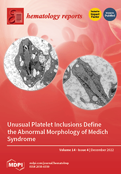
Medich syndrome is a rare macrothrombocytopenia with unusual scroll-like platelet inclusions in cross-sections; they are tubular or cone-shaped longitudinally and may deform a cell’s discoid shape. At the periphery of the inclusions, numerous layers of membranes and aggregates of glycogen are also present.  First described by Dr. James G. White in 2004, the condition also includes a deficiency of alpha granules and occasional giant alpha granules, as seen in chromosome 11q deletion syndrome. Of five cases reported to date, three presented with thrombocytopenia at birth, another at two weeks, and one at a year of age. All have been transfusion-dependent, and the diagnosis of Medich syndrome took years to establish. There is no specific genetic mutation identified yet for the syndrome. View this paper
  • Issues are regarded as officially published after their release is announced to the table of contents alert mailing list.
  • You may sign up for e-mail alerts to receive table of contents of newly released issues.
  • PDF is the official format for papers published in both, html and pdf forms. To view the papers in pdf format, click on the "PDF Full-text" link, and use the free Adobe Reader to open them.
Order results
Result details
Select all
Export citation of selected articles as:
12 pages, 924 KiB  
Article
Effectiveness of Convalescent Plasma Therapy in COVID-19 Patients with Hematological Malignancies: A Systematic Review
by Sapha Shibeeb, Ilham Ajaj, Hadeel Al-Jighefee and Atiyeh M. Abdallah
Hematol. Rep. 2022, 14(4), 377-388; https://doi.org/10.3390/hematolrep14040052 - 12 Dec 2022
Cited by 4 | Viewed by 1692
Abstract
Background: Immunocompromised patients, including those with hematological malignancies, are at a high risk of developing severe coronavirus disease 2019 (COVID-19) complications. Currently, there is a limited number of systematic reviews into the efficacy of convalescent plasma therapy (CPT) use in the treatment of [...] Read more.
Background: Immunocompromised patients, including those with hematological malignancies, are at a high risk of developing severe coronavirus disease 2019 (COVID-19) complications. Currently, there is a limited number of systematic reviews into the efficacy of convalescent plasma therapy (CPT) use in the treatment of COVID-19 patients with hematological malignancies. Therefore, the aim of this review was to systematically appraise the current evidence for the clinical benefits of this therapy in COVID-19 patients with hematological malignancies. Methods: A comprehensive search was conducted up to April 2022, using four databases: PubMed, Web of Science, Science Direct, and Scopus. Two reviewers independently assessed the quality of the included studies. Data collection analysis was performed using Microsoft Excel 365 and GraphPad Prism software. Results: 18 studies met the inclusion criteria; these records included 258 COVID-19 patients who had hematological malignancies and were treated with CPT. The main findings from the reviewed data suggest that CPT may be associated with improved clinical outcomes, including (a) higher survival rate, (b) improved SARS-CoV-2 clearance and presence of detectable anti-SARS-CoV-2 antibodies post CP transfusion, and (c) improved hospital discharge time and recovery after 1 month of CPT. Furthermore, treatment with convalescent plasma was not associated with the development of adverse events. Conclusions: CPT appears to be an effective supportive therapeutic option for hematological malignancy patients infected with COVID-19. To our knowledge, this is one of the first systematic reviews of the clinical benefits of CPT in COVID-19 patients with hematological malignancies. Full article
Show Figures

Figure 1

4 pages, 211 KiB  
Case Report
Safety and Immunogenicity of a Single Dose of BNT162b2 COVID-19 mRNA Vaccine in a Warfarin-Treated Protein S Deficient Patient: A Case Report and Literature Review
by Cucunawangsih Cucunawangsih, Ratna Sari Wijaya, Nata Pratama Hardjo Lugito and Ivet Suriapranata
Hematol. Rep. 2022, 14(4), 373-376; https://doi.org/10.3390/hematolrep14040051 - 05 Dec 2022
Viewed by 1481
Abstract
Patients with protein S (PS) deficiency possibly have a higher risk of developing severe COVID-19 disease. Therefore, vaccination against SARS-CoV-2 infections is recommended for PS-deficient patients. However, there are limited data regarding the safety and immunogenicity of the currently available COVID-19 mRNA vaccine [...] Read more.
Patients with protein S (PS) deficiency possibly have a higher risk of developing severe COVID-19 disease. Therefore, vaccination against SARS-CoV-2 infections is recommended for PS-deficient patients. However, there are limited data regarding the safety and immunogenicity of the currently available COVID-19 mRNA vaccine in PS-deficient patients. We report a case of monitoring the antibody response of a 40-year-old female diagnosed with PS deficiency and on warfarin treatment following a single dose of BNT162b2 mRNA vaccine. Antibody against the receptor-binding domain (RBD) of the SARS-CoV-2 spike (S) protein (anti-S) was measured on days 7, 14, and 21 after vaccination. Seroconversion was detected on day 21 but was possibly lower than the anti-S level previously reported in healthy individuals after receiving the first dose of the BNT162b2 mRNA vaccine. There were no local and systemic events reported up to 7 days in this patient after vaccination. This case highlights that the administration of the BNT162b2 vaccine had a favourable safety profile, and the second dose of the vaccine is required to provide the optimal protection against SARS-CoV-2 infection in PS-deficient patients. Full article
15 pages, 737 KiB  
Review
COVID-19 Adenoviral Vaccine-Induced Immune Thrombotic Thrombocytopenia (VITT), COVID-19-Related Thrombosis, and the Thrombotic Thrombocytopenic Syndromes
by Gewil Daniella Olipas Allas, Joekeem Del Rosario Arizala and Rafael Vincent Mercado Manalo
Hematol. Rep. 2022, 14(4), 358-372; https://doi.org/10.3390/hematolrep14040050 - 01 Dec 2022
Cited by 2 | Viewed by 2778
Abstract
Adenoviral-based vaccines such as ChadoX1 CoV-19 (AstraZeneca) and Ad26.COV2.S (J&J) were developed to prevent infection and reduce hospitalization or death in Coronavirus Disease 2019 (COVID-19) patients. Although these vaccines passed safety and efficacy trials with excellent neutralizing capabilities against SARS-CoV-2, very rare reports [...] Read more.
Adenoviral-based vaccines such as ChadoX1 CoV-19 (AstraZeneca) and Ad26.COV2.S (J&J) were developed to prevent infection and reduce hospitalization or death in Coronavirus Disease 2019 (COVID-19) patients. Although these vaccines passed safety and efficacy trials with excellent neutralizing capabilities against SARS-CoV-2, very rare reports of acute thrombotic thrombocytopenic events following administration emerged in certain populations, which triggered a series of clinical investigations that gave rise to a novel phenomenon called vaccine-induced immune thrombotic thrombocytopenia (VITT). Several converging pathways exist between VITT and other forms of thrombotic thrombocytopenic syndromes, specifically that of heparin-induced thrombocytopenia, which involves the formation of anti-PF4 antibodies and the activation of platelets leading to thrombocytopenia and thrombin-mediated clotting. Interestingly, certain differences in the presentation also exist in VITT, and guidelines have been published in recent months to assist clinicians in recognizing VITT to achieve desired outcomes. In this paper, we first discuss the clotting phenomenon in COVID-19 and delineate it from VITT, followed by a review of current knowledge on the clinical manifestations of VITT in lieu of other thrombotic thrombocytopenic syndromes. Likewise, emerging evidence on the role of adenoviral vectors and vaccine constituents is also discussed briefly. Full article
Show Figures

Figure 1

9 pages, 4668 KiB  
Case Report
Medich Giant Platelet Syndrome: An Evolving Qualitative and Quantitative Platelet Disorder
by Gita Massey, Laura Tyrrell, Yaser Diab and William T. Gunning III
Hematol. Rep. 2022, 14(4), 349-357; https://doi.org/10.3390/hematolrep14040049 - 01 Dec 2022
Viewed by 1910
Abstract
Qualitative platelet disorders remain rare and varied. We describe here 2 additional patients with giant platelets, thrombocytopenia, deficiency in alpha granules and the presence of membranous inclusions within the cytoplasm. Collectively known as Medich syndrome, we further elucidated structural and clinical features of [...] Read more.
Qualitative platelet disorders remain rare and varied. We describe here 2 additional patients with giant platelets, thrombocytopenia, deficiency in alpha granules and the presence of membranous inclusions within the cytoplasm. Collectively known as Medich syndrome, we further elucidated structural and clinical features of this rare syndrome. Platelets obtained from 2 patients with macro-thrombocytopenia were evaluated by electron microscopy. Structural findings were correlated with clinical characteristics. The defining morphologic feature found in the platelets of these patients is the presence of long, tubular inclusions consisting of several layers of membrane wrapped around a core of cytoplasm. These inclusions may deform the discoid shape of the platelet. In addition, abnormal giant alpha granules are present. Clinically all patients in the current report and review of the literature had mucosal bleeding and were often misdiagnosed as having immune related thrombocytopenia. To date five cases of Medich giant platelet syndrome have been reported. The cases are unified by the ultrastructural findings of abnormal alpha granules and unusual cytoplasmic scrolls. All patients experienced mucosal bleeding, however many clinical, biologic and genetic characteristics of this rare disorder remain to be determined. Full article
Show Figures

Figure 1

7 pages, 1156 KiB  
Article
Cutaquig® Is Well Tolerated in Immunodeficient Patients Who Did Not Tolerate Other Subcutaneous Immunoglobulin Products
by Sydney Brownlee, Crystal Allen, Mohammed F. Kana’an, D. William Cameron and Juthaporn Cowan
Hematol. Rep. 2022, 14(4), 342-348; https://doi.org/10.3390/hematolrep14040048 - 17 Nov 2022
Cited by 1 | Viewed by 1559
Abstract
Objective: Subcutaneous immunoglobulin (SCIG) treatment is generally tolerable, but some patients may experience adverse events to one or more SCIG products. We investigated whether 16.5% Cutaquig® treatment offered a tolerable and safe alternative treatment for immunodeficient patients. Methods: A one-year prospective cohort [...] Read more.
Objective: Subcutaneous immunoglobulin (SCIG) treatment is generally tolerable, but some patients may experience adverse events to one or more SCIG products. We investigated whether 16.5% Cutaquig® treatment offered a tolerable and safe alternative treatment for immunodeficient patients. Methods: A one-year prospective cohort study was conducted at a single center in Ottawa, Canada. Adult immunodeficient patients who reported previous intolerability, adverse events, or other difficulty to other 20% SCIG product(s) were recruited to start on 16.5% Cutaquig®. Treatment tolerability, safety, and quality of life were observed and described. Results: Seven out of ten patients tolerated Cutaquig®. There were no serious or severe adverse events related to the treatment. Three moderate infections were reported (two urinary tract infections and one injection site infection). The mean serum IgG level at the end of the study was comparable to baseline levels recorded before the study: 9.6 ± 4.5 vs. 7.6 ± 4.3 g/L, p = 0.07. The overall health and health domain changes in the SF-36 and quality of life tests using the EQ visual analog scale improved by 21.5% (p = 0.38), 16.7% (p = 0.29), and 7.7% (p = 0.23), respectively. Conclusions: Cutaquig® may be used as an alternative treatment option for patients who did not tolerate 20% SCIG products. Full article
Show Figures

Figure 1

7 pages, 1573 KiB  
Case Report
An Isolated Mesenteric Presentation of a Nodal Peripheral T Cell Lymphoma with T Follicular Helper Cell Phenotype
by Anna Keogh, Fiona Lynott, Antonios Papanicolau-Sengos, Mutaz Mohammed Nur, Aisling Spillane, Fiona Quinn, Ezzat ElHassadi, Elaine S. Jaffe and Richard Flavin
Hematol. Rep. 2022, 14(4), 335-341; https://doi.org/10.3390/hematolrep14040047 - 15 Nov 2022
Viewed by 1400
Abstract
Nodal peripheral T cell lymphoma (PTCL) with T follicular helper (TFH) cell phenotype is a provisional entity added to the 2016 revised WHO classification of haematological malignancies. These lymphomas have an aggressive clinical course and respond poorly to conventional treatments. Distinct histological features [...] Read more.
Nodal peripheral T cell lymphoma (PTCL) with T follicular helper (TFH) cell phenotype is a provisional entity added to the 2016 revised WHO classification of haematological malignancies. These lymphomas have an aggressive clinical course and respond poorly to conventional treatments. Distinct histological features have not been well described. Additionally, the minimum criteria for diagnosis is not well established but detection of at least two TFH markers in addition to CD4 is suggested to assign a TFH cell phenotype. Some pathological features of angioimmunoblastic T cell lymphoma (AITL) such as recurrent molecular alterations are commonly found. As the name suggests, these lymphomas are nodal in origin with patients presenting with widespread lymphadenopathy. We describe the first documented case of nodal PTCL with a TFH phenotype presenting as an isolated mesenteric mass with no nodal involvement. Full article
Show Figures

Figure 1

13 pages, 5355 KiB  
Case Report
Unusual Presentation of Hodgkin’s Lymphoma in Pregnancy: A Case Report and Systematic Review of Literature
by Joseph Delzotto, Tahira. S. Naqvi, Nnennaya. U. Opara, Anthony Delzotto and Andrew Morgan
Hematol. Rep. 2022, 14(4), 322-334; https://doi.org/10.3390/hematolrep14040046 - 03 Nov 2022
Viewed by 2285
Abstract
Diseases occurring during pregnancy create a dilemma of managing the patient without causing harm to the unborn child. Three percent of the peak incidence of Hodgkin lymphoma (HL) is congruent with the reproductive period, particularly with pregnancy. Pregnant patients with HL always require [...] Read more.
Diseases occurring during pregnancy create a dilemma of managing the patient without causing harm to the unborn child. Three percent of the peak incidence of Hodgkin lymphoma (HL) is congruent with the reproductive period, particularly with pregnancy. Pregnant patients with HL always require a team of medical experts ranging from a medical oncologist, high-risk obstetrician, and neonatologist. Effective communication with both the patient and family is also necessary. The treatment goal for these patients should focus on achieving complete remission for the mother while permitting the delivery of a healthy child. Pregnant patients diagnosed with HL should undergo similar clinical investigations as other non-pregnant patients with accurate disease staging and appropriate non-radiation imaging such as ultrasound while avoiding invasive procedures. Full article
Show Figures

Graphical abstract

12 pages, 1633 KiB  
Case Report
Acute Promyelocytic Leukemia in a Woman with Thalassemia Intermedia: Case Report and Review of Literature on Hematological Malignancies in β-Thalassemia Patients
by Claudio Pellegrino, Giulia Dragonetti, Patrizia Chiusolo, Monica Rossi, Nicoletta Orlando and Luciana Teofili
Hematol. Rep. 2022, 14(4), 310-321; https://doi.org/10.3390/hematolrep14040045 - 21 Oct 2022
Cited by 1 | Viewed by 5188
Abstract
Patients affected by transfusion-dependent β-thalassemia are prone to developing several clinical complications, mostly related to the iron overload. We report the case of a patient affected by transfusion-dependent β-thalassemia (TDT) developing acute promyelocytic leukemia (APL). In our case, the therapeutic management was significantly [...] Read more.
Patients affected by transfusion-dependent β-thalassemia are prone to developing several clinical complications, mostly related to the iron overload. We report the case of a patient affected by transfusion-dependent β-thalassemia (TDT) developing acute promyelocytic leukemia (APL). In our case, the therapeutic management was significantly complicated not only by myocardial dysfunction, but also by the occurrence of the differentiation syndrome following all-trans retinoic acid (ATRA) administration. We carried out a careful revision of the current literature on the occurrence of hematological malignancies in β-thalassemia patients to investigate the major complications so far described. APL occurrence in β-thalassemia patients has been very rarely reported, and our experience suggests that TDT patients suffering pre-existing comorbidities may develop a potentially fatal complication during ATRA therapy. Full article
Show Figures

Figure 1

5 pages, 10052 KiB  
Case Report
Immense Tumor of Maxillary Sinus with Exophthalmos—A Rare Underlying Cause
by Olga Lesniewska-Skowerska, Joanna Symela-Kaspera, Lucyna Klimczak-Gołąb, Wojciech Smolka and Jaroslaw Markowski
Hematol. Rep. 2022, 14(4), 305-309; https://doi.org/10.3390/hematolrep14040044 - 10 Oct 2022
Viewed by 1305
Abstract
Sinus tumors are arduous to diagnose due to often prolonging asymptomatic course until the infiltration of the adjacent structures occurs. Therefore, patients are diagnosed with advanced-stage disease, which negatively affects the treatment outcomes. A 60-year-old male was referred to our ward from an [...] Read more.
Sinus tumors are arduous to diagnose due to often prolonging asymptomatic course until the infiltration of the adjacent structures occurs. Therefore, patients are diagnosed with advanced-stage disease, which negatively affects the treatment outcomes. A 60-year-old male was referred to our ward from an outpatient clinic. The patient presented with double vision, right-side lacrimation, and exophthalmos. He also reported significant weight loss: 15 kg in 2 months. Physical examination revealed achiness and edema of the right temporal area, and subconjunctival hemorrhage of the right eye, and surprisingly no anosmia, no nasal obstruction, and no head/neck lymphadenopathy were present. The histopathology examination identified diffuse large B-cell lymphoma (CD20+ CD3- p63- bcl-2+ CD23-/+ bcl-6+ CD 10- MUM1+ Tdt- CD38- cyclin D1- CD30- c-myc+). The patient was qualified for R-CHOP immunochemotherapy (rituximab, cyclophosphamide, and hydroxydaunorubicin hydrochloride), which was well tolerated. After 3 months of treatment, all of the symptoms reversed and a whole-body PET scan showed no abnormal metabolic activity. Full article
Show Figures

Figure 1

5 pages, 1270 KiB  
Case Report
Case Report: α-Spectrin Mutation Associated with αLELY Polymorphism Responsible for Hereditary Pyropoikilocytosis
by María Sánchez Villalobos, Eduardo Salido Fiérrez, Jorge Martínez Nieto, Mª Carmen García Garay, Asunción Beltrán Videla, Ana Belen Pérez Oliva, Miguel Blanquer Blanquer and José María Moraleda Jiménez
Hematol. Rep. 2022, 14(4), 300-304; https://doi.org/10.3390/hematolrep14040043 - 08 Oct 2022
Viewed by 1968
Abstract
Hereditary pyropoikilocytosis (HPP) is characterised by severe hemolytic anemia due to membrane instability. We report the case of a 13-day-old boy with neonatal jaundice and severe hemolytic anemia. A peripheral smear examination showed severe anisopoikylocytosis. DNA sequencing revealed compound double heterozygous for mutant [...] Read more.
Hereditary pyropoikilocytosis (HPP) is characterised by severe hemolytic anemia due to membrane instability. We report the case of a 13-day-old boy with neonatal jaundice and severe hemolytic anemia. A peripheral smear examination showed severe anisopoikylocytosis. DNA sequencing revealed compound double heterozygous for mutant α-spectrin SPTA1 (Arg28His) and homozygous αLELY polymorphism (low expression α-spectrin allele), compatible with diagnosis of HPP.The patient required a blood transfusion initially, but spontaneously improved after two years. Our case illustrates that, despite the presence of the allele αLELY in homozygous, the clinical phenotype is similar to cases with a mutation in SPTA1 associated with αLELY in trans. Full article
Show Figures

Figure 1

6 pages, 1345 KiB  
Case Report
Late Presentation of Dyskeratosis Congenita: Germline Predisposition to Adult-Onset Secondary Acute Myeloid Leukemia
by Harry Ramos, Mai Mostafa Aly and Suresh Kumar Balasubramanian
Hematol. Rep. 2022, 14(4), 294-299; https://doi.org/10.3390/hematolrep14040042 - 02 Oct 2022
Cited by 1 | Viewed by 3488
Abstract
Classic dyskeratosis congenita is a hereditary disease where the majority of patients present with bone marrow failure and mucocutaneous changes: mainly skin pigmentation, nail dystrophy, oral premalignant leukoplakia, in addition to increased risk for malignancies. A 63-year-old man with a long history of [...] Read more.
Classic dyskeratosis congenita is a hereditary disease where the majority of patients present with bone marrow failure and mucocutaneous changes: mainly skin pigmentation, nail dystrophy, oral premalignant leukoplakia, in addition to increased risk for malignancies. A 63-year-old man with a long history of untreated chronic pulmonary disease, a smoker in the past, presented initially with pancytopenia and a clinical diagnosis of myelodysplastic syndrome with excess blasts returned a month later with leukocytosis (WBC 215.9 × 10⁶/μL) and diagnosed with acute myeloid leukemia (AML) with deletion of chromosome 7 and FLT3-TKD mutation. The patient’s mother and sister died in their 6th decade from rapidly progressing fulminant pulmonary fibrosis. He had abnormal skin pigmentation and oral leukoplakia on presentation. He was induced with 7 + 3 chemotherapy and started on midostaurin but experienced prolonged cytopenias, complicated by hypoxic acute on chronic respiratory failure requiring intubation and mechanical ventilation. D + 28 and D + 36 bone marrow examination showed trilineage hypoplasia but no blasts, though the D + 28 bone marrow biopsy revealed one metaphase with del (7) that was cleared on D + 35. The constellation of clinical features and strong family history along with del 7 and FLT3-TKD AML with preceding MDS highly suggests a germline predisposition state dyskeratosis congenita. Germline predispositions are often underrecognized as delayed onset conditions leading to AML and may have treatment and preventative implications especially genetic counseling for blood-related family members. Full article
Show Figures

Figure 1

4 pages, 216 KiB  
Case Report
Medication-Induced Factor V Inhibition in the Setting of Refractory Coagulopathy
by Brandon Travis Wiggins, Daniel Ramirez, Daniel Taylor, William Reichardt, Alyssa Kipke and Mark Minaudo
Hematol. Rep. 2022, 14(4), 290-293; https://doi.org/10.3390/hematolrep14040041 - 21 Sep 2022
Cited by 1 | Viewed by 1804
Abstract
Liver cirrhosis is commonly associated with coagulopathies, typically demonstrated by elevated prothrombin time, international normalized ratio, and partial thromboplastin time. In the setting of bleeding related to coagulopathies, oftentimes physicians try to reverse coagulopathy through a variety of methods including the use of [...] Read more.
Liver cirrhosis is commonly associated with coagulopathies, typically demonstrated by elevated prothrombin time, international normalized ratio, and partial thromboplastin time. In the setting of bleeding related to coagulopathies, oftentimes physicians try to reverse coagulopathy through a variety of methods including the use of vitamin K and fresh frozen plasma. Rarely, attempts at reversing coagulopathy are unsuccessful due to severe disease or factor inhibitors. The treatment of acquired factor V inhibitors is primarily performed through immunosuppression and supportive care for the initial bleeding episode. Early detection and treatment of factor V inhibition is challenging in a setting of underlying cirrhosis-related coagulopathy. Full article
4 pages, 210 KiB  
Case Report
A Case Report on Hepatic Extramedullary Hematopoiesis as the Manifestation of Progression to Secondary Myelofibrosis in a Patient with Essential Thrombocytopenia
by Kaitlin I. McArthur, Philip N. Papayanis, Michael H. K. Nguyen, Yahya Daneshbod and Mojtaba Akhtari
Hematol. Rep. 2022, 14(4), 286-289; https://doi.org/10.3390/hematolrep14040040 - 21 Sep 2022
Viewed by 1816
Abstract
Myeloproliferative neoplasms (MPN), which include primary myelofibrosis (PMF) and essential thrombocytopenia (ET), are characterized by the clonal proliferation of mature blood cells as a result of the overactivation of the JAK/STAT pathway. Extramedullary hematopoiesis (EMH), a common complication of PMF, occurs due to [...] Read more.
Myeloproliferative neoplasms (MPN), which include primary myelofibrosis (PMF) and essential thrombocytopenia (ET), are characterized by the clonal proliferation of mature blood cells as a result of the overactivation of the JAK/STAT pathway. Extramedullary hematopoiesis (EMH), a common complication of PMF, occurs due to the dysregulation of the bone marrow microenvironment. We report an interesting case of a 73-year-old female with a working diagnosis of ET who was found to have EMH in the liver on biopsy after she had newly onset elevated liver enzymes and her ET had progressed to secondary myelofibrosis. We conclude that in patients with MPN who have rising liver enzymes, EMH in the liver should be part of the differential diagnosis. In addition, we believe that EMH is a sign of progression from MPN to secondary myelofibrosis and that it is imperative for performing bone marrow aspiration and biopsy in order to reassess hematopoiesis and to look for bone marrow fibrosis as well as evidence of progression. Full article
Previous Issue
Next Issue
Back to TopTop