Article

Comparison of Point Shear Wave Elastography and 2-Dimensional Shear Wave Elastography Values of Liver Metastases from Colorectal Cancer

by
Emiliya Lyubomirova Nacheva-Georgieva
1,2,*,
Daniel Ilianov Doykov
1,2,
Bozhidar Krasimirov Hristov
1,2,
Katya Angelova Doykova
3 and
Mladen Ilianov Doykov
4
1
Second Department of Internal Diseases, Section of Gastroenterology, Medical University of Plovdiv, 4000 Plovdiv, Bulgaria
2
Clinic of Gastroenterology, University Hospital Kaspela-Plovdiv, 4001 Plovdiv, Bulgaria
3
Department of Radiology, Medical University of Plovdiv, 4000 Plovdiv, Bulgaria
4
Department of Urology and General Medicine, Medical Faculty, Medical University-Plovdiv, Clinic of Urology, University Hospital Kaspela-Plovdiv, 4001 Plovdiv, Bulgaria
*
Author to whom correspondence should be addressed.
Gastroenterol. Insights 2023, 14(3), 271-281; https://doi.org/10.3390/gastroent14030019
Submission received: 11 April 2023 / Revised: 19 June 2023 / Accepted: 26 June 2023 / Published: 3 July 2023
(This article belongs to the Special Issue Colorectal Cancer: From Molecular Mechanisms to Therapy Strategies)

Abstract

:
Nowadays ultrasound-based non-invasive techniques for the evaluation of tissue elasticity are becoming increasingly popular. A key determinant for the adequate treatment of focal liver lesions is on establishment of accurate diagnosis. Contemporary imaging modalities, particularly ultrasonographic, are widely accepted for assessing the elasticity of focal liver lesions but the investigation of their accuracy and differentiation potential is still ongoing. Aim: To compare the values of point shear wave elastography (pSWE) and two-dimensional shear wave elastography (2D-SWE) for liver metastases from colorectal cancer. Materials and Methods: A total of 31 adult patients with liver metastases from colorectal cancer (CRC) were included from the Department of Gastroenterology of University Hospital Kaspela, Plovdiv, Bulgaria, in the period June 2022 to November 2022. The men/women ratio of the participants was respectively 11 women and 20 men. For all of them point shear wave elastography (pSWE) and two-dimensional shear wave elastography (2D-SWE) were performed to evaluate the stiffness of liver metastases, by measuring the shear wave velocity (SWV) in a region of interest (ROI). Prior histological confirmation of colorectal cancer through diagnostic lower endoscopy was a prerequisite for inclusion in the study. Contrast-enhanced computer tomography (CECT) was used as a reference imaging modality to confirm the presence of lesions in the liver. All the images were evaluated by a radiologist with long-standing experience in liver imaging. Results: For point shear wave elastography (pSWE), the lower limit was found to be 1.80 m/s (90% CI: 1.39 to 2.32) and the upper limit was 4.21 m/s (90% CI: 3.69 to 4.60). For two-dimensional shear wave elastography (2D-SWE), the lower limit was determined to be 1.87 m/s (90% CI: 1.54 to 2.25) and the upper limit was 3.65 m/s (90% CI: 3.26 to 3.97). Conclusions: Point shear wave elastography (pSWE) and two-dimensional shear wave elastography (2D-SWE) could bring additional information about the stiffness of liver metastases from colorectal cancer but they are not to be considered a method to substitute biopsy of colorectal cancer during lower endoscopy.

1. Introduction

Ultrasound (US) is always the first method of choice in the assessment of the pathological state of the liver, in particular focal liver lesions (FLLs) [1]. Many of them are incidentally found during US but they remain undetected in standard B-mode. Some of them require liver biopsy as method of verification. The latter is associated with complications such as pain, hemorrhage- intraperitoneal; intrahepatic or subcapsular, haemobilia, bile peritonitis, pneumothorax and or pleural effusion, biopsy of adjacent organs, pain at the biopsy site. Bleeding is the most common complication that manifests on the background of an already damaged liver [2]. It is non-invasive techniques that patients and physicians tend to prefer when it comes to make the right clinical decisions.
CRC is one of the leading causes of cancer-related death worldwide. It is also the third most common cancer. The prognosis depends on the stage of the disease at the time of diagnosis [3]. Nowadays the gold standard for the investigation of colorectal cancer is lower endoscopy and biopsies of the tumor lesion. Colorectal cancer is a global burden [4].
Detecting it as well as its complications early is very important considering the negative effect on survival. Upon diagnosis, it is estimated that around 20% of patients with colorectal cancer already have liver metastases, and this proportion has remained stable over the past twenty years. By means of altering modifiable risk factors, as well as detecting and removing of precancerous lesions, colorectal cancer could be prevented [5,6,7]. Endoscopic removal of polyps has an important role on reducing the rate of colorectal cancer occurrence and colorectal cancer-related deaths. The lower endoscopy is still the most effective and widely used method of reducing colorectal incidence and mortality. Colonoscopy remains the best preventative procedure against the development of CRC. Additionally, continuous developments in colorectal cancer treatment have led to improvement in the survival rates of patients [8,9,10,11,12].
Conventional ultrasound imaging modalities do not provide additional information about tissue elasticity. Elastography was first reported in 1991 and the purpose was to replace the technique of palpation. At present, it is a principal method for diagnostics of many various diseases. Different conditions can result in tissue stiffness and elasticity. Elastographic techniques for non-invasive assessment of liver stiffness are transient elastography, strain imaging, endoscopic ultrasound elastography, MR elastography and shear wave imaging (SWE), which include point shear wave elastography (pSWE) and two-dimensional shear wave elastography (2D-SWE) [13,14,15,16]. pSWE and 2D-SWE have become more and more popular in the last ten years.

2. Aim

The principal objective of the current investigation is to compare the values of point shear wave elastography (pSWE) and two-dimensional shear wave elastography (2D-SWE) for liver metastases from colorectal cancer.

3. Materials and Methods

3.1. Patients

The research included 31 adult patients from the Clinic of Gastroenterology, of University Hospital Kaspela, Plovdiv city, Bulgaria, in the period June 2022 to November 2022. Design: Single-center prospective observational control trial. Each participant had a physical and ultrasound examination. We evaluate the liver structure, size, echogenicity, shape, outlines of the liver, number, and size of metastases, and their structure. We collect and obtain information about age, family history, risk factors, serum biomarkers, underlying liver disease and findings with other imaging techniques. Prior histological confirmation of colorectal cancer through diagnostic lower endoscopy was a prerequisite for inclusion in the study. Contrast-enhanced computer tomography (CECT) was used as a reference imaging modality to confirm the presence of metastatic lesions in the liver. The images from the latter were acquired according to standard protocols recommended by international guidelines. All the images were evaluated by a radiologist with the long-standing experience in liver imaging. Following the ethical recommendations of the Helsinki Declaration and Good Clinical Practice, all patients signed informed consent before entering the study.

3.2. Methods

The stiffness of metastatic liver lesions was determined by point shear wave elastography and two-dimensional shear wave elastography integrated in the same ultrasound machine, Esaote MyLab™ 9 Exp; Esaote, Genova, Italy. A convex C1-8IQ probe transducer was used. Our results are measured in units of m/s. The patients with liver metastases from colorectal cancer were recruited during US examinations at the Department of Gastroenterology of University Hospital Kaspela, Plovdiv, Bulgaria for surveillance of dyspeptic symptoms, abdominal pain or incidentally detected.
All participants were over 18 according to Bulgarian law and provided written informed consent to take part in the research. The time for the acquisition was defined as the time from the start to the end of obtaining 10 valid measurements. The upper edge of the measurement box should be placed 1.5 to 2.0 cm apart from the liver capsule to minimize the presence of artifacts. Ten elastographic measurements were performed with a validation criterion- median interquartile range (IQR/M) < 30 in accordance with recommendations [17]. Patients are required to fast for at least a minimum of two hours and rest for ten minutes before the procedure to avoid falsely elevated results of liver stiffness. During acquisition, patients need to hold their breath at mid-respiration to minimize breathing motion while avoiding deep inspiration or expiration, as advised by ultrasound elastography guidelines [18]. All patients included in our study were tested for the following laboratory parameters: alanine aminotransferase (ALAT), aspartate aminotransferase (ASAT), gamma-glutamyl transferase (GGT), alkaline phosphatase (ALP), total bilirubin and direct bilirubin. None of the patients included presented ascitic fluid into the peritoneal cavity at the time of acquisition and upon obtaining valid measurements. Provided a patient had multiple liver metastases with the same ultrasound characteristics, the lesion with the largest size was chosen. The size of the largest metastasis was 74/75 mm and that of the smallest was 23/29 mm. The youngest patient included in our study was a 37-years-old man. During the colonoscopy, we found colorectal cancer located into the transverse colon and the lesion was histologically proven by our pathologist. The result of the histology presented moderately differentiated adenocarcinoma (MD-AC). He had huge solitary liver metastasis with the size of 74/75 mm. Due to the large size of the hepatic lesion we also performed a liver biopsy, required by an oncologist in order to exclude another primary tumor. Liver ultrasound guided biopsy was performed using a Tru-cut 18 Ga, 22 mm biopsy needle TSK Laboratory, Tochigi-Shi, Japan. The needle was placed in the middle line of the ninth and eleventh intercostal space. The sample was fixed using formalin and stored in paraffin. Standard histological staining techniques (Hematoxylin and eosin and Trichrome Reticulin) was used to analyze the pathology of the liver sample. The histology confirmed the metastatic character of the liver lesion. The Tru-cut biopsy has been proved as a very helpful method for an accurate histological diagnosis. The oldest person included in our research is an 80-year old man.
Colonoscopies were performed by one of three of our experienced endoscopists. Each of them had performed more than 6000 colonoscopies. High-definition colonoscope (Olympus Evis Exera III 190 series) was used for all endoscopic procedures. The device has 330 degrees field of view with all the standard capabilities of the colonoscope. Increasing the vision field makes the method more precise. According to our work, colorectal cancers were most commonly found in the distal colon (75% of cases). 25% of cases were found proximal to the splenic flexure with 11 identified in the ceacum. Bowel preparation for colonoscopy included the adequate quantity of Fortrans (four sachet must be dissolved in 4 litres of water).
The inclusion criteria: patients were of age (>18 years); the presence of liver metastases from colorectal cancer histopathological proven by biopsies obtained during colonoscopy; lesions larger than 2 cm are included for more accurate measurement values, absence of any previous local treatments such as radiofrequency ablation or surgical resection.
Exclusion criteria for our research were: age < 18 years; liver lesions different than metastases from colorectal cancer; lesions less than 2 cm and deep-seated liver metastases localized in the left liver lobe, since this area is close to the heart and subjected to artificial motions due to heart pulsations.

3.3. Statistical Analysis

The data analysis was performed using the statistical software IBM SPSS version 27 (Chicago, IL, USA, 2020). Normally distributed continuous variables (Shapiro-Wilk test, p > 0.05) were described by their means and standard deviations, and between-group comparisons were performed through an independent-samples t-test. When the variables were non-normally distributed (Shapiro-Wilk test, p < 0.05), the data were presented through the medians and interquartile ranges (IQRs) and the Mann-Whitney U test was used for between group comparisons. Frequencies and percentages were used to describe categorical and ordinal variables, and the Fisher’s exact test was performed to test associations between them.
We used the MedCalc statistical software version 20.218 (MedCalc Software Ltd., Ostend, Belgium, 2023) to calculate the 95% reference intervals (RIs) for point shear wave elastography and 2-dimensional shear wave elastography, following the robust method, which according to the Clinical and Laboratory Standard Institute (CLSI) guidelines C28-A2 and C28-A3 (2008) is appropriate for small samples. The bootstrapped 90% confidence intervals for the lower and upper limits were calculated (10,000 iterations; random number seed: 978). Due to the lack of normal distributions (Shapiro-Wilk: point shear wave elastography, p < 0.001; 2-dimensional shear wave elastography, p < 0.001), the data was logarithmically transformation and back transformed afterwards. The level of agreement between the two elastography methods, namely pSWE and 2D-SWE, was established through Bland-Altman’s plot. For the continuously measured variables, when no outliers were detected, we used the Pearson correlation analysis to establish associations between variables of interest. For all other cases, including continuous variables with outliers and ordinal and categorical variables, the Spearman rank-order correlation analysis was performed. All statistical tests were two-tailed, and the results were interpreted as statistically significant at p < 0.05.

4. Results

4.1. Background Information about the Patients with Liver Metastases from Colorectal Cancer

The patients’ ages ranged from 30 to 80 years, with a mean age of 65.37 ± 9.17 years and a sex distribution of 62% men and 38% women (p = 0.114). Moderately differentiated adenocarcinoma constituted the most frequent type of malignant tumor in the whole sample (65.50%) as well as in men and women. Highly-differentiated adenocarcinoma was next in frequency, diagnosed in 24.15% of all patients, 11.10% of men, and 45.50% of women. The least frequent was low differentiated adenocarcinoma, which was found in 16.70% of the men and none of the women.
No significant sex differences were found in age (p = 0.261), histology (p > 0.05 for all grades); BMI (p = 0.278), GGT (p =0.102), ALP (p = 163), total bilirubin (p = 0.438), direct bilirubin (p = 0.759), ASAT (p = 0.465), ALAT (p = 0.206), pSWE velocity (p = 0.580), 2D-SWE velocity (p = 0.068), pSWE depth (p = 0.465), and 2-D-SWE depth (p = 0.412). No significant differences were found between the men and women patients with regard to risk factors like alcohol consumption (p = 0.119) and tobacco smoking (p = 1.00) (Table 1).

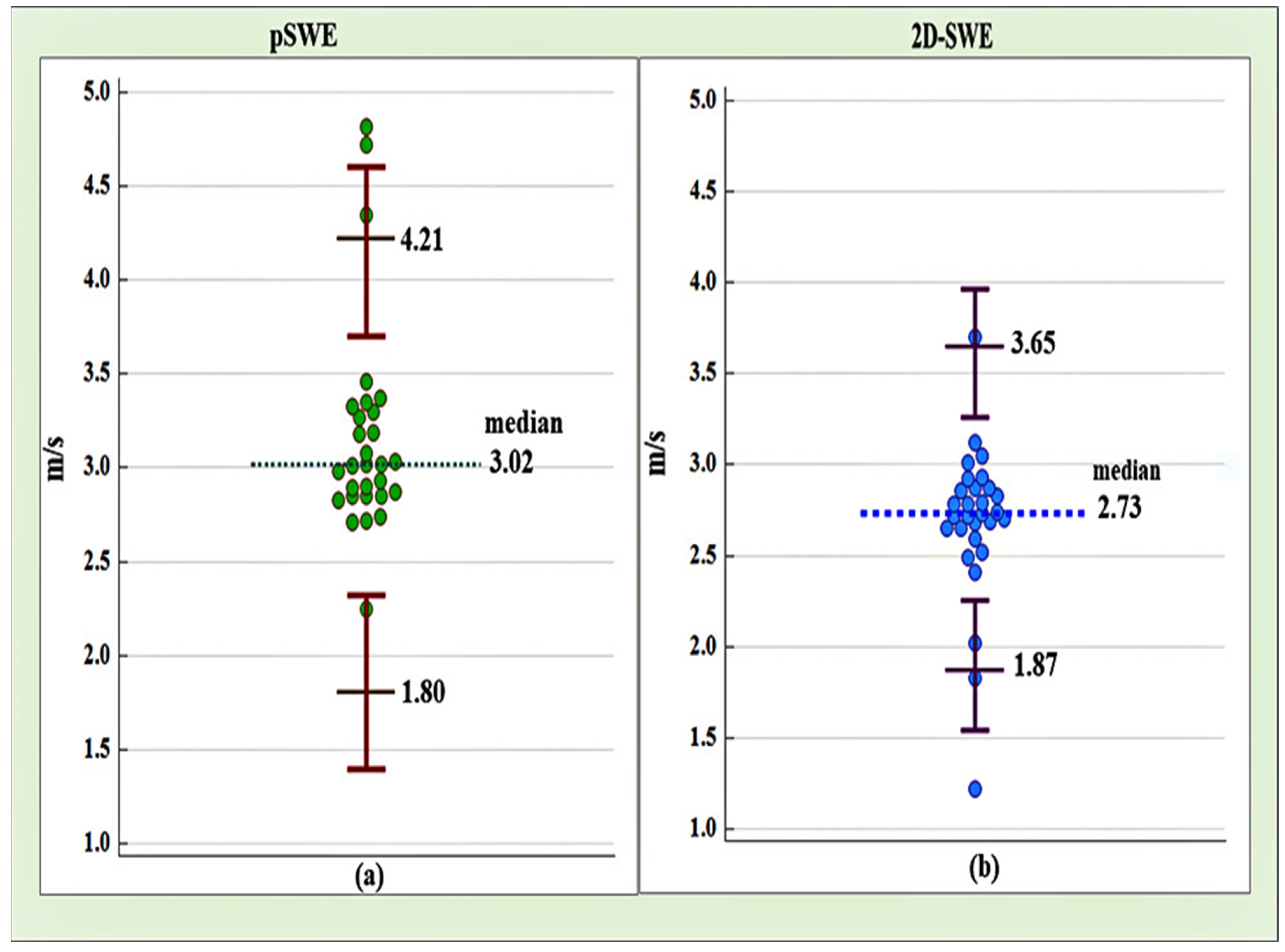
4.2. Reference Intervals Based on Point Shear Wave Elastography (pSWE) and Two-Dimensional Shear Wave Elastography (2D-SWE) for Liver Metastases from Colorectal Cancer

The reference intervals (RIs) for liver metastases from colorectal cancer based on pSWE and 2D-SWE data were calculated for the whole sample of 31 patients. Sex-related reference ranges were redundant because the target values did not vary significantly between men and women. For pSWE, the lower limit was found to be 1.80 m/s (90% CI: 1.39 to 2.32) and the upper limit was 4.21 m/s (90% CI: 3.69 to 4.60). For 2D-SWE, the lower limit was determined to be 1.87 m/s (90% CI: 1.54 to 2.25) and the upper limit was 3.65 m/s (90% CI: 3.26 to 3.97) (Figure 1).

4.3. Level of Agreement between Point Shear Wave Elastography (pSWE) and Two-Dimensional Shear Wave Elastography (2D-SWE)

The BlandAltman plot revealed an arithmetic mean difference of 0.50 m/s (95% CI- 0.143 to 0.821) between point shear wave elastography and two-dimensional shear wave elastography, with the first method tending to produce significantly higher values than the second (p = 0.006). As shown in the plot (Figure 2), the line of the mean difference between the two methods is situated 0.5 m/s above the line of zero difference.
We tested the association between metastases density as measured by point shear wave elastography and two-dimensional shear wave elastography and the demographic and clinical variables that are shown in Table 2. No significant associations were found between point shear wave elastography and the target variables (p > 0.05 for all tests). On the other hand, two-dimensional shear wave elastography correlated significantly with the patients’ age (p = 0.035), alkaline phosphatase (ALP) (p = 0.015), aspartate aminotransferase (ASAT) (p = 0.011), and alanine aminotransferase (ALAT) (p = 0.012), showing positive associations of moderate magnitude. The significant correlations are illustrated in Figure 3.

5. Discussion

Shear wave elastography imaging can be performed with a single conventional probe so that the investigator can have the opportunity to see the region of interest clearly and to precisely position it manually at the particular areas. The results from both elastographic techniques, point shear wave elastography, and two-dimensional shear wave elastography do not demonstrate significant differences between the men and women patients with regard to risk factors such as alcohol consumption and tobacco smoking. We found the lower and upper limit for liver metastases from colorectal cancer based on point shear wave elastography and two-dimensional shear wave elastography. The first method produced significantly higher values than the second (p = 0.006). The line of the mean difference between the two methods is situated 0.5 m/s above the line of zero difference. No significant associations were found between point shear wave elastography and aspartate aminotransferase (ASAT), alanine aminotransferase (ALAT), gamma-glutamyl transferase (GGT), alkaline phosphatase (ALP), total bilirubin and direct bilirubin. Two-dimensional shear wave elastography (2D-SWE) correlated significantly with the patients’ age, ALP, ASAT and ALAT (p = 0.012), and shows positive associations of moderate magnitude.
Few works have confirmed the diagnostic advantage of the point shear wave elastography using acoustic radiation force impulse (ARFI) [19,20] and two-dimensional shear wave elastography for the stiffness investigation of focal liver lesions [21].
Regardless of the promising results we expected to obtain from the study, there were some restrictions that needed to be taken into account. The maximum detection depth of shear wave elastography is limited and deep-seated liver metastases are difficult to assess [22]. In point shear wave elastography the allowed depth of transmission of an acoustic radiation impulse was only up to 10 cm from the skin. The maximum detection depth of two- dimensional shear wave elastography could be deeper compared to pSWE but depends on the type of the probe. Another disadvantage of shear wave elastography imaging is that the accuracy results of the shear wave’s speed decreased when the lesion is close to the heart and large vessels, or when patients had poor breath-holds.
Our research had several limitations, the first being that it involved only one type of liver metastases, namely from colorectal cancer.
Secondly, the number of involved patients could be larger. However, the number of participants is still statistically significant. We do not have enough patients belonging to other groups, such as metastases from esophageal cancer, gastric cancer, hepatocellular carcinoma, cholangiocarcinoma, pancreatic cancer.
Thirdly, the stiffness of liver metastases of colorectal cancer sizing under 2 cm was not measured but they could be evaluated by elastography. In the future, our work will continue in that respective direction. In medical clinical practice, imaging contributes considerably to the differentiating of hepatic lesions.
Conventional ultrasound examination is the first option for the objectification and diagnosis of liver lesions. There are some important advantages such as low cost, real-time imaging, and lack of radiation exposure. Computed tomography (CT) scans and MRI are highly sensitive but CT require ionizing radiation and magnetic resonance imaging (MRI) is contraindicated for patients with claustrophobia, cardiac pacemakers, implanted cardioverter- defibrillator, and metallic implants of foreign bodies. Both imaging modalities have higher cost compared to abdominal ultrasound. Another imaging technique that might be used is a combination of a positron emission tomography (PET) scanner and a computed tomography (CT) scanner in a single (PET/CT) device. PET/CT can often distinguish between benign and malignant lesions when CT and MRI cannot. PET/CT imaging is a non-invasive and painless procedure. This testing method uses radiolabelled molecules known as ‘radiopharmaceuticals’. The latter are used to detect or study abnormal cellular activity throughout the body. Fluorodeoxyglucose (18F), which is a radioactive form of glucose, is a commonly used radiotracer during PET/CT imaging. This radio-tracer is injected into the body before the scanning. A special camera is used to detect these radioactive emissions and produce images of the metabolic activity of the scanned region. A CT scan that is carried out simultaneously captures the X-ray images of internal organs from different angles. A computer then combines the data from PET and CT scans to produce 3-dimensional images of internal organs. Any abnormalities like tumors can be easily and accurately noticed through a PET/CT scan [23,24,25]. There are no major side effects associated with it. PET/CT, MRI, and liver biopsy are better for staging malignant lesions. There are some disadvantages such as higher cost; long duration of the procedure; radiation exposure and some technical difficulties. These may limit their daily use. Liver elastography compared to liver biopsy is a non-invasive method without any possible complications [26,27]. Liver biopsy should be in accordance with guidelines [28].
What stands out as the predominant tool in the diagnosis and management of patients with focal liver lesions is still liver biopsy [29]. Shear wave elastography is a widely used method for obtaining accurate stiffness information related to focal hepatic lesions [30,31]. The avoidance of invasive techniques should be seen as the main reason for performing elastography when it comes to the evaluation of focal liver lesions as they are commonly first detected on ultrasound examination.
Bearing in mind all the data up to now, the precision in diagnostics combined with the accuracy of elastography interpretation is of paramount interest. These techniques may be promising non-invasive methods for the evaluation of liver metastasis from colorectal cancer. Our investigation demonstrates that point shear wave elastography produces significantly higher values than two-dimensional shear wave elastography. Elastography is not to be considered a method to substitute histological confirmation of colorectal cancer by means of lower endoscopy or percutaneous liver biopsy. Pathomorphological assessment is currently the “gold” standard for the diagnosis of malignancies and determining the optimal therapeutic approach.

6. Conclusions

Final results show that both elastographic methods- point shear wave elastography and two-dimensional shear wave elastography can be considered to be promising in the diagnosis of focal liver lesions and in particular for liver metastases from colorectal cancer. Many investigations should be conducted before elastography could be put forward as a daily medical practice procedure for the differentiation of benign and malignant focal liver lesions and differentiation between different types of liver metastases according to their stiffness.

Author Contributions

D.I.D. and B.K.H.—supervision, K.A.D., E.L.N.-G. and M.I.D.—methodology. All authors have read and agreed to the published version of the manuscript.

Funding

This research received no external funding.

Institutional Review Board Statement

The study was conducted in accordance with the Declaration of Helsinki, and approved by the Institutional Review Board of University hospital Kaspela, Plovdiv city, Bulgaria, Protocol code-24/100322, Date of approval-3 October 2022.

Informed Consent Statement

Informed consent was obtained from all subjects involved in the study.

Data Availability Statement

No new data were created or analyzed in this study. Data sharing is not applicable to this article.

Conflicts of Interest

The authors declare no conflict of interest.

References

  1. Kamaya, A.; Machtaler, S.; Sanjani, S.S.; Nikoozadeh, A.; Sommer, F.G.; Khuri-Yakub, B.T.; Willmann, J.; Desser, T.S. New technologies in clinical ultrasound. Semin. Roentgenol. 2013, 48, 214–223. [Google Scholar] [CrossRef] [PubMed] [Green Version]
  2. Sherlock, S. Diseases of the Liver and Biliary System: Needle Biopsy of the Liver, 8th ed.; Blackwell Scientific Publications: Boston, MA, USA; Melbourne, Australia, 1989; pp. 36–48. [Google Scholar]
  3. Arnold, M.; Sierra, S.M.; Laversanne, M.; Soerjomataram, I.; Jemal, A.; Bray, F. Global patterns and trends in colorectal cancer incidence and mortality. Gut 2017, 66, 683–691. [Google Scholar] [CrossRef] [PubMed] [Green Version]
  4. Bray, F.; Ferlay, J.; Soerjomataram, I.; Siegel, R.L.; Torre, L.A.; Jemal, A. Global cancer statistics 2018: GLOBOCAN estimates of incidence and mortality worldwide for 36 cancers in 185 countries. CA Cancer J. Clin. 2018, 68, 394–424. [Google Scholar] [CrossRef] [PubMed] [Green Version]
  5. Bhat, S.K.; East, A.J. Colorectal cancer: Prevention and early diagnosis. Medicine 2015, 43, 295–298. [Google Scholar] [CrossRef]
  6. Wang, X.; O’Connell, K.; Jeon, J.; Song, M.; Hunter, D.; Hoffmeister, M.; Du, M. Combined effect of modifiable and non-modifiable risk factors for colorectal cancer risk in a pooled analysis of 11 population-based studies. BMJ Open Gastroenterol. 2019, 6, e000339. [Google Scholar] [CrossRef] [Green Version]
  7. Frampton, M.; Houlston, R.S. Modeling the prevention of colorectal cancer from the combined impact of host and behavioral risk factors. Genet Med. 2017, 19, 314–321. [Google Scholar] [CrossRef] [Green Version]
  8. Van Der Geest, L.G.M.; Lam-Boer, J.; Koopman, M.; Verhoef, C.; Elferink, M.A.G.; De Wilt, J.H.W. Nationwide trends in incidence, treatment and survival of colorectal cancer patients with synchronous metastases. Clin. Exp. Metastasis 2015, 32, 457–465. [Google Scholar] [CrossRef]
  9. Morris, E.J.; Forman, D.; Thomas, J.D.; Quirke, P.; Taylor, E.F.; Fairley, L.; Cottier, B.; Poston, G. Surgical management and outcomes of colorectal cancer liver metastases. Br. J. Surg. 2010, 97, 1110–1118. [Google Scholar] [CrossRef]
  10. Andres, A.; Mentha, G.; Adam, R.; Gerstel, E.; Skipenko, O.G.; Barroso, E.; Lopez-Ben, S.; Hubert, C.; Majno, P.E.; Toso, C. Surgical management of patients with colorectal cancer and simultaneous liver and lung metastases. Br. J. Surg. 2015, 102, 691–699. [Google Scholar] [CrossRef]
  11. De Baère, T.; Aupérin, A.; Deschamps, F.; Chevallier, P.; Gaubert, Y.; Boige, V.; Fonck, M.; Escudier, B.; Palussiére, J. Radiofrequency ablation is a valid treatment option for lung metastases: Experience in 566 patients with 1037 metastases. Ann. Oncol. 2015, 26, 987–991. [Google Scholar] [CrossRef]
  12. Klint, Å.; Engholm, G.; Storm, H.H.; Tryggvadóttir, L.; Gislum, M.; Hakulinen, T.; Bray, F. Trends in survival of patients diagnosed with cancer of the digestive organs in the Nordic countries 1964–2003 followed up to the end of 2006. Acta Oncol. 2010, 49, 578–607. [Google Scholar] [CrossRef] [PubMed]
  13. Stebbing, J.; Farouk, L.; Panos, G.; Anderson, M.; Jiao, L.R.; Mandalia, S.; Bower, M.; Gazzard, B.; Nelson, M. A meta-analysis of transient elastography for the detection of hepatic fibrosis. J. Clin. Gastroenterol. 2010, 44, 214–219. [Google Scholar] [CrossRef] [PubMed]
  14. Gennisson, J.L.; Deffieux, T.; Fink, M.; Tanter, M. Ultrasound elastography: Principles and techniques. Diagn. Interv. Imaging 2013, 94, 487–495. [Google Scholar] [CrossRef] [PubMed]
  15. Bamber, J.; Cosgrove, D.; Dietrich, C.F.; Fromageau, J.; Bojunga, J.; Calliada, F.; Piscaglia, F. EFSUMB guidelines and recommendations on the clinical use of ultrasound elastography. Part 1: Basic principles and technology. Ultraschall Der Med. Eur. J. Ultrasound 2013, 34, 169–184. [Google Scholar] [CrossRef]
  16. Tang, A.; Cloutier, G.; Szeverenyi, N.M.; Sirlin, C.B. Ultrasound Elastography and MR Elastography for Assessing Liver Fibrosis: Part 1, Principles and Techniques. Am. J. Roentgenol. 2015, 205, 22–32. [Google Scholar] [CrossRef] [Green Version]
  17. Shiina, T.; Nightingale, K.R.; Palmeri, M.L.; Hall, T.J.; Bamber, J.C.; Barr, R.G.; Castera, L.; Choi, B.I.; Chou, Y.H.; Cosgrove, D.; et al. WFUMB guidelines and recommendations for clinical use of ultrasound elastography: Part 1: Basis principals and terminology. Ultrasound Med. Biol. 2015, 41, 1126–1147. [Google Scholar] [CrossRef] [Green Version]
  18. Ferraioli, G.; Filice, C.; Castera, L.; Choi, B.I.; Sporea, I.; Wilson, S.R.; Cosgrove, D.; Dietrich, C.F.; Amy, D.; Bamber, J.C.; et al. WFUMB guidelines and recommendations for clinical use of ultrasound elastography: Part 3: Liver. Ultrasound Med. Biol. 2015, 41, 1161–1179. [Google Scholar] [CrossRef] [Green Version]
  19. Park, H.S.; Kim, Y.J.; Yu, M.H.; Jung, S.I.; Jeon, H.J. Shear Wave Elastography of Focal Liver Lesion: Intraobserver Reproducibility and Elasticity Characterization. Ultrasound Q. 2015, 31, 262–271. [Google Scholar] [CrossRef]
  20. Guibal, A.; Boularan, C.; Bruce, M.; Vallin, M.; Pilleul, F.; Walter, T.; Scoazec, J.Y.; Boublay, N.; Dumortier, J.; Lefort, T. Evaluation of shearwave elastography for the characterisation of focal liver lesions on ultrasound. Eur. Radiol. 2013, 23, 1138–1149. [Google Scholar] [CrossRef]
  21. Brunel, T.; Guibal, A.; Boularan, C.; Ducerf, C.; Mabrut, J.Y.; Bancel, B.; Boussel, L.; Rode, A. Focal nodular hyperplasia and hepatocellular adenoma: The value of shear wave elastography for differential diagnosis. Eur. J. Radiol. 2015, 84, 2059–2064. [Google Scholar] [CrossRef]
  22. Fahey, B.J.; Nightingale, K.R.; Nelson, R.C.; Palmeri, M.L.; Trahey, G.E. Acoustic radiation force impulse imaging of the abdomen: Demonstration of feasibility and utility. Ultrasound Med. Biol. 2005, 31, 1185–1198. [Google Scholar] [CrossRef]
  23. Piccinino, F.; Sagnelli, E.; Pasquale, G.; Giusti, G. Complications following percutaneous liver biopsy. A multicentre retrospective study on 68,276 biopsies. J. Hepatol. 1986, 2, 165–173. [Google Scholar] [CrossRef]
  24. McGill, D.B.; Rakela, J.; Zinsmeister, A.R.; Ott, B.J. A 21-year experience with major hemorrhage after percutaneous liver biopsy. Gastroenterology 1990, 99, 1396–1400. [Google Scholar] [CrossRef]
  25. Beyer, T.; Townsend, D.W.; Brun, T.; Kinahan, P.; Charron, M.; Roddy, R.; Jerin, J.; Young, J.; Byars, L.; Nutt, R. A combined PET/CT scanner for clinical oncology. J. Nucl. Med. 2000, 41, 1369–1379. [Google Scholar] [PubMed]
  26. Bar-Shalom, R.; Yefremov, N.; Guralnik, L.; Gaitini, D.; Frenkel, A.; Kuten, A.; Altman, H.; Keidar, Z.; Israel, O. Clinical performance of PET/CT in evaluation of cancer: Additional value for diagnostic imaging and patient management. J. Nucl. Med. 2003, 44, 1200–1209. [Google Scholar]
  27. Cohade, C.; Osman, M.; Leal, J.; Wahl, R.L. Direct comparison of 18F-FDG PET and PET/CT in patients with colorectal carcinoma. J. Nucl. Med. 2003, 44, 1797–1803. [Google Scholar]
  28. Grant, A.; Neuberger, J. Guidelines on the use of liver biopsy in clinical practice. Gut 1999, 45 (Suppl. 1), 1V1–1V11. [Google Scholar] [CrossRef] [PubMed]
  29. Rockey, D.C.; Caldwell, S.H.; Goodman, Z.D.; Nelson, R.C.; Smith, A.D. Liver biopsy. Hepatology 2009, 49, 1017–1044. [Google Scholar] [CrossRef] [PubMed]
  30. Ronot, M.; Di Renzo, S.; Gregoli, B.; Duran, R.; Castera, L.; Van Beers, B.E.; Vilgrain, V. Characterization of fortuitously discovered focal liver lesions: Additional information provided by shearwave elastography. Eur. Radiol. 2015, 25, 346–358. [Google Scholar] [CrossRef]
  31. Akdoğan, E.; Yılmaz, F.G. The role of acoustic radiation force impulse elastography in the differentiation of benign and malignant focal liver masses. Turk. J. Gastroenterol. 2018, 29, 456–463. [Google Scholar] [CrossRef] [Green Version]
Figure 1. Reference intervals based on pSWE (a) and 2D-SWE (b) for liver metastases from colorectal cancer, including medians and individual values.
Gastroent 14 00019 g001
Figure 2. Bland-Altman plot showing the limits of agreement between (pSWE) and (2D-SWE). The horizontal dotted line marks zero difference/perfect agreement.
Gastroent 14 00019 g002
Figure 3. Significant correlations between 2D-SWE and patients’ age (a), ALP (b), ASAT (c) and ALAT (d). *—Significant correlation at p < 0.05.
Gastroent 14 00019 g003
Table 1. Background information about the patients with liver metastases from colorectal cancer.
VariablesTotal
(n = 31)
Men
(n = 20)
Women
(n = 11)
p
Age
 ◦ mean ± SD65.37 ± 9.1766.90 ± 9.5262.90 ± 8.180.261 t
 ◦ minimum-maximum37–8037–8049–78
Histology n (%)
 ◦ LD-AC3 (10.35%)3 (16.70%)0 (0.00%)0.07 f
 ◦ MD-AC19 (65.50%)13 (72.20%)6 (54.50%)0.432 f
 ◦ HD-AC7 (24.15%)2 (11.10%)5 (45.50%)0.268 f
BMI
 ◦ mean ± SD22.02 ± 1.4122.25 ± 1.2821.65 ± 1.580.278 t
 ◦ minimum-maximum19.20 to 24.7020.40 to 24.7019.20 to 23.90
GGT median (IQR)56.00 (54.00)51.00 (42.50)76.00 (65.00)0.102 U
ALP mean ± SD298 ± 109.90275.90 ± 75.10335.20 ± 147.900.163 t
Total bilirubin median (IQR)17.20 (5.70)16.60 (5.10)18.30 (8.30)0.438 U
Direct bilirubin median (IQR)5.30 (3.60)5.30 (4.18)5.50 (3.80)0.759 U
ASAT median (IQR)44 (32.50)41.00 (22.00)45.00 (76.00)0.465 U
ALAT mean (±SD)47 (24.50)45.28 ± 18.1958.09 ± 28.790.206 t
pSWE velocity (m/s)
 ◦ median3.02 (0.47)3.02 (0.42)2.98 (0.52)0.580 U
 ◦ minimum-maximum2.25 to 4.822.25 to 4.822.71 to 4.72
2D-SWE (m/s)
 ◦ median (IQR)2.73 (0.25)2.76 (0.24)2.68 (0.27)0.068 U
 ◦ minimum-maximum1.22 to 3.701.22 to 3.121.83 to 3.70
pSWE depth (mm)
 ◦ median (IQR)47.72 (9.11)46.52 (9.24)50.70 (8.64)0.465 U
 ◦ minimum-maximum30.37 to 54.3331.49 to 51.9730.37 to 54.33
2-D-SWE depth (mm)
 ◦ median (IQR)46.80 (8.10)46.80 (6.05)50.20 (9.00)0.412 U
 ◦ minimum-maximum29.40 to 54.4032.00 to 53.4029.40 to 54.40
Alcohol n (%)17 (56.60%)13 (72.20%)4 (36.40%)0.119 f
Smoking n (%)19 (65.50%)12 (66.70%)7 (63.60%)1.000 f
IQR-interquartile range, GGT—Gamma-glutamyl transferase; ALP—Alkaline phosphatase; ASAT—Aspartate aminotransferase; ALAT—Alanine aminotransferase; t—t-test for independent samples; f—Fisher’s exact test; U—Mann-Whitney U test.
Table 2. Results from the correlation analysis for associations between metastasis density and demographic and clinical data.
VariablespSWE (m/s)2D-SWE (m/s)
Age
◦ Pearson r0.1140.392
◦ p0.5570.035
BMI
◦ Pearson r0.1620.091
◦ p0.4020.637
GGT
◦ Spearman rho0.195−0.130
◦ p0.3110.530
ALP
◦ Pearson r−0.2290.447
◦ p0.2320.015
Total bilirubin
◦ Pearson r−0.3360.256
◦ p0.0750.118
Direct bilirubin
◦ Pearson r−0.2540.070
◦ p0.1920.722
ASAT
◦ Pearson r−0.1740.463
◦ p0.3800.011
ALAT
◦ Pearson r−0.2670.459
◦ p0.1620.012
Tumor grade (histology)
◦ Spearman r−0.109−0.004
◦ p0.5720.984
Alcohol
◦ Spearman r0.130−0.059
◦ p0.5020.763
Smoking
◦ Spearman r−0.100−0.117
◦ p0.6070.704
Disclaimer/Publisher’s Note: The statements, opinions and data contained in all publications are solely those of the individual author(s) and contributor(s) and not of MDPI and/or the editor(s). MDPI and/or the editor(s) disclaim responsibility for any injury to people or property resulting from any ideas, methods, instructions or products referred to in the content.

