

Gastroenterol. Insights 2024, 15(2), 342-353; https://doi.org/10.3390/gastroent15020023 (registering DOI) - 24 Apr 2024
Abstract
We present a multicenter retrospective study of patients undergoing surgery for duodenal adenocarcinoma, from January 2010 to August 2020, in order to determine the epidemiological characteristics and the oncological results after surgical resection obtained in this rare tumor. Variables: demographics; tumor location; surgical
[...] Read more.
We present a multicenter retrospective study of patients undergoing surgery for duodenal adenocarcinoma, from January 2010 to August 2020, in order to determine the epidemiological characteristics and the oncological results after surgical resection obtained in this rare tumor. Variables: demographics; tumor location; surgical intervention and immediate postoperative period; and post-surgical follow-up information, such as recurrence, overall survival (OS), and disease-free survival (DFS). A total of 32 patients underwent surgery. The median age was 69.74 years (IQR 60.47–79.09) and the male/female distribution was 3:1. The surgeries performed were: pancreaticoduodenectomy (PD) in 16 (50%) patients, segmental resection in 13 (40.6%), and the local excision of the lesion in three (9.4%). The R0 rate was higher in PD (86.7% vs. 42.9%; p = 0.013). The OS and DFS rate at one, three and five years was 95%, 70%, and 60% and 86%, 55%, and 48%, respectively. There was a greater trend towards recurrence in patients who did not undergo PD (53.8% vs. 25%; p = 0.14) and conservative surgery seemed to be associated with more local recurrence than PD (57.1% vs. 33.3%; p = 0.49). PD and limited resection are both valid options in the cases of non-ampullary duodenal adenocarcinoma, although PD presented lower rates of loco-regional recurrence.
Full article
(This article belongs to the Section Gastrointestinal Disease)
►
Show Figures
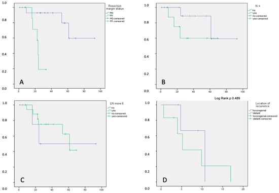
Figure 1


