Review

Diverticular Disease—An Updated Management Review

1
Albany Medical College, Albany, NY 12208, USA
2
Department of Surgery, Albany Medical Center Hospital, Albany, NY 12208, USA
3
Department of Internal Medicine, Albany Medical Center Hospital, Albany, NY 12208, USA
4
Department of Pathology, Albany Medical Center Hospital, Albany, NY 12208, USA
5
Department of Gastroenterology and Hepatology, Albany Medical Center Hospital, Albany, NY 12208, USA
*
Author to whom correspondence should be addressed.
Gastroenterol. Insights 2022, 13(4), 326-339; https://doi.org/10.3390/gastroent13040033
Submission received: 29 July 2022 / Revised: 14 September 2022 / Accepted: 27 September 2022 / Published: 30 September 2022
(This article belongs to the Special Issue Management and Treatment of Digestive Disorders)

Abstract

:
Diverticular disease is highly prevalent in the Western world, placing an increased burden on healthcare systems. This review clarifies the consensus in the literature on the disease’s classification, etiology, and management. Diverticular disease, caused by sac-like protrusions of colonic mucosa through the muscular colonic wall, has a varied disease course. Multiple theories contribute to our understanding of the etiology of the disease, with pathogenesis affected by age, diet, environmental conditions, lifestyle, the microbiome, genetics, and motility. The subtypes of diverticular disease in this review include symptomatic uncomplicated diverticular disease, segmental colitis associated with diverticulosis, and uncomplicated and complicated diverticulitis. We discuss emerging treatments and outline management options, such as supportive care, conservative management with or without antibiotics, and surgical intervention.

1. Introduction and Epidemiology

Diverticular disease and its complications remain a significant global burden on healthcare systems [1]. It is one of the most common gastrointestinal conditions among inpatients and outpatients in industrialized nations [1,2]. In such nations, diverticulosis of the sigmoid colon has prevalence rates between 5–70%, based on age and diagnostic modality [3,4]. Furthermore, colonic diverticulosis is the most common finding during routine colonoscopy [5], and in 2010, it was the eighth most common outpatient diagnosis in the United States, with 2,734,119 total outpatient visits [1].
The lifetime risk of diverticulitis in a person with diverticulosis, diagnosed based on colonoscopy or computed tomography (CT), is about 5% [2,5,6]. Even so, given that more than 50% of Americans over the age of 60 years have diverticulosis, diverticulitis is highly prevalent [2]. In 2012, it was estimated that diverticulitis alone accounted for 216,650 hospital admissions [1]. Furthermore, the incidence of diverticulitis is rising, with an increase of 26% in admissions for diverticulitis from 1998 to 2005 [3]; another study showed an increase of 26.8% for emergency room visits for diverticulitis from 2006 to 2013 [7]. In addition, the mean cost of these emergency room visits rose from $3061 to $4765 from 2006 to 2013, and this was adjusting for inflation [7]. The financial impact of diverticulitis is considerable, with hospital admissions costing an aggregate of $2.2 billion [1].
In this review article, we discuss emerging understandings of the etiology and management of diverticular disease. There are many contradictory findings in the literature, and this review highlights these controversies in order to best inform the reader. We offer a unique perspective from physicians across various specialties, including gastroenterology, pathology, surgery, and radiology, to enhance our review.

2. Definitions and Presentation

Diverticular disease is characterized by sac-like protrusions of the colonic wall. Left-sided colonic diverticula are more common in the West and are characterized by herniation of the mucosa through the muscularis propria at weak points in the wall where the vasa recti supply the mucosa and submucosa (Figure 1) [1,8,9]. Right-sided diverticula are generally true diverticula, more common in Asian countries, and herniate through all of the layers of the colon wall. For the purposes of this review, we will focus on left-sided “pseudo”-diverticula [4,10].
Diverticular disease can be classified on a continuum from diverticulosis to diverticulitis, with symptoms, inflammation, and complications largely affecting where on this continuum the disease exists. We describe the presentation and management for diverticulosis, symptomatic uncomplicated diverticular disease (SUDD), segmental colitis associated with diverticulosis (SCAD), and acute uncomplicated and complicated diverticulitis. See Table 1 for a summary overview of the location, epidemiology, presentation, diagnosis, pathogenesis, and management of each subtype.

2.1. Asymptomatic Diverticulosis

Asymptomatic diverticulosis is characterized by silent diverticula. It is frequently an incidental finding in patients undergoing imaging or endoscopy for other indications [1]. Histologically, asymptomatic diverticulosis shows non-inflamed colonic mucosa and submucosa protruding through the muscularis propria into the adventitia (subserosa) of the bowel wall (Figure 1). About 25% of people with asymptomatic colonic diverticulosis will experience an episode of symptomatic diverticular disease, and up to 5% will have an episode of acute diverticulitis, with or without complications [20,21].

2.2. Diverticular Bleeding

Diverticular bleeding is lower-gastrointestinal bleeding resulting from the rupture of diverticula-associated arteries [22]. Diverticular bleeding is considered a complicated form of diverticulosis. It presents as painless, intermittent, and often large-volume bleeding with left-sided bleeding bright-red and right-sided bleeding dark or melanotic [23]. It is not usually associated with diverticulitis [8].

2.3. Symptomatic Uncomplicated Diverticular Disease

SUDD is defined as chronic diverticulosis with associated chronic abdominal pain in the absence of acute symptoms of diverticulitis, overt colitis, or any leukocytosis. It develops in approximately 20% of patients with diverticulosis [1,24,25]. SUDD is diagnosed in patients with chronic diarrhea, crampy abdominal pain, and intermittent hematochezia in addition to inflammation seen via a colonoscopy and chronic inflammatory changes seen on a biopsy of the diverticulum [12]. Approximately 10–25% of patients with SUDD may develop acute diverticulitis [24].

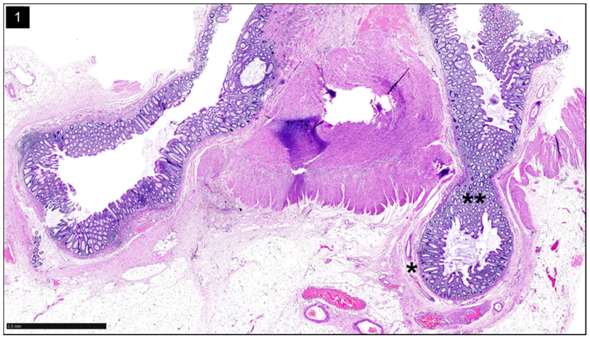
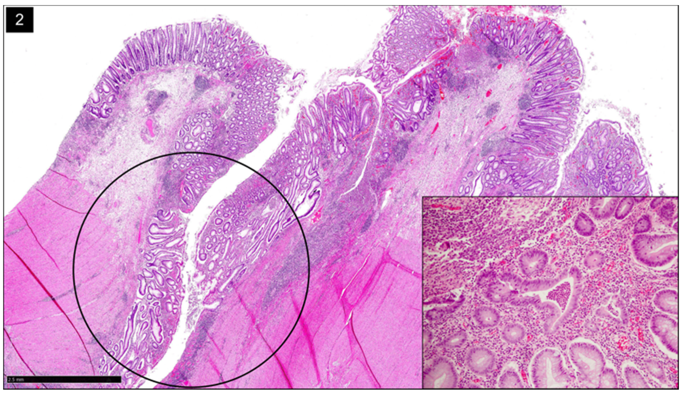
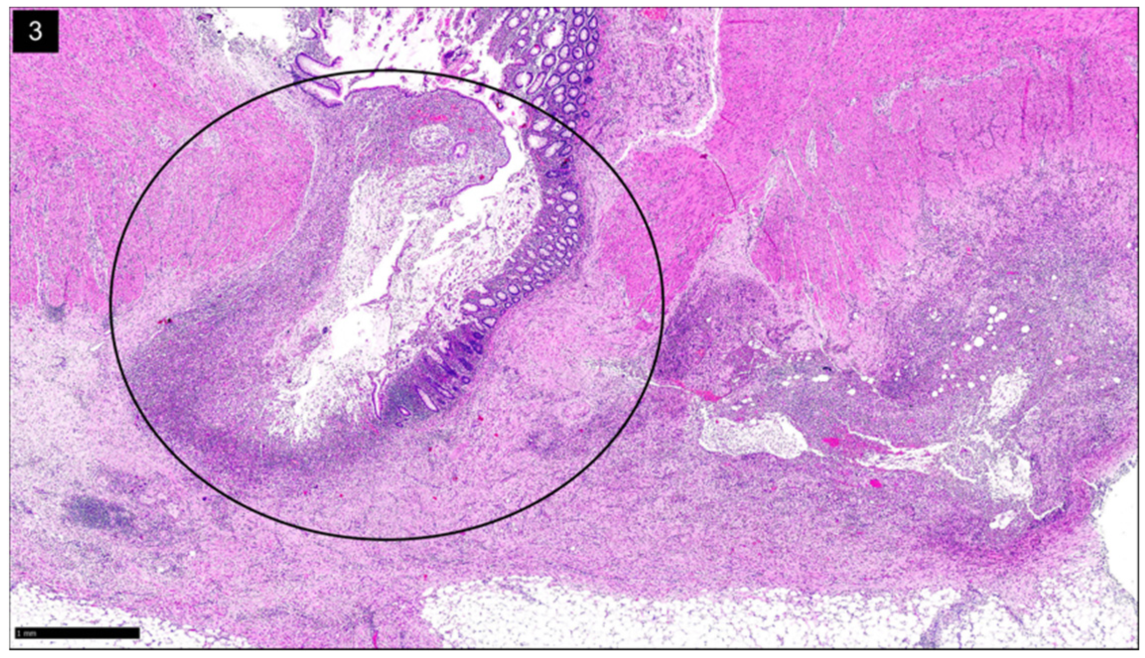
2.4. Segmental Colitis Associated with Diverticulosis
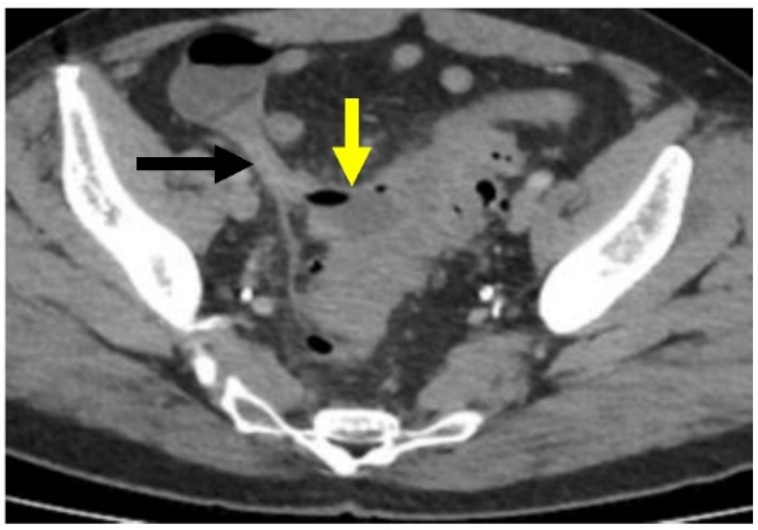
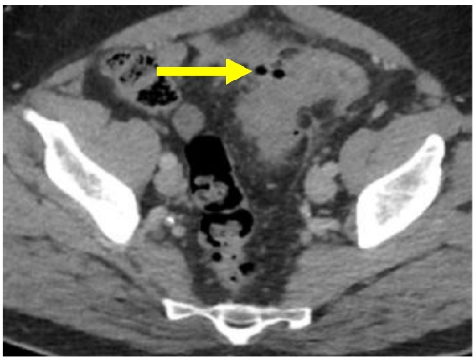
SCAD is characterized by nonspecific segmental inflammation in the sigmoid colon surrounded by multiple diverticula [4,11,12,25]. The initial presentation is often rectal bleeding and in some, diarrhea or abdominal pain [12]. SCAD is typically benign and self-limited [4]. SCAD differs from diverticulitis because the individual diverticula are not involved; histologically, this shows inflammation within the interdiverticular mucosa rather than the diverticular orifices [4,12]. Additionally, a chronic active colitis pattern of injury with crypt distortion and cryptitis is seen on histology (Figure 2). Its prevalence varies between 1.15% and 11.4% among those diagnosed with diverticular disease [11].

2.5. Diverticulitis

Acute diverticulitis is inflammation of a diverticulum [1,9,26]. It can be further sub classified into complicated and uncomplicated disease, depending on the presence or absence of perforation (respectively) [9].
Histologically, diverticulitis shows inflammation of the diverticular pouch with or without mucosal ulceration. When the diverticular pouch is ruptured, subserosal abscess can form (Figure 3). Computed Tomography (CT) with intravenous (IV) contrast is the imaging modality of choice to evaluate for acute diverticulitis and its complications [27].

2.6. Uncomplicated Diverticulitis

Most patients (~90%) will have uncomplicated diverticulitis, characterized by localized inflammation limited to the colonic wall and surrounding tissue. Patients present with fever, abdominal pain, and/or leukocytosis [4,26]. On CT imaging, there is evidence of inflammatory stranding within the peridiverticular fat without evidence of free air or an adjacent fluid collection (Figure 4).

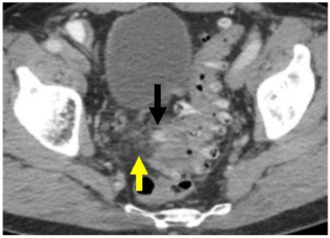
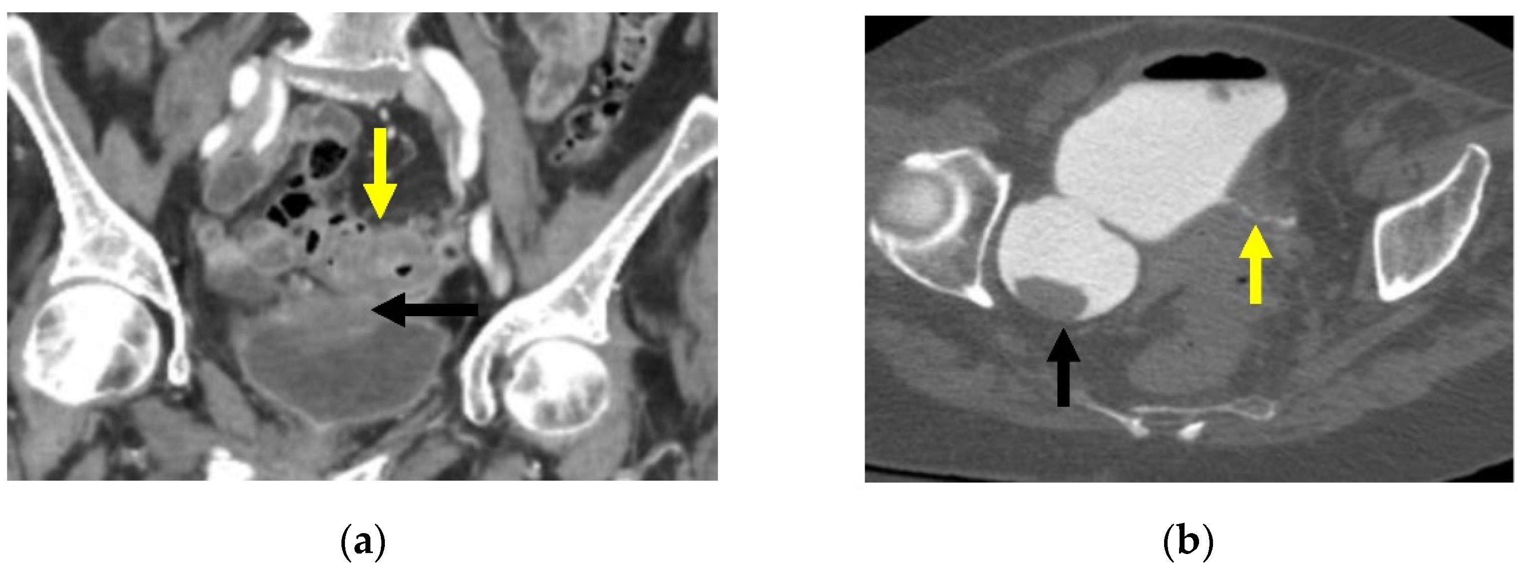
2.7. Complicated Diverticulitis

Complicated diverticulitis is defined as diverticulitis associated with localized or generalized perforation, localized or distant abscess, fistula, stricture, or obstruction [4,6,26]. Complications of diverticulitis occur in approximately 12% of patients; the most common complication is phlegmon or abscess, followed by peritonitis, obstruction, and fistula [2].
Acute complicated diverticulitis has been further subclassified in the surgical literature using the Hinchey Classification. Developed by Hinchey et al. in 1978 and later modified with the widespread usage of CT scanning (see Table 2), the Hinchey classification divides acute complicated diverticulitis into four categories based on whether the perforation is localized or not [28,29,30]. Classes I and II, a pericolic abscess or phlegmon (Figure 5) and distant pelvic intra-abdominal or retroperitoneal abscess, respectively, do not cause peritonitis or hemodynamic changes. Hinchey classes III and IV, generalized purulent and feculent peritonitis, respectively, often do. The modified Hinchey classification is a helpful adjunct to determine management, with purulent and fecal peritonitis (classes III-IV) most likely to lead to surgery [4,19].
CT scanning can show complicated disease as a peridiverticular mesenteric inflammatory reaction (Hinchey Ib) or microperforation with adjacent extraluminal air (Figure 6). Contrast is also helpful to evaluate for potential abscess (Hinchey Ia or II), where rim-enhancement will be seen (Figure 5).
The inflammatory reaction may progress to pericolic and mural phlegmonous infiltration as well as sealed perforation, free perforation, abscess, fistula (Figure 5 and Figure 7a,b), and a stenosing inflammatory sigmoid mass [9]. A fistula develops associated with diverticulitis when a fistulous tract, or connection, forms between the affected colon and another organ, whether that is the bladder, vagina, uterus, skin, or another part of the intestine (see Figure 5 and Figure 7a,b) [8,31]. These patients are often diagnosed clinically, with pneumaturia or a urinalysis growing gut microorganisms (colovesical fistula); foul-smelling vaginal discharge or air and stool from the vagina (colovaginal or colouterine fistula); presence of an enterocutaneous fistula with purulence or stool from the abdominal wall (colocutaneous fistula); or are asymptomatic or with vague abdominal symptoms (coloenteric fistula). On CT, enhancement is also seen along fistulous tracts, making them more conspicuous (Figure 7a). Retrograde opacification of the bladder during a cystogram (Figure 7b) or colon with rectal contrast may also be helpful to define fistulas [27].
Patients with stricture may present with obstruction, as the lumen of the colon narrows from the inflammatory reaction associated with diverticulitis. Patients typically have worsening constipation in the setting of long-standing diverticulitis. This can be seen as a dilated colon proximal to the stricture on CT scan [8]. For these patients, it is important to differentiate between a stricture from diverticulitis or from colorectal cancer, and they require a colonoscopy 6–8 weeks after the acute episode or resection (if the patient was treated with surgery) [4].
A subset of patients have ongoing or smoldering diverticulitis, defined as ongoing diverticulitis (pain with leukocytosis or inflammatory markers, fever, or CT evidence of inflammation) despite antibiotic treatment or exacerbations after cessation of treatment [2].

3. Etiology

Though still widely debated, the underlying pathological mechanisms that result in the formation of colonic diverticula and the subsets of diverticular disease are likely due to complex interactions among age, colonic motility, diet, colonic microbiota, genetic factors, and changes in colonic structure [1,6,10,16].

3.1. Risk Factors

Diet, lifestyle, and medications all play a role in the risk factors for diverticulitis [2,15]. Western dietary patterns, including diets high in red meat, fat, and refined grains, have been widely shown to increase the risk of diverticulitis [2]. A prospective cohort study of 47,888 men in the U.S. and the risk of diverticular complications (after adjustment for age, energy-adjusted total fat intake, and physical activity) found a positive association among high red meat, high-fat diets, and the risk of diverticular complications when compared with a low red meat, low-fat diet [12].
Although now widely contested (see Table 3) [32], a high-fiber diet has long been thought to play a major role in prevention of diverticular disease [33,34]. This observation was first made by Burkitt and Painter in 1971, who hypothesized that a high-fiber diet would facilitate a shorter transit time through the colon [33,34]. However, several studies have suggested no causative relationship exists between fiber and the disease’s development [4,6,34,35]. Similarly, nuts and seeds, once thought to exacerbate diverticulitis, do not appear to increase the risk [2].
Obesity, particularly central obesity, and smoking have been associated with an increased risk in diverticulitis [2].
People living in areas with low exposure to ultraviolet light or in rural areas were found to have a higher risk of diverticulitis-associated hospitalizations than people living in areas with high ultraviolet light exposure [21].
The use of nonsteroidal anti-inflammatory drugs (NSAIDs), steroids, and opioids has been found to increase the risk of diverticulitis and diverticular bleeding [2,4,12,21,22]. Though the mechanism by which they increase risk is not well understood, it has been proposed that NSAIDs may induce mucosal damage through reduced prostaglandin synthesis [21]. Immunosuppressive regimens are thought to increase the risk of diverticulitis due to impaired healing that may contribute to perforation [2]. Opioids affect gut motility, thereby potentially increasing the risk of diverticula formation. Additionally, several medications have been shown to potentially decrease the risk of diverticulitis including statins, calcium channel blockers, and metformin, but further studies are necessary to confirm these findings [2].

3.2. Pathogenesis

Diverticulitis is likely due to chronic inflammation, especially considering that many of its risk factors, such as obesity, lack of exercise, and consuming a processed-grain, high red meat diet are associated with systemic inflammation [2]. Diverticulitis has also been associated with an increased expression of matrix metalloproteinases and histamine, both of which are markers of intestinal inflammation [2]. While the debate has been ongoing, several theories have endured [16,17].
The “traumatic” theory suggests that the onset of diverticulitis is the result of an acute inflammation due to traumatic damage to a diverticulum [16,17]. Increased pressure within the colon is thought to push fecaliths into the diverticula, particularly large diverticula [16,17]. In doing so, the trapped stool erodes the mucosa of the diverticular sac, resulting in impaction, abrasion, inflammation, and potential bacterial overgrowth [16,17]. Additionally, if the proliferating bacteria breach the mucosal wall, the resulting waste and gas production may lead to bowel perforation [16,17]. Alternatively, damage from the trapped fecalith may irritate the mucosa with local inflammation, followed by vascular congestion and further obstruction, similarly resulting in stool trapping and bacterial overgrowth [17]. The “traumatic” theory may describe the general sequence of events that cause acute diverticulitis in older patients, as they have larger diverticula that are more likely to be obstructed by fecaliths [16].
The “ischemic” theory suggests that colonic motility, rather than fecal entrapment, causes diverticulitis [16,17]. The ischemic process is thought to be triggered by a long-lasting or potentially recurrent contractile impulse of the colon, resulting in persistent compression of the vasa recta at the diverticular neck and subsequent ischemia and microperforation [16,17]. This theory is supported by findings that patients with diverticular disease may have increased sensitivity to cholinergic denervation and thus excessive contractile impulses in the diverticular wall [16,17]. This may explain the potential cause of diverticulitis in younger patients, in whom colonic diverticula and fecalith entrapment is far less likely [16,17]. This theory has also been suggested as a potential mechanism for the development of SUDD, in which abdominal pain, potentially due to sustained contraction of the bowel wall, is present without acute symptoms of diverticulitis [16].
Studies have identified that the composition of colonic microbiota differs in patients with diverticular disease in comparison to those without [2,39]. Many of these studies have focused on the microbiota in patients diagnosed with SUDD; findings have suggested a decrease in bacteria that produce short-chain fatty acids (SCFAs) as well as a decrease in Akkermansia, which is a mucin-degrading bacteria that suppresses inflammation [2]. SCFAs supply the colonic epithelium as the primary energy source as well as increase mucus and antimicrobial peptide production, thus mediating immune homeostasis, further contributing to the theory of chronic inflammation [2]. Because there are significant differences in the diagnosis and presentation of SUDD and diverticulitis, these findings cannot be extrapolated to that of diverticulitis [2]. Even so, changes in the intestinal microbiome, particularly regarding bacteria involved in SCFA metabolism, are associated with the development of diverticulitis and symptoms associated with diverticulosis, such as SUDD [2,39].
Given the variation in theories of pathogenesis and onset, it seems critical to understand a genetic basis for the development of diverticular disease. The heritability of diverticular disease was estimated to be 53% in a Danish twin cohort and 40% in a Swedish study [21,40,41]. Furthermore, recent genome-wide association (GWAS) studies suggest that diverticular disease is primarily a disorder of the intestinal neuromuscular function, with impaired mesenteric vascular smooth muscle function and impaired connective fiber support [21]. In one retrospective GWAS of 28,000 patients admitted to a hospital with diverticular disease, 42 risk loci were identified [2,18]. These risk loci contained genes involved in immune regulation, maintenance of the extracellular matrix, cell adhesion, membrane transport, and intestinal motility [2,18]. These findings support the need to further understand underlying genetic mechanisms for the development of disease.

4. Management

4.1. Symptomatic Uncomplicated Diverticular Disease

Management of SUDD attempts to control symptoms, with the goal of preventing recurrences and avoiding the development of diverticulitis and other complications [24]. Current evidence suggests that rifaximin and mesalamine are beneficial in treating SUDD symptoms [24,25]. Fiber supplementation is still recommended despite a lack of recent evidence to support this use [24,25]. Studies have investigated the use of probiotics for the treatment of SUDD, although definitive evidence for its efficacy has not yet been established [25].

4.2. Segmental Colitis Associated with Diverticulosis

There are currently no guidelines for the treatment of SCAD [11]. Management is largely based on case series and adopted from inflammatory bowel disease (IBD) treatment [11]. Treatment generally includes the use of antibiotics, salicylates, or a combination of the two with a high-fiber diet [11]. Surgery is usually reserved for cases that are refractory to medical management [11].

4.3. Uncomplicated Diverticulitis

The treatment paradigm of uncomplicated diverticulitis is evolving. Outpatient management has traditionally included a clear liquid or regular diet, oral broad-spectrum antibiotics such as amoxicillin-clavulanic acid or ciprofloxacin and metronidazole for 7–14 days, and a close follow-up [2,25,26].
The use of antibiotics has recently been questioned [4]. A study randomized 623 patients with acute uncomplicated diverticulitis, who each received antibiotic treatment or a placebo. Antibiotic therapy did not accelerate recovery, prevent complications, or prevent recurrence, so the authors recommended reserving antibiotic use for the treatment of complicated diverticulitis [42]. This finding was further supported by a review of three randomized trials [26,43]. Based on this evidence, several European guidelines have stopped recommending antibiotics for uncomplicated diverticulitis [2]. Similarly, the American Society of Colon and Rectal Surgeons (ASCRS) gave a 1A recommendation to treat select patients with Hinchey Class I and Ia complicated diverticulitis without antibiotics, however they noted that patients treated without antibiotics were more likely to have elective surgery during follow-up [19]. The American Gastroenterological Association’s (AGA) 2015 guideline provided a conditional recommendation for selective rather than routine use of antibiotics in uncomplicated diverticulitis in the absence of severe disease, immunocompromised status, pregnancy, or significant comorbidity [2,32].
Outpatient treatment of uncomplicated diverticulitis is effective in most cases; less than 10% of patients are admitted to the emergency department for diverticulitis within 60 days of the initial evaluation [25]. The decision to hospitalize a patient with uncomplicated diverticulitis depends on the patient’s ability to tolerate oral intake, severity of illness, comorbidities, and outpatient support systems [26]. Hospitalization should be considered if patients have signs of peritonitis or if there is a concern for complicated diverticulitis [26]. Inpatient management includes no oral intake, IV fluid resuscitation, and IV antibiotics such as amoxicillin-clavulanic acid or ceftriaxone and metronidazole [26,44]. Outpatient follow-up with a surgeon after uncomplicated diverticulitis is not always necessary. Some patients with acute uncomplicated diverticulitis may desire surgery, however the ASCRS recommends against routine sigmoidectomy for uncomplicated disease. If the patient is experiencing persistent residual symptoms, recurrent episodes, or if the episodes significantly affect their quality of life, elective sigmoidectomy has been indicated to definitively treat the diverticulitis [19].

4.4. Complicated Diverticulitis

Admission to a hospital is recommended for all patients with complicated diverticulitis for bowel rest, IV antibiotics, IV fluid resuscitation, and possible intervention. Abscesses (Hinchey Ia or II) can be treated with CT-guided percutaneous drainage if the abscess is large enough, (>3 cm) and localized, assuming the patient is stable [4,19,26]. It is debated whether requiring a percutaneous drainage procedure leads to recurrent diverticulitis, with some studies indicating a lower recurrence rate than those treated conservatively without drainage, and others with a higher recurrence rate [8,19,26].
There is a large range in the rate of recurrence after conservatively managed acute diverticulitis: from 13–60.5% with most studies in the range of 25% of patients having a recurrent episode within 1–10 years [2,19,31]. Those with complicated diverticulitis, larger abscesses (>3–5 cm), and significant comorbid conditions were more likely to have a recurrent episode [4,19]. Regardless, the first episode of diverticulitis tends to have the highest risk of free perforation, and the risk of recurrence has been shown to increase with subsequent episodes [2,4].
Outpatient, elective sigmoidectomy with colorectal anastomosis is the surgical procedure of choice for hemodynamically stable patients who were treated with conservative management of their complicated or recurrent diverticulitis. The surgeon should weigh comorbid conditions, quality of life, recurrent episodes, and fitness for surgery in their decision to operate [19]. Whether elective or urgent/emergent, the resection should include portions of the colon affected by diverticulitis and not all segments with diverticulosis, so as to preserve colon length [8]. Patients with diverticula-associated fistula or stricture are considered to have chronic disease and can undergo an elective operation with a primary anastomosis [2,4].
For Hinchey III and IV diverticulitis, surgery during the acute hospital stay is up to the discretion of the surgeon. The ASCRS guidelines recommend surgical intervention if the patient has diffuse clinical peritonitis, hemodynamic instability, or if conservative management with bowel rest and IV antibiotics fails after several days [4,19]. The need for operative intervention and type of operation for patients with complicated diverticulitis is currently being debated. For many years, the standard of care was to offer a resection of the diseased segment with end-colostomy, as it can be performed safely, quickly (for a hemodynamically unstable patient), and does not include an anastomosis which inherently is at risk of breaking down in a septic patient undergoing surgery [8]. The rate of reversal of the ostomy after this procedure is low, however, with 20–50% of patients never being reversed [4]. Recently, multiple randomized controlled trials have shown the safety of performing a colorectal anastomosis with diverting loop ileostomy in a hemodynamically stable patient, and, in fact, the patients with primary anastomosis had a higher rate of subsequent ostomy reversal with fewer complications and shorter hospital stays [4,45,46]. It has emerged as a level IB recommendation by the ASCRS to perform a primary anastomosis in this patient population [19].
The current guidelines recommend that a colonoscopy be performed 6–8 weeks following recovery from an episode of diverticulitis in patients who have not had a recent colonoscopy. The recommendation is to wait several weeks for the colon to heal in order to reduce the risk of perforation. The risk of malignancy in uncomplicated diverticulitis is low, (~1.3%); however, a meta-analysis found that the prevalence of colorectal cancer was 7.9% in those with complicated diverticulitis [47]. A colonoscopy can evaluate for this, especially in the setting of preoperative preparation for sigmoidectomy.

5. Conclusions

It is critical to understand the etiology, classification, and management of diverticular disease due to the tremendous burden these subtypes place on healthcare systems. Categories of diverticular disease include uncomplicated and complicated diverticulitis, segmental colitis associated with diverticulosis, and symptomatic uncomplicated diverticular disease. Though subject to debate, it is thought that low fiber, high red meat, and high-fat diets, NSAIDs, low ultraviolet light exposure, smoking, and obesity are risk factors for diverticular disease. Treatment varies from conservative to surgical management, depending on the severity and presentation of the illness.

Author Contributions

S.K., K.O. and L.R. drafted and edited the manuscript. S.H.A., H.L., M.S., M.T. and B.V. provided critical revisions of the manuscript. All authors have read and agreed to the published version of the manuscript.

Funding

This research received no external funding.

Institutional Review Board Statement

Ethical review and approval were waived for this study because it did not meet the regulatory definitions of “human subjects.” Research activities did not involve obtaining and studying or analyzing biospecimens or information from living individuals.

Informed Consent Statement

Patient consent was waived because this study is not considered to be “human subjects” research. The de-identified images in this text were obtained from the physician library.

Data Availability Statement

Not applicable.

Conflicts of Interest

The authors declare no conflict of interest.

References

  1. Rezapour, M.; Ali, S.; Stollman, N. Diverticular Disease: An Update on Pathogenesis and Management. Gut Liver 2018, 12, 125–132. [Google Scholar] [CrossRef] [PubMed]
  2. Strate, L.L.; Morris, A.M. Epidemiology, Pathophysiology, and Treatment of Diverticulitis. Gastroenterology 2019, 156, 1282–1298.e1281. [Google Scholar] [CrossRef]
  3. Munie, S.T.; Nalamati, S.P.M. Epidemiology and Pathophysiology of Diverticular Disease. Clin. Colon. Rectal Surg. 2018, 31, 209–213. [Google Scholar] [CrossRef]
  4. Hawkins, A.T.; Wise, P.E.; Chan, T.; Lee, J.T.; Glyn, T.; Wood, V.; Eglinton, T.; Frizelle, F.; Khan, A.; Hall, J.; et al. Diverticulitis: An Update From the Age Old Paradigm. Curr. Probl. Surg. 2020, 57, 100862. [Google Scholar] [CrossRef] [PubMed]
  5. Shahedi, K.; Fuller, G.; Bolus, R.; Cohen, E.; Vu, M.; Shah, R.; Agarwal, N.; Kaneshiro, M.; Atia, M.; Sheen, V.; et al. Long-term risk of acute diverticulitis among patients with incidental diverticulosis found during colonoscopy. Clin. Gastroenterol. Hepatol. 2013, 11, 1609–1613. [Google Scholar] [CrossRef] [PubMed]
  6. Lamanna, L.; Moran, P.E. Diverticular Disease: Traditional and Evolving Paradigms. Gastroenterol. Nurs. 2018, 41, 111–119. [Google Scholar] [CrossRef]
  7. Bollom, A.; Austrie, J.; Hirsch, W.; Nee, J.; Friedlander, D.; Ellingson, K.; Cheng, V.; Lembo, A. Emergency Department Burden of Diverticulitis in the USA, 2006–2013. Dig. Dis. Sci. 2017, 62, 2694–2703. [Google Scholar] [CrossRef]
  8. Steele, S.R.; Steele, S.R.; Hull, T.L.; Hyman, N.; Maykel, J.A.; Read, T.E.; Whitlow, C.B. The ASCRS Textbook of Colon and Rectal Surgery, 4th ed.; Springer: Cham, Switzerland, 2022. [Google Scholar]
  9. Lembcke, B. Diagnosis, Differential Diagnoses, and Classification of Diverticular Disease. Viszeralmedizin 2015, 31, 95–102. [Google Scholar] [CrossRef]
  10. Tursi, A. Diverticulosis today: Unfashionable and still under-researched. Therap. Adv. Gastroenterol. 2016, 9, 213–228. [Google Scholar] [CrossRef]
  11. Schembri, J.; Bonello, J.; Christodoulou, D.K.; Katsanos, K.H.; Ellul, P. Segmental colitis associated with diverticulosis: Is it the coexistence of colonic diverticulosis and inflammatory bowel disease? Ann. Gastroenterol. 2017, 30, 257–261. [Google Scholar] [CrossRef] [Green Version]
  12. DiSiena, M.S.; Birk, J.W. Diverticular Disease: The Old, the New, and the Ever-Changing View. South Med. J. 2018, 111, 144–150. [Google Scholar] [CrossRef] [PubMed]
  13. Schultz, J.K.; Azhar, N.; Binda, G.A.; Barbara, G.; Biondo, S.; Boermeester, M.A.; Chabok, A.; Consten, E.C.J.; van Dijk, S.T.; Johanssen, A.; et al. European Society of Coloproctology: Guidelines for the management of diverticular disease of the colon. Colorectal. Dis. 2020, 22 (Suppl. 2), 5–28. [Google Scholar] [CrossRef] [PubMed]
  14. Hanna, M.H.; Kaiser, A.M. Update on the management of sigmoid diverticulitis. World J. Gastroenterol. 2021, 27, 760–781. [Google Scholar] [CrossRef]
  15. Peery, A.F.; Shaukat, A.; Strate, L.L. AGA Clinical Practice Update on Medical Management of Colonic Diverticulitis: Expert Review. Gastroenterology 2021, 160, 906–911.e901. [Google Scholar] [CrossRef] [PubMed]
  16. Naomi Piscopo, P.E. Diverticular Disease: A Review on Pathophysiology and Recent Evidence. Ulst. Med. J. 2020, 89, 83–88. [Google Scholar]
  17. Zullo, A. Medical hypothesis: Speculating on the pathogenesis of acute diverticulitis. Ann. Gastroenterol. 2018, 31, 747–749. [Google Scholar] [CrossRef]
  18. Maguire, L.H.; Handelman, S.K.; Du, X.; Chen, Y.; Pers, T.H.; Speliotes, E.K. Genome-wide association analyses identify 39 new susceptibility loci for diverticular disease. Nat. Genet. 2018, 50, 1359–1365. [Google Scholar] [CrossRef] [PubMed]
  19. Hall, J.; Hardiman, K.; Lee, S.; Lightner, A.; Stocchi, L.; Paquette, I.M.; Steele, S.R.; Feingold, D.L. The American Society of Colon and Rectal Surgeons Clinical Practice Guidelines for the Treatment of Left-Sided Colonic Diverticulitis. Dis. Colon Rectum 2020, 63, 728–747. [Google Scholar] [CrossRef]
  20. MD, A.T. Advances in the management of colonic diverticulitis. Can. Med. Assoc. J. 2012. [Google Scholar] [CrossRef]
  21. Tursi, A. Current and Evolving Concepts on the Pathogenesis of Diverticular Disease. J. Gastrointestin. Liver Dis. 2019, 28, 225–235. [Google Scholar] [CrossRef]
  22. Longo, S.; Altobelli, E.; Castellini, C.; Vernia, F.; Valvano, M.; Magistroni, M.; Mancusi, A.; Viscido, A.; Ashktorab, H.; Latella, G. Non-steroidal anti-inflammatory drugs and acetylsalicylic acid increase the risk of complications of diverticular disease: A meta-analysis of case-control and cohort studies. Int. J. Colorectal. Dis. 2022, 37, 521–529. [Google Scholar] [CrossRef] [PubMed]
  23. Mohammed Ilyas, M.I.; Szilagy, E.J. Management of Diverticular Bleeding: Evaluation, Stabilization, Intervention, and Recurrence of Bleeding and Indications for Resection after Control of Bleeding. Clin. Colon Rectal Surg. 2018, 31, 243–250. [Google Scholar] [CrossRef]
  24. Scaioli, E.; Colecchia, A.; Marasco, G.; Schiumerini, R.; Festi, D. Pathophysiology and Therapeutic Strategies for Symptomatic Uncomplicated Diverticular Disease of the Colon. Dig. Dis. Sci. 2016, 61, 673–683. [Google Scholar] [CrossRef]
  25. Walter Elisei, A.T. Recent advances in the treatment of colonic diverticular disease and prevention of acute diverticulitis. Ann. Gastroenterol. 2016, 29, 24–32. [Google Scholar]
  26. Wilkins, T.; Embry, K.; George, R. Diagnosis and Management of Acute Diverticulitis. Am. Fam. Physician 2013, 87, 612–620. [Google Scholar] [PubMed]
  27. Minordi, L.M.; Larosa, L.; Berte, G.; Pecere, S.; Manfredi, R. CT of the acute colonic diverticulitis: A pictorial essay. Diagn. Interv. Radiol. 2020, 26, 546–551. [Google Scholar] [CrossRef] [PubMed]
  28. Hinchey, E.J.; Schaal, P.G.; Richards, G.K. Treatment of perforated diverticular disease of the colon. Adv. Surg. 1978, 12, 85–109. [Google Scholar]
  29. Wasvary, H.; Turfah, F.; Kadro, O.; Beauregard, W. Same hospitalization resection for acute diverticulitis. Am. Surg. 1999, 65, 632–635; discussion 636. [Google Scholar]
  30. Kaiser, A.M.; Jiang, J.K.; Lake, J.P.; Ault, G.; Artinyan, A.; Gonzalez-Ruiz, C.; Essani, R.; Beart, R.W., Jr. The management of complicated diverticulitis and the role of computed tomography. Am. J. Gastroenterol. 2005, 100, 910–917. [Google Scholar] [CrossRef]
  31. Cameron, J.L.; Cameron, A.M. Current Surgical Therapy, 13th ed.; Elsevier: Philadelphia, PA, USA, 2020; 1508p. [Google Scholar]
  32. Stollman, N.; Smalley, W.; Hirano, I.; American Gastroenterological Association Institute Committee. American Gastroenterological Association Institute Guideline on the Management of Acute Diverticulitis. Gastroenterology 2015, 149, 1944–1949. [Google Scholar] [CrossRef]
  33. Neil, S.; Painter, D.P.B. Diverticular Disease of the Colon: A Deficiency Disease of Western Civilization. Br. Med. J. 1971, 4, 450–454. [Google Scholar]
  34. Barbaro, M.R.; Cremon, C.; Fuschi, D.; Marasco, G.; Palombo, M.; Stanghellini, V.; Barbara, G. Pathophysiology of Diverticular Disease: From Diverticula Formation to Symptom Generation. Int. J. Mol. Sci. 2022, 23, 6698. [Google Scholar] [CrossRef] [PubMed]
  35. Peery, A.F.; Sandler, R.S.; Ahnen, D.J.; Galanko, J.A.; Holm, A.N.; Shaukat, A.; Mott, L.A.; Barry, E.L.; Fried, D.A.; Baron, J.A. Constipation and a low-fiber diet are not associated with diverticulosis. Clin. Gastroenterol. Hepatol. 2013, 11, 1622–1627. [Google Scholar] [CrossRef] [PubMed]
  36. Ma, W.; Nguyen, L.H.; Song, M.; Jovani, M.; Liu, P.H.; Cao, Y.; Tam, I.; Wu, K.; Giovannucci, E.L.; Strate, L.L.; et al. Intake of Dietary Fiber, Fruits, and Vegetables and Risk of Diverticulitis. Am. J. Gastroenterol. 2019, 114, 1531–1538. [Google Scholar] [CrossRef] [PubMed]
  37. Carabotti, M.; Falangone, F.; Cuomo, R.; Annibale, B. Role of Dietary Habits in the Prevention of Diverticular Disease Complications: A Systematic Review. Nutrients 2021, 13, 1288. [Google Scholar] [CrossRef] [PubMed]
  38. Desai, M.; Fathallah, J.; Nutalapati, V.; Saligram, S. Antibiotics Versus No Antibiotics for Acute Uncomplicated Diverticulitis: A Systematic Review and Meta-analysis. Dis. Colon Rectum 2019, 62, 1005–1012. [Google Scholar] [CrossRef]
  39. Ticinesi, A.; Nouvenne, A.; Corrente, V.; Tana, C.; Di Mario, F.; Meschi, T. Diverticular Disease: A Gut Microbiota Perspective. J. Gastrointestin. Liver Dis. 2019, 28, 327–337. [Google Scholar] [CrossRef]
  40. Granlund, J.; Svensson, T.; Olen, O.; Hjern, F.; Pedersen, N.L.; Magnusson, P.K.; Schmidt, P.T. The genetic influence on diverticular disease--a twin study. Aliment. Pharmacol. Ther. 2012, 35, 1103–1107. [Google Scholar] [CrossRef]
  41. Strate, L.L.; Erichsen, R.; Baron, J.A.; Mortensen, J.; Pedersen, J.K.; Riis, A.H.; Christensen, K.; Sorensen, H.T. Heritability and familial aggregation of diverticular disease: A population-based study of twins and siblings. Gastroenterology 2013, 144, 736–742.e731; quiz e714. [Google Scholar] [CrossRef]
  42. Chabok, A.; Pahlman, L.; Hjern, F.; Haapaniemi, S.; Smedh, K.; Group, A.S. Randomized clinical trial of antibiotics in acute uncomplicated diverticulitis. Br. J. Surg. 2012, 99, 532–539. [Google Scholar] [CrossRef]
  43. Shabanzadeh, D.M.; Wille-Jorgensen, P. Antibiotics for uncomplicated diverticulitis. Cochrane Database Syst Rev 2012, 11, CD009092. [Google Scholar] [CrossRef] [PubMed]
  44. Ahmed, A.M.; Moahammed, A.T.; Mattar, O.M.; Mohamed, E.M.; Faraag, E.A.; AlSafadi, A.M.; Hirayama, K.; Huy, N.T. Surgical treatment of diverticulitis and its complications: A systematic review and meta-analysis of randomized control trials. Surgeon 2018, 16, 372–383. [Google Scholar] [CrossRef] [PubMed]
  45. Oberkofler, C.E.; Rickenbacher, A.; Raptis, D.A.; Lehmann, K.; Villiger, P.; Buchli, C.; Grieder, F.; Gelpke, H.; Decurtins, M.; Tempia-Caliera, A.A.; et al. A multicenter randomized clinical trial of primary anastomosis or Hartmann’s procedure for perforated left colonic diverticulitis with purulent or fecal peritonitis. Ann. Surg. 2012, 256, 819–826; discussion 826–817. [Google Scholar] [CrossRef] [PubMed]
  46. Binda, G.A.; Karas, J.R.; Serventi, A.; Sokmen, S.; Amato, A.; Hydo, L.; Bergamaschi, R.; Study Group on, D. Primary anastomosis vs nonrestorative resection for perforated diverticulitis with peritonitis: A prematurely terminated randomized controlled trial. Colorectal. Dis. 2012, 14, 1403–1410. [Google Scholar] [CrossRef] [PubMed]
  47. Meyer, J.; Orci, L.A.; Combescure, C.; Balaphas, A.; Morel, P.; Buchs, N.C.; Ris, F. Risk of Colorectal Cancer in Patients With Acute Diverticulitis: A Systematic Review and Meta-analysis of Observational Studies. Clin. Gastroenterol. Hepatol. 2019, 17, 1448–1456.e1417. [Google Scholar] [CrossRef] [Green Version]
Figure 1. Diverticulosis: Hematoxylin and eosin (H&E) stain shows the colonic mucosa (**) and submucosa (*) protruding through the muscularis propria in the sigmoid colon (H&E, ×15).
Gastroent 13 00033 g001
Figure 2. Diverticular Disease Associated Colitis, a.k.a. Segmental Colitis Associated with Diverticulosis (SCAD): H&E stain shows chronic active colitis (inset) in a segment of sigmoid colon with diverticular disease (Circle) (H&E, ×15).
Gastroent 13 00033 g002
Figure 3. Diverticulitis: H&E stain shows a diverticulum (circle) with acute inflammation and abscess formation in the peri colonic adipose tissue in the sigmoid colon (H&E, ×25).
Gastroent 13 00033 g003
Figure 4. Acute uncomplicated sigmoid diverticulitis. Axial CT image through the pelvis with oral and IV contrast. There is extensive sigmoid diverticulosis. There is inflammatory stranding within the fat (yellow arrow), which is centered on a sigmoid diverticulum (black arrow). There is no evidence for free air or adjacent fluid collection.
Gastroent 13 00033 g004
Figure 5. Acute sigmoid diverticulitis complicated by peridiverticular abscess. Axial CT with IV contrast through the pelvis. There is a rim-enhancing collection of fluid and air along the medial wall of the sigmoid colon (yellow arrow), compatible with abscess. This also abuts the appendix (black arrow), with possible fistulous communication.
Gastroent 13 00033 g005
Figure 6. Microperforation. Axial CT image through the pelvis with IV contrast. There is a small amount of extraluminal air (yellow arrow) along the medial wall of the sigmoid colon, without evidence for abscess. There is adjacent inflammation related to acute diverticulitis.
Gastroent 13 00033 g006
Figure 7. (a). Coronal CT image through the pelvis with IV contrast. There is segmental wall thickening of the sigmoid colon (yellow arrow) with multiple diverticulae. There is contiguous soft tissue density with the superior wall of the bladder (black arrow), suggestive of colovesicular fistula. (b). Axial image from CT cystogram. The bladder has been filled with contrast via Foley catheter. Tract is confirmed between the bladder and sigmoid colon (yellow arrow). There is also a large bladder diverticulum (black arrow).
Gastroent 13 00033 g007
Table 1. Summary overview of SCAD, SUDD, complicated diverticulitis, and uncomplicated diverticulitis.
SCADSUDDUncomplicated Diverticulitis Complicated Diverticulitis
LocationSigmoid colon [11]Nonluminal [12]Bowel wall and surrounding tissue [13]Bowel wall and surrounding tissue [13]
EpidemiologyMale > female, mean age 65 years [12]Male = female [12]Male > female in young patients, female > male in older patients [14]Male > female in young patients, female > male in older patients [14]
PresentationRectal sparing diverticular colitis [15] AsymptomaticThickening of colon wall and peri-colonic inflammatory changes [15]Abscess, peritonitis, obstruction, stricture, and/or fistula [2,15]
DiagnosisEndoscopy, confirmation with biopsy [12]Clinical: persistent abdominal pain without acute symptoms of diverticulitis [12,16]Clinical and/or computed tomography (CT) scan of abdomen and pelvis with contrast [15]Clinical and/or computed tomography (CT) scan of abdomen and pelvis with contrast [15]
Pathogenesis
  • Alterations in colonic microbiome, particularly a decrease in Akkermansia [2]
  • Low-grade chronic inflammation [6]
  • “Traumatic” theory suggests that diverticulitis onset is the result of an acute inflammation due to traumatic damage to a diverticulum [16,17]
  • “Ischemic” theory suggests that colonic motility is the cause for diverticulitis onset [16,17]
  • Alterations in colonic microbiome [2]
  • Genetic predisposition [18]
Management
  • Antibiotics, salicylates, or combination of the two + high-fiber diet [11]
  • Bowel rest, clear liquid diet [6]
  • Clear liquid diet +/− antibiotics during acute phase [6,15], for recurrent or smoldering diverticulitis or significant decrease in quality of life, elective surgery [19]
  • Operative regimen
Table 2. Hinchey Classification and Modified Hinchey Classification.
Hinchey Classification [28]Modified Hinchey Classification [4,29,31]
I—Pericolic abscess or phlegmonIa—Confined pericolic abscess
Ib—Confined pericolic phlegmon
II—Pelvic, abdominal, or retroperitoneal abscessII—Distant pelvic, intra-abdominal, or retroperitoneal abscess
III—Generalized purulent peritonitisIII—Generalized purulent peritonitis
IV—Generalized fecal peritonitisIV—Fecal peritonitis
Table 3. Controversial management issues in the treatment of diverticular disease.
For InterventionAgainst Intervention
High-fiber diets
  • A diet high in fruit, vegetables, and whole grains reduce the risk of diverticulitis [2,33,36]
  • Higher intake of fiber is associated with a decreased risk of diverticular complications [37]
  • Substitution of poultry or fish for one serving of unprocessed red meat per day is associated with a decreased risk of diverticulitis [37]
  • Guidelines recommend high fiber fruits, vegetables, whole grains, and legumes only in patients with a history of acute diverticulitis to reduce the risk of recurrence [34]
  • No increased risk of diverticulosis associated with a low-fiber diet [35]
  • No association found between nut, corn, or popcorn consumption and diverticulitis or diverticular bleeding [12]
Antibiotic use in the treatment of uncomplicated diverticulitis
  • Advised in those with Hinchey Class II or higher, immunosuppressed patients, those with comorbidities, refractory symptoms or vomiting, CRP >140 mg/L or baseline white blood cell count > 15 × 109 cells per liter [15,19]
  • Should be used selectively, rather than routinely, in immunocompetent patients [2,4,15]
  • Meta analysis of ~2200 patients illustrated no significant difference in clinical outcomes between patients with acute uncomplicated diverticulitis treated with antibiotics versus those who did not [38]
Elective surgery [19]
  • Recurrent symptoms, poor quality of life secondary to symptoms of diverticulitis, complicated diverticulitis treated conservatively (especially with abscess >3–5 cm)
  • Uncomplicated diverticulitis without sequelae of symptoms
  • Uncomplicated
  • Poor surgical candidate, multiple comorbidities, malnutrition, immunosuppression
Urgent/emergent surgery [19]
  • Hemodynamically unstable patient, diffuse clinical peritonitis, those with complicated diverticulitis and immunocompromised, those complicated diverticulitis failing conservative management
  • If bowel rest, IV antibiotics, and IV fluid resuscitation, +/− percutaneous drainage, improves symptoms of complicated diverticulitis
Type of surgery—primary anastomosis +/− diverting loop ileostomy (vs Hartmann’s procedure) [4]For primary anastomosis
  • Less comorbid ostomy reversal surgery or no ostomy reversal surgery required
  • Fewer stoma-related comorbidities
  • If stoma is present, higher rates of reversal
Against primary anastomosis
  • Concern for post-operative leak
  • Hemodynamically unstable patients should not undergo primary anastomosis
Publisher’s Note: MDPI stays neutral with regard to jurisdictional claims in published maps and institutional affiliations.

