
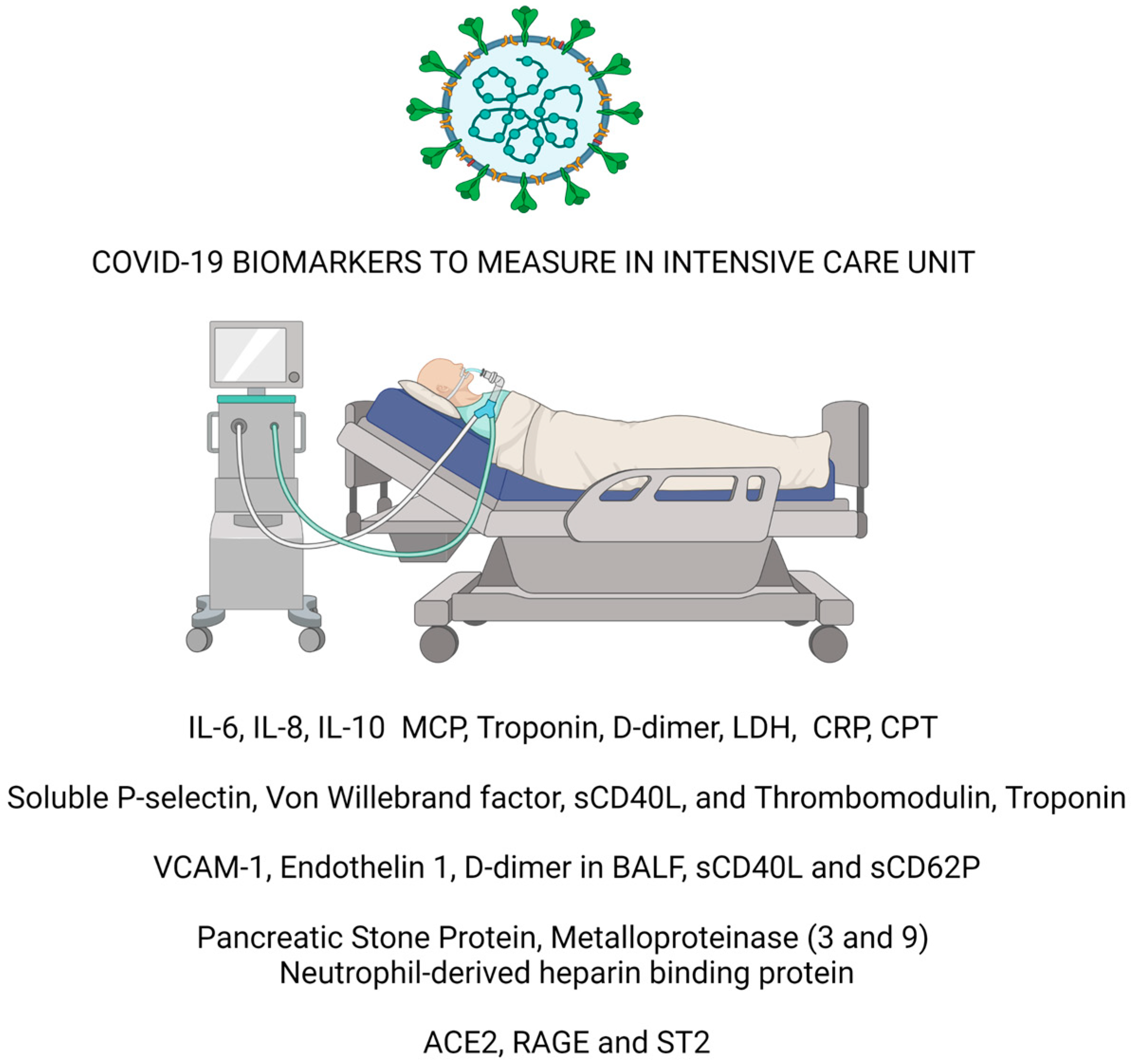
COVID-19 Biomarkers for Critically Ill Patients: A Compendium for the Physician
 ,
,
Abstract
:1. Introduction
Pathophysiological Determinants of Severe Forms of COVID-19 Infection
2. Materials and Methods
2.1. Cytokine Storm
2.2. Endothelium Dysfunction and Coagulation Biomarkers in COVID-19
2.3. Biomarker of Sepsis
2.4. Cardiovascular, Lung Biomarker, and New Perspectives in COVID-19
3. Limitations
4. Conclusions
Author Contributions
Funding
Institutional Review Board Statement
Informed Consent Statement
Data Availability Statement
Conflicts of Interest
Appendix A
- (1)
- Justification of the article’s importance for the readership. This review aims to summarize, for intensive care physicians, the current state of knowledge regarding known biomarkers for COVID-19 infection to identify predictors of the most critically ill patients.
- (2)
- Statement of concrete aims or formulation of questions. The aim of the review is to determine the main markers associated with the most severe forms of SARS-CoV-2 infection, to identify those patients at higher risk of death in the early stages of the infection.
- (3)
- Description of the literature search. We searched on PubMed® for the past three years, using the Boolean operators AND, OR, and NOT. We identified all results on the PubMed® database of all studies regarding COVID-19 biomarkers. We selected studies using Boolean operators’ endothelium, cytokines, bacterial infection, and the coagulation biomarker.
- (4)
- Referencing. The review considers and takes into consideration some of the most valuable articles in the field.
- (5)
- Scientific reasoning. To collect the data that we needed to build the article, we specifically used clinical trials regarding COVID-19 biomarkers.
- (6)
- Appropriate presentation of the data. In according to pathophysiological determinants of COVID-19 severity infection, we divided the results into four essential paragraphs: “Cytokine storm”, “Endothelium dysfunction and coagulation biomarkers in COVID-19”, “Biomarker of sepsis”, and “New perspective”.
References
- Melegari, G.; Giuliani, E.; Maini, G.; Barbieri, L.; Baffoni, P.; Bertellini, E.; Barbieri, A. Novel coronavirus (2019-nCov): Do you have enough intensive care units? Med. Intensiv. 2020, 44, 583–585. [Google Scholar] [CrossRef]
- Bivona, G.; Agnello, L.; Ciaccio, M. Biomarkers for Prognosis and Treatment Response in COVID-19 Patients. Ann. Lab. Med. 2021, 41, 540–548. [Google Scholar] [CrossRef]
- Barbieri, A.; Melegari, G.; Lob, V.; Mazzali, L.; D’Amelio, L.; Giovannoni, A.; Giuliani, E. Response by Twin Italian Hub Hospitals in a Double Seismic Event: A Retrospective Observational Investigation. Prehospital Emerg. Care 2018, 22, 353–360. [Google Scholar] [CrossRef] [Green Version]
- Allgaier, R.L.; Shaafi-Kabiri, N.; Romney, C.A.; Wallis, L.A.; Burke, J.J.; Bhangu, J.; Thomas, K.C. Use of Predictive Modeling to Plan for Special Event Medical Care During Mass Gathering Events. Disaster. Med. Public Health Prep. 2019, 13, 874–879. [Google Scholar] [CrossRef]
- Ceyhan, M.A.; Demir, G.G. Health Care Services in Shopping Centers: A Routine Mass-Gathering Event. Prehospital Disaster Med. 2020, 35, 669–675. [Google Scholar] [CrossRef]
- Cummings, M.J.; Baldwin, M.R.; Abrams, D.; Jacobson, S.D.; Meyer, B.J.; Balough, E.M.; Aaron, J.G.; Claassen, J.; Rabbani, L.E.; Hastie, J.; et al. Epidemiology, clinical course, and outcomes of critically ill adults with COVID-19 in New York City: A prospective cohort study. Lancet 2020, 395, 1763–1770. [Google Scholar] [CrossRef]
- Gallo Marin, B.; Aghagoli, G.; Lavine, K.; Yang, L.; Siff, E.J.; Chiang, S.S.; Salazar-Mather, T.P.; Dumenco, L.; Savaria, M.C.; Aung, S.N.; et al. Predictors of COVID-19 severity: A literature review. Rev. Med. Virol. 2021, 31, 1–10. [Google Scholar] [CrossRef]
- Wiersinga, W.J.; Rhodes, A.; Cheng, A.C.; Peacock, S.J.; Prescott, H.C. Pathophysiology, Transmission, Diagnosis, and Treatment of Coronavirus Disease 2019 (COVID-19): A Review. JAMA 2020, 324, 782–793. [Google Scholar] [CrossRef]
- Gintoni, I.; Adamopoulou, M.; Yapijakis, C. The Impact of ACE and ACE2 Gene Polymorphisms in Pulmonary Diseases Including COVID-19. In Vivo 2022, 36, 13–29. [Google Scholar] [CrossRef]
- Trougakos, I.P.; Stamatelopoulos, K.; Terpos, E.; Tsitsilonis, O.E.; Aivalioti, E.; Paraskevis, D.; Kastritis, E.; Pavlakis, G.N.; Dimopoulos, M.A. Insights to SARS-CoV-2 life cycle, pathophysiology, and rationalized treatments that target COVID-19 clinical complications. J. Biomed Sci. 2021, 28, 9. [Google Scholar] [CrossRef]
- Wallentin, L.; Lindbäck, J.; Eriksson, N.; Hijazi, Z.; Eikelboom, J.W.; Ezekowitz, M.D.; Granger, C.B.; Lopes, R.D.; Yusuf, S.; Oldgren, J.; et al. Angiotensin-converting enzyme 2 (ACE2) levels in relation to risk factors for COVID-19 in two large cohorts of patients with atrial fibrillation. Eur. Heart J. 2020, 41, 4037–4046. [Google Scholar] [CrossRef]
- Haffke, M.; Freitag, H.; Rudolf, G.; Seifert, M.; Doehner, W.; Scherbakov, N.; Hanitsch, L.; Wittke, K.; Bauer, S.; Konietschke, F.; et al. Endothelial dysfunction and altered endothelial biomarkers in patients with post-COVID-19 syndrome and chronic fatigue syndrome (ME/CFS). J. Transl. Med. 2022, 20, 138. [Google Scholar] [CrossRef]
- Paces, J.; Strizova, Z.; Smrz, D.; Cerny, J. COVID-19 and the immune system. Physiol. Res. 2020, 69, 379–388. [Google Scholar] [CrossRef]
- Iba, T.; Levy, J.H.; Levi, M.; Thachil, J. Coagulopathy in COVID-19. J. Thromb. Haemost. 2020, 18, 2103–2109. [Google Scholar] [CrossRef]
- Bonaventura, A.; Vecchié, A.; Dagna, L.; Martinod, K.; Dixon, D.L.; Van Tassell, B.W.; Dentali, F.; Montecucco, F.; Massberg, S.; Levi, M.; et al. Endothelial dysfunction and immunothrombosis as key pathogenic mechanisms in COVID-19. Nat. Rev. Immunol. 2021, 21, 319–329. [Google Scholar] [CrossRef]
- Varga, Z.; Flammer, A.J.; Steiger, P.; Haberecker, M.; Andermatt, R.; Zinkernagel, A.S.; Mehra, M.R.; Schuepbach, R.A.; Ruschitzka, F.; Moch, H. Endothelial cell infection and endotheliitis in COVID-19. Lancet 2020, 395, 1417–1418. [Google Scholar] [CrossRef]
- Baethge, C.; Goldbeck-Wood, S.; Mertens, S. SANRA—A scale for the quality assessment of narrative review articles. Res. Integr. Peer Rev. 2019, 4, 5. [Google Scholar] [CrossRef] [Green Version]
- Jøntvedt Jørgensen, M.; Holter, J.C.; Christensen, E.E.; Schjalm, C.; Tonby, K.; Pischke, S.E.; Jenum, S.; Skeie, L.G.; Nur, S.; Lind, A.; et al. Increased interleukin-6 and macrophage chemoattractant protein-1 are associated with respiratory failure in COVID-19. Sci. Rep. 2020, 10, 21697. [Google Scholar] [CrossRef]
- Pirabe, A.; Heber, S.; Schrottmaier, W.C.; Schmuckenschlager, A.; Treiber, S.; Pereyra, D.; Santol, J.; Pawelka, E.; Traugott, M.; Schörgenhofer, C.; et al. Age Related Differences in Monocyte Subsets and Cytokine Pattern during Acute COVID-19—A Prospective Observational Longitudinal Study. Cells 2021, 10, 3373. [Google Scholar] [CrossRef]
- Santa Cruz, A.; Mendes-Frias, A.; Oliveira, A.I.; Dias, L.; Matos, A.R.; Carvalho, A.; Capela, C.; Pedrosa, J.; Castro, A.G.; Silvestre, R. Interleukin-6 Is a Biomarker for the Development of Fatal Severe Acute Respiratory Syndrome Coronavirus 2 Pneumonia. Front. Immunol. 2021, 12, 613422. [Google Scholar] [CrossRef]
- Espíndola, O.M.; Gomes, Y.C.P.; Brandão, C.O.; Torres, R.C.; Siqueira, M.; Soares, C.N.; Lima, M.; Leite, A.; Venturotti, C.O.; Carvalho, A.J.C.; et al. Inflammatory Cytokine Patterns Associated with Neurological Diseases in Coronavirus Disease 2019. Ann. Neurol. 2021, 89, 1041–1045. [Google Scholar] [CrossRef]
- Van Singer, M.; Brahier, T.; Ngai, M.; Wright, J.; Weckman, A.M.; Erice, C.; Meuwly, J.Y.; Hugli, O.; Kain, K.C.; Boillat-Blanco, N. COVID-19 risk stratification algorithms based on sTREM-1 and IL-6 in emergency department. J. Allergy Clin. Immunol. 2021, 147, 99–106.e104. [Google Scholar] [CrossRef]
- Popadic, V.; Klasnja, S.; Milic, N.; Rajovic, N.; Aleksic, A.; Milenkovic, M.; Crnokrak, B.; Balint, B.; Todorovic-Balint, M.; Mrda, D.; et al. Predictors of Mortality in Critically Ill COVID-19 Patients Demanding High Oxygen Flow: A Thin Line between Inflammation, Cytokine Storm, and Coagulopathy. Oxidative Med. Cell. Longev. 2021, 2021, 6648199. [Google Scholar] [CrossRef]
- Galván-Román, J.M.; Rodríguez-García, S.C.; Roy-Vallejo, E.; Marcos-Jiménez, A.; Sánchez-Alonso, S.; Fernández-Díaz, C.; Alcaraz-Serna, A.; Mateu-Albero, T.; Rodríguez-Cortes, P.; Sánchez-Cerrillo, I.; et al. IL-6 serum levels predict severity and response to tocilizumab in COVID-19: An observational study. J. Allergy Clin. Immunol. 2021, 147, 72–80.e78. [Google Scholar] [CrossRef]
- Gordon, A.C.; Mouncey, P.R.; Al-Beidh, F.; Rowan, K.M.; Nichol, A.D.; Arabi, Y.M.; Annane, D.; Beane, A.; van Bentum-Puijk, W.; Berry, L.R.; et al. Interleukin-6 Receptor Antagonists in Critically Ill Patients with Covid-19. N. Engl. J. Med. 2021, 384, 1491–1502. [Google Scholar] [CrossRef]
- Salama, C.; Han, J.; Yau, L.; Reiss, W.G.; Kramer, B.; Neidhart, J.D.; Criner, G.J.; Kaplan-Lewis, E.; Baden, R.; Pandit, L.; et al. Tocilizumab in Patients Hospitalized with Covid-19 Pneumonia. N. Engl. J. Med. 2021, 384, 20–30. [Google Scholar] [CrossRef]
- Queiroz, M.A.F.; Neves, P.; Lima, S.S.; Lopes, J.D.C.; Torres, M.; Vallinoto, I.; Bichara, C.D.A.; Dos Santos, E.F.; de Brito, M.; da Silva, A.L.S.; et al. Cytokine Profiles Associated With Acute COVID-19 and Long COVID-19 Syndrome. Front. Cell. Infect. Microbiol. 2022, 12, 922422. [Google Scholar] [CrossRef]
- Schultheiß, C.; Willscher, E.; Paschold, L.; Gottschick, C.; Klee, B.; Henkes, S.S.; Bosurgi, L.; Dutzmann, J.; Sedding, D.; Frese, T.; et al. The IL-1β, IL-6, and TNF cytokine triad is associated with post-acute sequelae of COVID-19. Cell Rep. Med. 2022, 3, 100663. [Google Scholar] [CrossRef]
- Melero, I.; Villalba-Esparza, M.; Recalde-Zamacona, B.; Jiménez-Sánchez, D.; Teijeira, Á.; Argueta, A.; García-Tobar, L.; Álvarez-Gigli, L.; Sainz, C.; Garcia-Ros, D.; et al. Neutrophil Extracellular Traps, Local IL-8 Expression, and Cytotoxic T-Lymphocyte Response in the Lungs of Patients With Fatal COVID-19. Chest 2022, 162, 1006–1016. [Google Scholar] [CrossRef]
- Bain, W.; Yang, H.; Shah, F.A.; Suber, T.; Drohan, C.; Al-Yousif, N.; DeSensi, R.S.; Bensen, N.; Schaefer, C.; Rosborough, B.R.; et al. COVID-19 versus Non-COVID-19 Acute Respiratory Distress Syndrome: Comparison of Demographics, Physiologic Parameters, Inflammatory Biomarkers, and Clinical Outcomes. Ann. Am. Thorac. Soc. 2021, 18, 1202–1210. [Google Scholar] [CrossRef]
- Guasp, M.; Muñoz-Sánchez, G.; Martínez-Hernández, E.; Santana, D.; Carbayo, Á.; Naranjo, L.; Bolós, U.; Framil, M.; Saiz, A.; Balasa, M.; et al. CSF Biomarkers in COVID-19 Associated Encephalopathy and Encephalitis Predict Long-Term Outcome. Front. Immunol. 2022, 13, 866153. [Google Scholar] [CrossRef]
- Han, H.; Ma, Q.; Li, C.; Liu, R.; Zhao, L.; Wang, W.; Zhang, P.; Liu, X.; Gao, G.; Liu, F.; et al. Profiling serum cytokines in COVID-19 patients reveals IL-6 and IL-10 are disease severity predictors. Emerg. Microbes Infect. 2020, 9, 1123–1130. [Google Scholar] [CrossRef]
- Goshua, G.; Pine, A.B.; Meizlish, M.L.; Chang, C.H.; Zhang, H.; Bahel, P.; Baluha, A.; Bar, N.; Bona, R.D.; Burns, A.J.; et al. Endotheliopathy in COVID-19-associated coagulopathy: Evidence from a single-centre, cross-sectional study. Lancet Haematol. 2020, 7, e575–e582. [Google Scholar] [CrossRef]
- Vieceli Dalla Sega, F.; Fortini, F.; Spadaro, S.; Ronzoni, L.; Zucchetti, O.; Manfrini, M.; Mikus, E.; Fogagnolo, A.; Torsani, F.; Pavasini, R.; et al. Time course of endothelial dysfunction markers and mortality in COVID-19 patients: A pilot study. Clin. Transl. Med. 2021, 11, e283. [Google Scholar] [CrossRef]
- Al-Samkari, H.; Karp Leaf, R.S.; Dzik, W.H.; Carlson, J.C.T.; Fogerty, A.E.; Waheed, A.; Goodarzi, K.; Bendapudi, P.K.; Bornikova, L.; Gupta, S.; et al. COVID-19 and coagulation: Bleeding and thrombotic manifestations of SARS-CoV-2 infection. Blood 2020, 136, 489–500. [Google Scholar] [CrossRef]
- Nossent, E.J.; Schuurman, A.R.; Reijnders, T.D.Y.; Saris, A.; Jongerius, I.; Blok, S.G.; de Vries, H.; Duitman, J.; Vonk Noordegraaf, A.; Meijboom, L.J.; et al. Pulmonary Procoagulant and Innate Immune Responses in Critically Ill COVID-19 Patients. Front. Immunol. 2021, 12, 664209. [Google Scholar] [CrossRef]
- Hamzeh-Cognasse, H.; Mansour, A.; Reizine, F.; Mismetti, P.; Gouin-Thibault, I.; Cognasse, F. Platelet-derived sCD40L: Specific inflammatory marker for early-stage severe acute respiratory syndrome coronavirus 2 infection. Virol. J. 2021, 18, 211. [Google Scholar] [CrossRef]
- Price, D.R.; Benedetti, E.; Hoffman, K.L.; Gomez-Escobar, L.; Alvarez-Mulett, S.; Capili, A.; Sarwath, H.; Parkhurst, C.N.; Lafond, E.; Weidman, K.; et al. Angiopoietin 2 Is Associated with Vascular Necroptosis Induction in Coronavirus Disease 2019 Acute Respiratory Distress Syndrome. Am. J. Pathol. 2022, 192, 1001–1015. [Google Scholar] [CrossRef]
- Villa, E.; Critelli, R.; Lasagni, S.; Melegari, A.; Curatolo, A.; Celsa, C.; Romagnoli, D.; Melegari, G.; Pivetti, A.; Di Marco, L.; et al. Dynamic angiopoietin-2 assessment predicts survival and chronic course in hospitalized patients with COVID-19. Blood Adv. 2021, 5, 662–673. [Google Scholar] [CrossRef]
- Pine, A.B.; Meizlish, M.L.; Goshua, G.; Chang, C.H.; Zhang, H.; Bishai, J.; Bahel, P.; Patel, A.; Gbyli, R.; Kwan, J.M.; et al. Circulating markers of angiogenesis and endotheliopathy in COVID-19. Pulm. Circ. 2020, 10, 2045894020966547. [Google Scholar] [CrossRef]
- Melegari, G.; Critelli, R.M.; Lasagni, S.; Romagnoli, D.; Bertellini, E.; Villa, E. Dynamic Angiopoietin-2 Serum Level as Endothelial Damage Marker and Potential Therapeutic Target. Am. J. Pathol. 2022, 192, 1336–1337. [Google Scholar] [CrossRef]
- Smadja, D.M.; Guerin, C.L.; Chocron, R.; Yatim, N.; Boussier, J.; Gendron, N.; Khider, L.; Hadjadj, J.; Goudot, G.; Debuc, B.; et al. Angiopoietin-2 as a marker of endothelial activation is a good predictor factor for intensive care unit admission of COVID-19 patients. Angiogenesis 2020, 23, 611–620. [Google Scholar] [CrossRef]
- Vassiliou, A.G.; Kotanidou, A.; Dimopoulou, I.; Orfanos, S.E. Endothelial Damage in Acute Respiratory Distress Syndrome. Int. J. Mol. Sci. 2020, 21, 8793. [Google Scholar] [CrossRef]
- Flaumenhaft, R.; Enjyoji, K.; Schmaier, A.A. Vasculopathy in COVID-19. Blood 2022, 140, 222–235. [Google Scholar] [CrossRef]
- Andrianto; Al-Farabi, M.J.; Nugraha, R.A.; Marsudi, B.A.; Azmi, Y. Biomarkers of endothelial dysfunction and outcomes in coronavirus disease 2019 (COVID-19) patients: A systematic review and meta-analysis. Microvasc. Res. 2021, 138, 104224. [Google Scholar] [CrossRef]
- Li, Y.; Zhao, K.; Wei, H.; Chen, W.; Wang, W.; Jia, L.; Liu, Q.; Zhang, J.; Shan, T.; Peng, Z.; et al. Dynamic relationship between D-dimer and COVID-19 severity. Br. J. Haematol. 2020, 190, e24–e27. [Google Scholar] [CrossRef]
- Al Otair, H.; AlSaleh, K.; AlQahtany, F.S.; Al Ayed, K.; Al Ammar, H.; Al Mefgai, N.; Al Zeer, F. The Level of vWF Antigen and Coagulation Markers in Hospitalized Patients with Covid-19. J. Blood Med. 2021, 12, 809–817. [Google Scholar] [CrossRef]
- Venet, F.; Cour, M.; Rimmelé, T.; Viel, S.; Yonis, H.; Coudereau, R.; Amaz, C.; Abraham, P.; Monard, C.; Casalegno, J.S.; et al. Longitudinal assessment of IFN-I activity and immune profile in critically ill COVID-19 patients with acute respiratory distress syndrome. Crit. Care 2021, 25, 140. [Google Scholar] [CrossRef]
- Mellhammar, L.; Thelaus, L.; Elén, S.; Fisher, J.; Linder, A. Heparin binding protein in severe COVID-19—A prospective observational cohort study. PLoS ONE 2021, 16, e0249570. [Google Scholar] [CrossRef]
- Voiriot, G.; Fartoukh, M.; Durand-Zaleski, I.; Berard, L.; Rousseau, A.; Armand-Lefevre, L.; Verdet, C.; Argaud, L.; Klouche, K.; Megarbane, B.; et al. Combined use of a broad-panel respiratory multiplex PCR and procalcitonin to reduce duration of antibiotics exposure in patients with severe community-acquired pneumonia (MULTI-CAP): A multicentre, parallel-group, open-label, individual randomised trial conducted in French intensive care units. BMJ Open 2021, 11, e048187. [Google Scholar] [CrossRef]
- Pink, I.; Raupach, D.; Fuge, J.; Vonberg, R.P.; Hoeper, M.M.; Welte, T.; Rademacher, J. C-reactive protein and procalcitonin for antimicrobial stewardship in COVID-19. Infection 2021, 49, 935–943. [Google Scholar] [CrossRef]
- Smilowitz, N.R.; Kunichoff, D.; Garshick, M.; Shah, B.; Pillinger, M.; Hochman, J.S.; Berger, J.S. C-reactive protein and clinical outcomes in patients with COVID-19. Eur. Heart J. 2021, 42, 2270–2279. [Google Scholar] [CrossRef]
- Bray, C.; Bell, L.N.; Liang, H.; Haykal, R.; Kaiksow, F.; Mazza, J.J.; Yale, S.H. Erythrocyte Sedimentation Rate and C-reactive Protein Measurements and Their Relevance in Clinical Medicine. WMJ 2016, 115, 317–321. [Google Scholar]
- Pierrakos, C.; Vincent, J.L. Sepsis biomarkers: A review. Crit. Care 2010, 14, R15. [Google Scholar] [CrossRef] [Green Version]
- Carbonell, R.; Urgelés, S.; Salgado, M.; Rodríguez, A.; Reyes, L.F.; Fuentes, Y.V.; Serrano, C.C.; Caceres, E.L.; Bodí, M.; Martín-Loeches, I.; et al. Negative predictive value of procalcitonin to rule out bacterial respiratory co-infection in critical COVID-19 patients. J. Infect 2022, 85, 374–381. [Google Scholar] [CrossRef]
- Pugin, J.; Daix, T.; Pagani, J.L.; Morri, D.; Giacomucci, A.; Dequin, P.F.; Guitton, C.; Que, Y.A.; Zani, G.; Brealey, D.; et al. Serial measurement of pancreatic stone protein for the early detection of sepsis in intensive care unit patients: A prospective multicentric study. Crit. Care 2021, 25, 151. [Google Scholar] [CrossRef]
- Van Singer, M.; Brahier, T.; Brochu Vez, M.J.; Gerhard Donnet, H.; Hugli, O.; Boillat-Blanco, N. Pancreatic stone protein for early mortality prediction in COVID-19 patients. Crit. Care 2021, 25, 267. [Google Scholar] [CrossRef]
- Lagadinou, M.; Paraskevas, T.; Velissaris, D.; Michailides, C.; Eleftherakis, G.; Sampsonas, F.; Siakallis, G.; Assimakopoulos, S.F.; Marangos, M. The role of pancreatic stone protein as a prognostic factor for COVID-19 patients. Eur. Rev. Med. Pharmacol. Sci. 2022, 26, 6391–6395. [Google Scholar] [CrossRef]
- Melegari, G.; Giuliani, E.; Di Pietro, G.; Alberti, F.; Campitiello, M.; Bertellini, E.; Barbieri, A. Point-of-care pancreatic stone protein measurement in critically ill COVID-19 patients. BMC Anesthesiol. 2023, 23, 226. [Google Scholar]
- Huang, M.; Yang, Y.; Shang, F.; Zheng, Y.; Zhao, W.; Luo, L.; Han, X.; Lin, A.; Zhao, H.; Gu, Q.; et al. Clinical Characteristics and Predictors of Disease Progression in Severe Patients with COVID-19 Infection in Jiangsu Province, China: A Descriptive Study. Am. J. Med. Sci. 2020, 360, 120–128. [Google Scholar]
- Liaqat, A.; Ali-Khan, R.S.; Asad, M.; Rafique, Z. Evaluation of myocardial injury patterns and ST changes among critical and non-critical patients with coronavirus-19 disease. Sci. Rep. 2021, 11, 4828. [Google Scholar] [CrossRef] [PubMed]
- Ileri, C.; Dogan, Z.; Ozben, B.; Karaoglu, C.; Gunay, N.; Tigen, K.; Basat, S.; Uyan, C. Evaluation of the relation between cardiac biomarkers and thorax computed tomography findings in COVID-19 patients. Biomark. Med. 2021, 15, 285–293. [Google Scholar] [CrossRef] [PubMed]
- Pérez-Mies, B.; Gómez-Rojo, M.; Carretero-Barrio, I.; Bardi, T.; Benito, A.; García-Cosío, M.; Caballero, Á.; de Pablo, R.; Galán, J.C.; Pestaña, D.; et al. Pulmonary vascular proliferation in patients with severe COVID-19: An autopsy study. Thorax 2021, 76, 1044–1046. [Google Scholar] [CrossRef] [PubMed]
- Gelzo, M.; Cacciapuoti, S.; Pinchera, B.; De Rosa, A.; Cernera, G.; Scialò, F.; Comegna, M.; Mormile, M.; Fabbrocini, G.; Parrella, R.; et al. Matrix metalloproteinases (MMP) 3 and 9 as biomarkers of severity in COVID-19 patients. Sci. Rep. 2022, 12, 1212. [Google Scholar] [CrossRef]
- Danlos, F.X.; Grajeda-Iglesias, C.; Durand, S.; Sauvat, A.; Roumier, M.; Cantin, D.; Colomba, E.; Rohmer, J.; Pommeret, F.; Baciarello, G.; et al. Metabolomic analyses of COVID-19 patients unravel stage-dependent and prognostic biomarkers. Cell Death Dis. 2021, 12, 258. [Google Scholar] [CrossRef]
- Wick, K.D.; Siegel, L.; Neaton, J.D.; Oldmixon, C.; Lundgren, J.; Dewar, R.L.; Lane, H.C.; Thompson, B.T.; Matthay, M.A. RAGE has potential pathogenetic and prognostic value in nonintubated hospitalized patients with COVID-19. JCI Insight 2022, 7, e157499. [Google Scholar] [CrossRef]
- Zeng, Z.; Hong, X.Y.; Li, Y.; Chen, W.; Ye, G.; Li, Y.; Luo, Y. Serum-soluble ST2 as a novel biomarker reflecting inflammatory status and illness severity in patients with COVID-19. Biomark. Med. 2020, 14, 1619–1629. [Google Scholar] [CrossRef]


| Authors/Year | Type of Study | Biomarker | Patients (Sample) | Outcome |
|---|---|---|---|---|
| Jøntvedt Jørgensen et al. [18] Year 2020 | Prospective studies (Classified also as Trial on Pubmed) | Il-6 and MCP | 34 patients | IL-6 and MCP-1 were inversely correlated with P/F |
| Pirabe et al. [19] Year 2021 | Prospective studies (Classified also as Trial on Pubmed) | IL-6, IL-8 and tumor necrosis factor (TNF) | 110 patients | Adverse outcomes in elderly are associated with an inappropriate immune response, |
| Santa Cruz et al. [20] Year 2021 | Prospective studies (Classified also as Trial on Pubmed) | Il-6 | 46 Patients | IL-6 level was the most significant predictor of the non-survivors group, |
| Espindola et al. [21] Year 2021 | Prospective studies (Classified also as Trial on Pubmed) | Il-6 in cerebrum spinal fluid (CSF) | 48 patients | Neurological syndromes related to SARS-CoV-2 were associated with high CSF levels of IL-6 |
| Van singer et al. [22] Year 2020 | Prospective studies (Classified also as Trial on Pubmed) | Il-6 and endothelial dysfunction biomarkers And TREM-1 myeloid receptor | 76 patients | IL-6 measured at presentation to the ED had the best accuracy for 30-day oxygen requirement |
| Popadic et al. [23] Year 2021 | Prospectives studies | Serum albumin, D-dimer, and IL-6 | 160 patients | Serum albumin, D-dimer, and IL-6 at admission to ICU were independently associated with mortality |
| Galván-Román et al. [24] Year 2021 | Prospectives studies | Il-6 and Tocilizumab response | 146 patients | IL-6 greater than 30 pg/mL predicts IMV requirement and it helps in tocilizumab choice |
| Gordon et al. [25] Year 2021 | Clinical Trials | Il-6 and Tocilizumab response | 353 patients | Il-6 reduction is associated with tocilizumab response and outcome |
| Salama et al. [26] Year 2021 | Clinical Trials | Il-6 and Tocilizumab response | 389 patients | Il-6 reduction is associated with tocilizumab response and outcome |
| Schultheiß et al. [27] Year 2022 | Prospectives studies | Il-6 and long term sequelae | 318 patients | Il-6 monitoring is useful for long term sequelae |
| Queiroz et al. [28] Year 2022 | Prospectives studies | Il-6 and long term complications | 317 patients | Il-6 monitoring is useful for long term complications |
| Melero et al. [29] Year 2022 | Prospectives studies | IL-8 messenger RNA (mRNA) | Lung Biopsy from 16 patients | Il-8 is associated to nflammatory infiltrates and neutrophil extracellular traps |
| Bain et al. [30] Year 2021 | Prospectives studies | Il-6, Il-8, and Il-10 | 92 patients | Conclusions: COVID-19 ARDS bears several similarities to viral ARDS |
| Guasp et al. [31] Year 2022 | Prospectives studies | Il-6, Il-8, and Il-10 IL-10, Il-1RA, IP-10 | 60 patients | levels of pro-inflammatory cytokines do not predict the long-term functional outcome |
| Han et al. [32] Year 2020 | Prospectives studies | Il-6 and Il-10 | 102 patients | IL-6 and IL-10 can be used as predictors for patients with higher risk of disease deterioration. |
| Authors | Type of Study | Biomarker | Patients | Outcome |
|---|---|---|---|---|
| Goshua et al. [33] Year 2020 | Prospective studies (Classified also as Trial on Pubmed) | Endothelial biomarker: P-selectin, Von Willebrand factor (VWF) sCD40L, thrombomodulin | 68 patients | Endotheliopathy is present in COVID-19 and is likely to be associated with critical illness and death |
| Vieceli Dalla Sega et al. [34] Year 2021 | Prospective studies (Classified also as Trial on Pubmed) | VCAM-1, endothelin-1 and thrombomodulin | 54 patients | Endothelin-1 remained stable in nonsurvivors but increased over time in survivors |
| Al-Samkari et al. [35] Year 2020 | Prospective studies (Classified also as Trial on Pubmed) | D-Dimer | 400 patients | Elevated D-dimer at initial presentation was predictive of coagulation-associated complications |
| Nossent et al. [36] Year 2021 | Prospective studies | D-dimer and thrombin-antithrombin complexes, in bronchoalveolar lavage fluid | 17 patients | Critically ill, with COVID-19 show strong complement system, cytokines, chemokines and growth factors in the bronchoalveolar compartment |
| Hamzeh-Cognasse et al. [37] Year 2021 | Prospective studies (Classified also as Trial on Pubmed) | sCD40L and sCD62P | 55 patients | there is a platelet signature of inflammatory response to SARS-CoV-2 infection which varies overtime |
| Price et al. [38] Year 2022 | Prospective studies | Angipoietin 2 (ANGPT2) | 102 Patients | COVID-19 ARDS lung autopsy confirmed a link between vascular injury (ANGPT2) and platelet-rich microthrombi |
| Villa et al. [39] Year 2022 | Prospective studies | Angipoietin 2 | 187 patients | Angiopoietin-2 may be an early and useful predictor of COVID-19 clinical course |
| Pine et al. [40] Year 2022 | Prospective studies | angiopoietin-2, follistatin, and plasminogen activator inhibitor-1 (PAI-1) | 49 patients | Elevated markers of endothelial injury were strongly predictive of in-hospital mortality |
| Smadja et al. [42] Yaer 2020 | Prospective studies | Angipoietin 2 | 40 Patients | Angiopoietin-2 is a relevant predictive factor for ICU direct admission in COVID-19 patients. |
| Al Otair et al. [47] Year 2021 | Prospective studies | Protein C, protein S, antithrombin (AT) III, clotting factor (F) VIII, von Willebrand factor (vWF) and coagulation screening tests (PT and a PTT), fibrinogen, D-dimer | 68 patients | The level of vWF is increased early in the course of COVID-19 infection. This can be used as a biomarker for endothelial injury. |
| Authors | Type of Study | Biomarker | Patients | Outcome |
|---|---|---|---|---|
| Venet et al. [48] Year 2021 | Prospective studies (Classified also as Trial on Pubmed) | Plasma IFNα2 levels and IFN-stimulated genes | 64 patients | ARDS in SARS-CoV-2 infection appears to be associated with the intensity of immune alterations upon ICU admission |
| Mellhammar et al. [49] Year 2021 | Prospective studies (Classified also as Trial on Pubmed) | Neutrophil-derived heparin binding protein (HBP; | 35 patients | HBP is elevated prior to onset of organ dysfunction in patients with severe COVID-19 |
| Smilowitz et al. [52] Year 2021 | Prospective studies (Classified also as Trial on Pubmed) | C reactive protein (CRP) | 2872 patients | CRP is strongly associated critical illness, and mortality in COVID-19. |
| Van Singer et al. [57] Year 2022 | Prospective studies | Pancreatic Stone Protein | 107 patients | CRB-65, CRP and PSP have an excellent accuracy to rule out early mortality in COVID-19. |
| Lagadinou et al. [58] Year 2022 | Prospective studies | Pancreatic Stone Protein | 55 patients | The optimal cut-off value to predict prolonged hospital stay was 51 ng/dL |
| Melegari et al. [59] Year 2023 | Prospective studies | Pancreatic Stone Protein | 21 Patients | Monitoring PSP plasma levels could be useful in the absence of a specific COVID-19 |
| Authors | Type of Study | Biomarker | Patients | Outcome |
|---|---|---|---|---|
| Huang et al. [60] Year 2020 | Prospective studies (Classified also as Trial on Pubmed) | Troponin and Lymphocyte count | 60 patients | The higher levels of troponin T and lower lymphocyte count were predictors of disease progression. |
| Liaqat et al. [61] Year 2021 | Prospective studies (Classified also as Trial on Pubmed) | Troponin and Lymphocyte count | 201 patients | COVID-19 disease favors cardiovascular injury among critical and non-critical patients. |
| Ileri et al. [62] Year 2021 | Propsective study | Troponin | 74 patients | COVID-19 patients with severe CT findings and progressive disease had higher hs-cTnI levels |
| Perez et al. [63] Year 2021 | Propsective study | CD31, CD34 and vascular endothelial cadherin. Platelet-derived growth factor receptor-β | 16 patients (lung biopsy) | These vascular alterations may contribute to the severe and refractory hypoxaemia in COVID-19 |
| Gelzo et al. [64] Year 2022 | Prospective studies (Classified also as Trial on Pubmed) | Matrix metalloproteinases (MMP) 3 and 9 | 108 patients | MMP3 may help to early predict the severity of COVID-19 |
| Danlos et al. [65] | Prospective studies (Classified also as Trial on Pubmed) | Metabolome | 72 patients | Metabolome are associated with COVID-19 severity of disease and possible target |
| Wick et al. [66] | Prospective studies (Classified also as Trial on Pubmed) | RAGE | 277 patients | Plasma sRAGE may be a promising biomarker for COVID-19 prognostication |
| Zeng et al. [67] | Prospective studies (Classified also as Trial on Pubmed) | Serum sST2 | 80 patients | Serum sST2 levels in nonsurviving cases were persistently high in COVID-19 patients |
Disclaimer/Publisher’s Note: The statements, opinions and data contained in all publications are solely those of the individual author(s) and contributor(s) and not of MDPI and/or the editor(s). MDPI and/or the editor(s) disclaim responsibility for any injury to people or property resulting from any ideas, methods, instructions or products referred to in the content. |
© 2023 by the authors. Licensee MDPI, Basel, Switzerland. This article is an open access article distributed under the terms and conditions of the Creative Commons Attribution (CC BY) license (https://creativecommons.org/licenses/by/4.0/).
Share and Cite
Arturi, F.; Melegari, G.; Giansante, A.; Giuliani, E.; Bertellini, E.; Barbieri, A. COVID-19 Biomarkers for Critically Ill Patients: A Compendium for the Physician. Neurol. Int. 2023, 15, 881-895. https://doi.org/10.3390/neurolint15030056
Arturi F, Melegari G, Giansante A, Giuliani E, Bertellini E, Barbieri A. COVID-19 Biomarkers for Critically Ill Patients: A Compendium for the Physician. Neurology International. 2023; 15(3):881-895. https://doi.org/10.3390/neurolint15030056
Chicago/Turabian StyleArturi, Federica, Gabriele Melegari, Antonio Giansante, Enrico Giuliani, Elisabetta Bertellini, and Alberto Barbieri. 2023. "COVID-19 Biomarkers for Critically Ill Patients: A Compendium for the Physician" Neurology International 15, no. 3: 881-895. https://doi.org/10.3390/neurolint15030056