Article

Microbiota and Mitochondrial Sex-Dependent Imbalance in Fibromyalgia: A Pilot Descriptive Study

by
Jorge A. Ramírez-Tejero
1,
Elena Durán-González
1,
Antonio Martínez-Lara
1,
Laura Lucena del Amo
1,
Isabel Sepúlveda
1,
Andrés Huancas-Díaz
2,
Marco Carvajal
2,* and
David Cotán
1,*
1
Pronacera Therapeutics S.L., 41015 Sevilla, Spain
2
Instituto de Medicina Funcional e Integral de Perú, Lima 15073, Peru
*
Authors to whom correspondence should be addressed.
Neurol. Int. 2023, 15(3), 868-880; https://doi.org/10.3390/neurolint15030055
Submission received: 11 May 2023 / Revised: 23 June 2023 / Accepted: 25 June 2023 / Published: 12 July 2023

Abstract

:
Fibromyalgia is a widespread chronic condition characterized by pain and fatigue. Among the long list of physiological disturbances linked to this syndrome, mitochondrial imbalance and oxidative stress stand out. Recently, the crosstalk between mitochondria and intestinal microbiota has caught the attention of biomedical researchers, who have found connections between this axis and several inflammatory and pain-related conditions. Hence, this pilot descriptive study focused on characterizing the mitochondrial mass/mitophagy ratio and total antioxidant capacity in PBMCs, as well as some microbiota components in feces, from a Peruvian cohort of 19 females and 7 males with FM. Through Western blotting, electrochemical oxidation, ELISA, and real-time qPCR, we determined VDAC1 and MAP1LC3B protein levels; total antioxidant capacity; secretory immunoglobulin A (sIgA) levels; and Firmicutes/Bacteroidetes, Bacteroides/Prevotella, and Roseburia/Eubacterium ratios; as well as Ruminococcus spp., Pseudomonas spp., and Akkermansia muciniphila levels, respectively. We found statistically significant differences in Ruminococcus spp. and Pseudomonas spp. levels between females and males, as well as a marked polarization in mitochondrial mass in both groups. Taken together, our results point to a mitochondrial imbalance in FM patients, as well as a sex-dependent difference in intestinal microbiota composition.

Graphical Abstract

1. Introduction

Fibromyalgia (FM) is a widespread chronic condition first introduced in the WHO- ICD in 1992 and currently coded in WHO ICD-11 as MG-30.01. Its course demonstrates two main symptoms: widespread chronic pain and long-lasting fatigue. Nonetheless, a plethora of physical and psychological traits have been linked to this chronic disease, with those affecting the musculoskeletal and nervous systems standing out [1]. Additional common features of FM, such as joint stiffness, muscle twitches, restless sleep, depression, stress, and anxiety, strongly affect the quality of life of these patients, who represent 6% of the global population, most of them middle-aged women from urban areas [2]. Despite the high prevalence, no effective molecular diagnosis for FM has been developed to date. Clinical self-questionnaires, such as the Fibromyalgia Impact Questionnaire (FIQ) and Symptoms Scale (SS), are combined with a physical evaluation of pain through the Widespread Pain Index (WPI), the established positive diagnostic threshold being WPI ≥ 7 and SS ≥ 5 or WPI between 3 and 6 and SS ≥ 9 [3]. This vague diagnostic is supported by a confusing etiopathogenesis that has been deeply reviewed in the last two decades and recently summarized by Sarzi-Puttini et al. [4]. These authors grouped all the related symptoms into two differentiated groups: cardinal and common features. While the first category corresponds to those symptoms that are always present in patients, such as generalized pain, fatigue, and sleep disturbances, the second includes those symptoms shared with other comorbidities, such as psychological affections, regional pain, and hyperalgesia/allodynia. Current screening tools are based on clinical questionnaires that only determine physical and psychological performance, as well as the presence of widespread pain [4], due to the lack of biomarkers. For this reason, the research on biomarkers for FM has focused on an endless list of putative candidates related to clinical imaging techniques, genetic alterations, the immune system, mitochondrial metabolism, hormones and neurological signaling [5]. Although it is difficult to clarify such a controversial topic, it makes sense to deepen research on the gut–mitochondria axis, since a profound lack of energy in the muscle and central nervous system is reported by FM patients [6,7], and this axis has a crucial role in nutrient digestion and energy production.
Microbiota are ubiquitous in the human body. Several genera from the Bacteria, Archaea, and Eukarya kingdoms colonize not only the outer parts of the body but also the inner ones. In this sense, intestinal microbiota are one of the most abundant and diverse communities in the body [8], with a direct impact on the maintenance of health and energy balance. Although this community is quite unstable in early life, intestinal microbiota are more balanced and well-documented in late adulthood, a period of life when only long-term factors, such as weight gain, travel, or diseases, can disrupt their stability [9]. This equilibrium depends on the distribution of the most abundant phyla in the intestinal microbiota: Firmicutes, Bacteroidetes, Actinobacteria, Verrucomicrobia, and Proteobacteria. The measurement of these phyla/genera and their ratios are useful to determine intestinal microbiota health (e.g., Firmicutes/Bacteroidetes and Bacteroides/Prevotella) [10]. When the distribution of these phyla/genera is altered, intestinal dysbiosis appears and triggers a negative feedback loop that underlies a broad spectrum of diseases located not only in the intestinal system but also in the immune [11], cardiovascular [12,13], and musculoskeletal [14,15] systems, paving the way for the appearance of allergies, cardiovascular pathologies, and a broad spectrum of rheumatic diseases, respectively. Strikingly, the vast majority of these effects are closely related to FM symptoms, as reported in the EPIFFAC study [16]. Thus, some authors have pointed to intestinal microbiota as a main actor in the FM condition [17,18]; they have been proposed as a biomarker [19] and are even emerging as a promising therapeutic target for supplementation and diet [20].
The mitochondria is the powerhouse of the cell. While intestinal microbiota are the first line in food digestion and nutrient processing, the final aim of the entire process is to provide energy to the cells through ATP production—a molecular pathway that takes place in the mitochondria. Thus, even though this organelle is heterogeneously present in human cells, it is especially abundant in those tissues with higher energy demand, such as the muscles and brain [21], both strongly affected by the etiology of FM [22]. In fact, several authors have highlighted the implication of mitochondrial imbalance in patients affecting both mtDNA and several mitochondrial pathways, such as the electron transport chain, tricarboxylic acid cycle, and fatty acid beta-oxidation [23,24,25,26,27,28]. As a result, FM patients have shown an increased level of oxidative stress [29,30] that, later, causes difficulties at multiple levels, such as mitochondrial biogenesis and selective mitochondrial autophagy (mitophagy). Furthermore, the crosstalk between mitochondria and microbiota, also defined as the gut–mitochondria axis, is involved in inflammation [31], a hallmark of FM.
Hence, the main objective of this retrospective, observational pilot study was to characterize the composition of the intestinal microbiota, as well as the mitochondrial balance, to explore the differences between male and female FM patients.

2. Materials and Methods
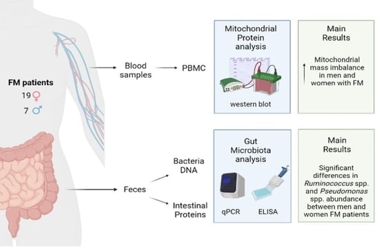
2.1. Patients

This retrospective, observational pilot study was carried out on 26 patients from the Institute for Functional Medicine (IFM) in Lima (Peru). The medical records were coded and anonymized to comply with Regulation 2016/079 of the European General Data Protection Regulations. The patients who were considered for this study had already been diagnosed with primary FM by an experienced practitioner according to the 2010 criteria of the American College of Rheumatology [3]. Inclusion criteria were based on age (only adult patients were included) and lifestyle habits (smokers and morbidly obese patients were not allowed to participate because of the deleterious effect of both). All the patients signed an informed consent form to participate in this study, which was approved by an ethics committee and conducted according to the Declaration of Helsinki from the World Medical Association.

2.2. Health Questionnaires

The patients filled out the clinical symptoms sheet, a self-questionnaire from the Cleveland Clinic Center for Functional Medicine and IFM (Cleveland, OH, USA) which is used to assess the impact of the condition on overall health (Supplementary File S1). Statistical analysis was performed using the statistical software STATA v.15® (StataCorp 2015, Stata Statistical Software, College Station, TX, USA). The IFM questionnaire has 15 items describing physical and psychological health. The answers refer to the feelings that the patient has experienced during the last two weeks, and the rating scale ranges from 0 to 4 points: 0 means “never or almost never experienced the symptom”; 1 means “occasionally experienced it, but the effect is not serious”; 2 means “experienced it occasionally and the effect is severe”; 3 means “experienced it frequently, but the effect is not serious”; and 4 means “frequently suffered it and the effect is serious”. The final score is divided into five groups: from 0–10 as “good health”; from 10–20 as “mild dysfunction”; from 20–50 as “moderate dysfunction”; from 50–100 as “severe dysfunction”, and more than 100 as “critical dysfunction”.

2.3. Determination of Mitochondrial Mass and Total Antioxidant Capacity

Peripheral blood samples were collected from patients in the early morning using a BD Vacutainer® Safety-Lok™ blood collection set and two BD Vacutainer® Lithium Heparin Tubes (Becton Dickinson, Franklin Lakes, NJ, USA). One of the tubes was used to determine the mitochondrial ratio and total antioxidant capacity (TAC), while the other one was used for peripheral blood mononuclear cells protein extraction (PBMC). For the isolation of PBMCs, 5 mL of whole blood was mixed with 45 mL of ammonium chloride lysis buffer 0.17 M (Thermo Fisher Scientific, Waltham, ME, USA). After shaking for 5 min at room temperature, the tubes were centrifuged for 10 min at 600 FCR, recovering the isolated PBMC pellet. Cells were then lysed with a buffer containing 0.32 M sucrose, 10 mM tris-HCl, 5 mM MgCl2, Triton X-100 1%, and proteinase inhibitor. After a 20 min incubation period on ice, samples were sonicated for better cell lysis. The subsequent whole protein quantification was performed using the Lowry method at 750 nm. The mitochondrial mass was determined using the Western blotting technique in PBMC, as described by Martínez-Lara et al. [28] Hence, mitochondrial mass was evaluated through immunodetection of voltage-dependent anion-selective channel protein 1 (VDAC1; UniProt Accession P21796), while mitophagy was measured through microtubule-associated proteins 1A/1B light chain 3B (MAP1LC3B; UniProt Accession Q9GZQ8) using specific monoclonal antibodies (Abcam, Cambridge, UK). For TAC measurement, an e-BQC lab device (Bioquochem SL, Oviedo, Asturias, Spain) was used. e-BQC lab is an electroanalytical system designed for measuring the antioxidant capacity of biomedical samples [32,33]. The device provided TAC values in terms of electric charge units (Q, µC). The e-BQC lab TAC results were transformed into uric acid equivalents (mM) using a standard curve with a wide range of uric acid dilutions (0/0.02/0.03/0.04/0.05/0.07/0.10/0.15/0.20/0.25/0.30 mM), as shown in Figure 1.

2.4. Microbiota Analysis

Fecal samples were collected with a DANASTOOL Sample Collection MICROBIOME Kit (DANAGENE, Barcelona, Spain) containing a DNA stabilization solution for sample shipment to avoid degradation. DNA extraction was performed in 0.5–1 g of the sample with a DANAGENE Microbiome Fecal DNA kit (DANAGENE, Barcelona, Spain) via a cetyltrimethylammonium bromide (CTAB) buffer-based procedure. Briefly, a portion of the sample was placed into a 2 mL Eppendorf® microtube with 1 mL of CTAB. Then, the mixture was roughly homogenized by pipetting to avoid foam and incubated for 10 min at 70 °C. Next, homogenization of the sample took place on a Vortex Genie 2 for 10 min at maximum speed, and, after that, it was centrifuged at 14,000 rpm for 5 min. Then, 600 µL of the supernatant was transferred to a 1.5 mL Eppendorf® microtube with 200 µL of EC buffer to remove contaminants, and subsequently mixed in vortex. The following steps included 5 min of incubation at 4 °C and centrifugation at 14,000 rpm for 5 min in order to transfer 500 µL of supernatant into a new 1.5 Eppendorf® microtube. Before transferring the liquid to the MicroSpin column, Proteinase K incubation was performed (25 µL, 70 °C, 10 min). The obtained product was placed into a reservoir of the MicroSpin column for the binding, washing, and elution steps, with rounds of centrifugation of 10,000 rpm for 60 s. The quality and purity of the extracted DNA were assessed by absorbance at 260 nm with a µDro Duo plate in a Multiskan SkyHigh Plate Reader (Thermo Fisher Scientifics, Waltham, ME, USA). Bacterial abundances were quantified by real-time quantitative PCR and then used to calculate the ratios of Firmicutes/Bacteroidetes, Bacteroides/Prevotella, and Roseburia/Eubacterium, as well as the presence of Ruminococcus, Pseudomonas, and Akkermansia muciniphila. The primer designs are described in Table 1.
Additionally, intestinal secretory IgA was determined with an ELISA kit (Eagle Bioscience, Amherst, NH, USA), following the recommendations of the manufacturer.

3. Results

3.1. Demographic Profile of Patients

Of the patients included in the study, 19 (73%) were female and 7 (27%) were male. The age range was between 43 and 55, with a mean age of 47 years. Patients did not have any nutritional habits (vegetarian, vegan, etc.), nor any therapeutic regimens (ketogenic diets, elimination diets, or similar). More than half of the patients (62%) engaged in aerobic exercise (walking, running, swimming, or stationary cycling) or mild vigorous exercise (yoga, Pilates) for at least 1 h 3 times per week. Of these patients, 62% did not undertake any psychological or emotion-focused therapy. Additionally, up to 68% of the patients were consuming non-steroidal anti-inflammatory drugs (NSAIDs). As Table 2 demonstrates, 82% of the patients presented with slight to critical dysfunction. The categorical variables, such as sex, type of nutrition, physical exercise, and drug consumption, are expressed by count. For numerical variables such as age, the mean and standard deviation are expressed in brackets, and the p-values, as calculated using Student’s t-test, are shown.

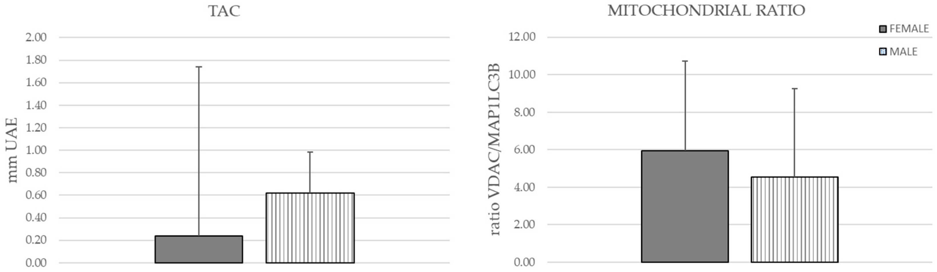
3.2. Mitochondrial Mass and Total Antioxidant Capacity

The mitochondrial ratio was imbalanced compared to the mitochondrial mass, showing higher VDAC protein levels in both groups (Figure 2). Similarly, the TAC mean showed no statistically significant differences between male and female patients (Figure 2).

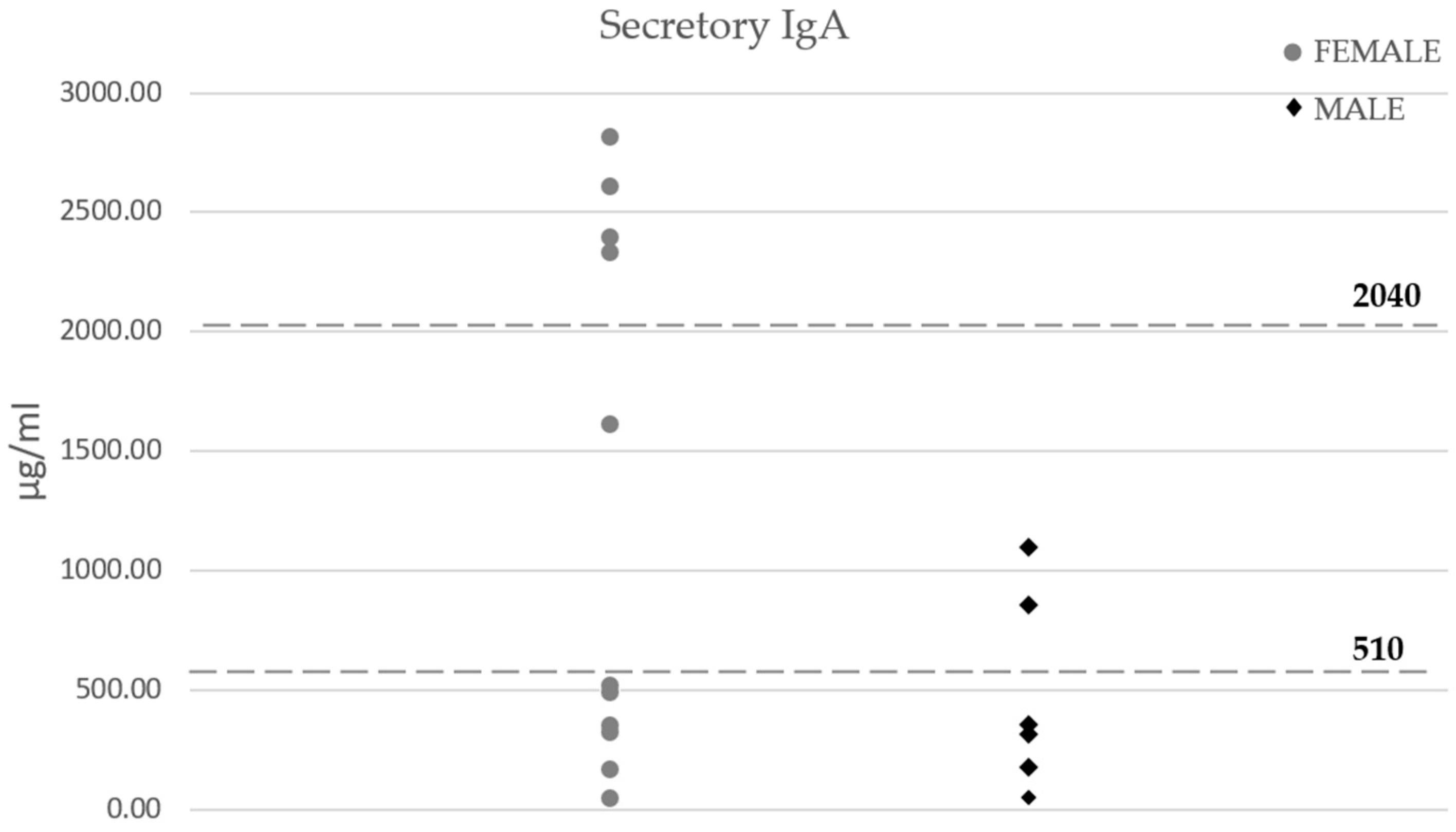
3.3. Microbiota Composition

None of the ratios showed differences between male and female patients. However, female patients showed higher levels of Ruminococcus spp. and Pseudomonas spp. (Figure 3).
The mean secretory IgA levels in women were double compared with those found in men, although there were no statistically significant differences. Interestingly, most of the patients (85%) showed out-of-range levels in both groups (Figure 4).

4. Discussion

The current observational pilot study analyzed mitochondrial and microbiota molecular markers in a Peruvian cohort of female and male FM patients. Although there were no differences in the mitochondrial mass or the TAC, the results showed Pseudomonas and Ruminococcus to be differentially expressed bacteria between both groups.
Chronic conditions are usually difficult to tackle, not only because of the numerous factors that contribute to their development but also due to the heterogeneous group of patients. FM is one of these diseases. In fact, recent publications have proposed different groups of FM patients according to their pain symptoms [43]. Despite this, several research groups have been trying to find molecular biomarkers that could be useful for the diagnostics, treatment tracking, and classification of these patients. In this sense, FM has been traditionally linked to oxidative stress and mitochondria. Several authors have found mitochondrial dysfunction, mtDNA mutations, and different oxidative stress markers in FM patients [23,24,25,26,27,28,30,44]. Later, these problems underlie to chronic and neuropathic pain, as well as inflammation, not only in FM but also in a plethora of complex syndromes [45,46,47,48,49]. In line with this hypothesis, our results showed VDAC1 protein levels that were higher than MAP1LC3B levels in female and male patients. VDAC1 is a mitochondrial membrane porin that is the predominantly expressed form in this organelle [50], while MAP1LC3B is closely related to mitophagy since it is the first protein to trigger the molecular signaling that ends in mitochondrial autophagy [51,52]. These results suggest that FM patients could have a sex-independent marked polarization to mitochondrial accumulation and a subsequent unbalanced oxidative metabolism, even though the lack of healthy volunteers in our study makes it difficult to support the hypothesis that points to mitochondria and oxidative stress as hallmarks of this syndrome. Additionally, the high number of patients consuming drugs such as NSAIDs and analgesics in our cohort might be a confounding factor in determining both parameters, since a huge number of pharmacological therapies affect mitochondrial function [53]. Indeed, these drugs not only influence the mitochondria and oxidative metabolism enzymes (such as COX-1 and COX-2), but also on gastrointestinal homeostasis, especially in microbiota [53]. Hence, as discussed later in this section, the higher number of female patients taking NSAIDs might have an influence on both areas. To solve this, further research with more patients and better-defined drug consumption is needed.
Recently, a growing number of researchers have been attempting to find connections between microbiota and many chronic illnesses. In fact, the interplay between microbiota and mitochondria is rising as a promising diagnostic and therapeutic target for pain, neurodegeneration, and metabolic affections such as type 2 diabetes and obesity, among others [54,55,56]. In particular, chronic pain-related conditions are closely connected to gastrointestinal disturbances and the gut–brain axis [57]. Concretely, several neurotransmitters and metabolites related to pain processing and inflammation are produced in the intestine by a long list of microbes that colonize it [58,59,60]. Therefore, this could be a connection to be analyzed in chronic pain-related conditions such as FM. Our results did not show statistically significant differences between female and male patients in the three microbiota ratios which we analyzed, but the Firmicutes/Bacteroidetes ratio was below the normal range found in healthy volunteers in a previous study [40]. This ratio seems to positively correlate with BMI and type 2 diabetes [61,62,63], although different cohorts of individuals from different countries showed opposite results [40]. Nonetheless, both genera exert interesting metabolic activities which could be key to their health impact. Firmicutes and Bacteroidetes have huge gene clusters coding for P450 monooxygenases, ferredoxins, and other secondary metabolites with relevant roles in host metabolism, mostly in oxidative pathways. These clusters are more abundant in Bacteroidetes species, and have been proposed as factors responsible for health changes attributed to this ratio [64]. Given that several FM studies have shown oxidative stress and mitochondrial dysfunction markers in these patients [23,24,25,26,27,28,30,49], the low Firmicutes/Bacteroidetes ratio found in our cohort could somehow be linked to mitochondrial imbalance, which was also present in this group. Nonetheless, as previously pointed out, our patients are widely exposed to pharmacological treatment, with a relevant number of participants using NSAIDs or analgesic treatment, especially women. A plethora of authors have highlighted the strong impact of NSAID treatments on intestinal homeostasis and microbiota, as widely reviewed by Maseda and Ricciotti as well as Zádori et al., among others [65,66]. For instance, the use of well-known NSAIDs such as indomethacin [67,68,69,70], diclofenac [71], or ibuprofen [72] has been shown to influence the abundance of Bacteroides in different cohorts of human subjects and animal models. Surprisingly, these are some of the most used NSAIDs among FM patients, so microbiota alterations within this pathology could be linked to the treatment. A detailed discussion regarding NSAIDs' full repertoire of microbiota-related effects can be found in the review by Zadori et al. [66]. As a result, these findings provide a starting point to shed some light on the differences in microbiota composition between FM patients and healthy volunteers.
Focusing on the specific genera, while Akkermansia muciniphila showed no differences, Ruminococcus spp. and Pseudomonas spp. did demonstrate statistically significant differences between female and male patients with FM. Ruminococcus spp., an anaerobic gram-positive coccus, was lower in women. This genus was found to be positively correlated with BMI [73], and, in our cohort, men had a slightly higher BMI. Thus, BMI itself could be responsible for this difference between the two groups. R. gnavus has been identified as an inflammatory LPS-producing bacterium that is able to increase TNF-alpha production [74]. Since low-grade inflammation is a common feature of FM, the role of Ruminococcus spp. in this condition should be studied. In contrast, Yunus et al. [75] demonstrated that male FM patients had lower tender points, pain and gastrointestinal symptoms than women. According to this, the differences found in these symptoms could not be supported by Ruminococcus spp. differences, although further research is needed. Strikingly, this genus was found to be decreased in feces from an animal model of endometriosis [76], a pain-related condition that has been identified as a comorbidity of FM [77]. In line with this, the Ruminococcaceae family was found to be negatively correlated with enterocyte apoptosis and IL-6 levels in mice, indicating a potential protective role of this bacterium against intestinal inflammation [78]. Furthermore, Ruminococcus has been studied as a key bacterium in estrobolome, the group of enzymes and metabolic reactions that depend on microbiota and that impact estrogens levels. In fact, β-glucuronidase from Ruminococcus has shown its capability to reactivate estrogens in the gut lumen, allowing them to re-enter the bloodstream in their active form [79]. Similarly, the G-protein-coupled estrogen receptor has been proposed as a differential biomarker between FM patients and healthy controls, with higher levels in patients being negatively correlated with prolactin levels [80]. Taken together, these results highlight the promising role of hormonal imbalances and the abundance of Ruminococcus in the differential prevalence of FM between males and females, since lower levels of this bacterium might have an indirect impact on inflammation and the well-known differences in estrogen-related pain signaling between males and females.
Pseudomonas spp. also showed statistically significant differences in our result, proving to be lower in male patients. Pseudomonas are an aerobic group of gram-negative bacilli that act mainly as opportunistic microorganisms, causing pathogenic disturbances in the intestine [81]. Taking into account that gastrointestinal symptoms seem to be more frequent in females [75], Pseudomonas could be one of the genera responsible for this situation. In fact, it has been demonstrated that, in chronic fatigue syndrome, serum IgA anti-Pseudomonas LPS increases compared to healthy controls, and their levels positively correlate with the clinical symptoms of these patients [82]. Secretory IgA has been traditionally studied at a gastrointestinal level as a master regulator of intestinal microbiota, being responsible for the immune responses to its changes [83]. This antibody did not show differences between female and male feces in our FM cohort, but 85% of them had out-of-range sIgA concentrations. Although congenital immunodeficiency and other influential factors must be discarded, these changes could be behind the shifts in microbiota between the two groups of Peruvian patients. Maes et al. [82] suggested that antioxidants may be useful in chronic fatigue syndrome to repair the intestinal barrier, whose increased permeability facilitates the blood circulation of LPS. Whether this intervention could help FM patients or not to control intestinal dysbiosis and the intestinal barrier needs to be addressed. Moreover, further research is needed in order to understand the influence of sex, drugs, nutrition, supplements, and life habits on gastrointestinal health in the FM context.

5. Conclusions

Taken together, our results suggest that the microbiota–mitochondria axis can help in the diagnostics, treatment tracking, and classification of FM patients. sIgA, Ruminoccocus spp., and Pseudomonas spp. might be useful markers not only to diagnose FM, but also to differentiate between male and female patients. However, to properly define the roles of these markers and the microbiota–mitochondria axis, further research with a higher and better-defined number of patients is necessary.

Supplementary Materials

The following supporting information can be downloaded at: https://www.mdpi.com/article/10.3390/neurolint15030055/s1, Supplementary File S1: Cleveland Clinic Center for Functional Medicine and IFM (Cleveland, OH, USA) Health Questionnaire, used to assess the overall health self-perception of patients.

Author Contributions

Conceptualization, J.A.R.-T., M.C. and D.C.; methodology, J.A.R.-T., E.D.-G., L.L.d.A., I.S. and A.M.-L.; resources, A.H.-D. and M.C.; writing—original draft preparation, J.A.R.-T., E.D.-G., L.L.d.A., I.S. and A.M.-L.; writing—review and editing, J.A.R.-T., A.M.-L. and D.C.; project administration, M.C. and D.C.; funding acquisition, M.C. and D.C. All authors have read and agreed to the published version of the manuscript.

Funding

This research received no external funding.

Institutional Review Board Statement

The study was conducted in accordance with the Declaration of Helsinki and approved by the Ethics Committee of Quirónsalud-catalunya hospitality group (protocol code IDI-20210749 approved on 11 January 2022).

Informed Consent Statement

Informed consent was obtained from all subjects involved in the study.

Data Availability Statement

Data sharing is not applicable.

Acknowledgments

We would like to thank Hermani Lab Genetics (Lima, Peru) for its involvement and management in sample shipment and patient coordination.

Conflicts of Interest

Pronacera Therapeutics S.L. develops and provides molecular tests for the prevention, diagnosis, and personalized treatment of several conditions that involve the analysis of genes, proteins, and microbes in human samples. Neither the patients nor the healthcare professionals participating in this study have been paid or influenced whatsoever. Hence, the study was conducted with the sole resources of Pronacera Therapeutics S.L.

References

  1. Longley, K. Fibromyalgia: Aetiology, Diagnosis, Symptoms and Management. Br. J. Nurs. 2006, 15, 729–733. [Google Scholar] [CrossRef] [PubMed]
  2. Marques, A.P.; Santo, A.D.S.D.E.; Berssaneti, A.A.; Matsutani, L.A.; Yuan, S.L.K. Prevalence of Fibromyalgia: Literature Review Update. Rev. Bras. Reumatol. 2017, 57, 356–363. [Google Scholar] [CrossRef]
  3. Wolfe, F.; Clauw, D.J.; Fitzcharles, M.A.; Goldenberg, D.L.; Katz, R.S.; Mease, P.; Russell, A.S.; Russell, I.J.; Winfield, J.B.; Yunus, M.B. The American College of Rheumatology Preliminary Diagnostic Criteria for Fibromyalgia and Measurement of Symptom Severity. Arthritis Care Res. 2010, 62, 600–610. [Google Scholar] [CrossRef] [PubMed]
  4. Sarzi-Puttini, P.; Giorgi, V.; Marotto, D.; Atzeni, F. Fibromyalgia: An Update on Clinical Characteristics, Aetiopathogenesis and Treatment. Nat. Rev. Rheumatol. 2020, 16, 645–660. [Google Scholar] [CrossRef]
  5. Hackshaw, K.V. The Search for Biomarkers in Fibromyalgia. Diagnostics 2021, 11, 156. [Google Scholar] [CrossRef]
  6. Gerdle, B.; Ghafouri, B.; Lund, E.; Bengtsson, A.; Lundberg, P.; van Ettinger-Veenstra, H.; Leinhard, O.D.; Forsgren, M.F. Evidence of Mitochondrial Dysfunction in Fibromyalgia: Deviating Muscle Energy Metabolism Detected Using Microdialysis and Magnetic Resonance. J. Clin. Med. 2020, 9, 3527. [Google Scholar] [CrossRef]
  7. Jung, Y.H.; Kim, H.; Lee, D.; Lee, J.Y.; Moon, J.Y.; Choi, S.H.; Kang, D.H. Dysfunctional Energy Metabolisms in Fibromyalgia Compared with Healthy Subjects. Mol. Pain. 2021, 17, 17448069211012833. [Google Scholar] [CrossRef]
  8. Huttenhower, C.; Gevers, D.; Knight, R.; Abubucker, S.; Badger, J.H.; Chinwalla, A.T.; Creasy, H.H.; Earl, A.M.; Fitzgerald, M.G.; Fulton, R.S.; et al. Structure, Function and Diversity of the Healthy Human Microbiome. Nature 2012, 486, 207. [Google Scholar] [CrossRef]
  9. Spor, A.; Koren, O.; Ley, R. Unravelling the Effects of the Environment and Host Genotype on the Gut Microbiome. Nat. Rev. Microbiol. 2011, 9, 279–290. [Google Scholar] [CrossRef]
  10. Larsen, N.; Vogensen, F.K.; van den Berg, F.W.J.; Nielsen, D.S.; Andreasen, A.S.; Pedersen, B.K.; Al-Soud, W.A.; Sørensen, S.J.; Hansen, L.H.; Jakobsen, M. Gut Microbiota in Human Adults with Type 2 Diabetes Differs from Non-Diabetic Adults. PLoS ONE 2010, 5, e9085. [Google Scholar] [CrossRef]
  11. Vatanen, T.; Kostic, A.D.; D’Hennezel, E.; Siljander, H.; Franzosa, E.A.; Yassour, M.; Kolde, R.; Vlamakis, H.; Arthur, T.D.; Hämäläinen, A.M.; et al. Variation in Microbiome LPS Immunogenicity Contributes to Autoimmunity in Humans. Cell 2016, 165, 842–853. [Google Scholar] [CrossRef]
  12. Koeth, R.A.; Wang, Z.; Levison, B.S.; Buffa, J.A.; Org, E.; Sheehy, B.T.; Britt, E.B.; Fu, X.; Wu, Y.; Li, L.; et al. Intestinal Microbiota Metabolism of L-Carnitine, a Nutrient in Red Meat, Promotes Atherosclerosis. Nat. Med. 2013, 19, 576–585. [Google Scholar] [CrossRef]
  13. Tilg, H. A Gut Feeling about Thrombosis. N. Engl. J. Med. 2016, 374, 2494–2496. [Google Scholar] [CrossRef]
  14. Jangi, S.; Gandhi, R.; Cox, L.M.; Li, N.; von Glehn, F.; Yan, R.; Patel, B.; Mazzola, M.A.; Liu, S.; Glanz, B.L.; et al. Alterations of the Human Gut Microbiome in Multiple Sclerosis. Nat. Commun. 2016, 7, 12015. [Google Scholar] [CrossRef]
  15. Chen, J.; Wright, K.; Davis, J.M.; Jeraldo, P.; Marietta, E.V.; Murray, J.; Nelson, H.; Matteson, E.L.; Taneja, V. An Expansion of Rare Lineage Intestinal Microbes Characterizes Rheumatoid Arthritis. Genome Med. 2016, 8, 43. [Google Scholar] [CrossRef]
  16. Collado, A.; Gomez, E.; Coscolla, R.; Sunyol, R.; Solé, E.; Rivera, J.; Altarriba, E.; Carbonell, J.; Castells, X. Work, Family and Social Environment in Patients with Fibromyalgia in Spain: An Epidemiological Study: EPIFFAC Study. BMC Health Serv. Res. 2014, 14, 513. [Google Scholar] [CrossRef]
  17. Minerbi, A.; Gonzalez, E.; Brereton, N.J.B.; Anjarkouchian, A.; Dewar, K.; Fitzcharles, M.A.; Chevalier, S.; Shir, Y. Altered Microbiome Composition in Individuals with Fibromyalgia. Pain 2019, 160, 2589–2602. [Google Scholar] [CrossRef]
  18. Albayrak, B.; Süsgün, S.; Küçükakkaş, O.; Akbaş, F.; Yabaci, A.; Özçelik, S. Investigating of Relation Between Fibromyalgia Syndrome and Intestinal Microbiota. Mikrobiyol. Bul. 2021, 55, 146–160. [Google Scholar] [CrossRef]
  19. Clos-Garcia, M.; Andrés-Marin, N.; Fernández-Eulate, G.; Abecia, L.; Lavín, J.L.; van Liempd, S.; Cabrera, D.; Royo, F.; Valero, A.; Errazquin, N.; et al. Gut Microbiome and Serum Metabolome Analyses Identify Molecular Biomarkers and Altered Glutamate Metabolism in Fibromyalgia. EBioMedicine 2019, 46, 499–511. [Google Scholar] [CrossRef]
  20. Baldi, S.; Pagliai, G.; Dinu, M.; di Gloria, L.; Nannini, G.; Curini, L.; Pallecchi, M.; Russo, E.; Niccolai, E.; Danza, G.; et al. Effect of Ancient Khorasan Wheat on Gut Microbiota, Inflammation, and Short-Chain Fatty Acid Production in Patients with Fibromyalgia. World J. Gastroenterol. 2022, 28, 1965–1980. [Google Scholar] [CrossRef]
  21. D’Erchia, A.M.; Atlante, A.; Gadaleta, G.; Pavesi, G.; Chiara, M.; de Virgilio, C.; Manzari, C.; Mastropasqua, F.; Prazzoli, G.M.; Picardi, E.; et al. Tissue-Specific MtDNA Abundance from Exome Data and Its Correlation with Mitochondrial Transcription, Mass and Respiratory Activity. Mitochondrion 2015, 20, 13–21. [Google Scholar] [CrossRef] [PubMed]
  22. Siracusa, R.; di Paola, R.; Cuzzocrea, S.; Impellizzeri, D. Fibromyalgia: Pathogenesis, Mechanisms, Diagnosis and Treatment Options Update. Int. J. Mol. Sci. 2021, 22, 3891. [Google Scholar] [CrossRef] [PubMed]
  23. Cordero, M.D.; Díaz-Parrado, E.; Carrión, A.M.; Alfonsi, S.; Sánchez-Alcazar, J.A.; Bullón, P.; Battino, M.; de Miguel, M. Is Inflammation a Mitochondrial Dysfunction-Dependent Event in Fibromyalgia? Antioxid Redox Signal 2013, 18, 800–807. [Google Scholar] [CrossRef] [PubMed]
  24. Danda, S.; Thomas, B.; Paramasivam, G.; Thomas, R.; Mathew, J.; Danda, D. A Descriptive Pilot Study of Mitochondrial Mutations & Clinical Phenotype in Fibromyalgia Syndrome. Indian J. Med. Res. 2019, 149, 47–50. [Google Scholar] [CrossRef] [PubMed]
  25. Cordero, M.D.; de Miguel, M.; Carmona-López, I.; Bonal, P.; Campa, F.; Moreno-Fernández, A.M. Oxidative Stress and Mitochondrial Dysfunction in Fibromyalgia. Neuroendocrinol. Lett. 2010, 31, 169–173. [Google Scholar]
  26. Cordero, M.D.; de Miguel, M.; Moreno Fernández, A.M.; Carmona López, I.M.; Garrido Maraver, J.; Cotán, D.; Gómez Izquierdo, L.; Bonal, P.; Campa, F.; Bullon, P.; et al. Mitochondrial Dysfunction and Mitophagy Activation in Blood Mononuclear Cells of Fibromyalgia Patients: Implications in the Pathogenesis of the Disease. Arthritis Res. Ther. 2010, 12, R17. [Google Scholar] [CrossRef]
  27. Meeus, M.; Nijs, J.; Hermans, L.; Goubert, D.; Calders, P. The Role of Mitochondrial Dysfunctions Due to Oxidative and Nitrosative Stress in the Chronic Pain or Chronic Fatigue Syndromes and Fibromyalgia Patients: Peripheral and Central Mechanisms as Therapeutic Targets? Expert Opin. Ther. Targets 2013, 17, 1081–1089. [Google Scholar] [CrossRef]
  28. Martínez-Lara, A.; Moreno-Fernández, A.M.; Jiménez-Guerrero, M.; Díaz-López, C.; De-Miguel, M.; Cotán, D.; Sánchez-Alcázar, J.A. Mitochondrial Imbalance as a New Approach to the Study of Fibromyalgia. Open Access Rheumatol. 2020, 12, 175–185. [Google Scholar] [CrossRef]
  29. Fatima, G.; Das, S.K.; Mahdi, A.A. Some Oxidative and Antioxidative Parameters and Their Relationship with Clinical Symptoms in Women with Fibromyalgia Syndrome. Int. J. Rheum. Dis. 2017, 20, 39–45. [Google Scholar] [CrossRef]
  30. Sánchez-Domínguez, B.; Bullón, P.; Román-Malo, L.; Marín-Aguilar, F.; Alcocer-Gómez, E.; Carrión, A.M.; Sánchez-Alcazar, J.A.; Cordero, M.D. Oxidative Stress, Mitochondrial Dysfunction and, Inflammation Common Events in Skin of Patients with Fibromyalgia. Mitochondrion 2015, 21, 69–75. [Google Scholar] [CrossRef]
  31. Jackson, D.N.; Theiss, A.L. Gut Bacteria Signaling to Mitochondria in Intestinal Inflammation and Cancer. Gut Microbes 2020, 11, 285–304. [Google Scholar] [CrossRef]
  32. Serrano-Serra, J.P.; Montero-Vilchez, T.; Buendia-Eisman, A.; Arias-Santiago, S. Epidermal Barrier Function and Skin Homeostasis in Skin with Permanent and Adhesive Tattoos: A Cross-Sectional Study. J. Clin. Med. 2021, 10, 888. [Google Scholar] [CrossRef]
  33. Aranaz, M.; Costas-Rodríguez, M.; Lobo, L.; García, M.; González-Iglesias, H.; Pereiro, R.; Vanhaecke, F. Homeostatic Alterations Related to Total Antioxidant Capacity, Elemental Concentrations and Isotopic Compositions in Aqueous Humor of Glaucoma Patients. Anal. Bioanal. Chem. 2022, 414, 515–524. [Google Scholar] [CrossRef]
  34. Walter, J.; Hertel, C.; Tannock, G.W.; Lis, C.M.; Munro, K.; Hammes, W.P. Detection of Lactobacillus, Pediococcus, Leuconostoc, and Weissella Species in Human Feces by Using Group-Specific PCR Primers and Denaturing Gradient Gel Electrophoresis. Appl. Environ. Microbiol. 2001, 67, 2578–2585. [Google Scholar] [CrossRef]
  35. Bartosch, S.; Fite, A.; Macfarlane, G.T.; McMurdo, M.E.T. Characterization of Bacterial Communities in Feces from Healthy Elderly Volunteers and Hospitalized Elderly Patients by Using Real-Time PCR and Effects of Antibiotic Treatment on the Fecal Microbiota. Appl. Environ. Microbiol. 2004, 70, 3575–3581. [Google Scholar] [CrossRef]
  36. Rekha, R.; Rizvi, M.A.; Jaishree, P. Designing and Validation of Genus-Specific Primers for Human Gut Flora Study. Electron. J. Biotechnol. 2006, 9, 505–511. [Google Scholar] [CrossRef]
  37. Derrien, M. Mucin Utilisation and Host Interactions of the Novel Intestinal Microbe Akkermansia Muciniphila; Wageningen University: Wageningen, The Netherlands, 2007. [Google Scholar]
  38. Guo, X.; Xia, X.; Tang, R.; Zhou, J.; Zhao, H.; Wang, K. Development of a Real-Time PCR Method for Firmicutes and Bacteroidetes in Faeces and Its Application to Quantify Intestinal Population of Obese and Lean Pigs. Lett. Appl. Microbiol. 2008, 47, 367–373. [Google Scholar] [CrossRef]
  39. Lauritsen, J.G.; Hansen, M.L.; Bech, P.K.; Jelsbak, L.; Gram, L.; Strube, M.L. Identification and Differentiation of Pseudomonas Species in Field Samples Using an RpoD Amplicon Sequencing Methodology. mSystems 2021, 6, e0070421. [Google Scholar] [CrossRef]
  40. Magne, F.; Gotteland, M.; Gauthier, L.; Zazueta, A.; Pesoa, S.; Navarrete, P.; Balamurugan, R. The Firmicutes/Bacteroidetes Ratio: A Relevant Marker of Gut Dysbiosis in Obese Patients? Nutrients 2020, 12, 1474. [Google Scholar] [CrossRef]
  41. Hjorth, M.F.; Blædel, T.; Bendtsen, L.Q.; Lorenzen, J.K.; Holm, J.B.; Kiilerich, P.; Roager, H.M.; Kristiansen, K.; Larsen, L.H.; Astrup, A. Prevotella-to-Bacteroides Ratio Predicts Body Weight and Fat Loss Success on 24-Week Diets Varying in Macronutrient Composition and Dietary Fiber: Results from a Post-Hoc Analysis. Int. J. Obes. 2018, 43, 149–157. [Google Scholar] [CrossRef]
  42. Aminov, R.I.; Walker, A.W.; Duncan, S.H.; Harmsen, H.J.M.; Welling, G.W.; Flint, H.J. Molecular Diversity, Cultivation, and Improved Detection by Fluorescent in Situ Hybridization of a Dominant Group of Human Gut Bacteria Related to Roseburia Spp. or Eubacterium Rectale. Appl. Environ. Microbiol. 2006, 72, 6371–6376. [Google Scholar] [CrossRef] [PubMed]
  43. Martínez, M.P.; Sánchez, A.I.; Prados, G.; Lami, M.J.; Villar, B.; Miró, E. Fibromyalgia as a Heterogeneous Condition: Subgroups of Patients Based on Physical Symptoms and Cognitive-Affective Variables Related to Pain. Span J. Psychol. 2021, 24, e33. [Google Scholar] [CrossRef] [PubMed]
  44. Castro-Marrero, J.; Cordero, M.D.; Sáez-Francas, N.; Jimenez-Gutierrez, C.; Aguilar-Montilla, F.J.; Aliste, L.; Alegre-Martin, J. Could Mitochondrial Dysfunction Be a Differentiating Marker between Chronic Fatigue Syndrome and Fibromyalgia? Antioxid. Redox Signal. 2013, 19, 1855–1860. [Google Scholar] [CrossRef]
  45. Flatters, S.J.L. The Contribution of Mitochondria to Sensory Processing and Pain. Prog. Mol. Biol. Transl. Sci. 2015, 131, 119–146. [Google Scholar] [CrossRef] [PubMed]
  46. Marchev, A.S.; Dimitrova, P.A.; Burns, A.J.; Kostov, R.V.; Dinkova-Kostova, A.T.; Georgiev, M.I. Oxidative Stress and Chronic Inflammation in Osteoarthritis: Can NRF2 Counteract These Partners in Crime? Ann. N. Y. Acad. Sci. 2017, 1401, 114–135. [Google Scholar] [CrossRef]
  47. Shim, H.S.; Bae, C.; Wang, J.; Lee, K.H.; Hankerd, K.M.; Kim, H.K.; Chung, J.M.; La, J.H. Peripheral and Central Oxidative Stress in Chemotherapy-Induced Neuropathic Pain. Mol. Pain. 2019, 15, 1744806919840098. [Google Scholar] [CrossRef]
  48. Dai, C.Q.; Guo, Y.; Chu, X.Y. Neuropathic Pain: The Dysfunction of Drp1, Mitochondria, and ROS Homeostasis. Neurotox Res. 2020, 38, 553–563. [Google Scholar] [CrossRef]
  49. dos Santos, J.M.; Lacerda, A.C.R.; Ribeiro, V.G.C.; Figueiredo, P.H.S.; Fonseca, S.F.; da Lage, V.K.S.; Costa, H.S.; Lima, V.P.; Sañudo, B.; Bernardo-Filho, M.; et al. Oxidative Stress Biomarkers and Quality of Life Are Contributing Factors of Muscle Pain and Lean Body Mass in Patients with Fibromyalgia. Biology 2022, 11, 935. [Google Scholar] [CrossRef]
  50. Yamamoto, T.; Yamada, A.; Watanabe, M.; Yoshimura, Y.; Yamazaki, N.; Yoshimura, Y.; Yamauchi, T.; Kataoka, M.; Nagata, T.; Terada, H.; et al. VDAC1, Having a Shorter N-Terminus than VDAC2 but Showing the Same Migration in an SDS-Polyacrylamide Gel, Is the Predominant Form Expressed in Mitochondria of Various Tissues. J. Proteome Res. 2006, 5, 3336–3344. [Google Scholar] [CrossRef]
  51. Tanida, I.; Ueno, T.; Kominami, E. LC3 and Autophagy. Methods Mol. Biol. 2008, 445, 77–88. [Google Scholar] [CrossRef]
  52. Huang, R.; Liu, W. Identifying an Essential Role of Nuclear LC3 for Autophagy. Autophagy 2015, 11, 852–853. [Google Scholar] [CrossRef]
  53. Lyakhovich, A.; Graifer, D. Mitochondria-Mediated Oxidative Stress: Old Target for New Drugs. Curr. Med. Chem. 2015, 22, 3040–3053. [Google Scholar] [CrossRef]
  54. Vezza, T.; Abad-Jiménez, Z.; Marti-Cabrera, M.; Rocha, M.; Víctor, V.M. Microbiota-Mitochondria Inter-Talk: A Potential Therapeutic Strategy in Obesity and Type 2 Diabetes. Antioxidants 2020, 9, 848. [Google Scholar] [CrossRef]
  55. Kramer, P. Mitochondria-Microbiota Interaction in Neurodegeneration. Front. Aging Neurosci. 2021, 13, 776936. [Google Scholar] [CrossRef]
  56. Zhang, Y.; Zhang, J.; Duan, L. The Role of Microbiota-Mitochondria Crosstalk in Pathogenesis and Therapy of Intestinal Diseases. Pharmacol. Res. 2022, 186, 106530. [Google Scholar] [CrossRef]
  57. Russo, R.; Cristiano, C.; Avagliano, C.; de Caro, C.; la Rana, G.; Raso, G.M.; Canani, R.B.; Meli, R.; Calignano, A. Gut-Brain Axis: Role of Lipids in the Regulation of Inflammation, Pain and CNS Diseases. Curr. Med. Chem. 2018, 25, 3930–3952. [Google Scholar] [CrossRef]
  58. Auteri, M.; Zizzo, M.G.; Serio, R. GABA and GABA Receptors in the Gastrointestinal Tract: From Motility to Inflammation. Pharmacol. Res. 2015, 93, 11–21. [Google Scholar] [CrossRef]
  59. Otaru, N.; Ye, K.; Mujezinovic, D.; Berchtold, L.; Constancias, F.; Cornejo, F.A.; Krzystek, A.; de Wouters, T.; Braegger, C.; Lacroix, C.; et al. GABA Production by Human Intestinal Bacteroides Spp.: Prevalence, Regulation, and Role in Acid Stress Tolerance. Front. Microbiol. 2021, 12, 656895. [Google Scholar] [CrossRef]
  60. Guo, R.; Chen, L.H.; Xing, C.; Liu, T. Pain Regulation by Gut Microbiota: Molecular Mechanisms and Therapeutic Potential. Br. J. Anaesth 2019, 123, 637–654. [Google Scholar] [CrossRef]
  61. Koliada, A.; Syzenko, G.; Moseiko, V.; Budovska, L.; Puchkov, K.; Perederiy, V.; Gavalko, Y.; Dorofeyev, A.; Romanenko, M.; Tkach, S.; et al. Association between Body Mass Index and Firmicutes/Bacteroidetes Ratio in an Adult Ukrainian Population. BMC Microbiol. 2017, 17, 120. [Google Scholar] [CrossRef]
  62. Qadir, R.M.; Assafi, M.S. The Association between Body Mass Index and the Oral Firmicutes and Bacteroidetes Profiles of Healthy Individuals. Malays. Fam. Physician 2021, 16, 36–43. [Google Scholar] [CrossRef] [PubMed]
  63. Bahar-Tokman, H.; Demirci, M.; Keskin, F.E.; Cagatay, P.; Taner, Z.; Ozturk-Bakar, Y.; Ozyazar, M.; Kiraz, N.; Kocazeybek, B.S. Firmicutes/Bacteroidetes Ratio in the Gut Microbiota and IL-1β, IL-6, IL-8, TLR2, TLR4, TLR5 Gene Expressions in Type 2 Diabetes. Clin. Lab. 2022, 68, 1903–1910. [Google Scholar] [CrossRef] [PubMed]
  64. Nkosi, B.V.Z.; Padayachee, T.; Gront, D.; Nelson, D.R.; Syed, K. Contrasting Health Effects of Bacteroidetes and Firmicutes Lies in Their Genomes: Analysis of P450s, Ferredoxins, and Secondary Metabolite Clusters. Int. J. Mol. Sci. 2022, 23, 5057. [Google Scholar] [CrossRef]
  65. Maseda, D.; Ricciotti, E. NSAID-Gut Microbiota Interactions. Front. Pharmacol. 2020, 11, 1153. [Google Scholar] [CrossRef] [PubMed]
  66. Zádori, Z.S.; Király, K.; Al-Khrasani, M.; Gyires, K. Interactions between NSAIDs, opioids and the gut microbiota—Future perspectives in the management of inflammation and pain. Pharmacol. Ther. 2023, 241, 108327. [Google Scholar] [CrossRef]
  67. Craven, M.; Egan, C.E.; Dowd, S.E.; McDonough, S.P.; Dogan, B.; Denkers, E.Y.; Bowman, D.; Scherl, E.J.; Simpson, K.W. Inflammation drives dysbiosis and bacterial invasion in murine models of ileal Crohn's disease. PLoS ONE 2012, 7, e41594. [Google Scholar] [CrossRef]
  68. Lázár, B.; László, S.B.; Hutka, B.; Tóth, A.S.; Mohammadzadeh, A.; Berekméri, E.; Ágg, B.; Balogh, M.; Sajtos, V.; Király, K.; et al. A comprehensive time course and correlation analysis of indomethacin-induced inflammation, bile acid alterations and dysbiosis in the rat small intestine. Biochem. Pharmacol. 2021, 190, 114590. [Google Scholar] [CrossRef]
  69. Maseda, D.; Zackular, J.P.; Trindade, B.; Kirk, L.; Roxas, J.L.; Rogers, L.M.; Washington, M.K.; Du, L.; Koyama, T.; Viswanathan, V.K.; et al. Nonsteroidal Anti-inflammatory Drugs Alter the Microbiota and Exacerbate Clostridium difficile Colitis while Dysregulating the Inflammatory Response. mBio 2019, 10, e02282-18. [Google Scholar] [CrossRef]
  70. Terán-Ventura, E.; Aguilera, M.; Vergara, P.; Martínez, V. Specific changes of gut commensal microbiota and TLRs during indomethacin-induced acute intestinal inflammation in rats. J. Crohn’s Colitis 2014, 8, 1043–1054. [Google Scholar] [CrossRef]
  71. Colucci, R.; Pellegrini, C.; Fornai, M.; Tirotta, E.; Antonioli, L.; Renzulli, C.; Ghelardi, E.; Piccoli, E.; Gentile, D.; Benvenuti, L.; et al. Pathophysiology of NSAID-Associated Intestinal Lesions in the Rat: Luminal Bacteria and Mucosal Inflammation as Targets for Prevention. Front. Pharmacol. 2018, 9, 1340. [Google Scholar] [CrossRef]
  72. Lu, Z.; Lu, Y.; Wang, X.; Wang, F.; Zhang, Y. Activation of intestinal GR-FXR and PPARα-UGT signaling exacerbates ibuprofen-induced enteropathy in mice. Arch. Toxicol. 2018, 92, 1249–1265. [Google Scholar] [CrossRef]
  73. Xu, Z.; Jiang, W.; Huang, W.; Lin, Y.; Chan, F.K.L.; Ng, S.C. Gut microbiota in patients with obesity and metabolic disorders—a systematic review. Genes Nutr. 2022, 17, 2. [Google Scholar] [CrossRef]
  74. Henke, M.T.; Kenny, D.J.; Cassilly, C.D.; Vlamakis, H.; Xavier, R.J.; Clardy, J. Ruminococcus Gnavus, a Member of the Human Gut Microbiome Associated with Crohn’s Disease, Produces an Inflammatory Polysaccharide. Proc. Natl. Acad. Sci. USA 2019, 116, 12672–12677. [Google Scholar] [CrossRef]
  75. Yunus, M.B.; Inanici, F.; Aldag, J.C.; Mangold, R.F. Fibromyalgia in Men: Comparison of Clinical Features with Women. J. Rheumatol. 2000, 27, 485–490. [Google Scholar]
  76. Cao, Y.; Jiang, C.; Jia, Y.; Xu, D.; Yu, Y. Letrozole and the Traditional Chinese Medicine, Shaofu Zhuyu Decoction, Reduce Endometriotic Disease Progression in Rats: A Potential Role for Gut Microbiota. Evid. Based Complement. Alternat. Med. 2020, 2020, 3687498. [Google Scholar] [CrossRef]
  77. Greenbaum, H.; Weil, C.; Chodick, G.; Shalev, V.; Eisenberg, V.H. Evidence for an Association between Endometriosis, Fibromyalgia, and Autoimmune Diseases. Am. J. Reprod. Immunol. 2019, 81, e13095. [Google Scholar] [CrossRef]
  78. Meng, J.; Banerjee, S.; Zhang, L.; Sindberg, G.; Moidunny, S.; Li, B.; Robbins, D.J.; Girotra, M.; Segura, B.; Ramakrishnan, S.; et al. Opioids Impair Intestinal Epithelial Repair in HIV-Infected Humanized Mice. Front. Immunol. 2020, 10, 2999. [Google Scholar] [CrossRef]
  79. Ervin, S.M.; Li, H.; Lim, L.; Roberts, L.R.; Liang, X.; Mani, S.; Redinbo, M.R. Gut Microbial β-Glucuronidases Reactivate Estrogens as Components of the Estrobolome That Reactivate Estrogens. J. Biol. Chem. 2019, 294, 18586. [Google Scholar] [CrossRef]
  80. Koca, T.; Koçyiğit, B.; Seyithanoğlu, M.; Berk, E. The Importance of G-Protein Coupled Estrogen Receptor in Patients With Fibromyalgia. Arch. Rheumatol. 2019, 34, 419. [Google Scholar] [CrossRef]
  81. Hu, Y.; Qing, Y.; Chen, J.; Liu, C.; Lu, J.; Wang, Q.; Zhen, S.; Zhou, H.; Huang, L.; Zhang, R. Prevalence, Risk Factors, and Molecular Epidemiology of Intestinal Carbapenem-Resistant Pseudomonas Aeruginosa. Microbiol. Spectr. 2021, 9, e01344-21. [Google Scholar] [CrossRef]
  82. Maes, M.; Mihaylova, I.; Leunis, J.C. Increased Serum IgA and IgM against LPS of Enterobacteria in Chronic Fatigue Syndrome (CFS): Indication for the Involvement of Gram-Negative Enterobacteria in the Etiology of CFS and for the Presence of an Increased Gut-Intestinal Permeability. J. Affect Disord. 2007, 99, 237–240. [Google Scholar] [CrossRef] [PubMed]
  83. Pabst, O.; Slack, E. IgA and the Intestinal Microbiota: The Importance of Being Specific. Mucosal Immunol. 2019, 13, 12–21. [Google Scholar] [CrossRef] [PubMed]
Figure 1. Pattern of uric acid used in eBQC TAC measurement.
Neurolint 15 00055 g001
Figure 2. Total antioxidant capacity and mitochondrial ratio comparison between male and female patients.
Neurolint 15 00055 g002
Figure 3. Intestinal microbiome measurements. * p-value < 0.05. For the Firmicutes/Bacteroidetes, Bacteroides/Prevotella, and Roseburia/Eubacterium plots, dashed lines represent the minimal ratio found in healthy volunteers across the bibliography [40,41,42]. For the Pseudomonas, Ruminococcus, and Akkermansia graphs, box-and-whisker plots show the median (horizontal line inside the box), the mean (crosses inside the box), the lower and upper quartiles (lower and upper box limits), and the maximum and minimum values (the whiskers).
Neurolint 15 00055 g003
Figure 4. Secretory IgA levels determined with ELISA. The dashed line represents the normal range according to the manufacturer.
Neurolint 15 00055 g004
Table 1. Primer designs for the characterization of intestinal microbiota.
BacteriaPrimer PairReferences
Akkermansia muciniphilaAkkF, AkkR[10,34,35,36,37,38,39]
Bacteroides spp.BacF 1, BacR 1
Bacteroidetes spp.BatsF, BatsR
Eubacterium spp.EuF A, EuR A
Firmicutes spp.FirmF, FirmR
Prevotella spp.PrevF 2, PrevR 2
Pseudomonas spp.PseF A, PseR A
Roseburia spp.RosF A, RosR A
Ruminococcus spp.RumF 3, RumR 3
All bacteriaAllF A, AllR A
1Bacteroides spp. targeted B. acidifaciens, B. caccae, B. clarus, B. faecalis, B. intestinalis, B. oleiciplenus, B. rodentium, B. stercoris, B. uniformis. 2 Prevotella spp. targeted P. corporis, P. buccae, P. buccalis, P. dentalis, P. dentasini, P. denticola, P. enoeca, P. histicola, P. intermedia, P. loescheii, P. maculosa, P. oralis, P. phocaeensis. 3 Ruminococcus spp. targeted mainly, R. gnavus. A Species targeted by these primers can be checked in the list of references.
Table 2. Demographic and health habit profiles of patients.
VariableOutcomeMale (n = 7)Female (n = 18)p-Value
Age 45.43 (±8.96)50.28 (±10.21)0.26
Body Mass Index (BMI) 26.26 (±3.31)24.97 (3.07)0.39
IFM questionnaire score 51.80 (±15.87)62.17 (±35.37)0.06
NutritionNot controlled718n.a. *
Under control00
Physical exerciseNone37n.a. *
Yes411
Psychological therapyNone47n.a. *
Yes311
Drugs 1NSAIDs414n.a. *
Antidepressants410
Neuropathic drugs17
Analgesics515
Antibiotics 222
Probiotics 225
1 Refers to the moment in which the samples were taken, except for antibiotics and probiotics, which refer to the last 3 months. 2 Antibiotic and probiotic treatments were suspended at least 15 days before sampling. * Not applicable.
Disclaimer/Publisher’s Note: The statements, opinions and data contained in all publications are solely those of the individual author(s) and contributor(s) and not of MDPI and/or the editor(s). MDPI and/or the editor(s) disclaim responsibility for any injury to people or property resulting from any ideas, methods, instructions or products referred to in the content.

