


Neurol. Int. 2024, 16(2), 459-469; https://doi.org/10.3390/neurolint16020034 - 18 Apr 2024
Abstract
►
Show Figures
While total knee arthroplasties (TKAs) are performed with the intent to reduce pain, chronic postsurgical pain (CPSP) is one of the most well-documented complications that can occur following surgery. This study aimed to assess whether perioperative factors, focusing on acute postsurgical pain and
[...] Read more.
While total knee arthroplasties (TKAs) are performed with the intent to reduce pain, chronic postsurgical pain (CPSP) is one of the most well-documented complications that can occur following surgery. This study aimed to assess whether perioperative factors, focusing on acute postsurgical pain and perioperative opioid consumption, were associated with the development of chronic postsurgical pain. Under general anesthesia, 108 patients underwent TKA and were treated postoperatively with a multimodal analgesia approach. Numeric Rating Scale (NRS) pain scores at rest and with movement were recorded on postoperative days 0–3, 7, 14, and 30. Patients were sent a survey to assess chronic pain at months 22–66, which was examined as a single-group post hoc analysis. Based on the responses, patients were either classified into the CPSP or non-CPSP patient group. Chronic postsurgical pain was defined as an NRS score ≥ 4 with movement and the presence of resting pain. The primary outcome was a change in NRS. There were no differences in NRS pain scores with movement in the first 30 days postoperatively between patients with CPSP and without CPSP. Each unit increase in resting pain on postoperative days 3 and 14 was associated with significantly greater odds of CPSP presence (OR = 1.52; OR = 1.61, respectively), with a trend towards greater odds of CPSP at days 7 and 30 (OR = 1.33; OR = 1.43, respectively). We found that very intense pain in the initial phase seems to be related to the development of CPSP after TKA.
Full article
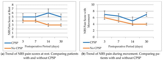
Figure 1

