
Tracing the History of Hepatitis E Virus Infection in Mexico: From the Enigmatic Genotype 2 to the Current Disease Situation

Abstract
:1. Introduction
2. HEV-Gt2: Its Emergence in Mexico
3. The Enigmatic Disappearance of HEV-Gt2 in Mexico
4. HEV Seroprevalence in Mexico
5. HEV in Risk Groups
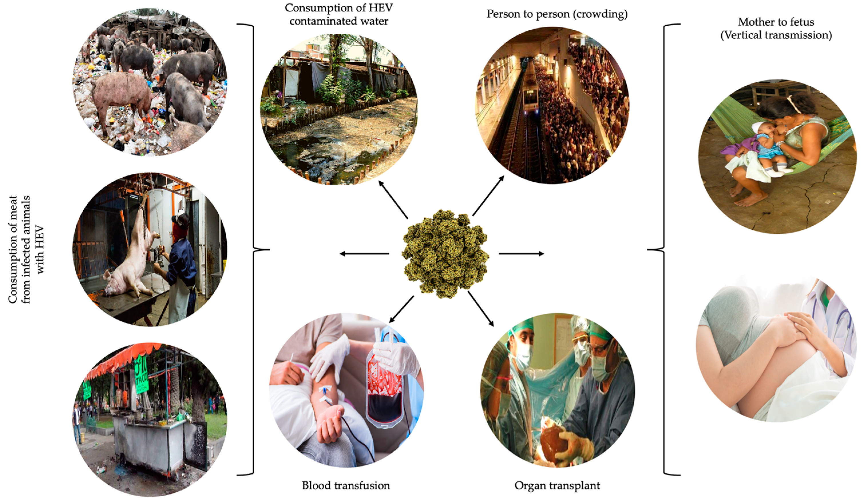
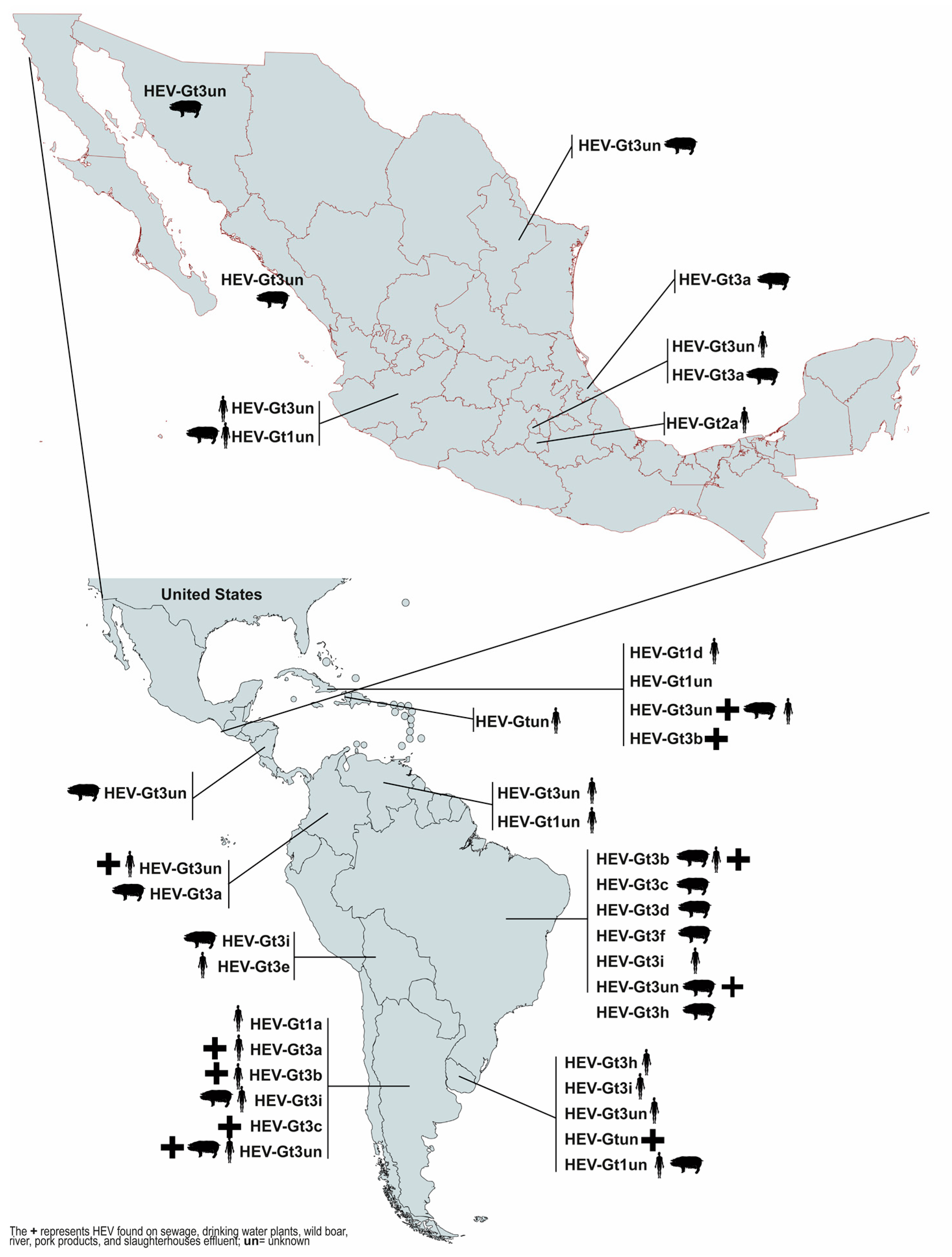
6. Potential Sources of HEV in Mexico
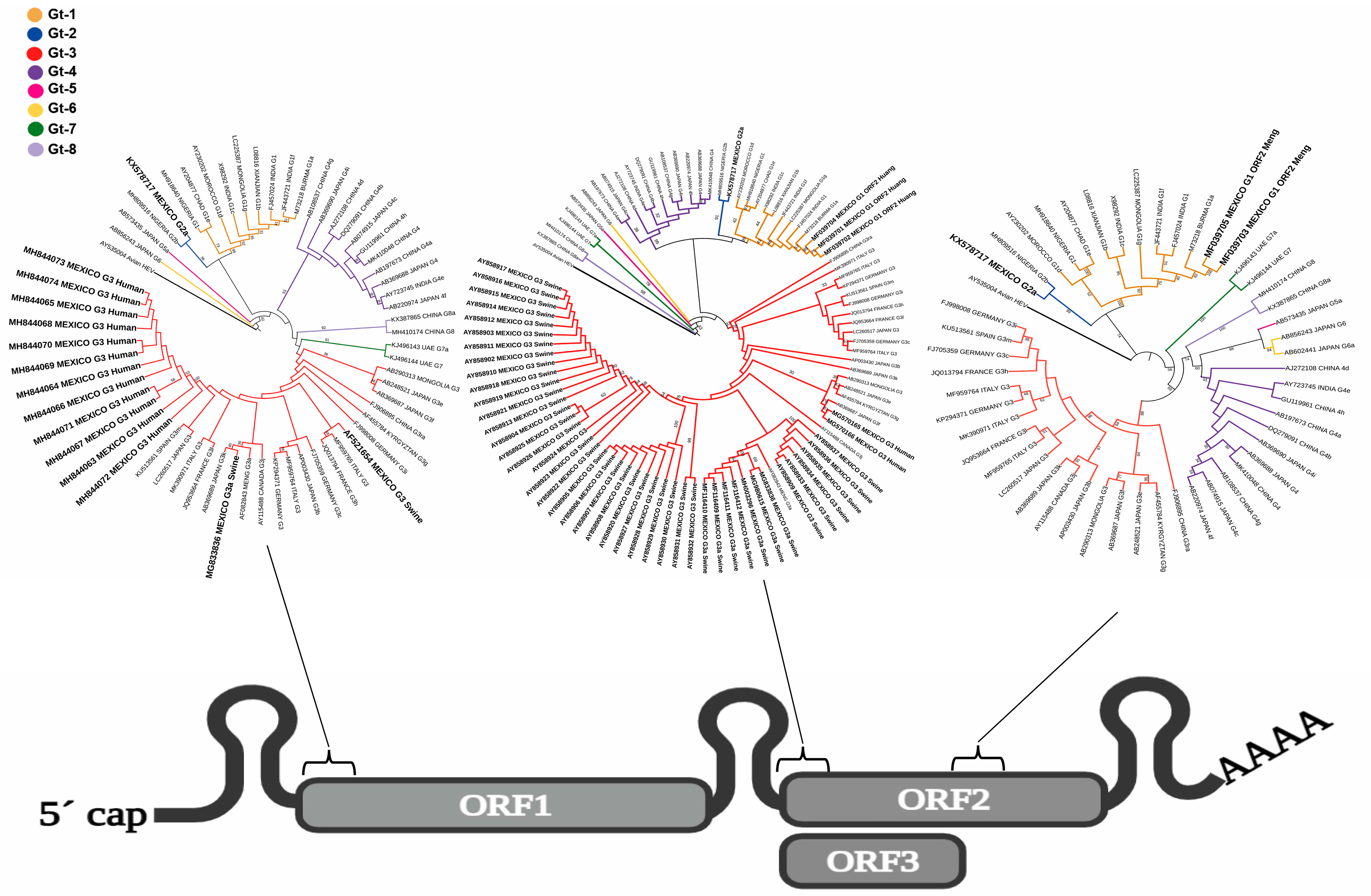
7. The Molecular Epidemiology of HEV in Mexico: A Challenge to Work on
8. Conclusions
Supplementary Materials
Author Contributions
Funding
Acknowledgments
Conflicts of Interest
References
- Pallerla, S.R.; Harms, D.; Johne, R.; Todt, D.; Steinmann, E.; Schemmerer, M.; Wenzel, J.J.; Hofmann, J.; Shih, J.W.K.; Wedemeyer, H.; et al. Hepatitis E Virus Infection: Circulation, Molecular Epidemiology, and Impact on Global Health. Pathogens 2020, 9, 856. [Google Scholar] [CrossRef]
- Willauer, A.N.; Sherman, K.E. Hepatitis E virus: Has anything changed? Curr. Opin. Gastroenterol. 2023, 39, 169–174. [Google Scholar] [CrossRef]
- Pischke, S.; Hartl, J.; Pas, S.D.; Lohse, A.W.; Jacobs, B.C.; Van der Eijk, A.A. Hepatitis E virus: Infection beyond the liver? J. Hepatol. 2017, 66, 1082–1095. [Google Scholar] [CrossRef] [PubMed]
- Rawla, P.; Raj, J.P.; Kannemkuzhiyil, A.J.; Aluru, J.S.; Thandra, K.C.; Gajendran, M. A Systematic Review of the Extra-Hepatic Manifestations of Hepatitis E Virus Infection. Med. Sci. 2020, 8, 9. [Google Scholar] [CrossRef] [PubMed]
- Sayed, I.M.; Abd Elhameed, Z.A.; Abd El-Kareem, D.M.; Abdel-Malek, M.A.Y.; Ali, M.E.; Ibrahim, M.A.; Sayed, A.A.; Khalaf, K.A.B.; Abdel-Wahid, L.; El-Mokhtar, M.A. Hepatitis E Virus Persistence and/or Replication in the Peripheral Blood Mononuclear Cells of Acute HEV-Infected Patients. Front. Microbiol. 2021, 16, 696680. [Google Scholar] [CrossRef]
- Sayed, I.M.; Seddik, M.I.; Gaber, M.A.; Saber, S.H.; Mandour, S.A.; El-Mokhtar, M.A. Replication of Hepatitis E Virus (HEV) in Primary Human-Derived Monocytes and Macrophages In Vitro. Vaccines 2020, 8, 239. [Google Scholar] [CrossRef]
- Bose, P.D.; Das, B.C.; Hazam, R.K.; Kumar, A.; Medhi, S.; Kar, P. Evidence of extrahepatic replication of hepatitis E virus in human placenta. J. Gen. Virol. 2014, 95 Pt 6, 1266–1271. [Google Scholar] [CrossRef]
- Drave, S.A.; Debing, Y.; Walter, S.; Todt, D.; Engelmann, M.; Friesland, M.; Wedemeyer, H.; Neyts, J.; Behrendt, P.; Steinmann, E. Extrahepatic replication and infection of hepatitis E virus in neuronal-derived cells. J. Viral. Hepat. 2016, 23, 512–521. [Google Scholar] [CrossRef]
- Marion, O.; Lhomme, S.; Nayrac, M.; Dubois, M.; Pucelle, M.; Requena, M.; Migueres, M.; Abravanel, F.; Peron, J.M.; Carrere, N.; et al. Hepatitis E virus replication in human intestinal cells. Gut 2020, 69, 901–910. [Google Scholar] [CrossRef] [PubMed]
- Kenney, S.P.; Meng, X.J. Hepatitis E Virus Genome Structure and Replication Strategy. Cold Spring Harb. Perspect Med. 2019, 9, a031724. [Google Scholar] [CrossRef]
- Nair, V.P.; Anang, S.; Subramani, C.; Madhvi, A.; Bakshi, K.; Srivastava, A.; Shalimar; Nayak, B.; Ranjith Kumar, C.T.; Surjit, M. Endoplasmic Reticulum Stress induced Synthesis of a Novel Viral Factor Mediates Efficient Replication of Genotype-1 Hepatitis E Virus. PloS Pathog. 2016, 12, e1005521. [Google Scholar] [CrossRef]
- Purdy, M.A.; Drexler, J.F.; Meng, X.J.; Norder, H.; Okamoto, H.; Van der Poel, W.H.M.; Reuter, G.; de Souza, W.M.; Ulrich, R.G.; Smith, D.B. ICTV Virus Taxonomy Profile: Hepeviridae 2022. J. Gen. Virol. 2022, 103, 001778. [Google Scholar] [CrossRef] [PubMed]
- Wang, B.; Meng, X.J. Hepatitis E virus: Host tropism and zoonotic infection. Curr. Opin. Microbiol. 2021, 59, 8–15. [Google Scholar] [CrossRef]
- Raji, Y.E.; Toung, O.P.; Taib, N.M.; Sekawi, Z.B. Hepatitis E Virus: An emerging enigmatic and underestimated pathogen. Saudi J. Biol. Sci. 2022, 29, 499–512. [Google Scholar] [CrossRef]
- Porea, D.; Raileanu, C.; Crivei, L.A.; Gotu, V.; Savuta, G.; Pavio, N. First Detection of Hepatitis E Virus (Rocahepevirus ratti Genotype C1) in Synanthropic Norway Rats (Rattus norvegicus) in Romania. Viruses 2023, 15, 1337. [Google Scholar] [CrossRef] [PubMed]
- Bi, H.; Yang, R.; Wu, C.; Xia, J. Hepatitis E virus and blood transfusion safety. Epidemiol. Infect. 2020, 148, e158. [Google Scholar] [CrossRef] [PubMed]
- Kamar, N.; Izopet, J.; Pavio, N.; Aggarwal, R.; Labrique, A.; Wedemeyer, H.; Dalton, H.R. Hepatitis E virus infection. Nat Rev. Dis. Primer. 2017, 3, 17086. [Google Scholar] [CrossRef]
- Kar, P.; Karna, R. A Review of the Diagnosis and Management of Hepatitis, E. Curr. Treat. Options Infect. Dis. 2020, 12, 310–320. [Google Scholar] [CrossRef]
- Lemon, S.M.; Walker, C.M. Enterically Transmitted Non-A, Non-B Hepatitis and the Discovery of Hepatitis E Virus. Cold Spring Harb. Perspect Med. 2019, 9, a033449. [Google Scholar] [CrossRef]
- Programme UND. Trapped: High Inequality and Low Growth in Latin America and the Caribbean: Regional Human Development Report 2021. United Nations. Available online: https://www.un-ilibrary.org/content/books/9789210057844 (accessed on 14 August 2023).
- Arankalle, V.A.; Ticehurst, J.; Sreenivasan, M.A.; Kapikian, A.Z.; Popper, H.; Pavri, K.M.; Purcell, R.H. Aetiological association of a virus-like particle with enterically transmitted non-A., non-B hepatitis. Lancet Lond Engl. 1988, 1, 550–554. [Google Scholar] [CrossRef]
- Ticehurst, J.; Rhodes, L.L., Jr.; Krawczynski, K.; Asher, L.V.S.; Engler, W.F.; Mensing, T.L.; Caudill, J.D.; Sjogren, M.H.; Hoke, C.H., Jr.; LeDuc, J.W.; et al. Infection of Owl Monkeys (Aotus trivirgatus) and Cynomolgus Monkeys (Macaca fascicularist) with Hepatitis E Virus from Mexico. J. Infect. Dis. 1992, 165, 835–845. [Google Scholar] [CrossRef]
- Bradley, D.W.; Balayan, M.S. Virus of enterically transmitted non-A., non-B hepatitis. Lancet Lond Engl. 1988, 1, 819. [Google Scholar] [CrossRef]
- Huang, C.C.; Nguyen, D.; Fernandez, J.; Yun, K.Y.; Fry, K.E.; Bradley, D.W.; Tam, A.W.; Reyes, G.R. Molecular cloning and sequencing of the Mexico isolate of hepatitis E virus (HEV). Virology 1992, 191, 550–558. [Google Scholar] [CrossRef]
- Cooper, K.; Huang, F.F.; Batista, L.; Rayo, C.D.; Bezanilla, J.C.; Toth, T.E.; Meng, X.J. Identification of Genotype 3 Hepatitis E Virus (HEV) in Serum and Fecal Samples from Pigs in Thailand and Mexico, Where Genotype 1 and 2 HEV Strains Are Prevalent in the Respective Human Populations. J. Clin. Microbiol. 2005, 43, 1684–1688. [Google Scholar] [CrossRef]
- Realpe-Quintero, M.; Copado-Villagrana, E.D.; Trujillo-Ochoa, J.L.; Alvarez, A.H.; Panduro, A.; Fierro, N.A. Cytokine Signatures Discriminate Highly Frequent Acute Hepatitis a Virus and Hepatitis E Virus Coinfections from Monoinfections in Mexican Pediatric Patients. Pediatr. Infect. Dis. J. 2017, 36, 689–692. [Google Scholar] [CrossRef]
- Realpe-Quintero, M.; Mirazo, S.; Viera-Segura, O.; Copado-Villagrana, E.D.; Panduro, A.; Roman, S.; Arbiza, J.; Fierro, N.A. Hepatitis E Virus Genotype 1 and Hepatitis A Virus Dual Infection in Pediatric Patients with a Low Socioeconomic Status from Mexico. Intervirology 2018, 61, 105–110. [Google Scholar] [CrossRef]
- Viera-Segura, O.; Realpe-Quintero, M.; Panduro, A.; Roman, S.; Jose-Abrego, A.; Gonzalez-Aldaco, K.; Trujillo-Ochoa, J.L.; Fierro, N.A. First detection of hepatitis E virus genotype 3 as a common infectious agent in patients with chronic liver damage in Mexico. Ann. Hepatol. 2019, 18, 571–577. [Google Scholar] [CrossRef]
- López-Santaella, T.; Álvarez YMuñoz, T.; Medeiros-Domingo, M.; Moreno-Espinosa, S.; Consuelo-Sánchez, A.; Muñoz-Hernández, O.; Sarmiento-Silva, R.E.; Sotomayor-González, A.; Trujillo-Ortega, M.E.; García-Hernández, M.E.; et al. Serological and molecular study of Hepatitis E virus in pediatric patients in Mexico. Ann. Hepatol. 2020, 19, 295–301. [Google Scholar] [CrossRef] [PubMed]
- Velazquez, O.; Harrison, C.; Stetler, H.C.; Avila, C.; Ornelas, G.; Hadler, C. Transmitted Non-A, Non-B Hepatitis in Mexico, 1986–1987. JAMA 1990, 263, 3281–3285. [Google Scholar] [CrossRef] [PubMed]
- Yarbough, P.O.; Tam, A.W.; Fry, K.E.; Krawczynski, K.; McCaustland, K.A.; Bradley, D.W.; Reyes, G.R. Hepatitis E virus: Identification of type-common epitopes. J. Virol. 1991, 65, 5790–5797. [Google Scholar] [CrossRef]
- Balayan, M.S.; Andjaparidze, A.G.; Savinskaya, S.S.; Ketiladze, E.S.; Braginsky, D.M.; Savinov, A.P.; Poleschuk, V.F. Evidence for a virus in non-A, non-B hepatitis transmitted via the fecal-oral route. Intervirology 1983, 20, 23–31. [Google Scholar]
- Reyes, G.R.; Purdy, M.A.; Kim, J.P.; Luk, K.C.; Young, L.M.; Fry, K.E.; Bradley, D.W. Isolation of a cDNA from the virus responsible for enterically transmitted non-A., non-B hepatitis. Science 1990, 247, 1335–1339. [Google Scholar] [CrossRef]
- Tam, A.W.; Smith, M.M.; Guerra, M.E.; Huang, C.C.; Bradley, D.W.; Fry, K.E.; Reyes, G.R. Hepatitis E virus (HEV): Molecular cloning and sequencing of the full-length viral genome. Virology 1991, 185, 120–131. [Google Scholar] [CrossRef]
- Tsarev, S.A.; Emerson, S.U.; Reyes, G.R.; Tsareva, T.S.; Legters, L.J.; Malik, I.A.; Iqbal, M.; Purcell, R.H. Characterization of a prototype strain of hepatitis E virus. Proc. Natl. Acad. Sci. USA 1992, 89, 559–563. [Google Scholar] [CrossRef]
- Lu, L.; Li, C.; Hagedorn, C.H. Phylogenetic analysis of global hepatitis E virus sequences: Genetic diversity, subtypes and zoonosis. Rev. Med. Virol. 2006, 16, 5–36. [Google Scholar] [CrossRef]
- Zhai, L.; Dai, X.; Meng, J. Hepatitis E virus genotyping based on full-length genome and partial genomic regions. Virus Res. 2006, 120, 57–69. [Google Scholar] [CrossRef]
- Smith, D.B.; Simmonds, P.; Izopet, J.; Oliveira-Filho, E.F.; Ulrich, R.G.; Johne, R.; Koenig, M.; Jameel, S.; Harrison, T.J.; Meng, X.J. Proposed reference sequences for hepatitis E virus subtypes. J. Gen. Virol. 2016, 97, 537–542. [Google Scholar] [CrossRef]
- Smith, D.B.; Izopet, J.; Nicot, F.; Simmonds, P.; Jameel, S.; Meng, X.J.; Norder, H.; Okamoto, H.; van der Poel, W.H.M.; Reuter, G.; et al. Update: Proposed reference sequences for subtypes of hepatitis E virus (species Orthohepevirus A). J. Gen. Virol. 2020, 101, 692–698. [Google Scholar] [CrossRef]
- Kaiser, M.; Kamili, S.; Hayden, T.; Blümel, J.; Baylis, S.A. Genome Sequence of a Genotype 2 Hepatitis E Virus World Health Organization Reference Strain. Genome Announc. 2017, 5, e01664-16. [Google Scholar] [CrossRef]
- Buisson, Y.; Grandadam, M.; Nicand, E.; Cheval, P.; van Cuyck-Gandre, H.; Innis, B.; Rehel, P.; Coursaget, P.; Teyssou, R.; Tsarev, S. Identification of a novel hepatitis E virus in Nigeria. J. Gen. Virol. 2000, 81, 903–909. [Google Scholar] [CrossRef]
- Desai, A.N.; Mohareb, A.M.; Elkarsany, M.M.; Desalegn, H.; Madoff, L.C.; Lassmann, B. Viral Hepatitis E Outbreaks in Refugees and Internally Displaced Populations, sub-Saharan Africa, 2010–2020. Emerg. Infect. Dis. 2022, 28, 1074–1076. [Google Scholar] [CrossRef]
- Escribà, J.M.; Nakoune, E.; Recio, C.; Massamba, P.M.; Matsika-Claquin, M.D.; Goumba, C.; Rose, A.M.; Nicand, E.; García, E.; Leklegban, C.; et al. Hepatitis E, Central African Republic. Emerg. Infect. Dis. 2008, 14, 681–683. [Google Scholar] [CrossRef]
- Kim, J.H.; Nelson, K.E.; Panzner, U.; Kasture, Y.; Labrique, A.B.; Wierzba, T.F. A systematic review of the epidemiology of hepatitis E virus in Africa. BMC Infect. Dis. 2014, 14, 308. [Google Scholar] [CrossRef]
- Maila, H.T.; Bowyer, S.M.; Swanepoel, R. Identification of a new strain of hepatitis E virus from an outbreak in Namibia in 1995. J. Gen. Virol. 2004, 85, 89–95. [Google Scholar] [CrossRef]
- Nicand, E.; Armstrong, G.L.; Enouf, V.; Guthmann, J.P.; Guerin, J.P.; Caron, M.; Nizou, J.Y.; Andraghetti, R. Genetic heterogeneity of hepatitis E virus in Darfur, Sudan, and neighboring Chad. J. Med. Virol. 2005, 77, 519–521. [Google Scholar] [CrossRef]
- Sadio, B.D.; Faye, M.; Kaiser, M.; Diarra, M.; Balique, F.; Diagne, C.T.; Faye, O.; Diagne, M.M.; Fall, G.; Ndiaye, O.; et al. First hepatitis E outbreak in Southeastern Senegal. Sci. Rep. 2022, 12, 17878. [Google Scholar] [CrossRef]
- Wang, B.; Akanbi, O.A.; Harms, D.; Adesina, O.; Osundare, F.A.; Naidoo, D.; Deveaux, I.; Ogundiran, O.; Ugochukwu, U.; Mba, N.; et al. A new hepatitis E virus genotype 2 strain identified from an outbreak in Nigeria, 2017. Virol. J. 2018, 15, 163. [Google Scholar] [CrossRef]
- Realpe-Quintero, M.; Viera-Segura, O.; Fierro, N.A. Hepatitis E Virus: Still an Enigma in Mexico. Ann. Hepatol. 2018, 17, 544–546. [Google Scholar] [CrossRef]
- Lisker, M.; Dawson, G.; Sixtos, S. Endemicidad de la hepatitis E. Rev. Gastroenterol. Mex. 1994, 59, 155. [Google Scholar]
- Barriga-Angulo, G.; Castillo-Torres, N.P.; Dehesa-Teran, R. Seroepidemiología de la Hepatitis Viral de Tipo E en México. 1996. Available online: https://www.imbiomed.com.mx/articulo.php?id=25358 (accessed on 10 July 2023).
- Bernal Reyes, R.; Licona Solís, J.E. Seroepidemiology of hepatitis E in the State of Hidalgo. Rev. Gastroenterol. Mex. 1996, 61, 233–238. [Google Scholar]
- Alvarez-Muñoz, M.T.; Torres, J.; Damasio, L.; Gómez, A.; Tapia-Conyer, R.; Muñoz, O. Seroepidemiology of Hepatitis E Virus Infection in Mexican Subjects 1 to 29 Years of Age. Arch. Med. Res. 1999, 30, 251–254. [Google Scholar] [CrossRef] [PubMed]
- Berebichez-Fridman, R.; Vázquez-Campuzano, R.; Galnares-Olalde, J.A.; Blachman-Braun, R. Hepatitis E virus incidence in patients with non-identified acute viral hepatitis in Mexico. Rev. Médica Hosp. Gen. México 2016, 79, 150–154. [Google Scholar] [CrossRef]
- Alvarado-Esquivel, C.; Sandoval-Carrillo, A.A.; Salas-Pacheco, J.M.; Antuna-Salcido, E.I.; Castro-Martínez, K.S.; Ortiz-Montaño, D.S.; Beristain-Garcia, I.; Ramos-Nevarez, A.; Sifuentes-Alvarez, A.; Rábago-Sánchez, E.; et al. Hepatitis E Virus Infection in a Northern Mexican City: A Cross-Sectional Seroprevalence Study. Iran J Public Health. 2021. Available online: https://publish.kne-publishing.com/index.php/ijph/article/view/5088 (accessed on 10 July 2023).
- Alvarado-Esquivel, C.; Sanchez-Anguiano, L.F.; Hernandez-Tinoco, J. Seroepidemiology of Hepatitis E Virus Infection in Mennonites in Mexico. J. Clin. Med. Res. 2015, 7, 103–108. [Google Scholar] [CrossRef]
- Alvarado-Esquivel, C.; Gutierrez-Martinez, V.D.; Ramírez-Valles, E.G.; Sifuentes-Alvarez, A. Low prevalence of anti-hepatitis E virus IgG antibodies in Tepehuanos in Mexico. Ann. Hepatol. 2020, 19, 186–189. [Google Scholar] [CrossRef]
- Al-Shimari, F.H.; Rencken, C.A.; Kirkwood, C.D.; Kumar, R.; Vannice, K.S.; Stewart, B.T. Systematic review of global hepatitis E outbreaks to inform response and coordination initiatives. BMC Public Health 2023, 23, 1120. [Google Scholar] [CrossRef]
- Trujillo-Ochoa, J.L.; Viera-Segura, O.; Fierro, N.A. Challenges in Management of Hepatitis A Virus Epidemiological Transition in Mexico. Ann. Hepatol. 2019, 18, 14–22. [Google Scholar] [CrossRef] [PubMed]
- Copado-Villagrana, E.D.; Anaya-Covarrubias, J.Y.; Viera-Segura, O.; Trujillo-Ochoa, J.L.; Panduro, A.; José-Abrego, A.; Roman, S.; Fierro, N.A. Spatial and Temporal Distribution of Hepatitis A Virus and Hepatitis E Virus Among Children with Acute Hepatitis in Mexico. Viral. Immunol. 2021, 34, 653–657. [Google Scholar] [CrossRef] [PubMed]
- Horvatits, T.; Schulze zur Wiesch, J.; Lütgehetmann, M.; Lohse, A.W.; Pischke, S. The Clinical Perspective on Hepatitis, E. Viruses 2019, 11, 617. [Google Scholar] [CrossRef]
- Statista. Length of Longest International Land Borders Worldwide. 2019. Available online: https://www.statista.com/statistics/1103985/border-length-between-countries/ (accessed on 10 July 2023).
- Fortier, D.; Treadwell, T.L.; Koff, R.S. Enterically transmitted non-A, non-B hepatitis: Importation from Mexico to Massachusetts. N. Engl. J. Med. 1989, 320, 1281–1282. [Google Scholar]
- Bader, T.F.; Krawczynski, K.; Polish, L.B.; Favorov, M.O. Hepatitis E in a U.S. traveler to Mexico. N. Engl. J. Med. 1991, 325, 1659. [Google Scholar] [PubMed]
- Wu, J.C.; Sheen, I.J.; Chiang, T.Y.; Sheng, W.Y.; Wang, Y.J.; Chan, C.Y.; Lee, S.D. The impact of traveling to endemic areas on the spread of hepatitis E virus infection: Epidemiological and molecular analyses. Hepatology 1998, 27, 1415–1420. [Google Scholar] [CrossRef] [PubMed]
- Gawoski, J.M.; Ooi, W.W.; Pankey, G.A.; Yarbough, P.O. Hepatitis E seroconversion in United States travelers abroad. Am. J. Trop. Med. Hyg. 1999, 61, 822–824. [Google Scholar]
- Piper-Jenks, N.; Horowitz, H.W.; Schwartz, E. Risk of Hepatitis E Infection to Travelers. J. Travel Med. 2006, 7, 194–199. [Google Scholar] [CrossRef]
- Boggild, A.; Geduld, J.; Libman, M.; Ward, B.; McCarthy, A.; Hajek, J.; Ghesquiere, W.; Vincelette, J.; Kuhn, S.; Freedman, D.O.; et al. Travel-acquired infections in Canada: CanTravNet 2011–2012. Can. Commun. Dis. Rep. 2014, 40, 313–325. [Google Scholar] [CrossRef]
- Wu, C.; Wu, X.; Xia, J. Hepatitis E virus infection during pregnancy. Virol. J. 2020, 17, 73. [Google Scholar] [CrossRef] [PubMed]
- Alvarado-Esquivel, C.; Sánchez-Anguiano, L.F.; Hernández-Tinoco, J. Hepatitis E virus exposure in pregnant women in rural Durango, Mexico. Ann. Hepatol. 2014, 13, 510–517. [Google Scholar] [CrossRef]
- Baptista-González, H.; Trueba-Gómez, R.; Rosenfeld-Mann, F.; Roque-Álvarez, E.; Méndez-Sánchez, N. Low prevalence of IgG antibodies against antigens of HEV genotypes 1 and 3 in women with a high-risk pregnancy. J. Med. Virol. 2017, 89, 2051–2054. [Google Scholar] [CrossRef]
- Redlinger, T.; O’Rourke, K.; Nickey, L.; Martinez, G. Elevated hepatitis A and E seroprevalence rates in a Texas/Mexico border community. Tex. Med. 1998, 94, 68–71. [Google Scholar]
- Chaires, L.G.; Garcia-juarez, I.; Sanchez-Avila, J.F.; Kamili, S.; Drobeniuc, J.; Tejada, A.; Aftab, M.; Tepox, A.; Muñoz-Martínez, S.G. Prevalence of Hepatitis E Virus Infection in a Cohort of Patients with Liver Transplantation in a High-Volume Center in Mexico. Gastroenterology 2019, 156, S-1054. [Google Scholar] [CrossRef]
- Alvarado-Esquivel, C.; Gutierrez-Martinez, V.D.; Ramírez-Valles, E.G.; Sifuentes-Alvarez, A. Hepatitis E virus infection and waste pickers: A case-control seroprevalence study. J. Med. Virol. 2021, 93, 3779–3785. [Google Scholar] [CrossRef]
- Alvarado-Esquivel, C.; Gutierrez-Martinez, V.D.; Ramirez-Valles, E.G.; Sifuentes-Alvarez, A. Hepatitis E Virus Infection and Butchers: A Case-Control Seroprevalence Study. Gastroenterol. Res. 2021, 14, 96–103. [Google Scholar] [CrossRef] [PubMed]
- Songtanin, B.; Molehin, A.J.; Brittan, K.; Manatsathit, W.; Nugent, K. Hepatitis E Virus Infections: Epidemiology, Genetic Diversity, and Clinical Considerations. Viruses 2023, 15, 1389. [Google Scholar] [CrossRef] [PubMed]
- Viera-Segura, O.; Panduro, A.; Trujillo-Ochoa, J.L.; Copado-Villagrana, E.; Torres-Valadez, R.; Sepulveda-Villegas, M.; Roman, S.; Fierro, N.A. Evidence for Increased Inflammatory Cytokine Profile in Hepatitis E Virus-Infected Obese Patients: Implications for Chronic Liver Disease. Viral. Immunol. 2020, 33, 600–609. [Google Scholar] [CrossRef] [PubMed]
- Panduro, A.; Escobedo Meléndez, G.; Fierro, N.A.; Ruiz Madrigal, B.; Zepeda-Carrillo, E.A.; Román, S. Epidemiology of viral hepatitis in Mexico. Salud Publica Mex. 2011, 53 (Suppl. S1), S37–S45. [Google Scholar]
- Copado-Villagrana, E.D.; Pizuorno, A.; García-Suárez, A.; Abarca, J.C.; DuPont, G.; Jaramillo-Bueno, S.; Fierro, N.A. IL-18 discriminates highly frequent hepatitis E virus positive from negative blood donors in Mexico. Ann. Hepatol. 2023, 28, 101117. [Google Scholar] [CrossRef]
- Wolski, A.; Pischke, S.; Ozga, S.K.; Addo, M.M.; Horvatits, T. Higher Risk of HEV Transmission and Exposure among Blood Donors in Europe and Asia in Comparison to North America: A Meta-Analysis. Pathogens 2023, 12, 425. [Google Scholar] [CrossRef]
- Nan, Y.; Wu, C.; Zhao, Q.; Zhou, E.M. Zoonotic Hepatitis E Virus: An Ignored Risk for Public Health. Front. Microbiol. 2017, 8, 2396. [Google Scholar] [CrossRef]
- Medrano, C.; Boadella, M.; Barrios, H.; Cantú, A.; García, Z.; De La Fuente, J.; Gortazar, C. Zoonotic Pathogens among White-Tailed Deer, Northern Mexico, 2004–2009. Emerg. Infect. Dis. 2012, 18, 1372–1374. Available online: http://wwwnc.cdc.gov/eid/article/18/8/11-1902_article.htm (accessed on 10 July 2023). [CrossRef]
- Alvarado-Esquivel, C.; Sánchez-Anguiano, L.F.; Hernández-Tinoco, J.; Alvarado-Félix, Á.O. Seroepidemiology of hepatitis E virus infection in pigs in Durango State, Mexico. J. Med. Virol. 2018, 90, 328–333. [Google Scholar] [CrossRef]
- Merino-Ramos, T.; Martín-Acebes, M.A.; Casal, J.; Saiz, J.C.; Loza-Rubio, E. Prevalence of Hepatitis E Virus (HEV) Antibodies in Mexican Pigs. Food Environ. Virol. 2016, 8, 156–159. [Google Scholar] [CrossRef]
- García-Hernández, M.E.; Cruz-Rivera, M.; Sánchez-Betancourt, J.I.; Rico-Chávez, O.; Vergara-Castañeda, A.; Trujillo, M.E.; Sarmiento-Silva, R.E. Seroprevalence of anti-hepatitis E virus antibodies in domestic pigs in Mexico. BMC Vet. Res. 2017, 13, 289. [Google Scholar] [CrossRef] [PubMed]
- Statista. 2022. Available online: https://www.statista.com/statistics/955170/mexico-swine-production-volume/ (accessed on 10 July 2023).
- Meng, X.J.; Purcell, R.H.; Halbur, P.G.; Lehman, J.R.; Webb, D.M.; Tsareva, T.S.; Haynes, J.S.; Thacker, B.J.; Emerson, S.U. A novel virus in swine is closely related to the human hepatitis E virus. Proc. Natl. Acad. Sci. USA 1997, 94, 9860–9865. [Google Scholar] [CrossRef] [PubMed]
- Cantú-Martínez, M.A.; Roig-Sagués, A.X.; Cedillo-Rosales, S.; Zamora-Ávila, D.E.; Ávalos-Ramírez, R. Detección molecular del virus de la hepatitis E en hígados de cerdo destinados al consumo humano en el estado de Nuevo León, México. Salud Pública México 2013, 55, 193–195. [Google Scholar] [CrossRef] [PubMed]
- Sotomayor-González, A.; Trujillo-Ortega, M.; Taboada-Ramírez, B.; Sandoval-Jaime, C.; Sarmiento-Silva, R. Phylogenetic Analysis and Characterization of the Complete Hepatitis E Virus Genome (Zoonotic Genotype 3) in Swine Samples from Mexico. Viruses 2018, 10, 391. [Google Scholar] [CrossRef]
- Pisano, M.B.; Mirazo, S.; Re, V.E. Hepatitis E Virus Infection: Is It Really a Problem in Latin America? Clin. Liver Dis. 2020, 16, 108–113. [Google Scholar] [CrossRef]
- Pisano, M.B.; Martinez-Wassaf, M.G.; Mirazo, S.; Fantilli, A.; Arbiza, J.; Debes, J.D.; Ré, V.E. Hepatitis E virus in South America: The current scenario. Liver Int. 2018, 38, 1536–1546. [Google Scholar] [CrossRef]
- Remondegui, C.; Ceballos, S.; Arce, L.P.; Pintado, E.; Vidaurre, R.; Nitschko, H.; Osterman, A.; Vizoso Pinto, M.G. Serologic evidence of the circulation of the hepatitis E virus and the prevalence of antibodies against hepatitis An in an indigenous population in northern Argentina. Rev. Argent. Microbiol. 2021, 53, 314–324. [Google Scholar] [CrossRef]
- Pisano, M.B.; Lugo, B.C.; Poma, R.; Cristóbal, H.A.; Raskovsky, V.; Martínez Wassaf, M.G.; Rajal, V.B.; Ré, V.E. Environmental Hepatitis E Virus Detection Supported by Serological Evidence in the Northwest of Argentina. Trans. R. Soc. Trop. Med. Hyg. 2018, 112, 181–187. [Google Scholar] [CrossRef]
- Munné, M.S.; Vladimirsky, S.; Otegui, L.; Castro, R.; Brajterman, L.; Soto, S.; Guarnera, E.; Molina, V.; Monfellano, M.; Schlauder, G.G.; et al. Identification of the First Strain of Swine Hepatitis E Virus in South America and Prevalence of Anti-HEV Antibodies in Swine in Argentina. J. Med. Virol. 2006, 78, 1579–1583. [Google Scholar] [CrossRef]
- Munné, M.S.; Altabert, N.R.; Otegui, M.L.O.; Vladimirsky, S.N.; Moreiro, R.; Espul, M.P.; Espul, C.; Manzur, A.; Soto, S.S.; Brajterman, L.S.; et al. Updating the Knowledge of Hepatitis E: New Variants and Higher Prevalence of Anti-HEV in Argentina. Ann. Hepatol. 2014, 13, 496–502. [Google Scholar] [CrossRef]
- Martínez Wassaf, M.G.; Pisano, M.B.; Barril, P.A.; Elbarcha, O.C.; Pinto, M.A.; Mendes de Oliveira, J.; DiGiusto, P.; Nates, S.V.; Ré, V.E. First Detection of Hepatitis E Virus in Central Argentina: Environmental and Serological Survey. J. Clin. Virol. 2014, 61, 334–339. [Google Scholar] [CrossRef] [PubMed]
- Schlauder, G.G.; Frider, B.; Sookoian, S.; Castaño, G.C.; Mushahwar, I.K. Identification of 2 Novel Isolates of Hepatitis E Virus in Argentina. J. Infect. Dis. 2000, 182, 294–297. [Google Scholar] [CrossRef] [PubMed]
- Munné, M.S.; Altabert, N.R.; Vladimirsky, S.N.; Moreiro, R.; Mares, L.O.O.; Soto, S.S.; Brajterman, L.S.; Castro, R.E.; González, J.E. Identifications of Polyphyletic Variants in Acute Hepatitis Suggest an Underdiagnosed Circulation of Hepatitis E Virus in Argentina. J. Clin. Virol. 2011, 52, 138–141. [Google Scholar] [CrossRef]
- Munné, M.S.; Vladimirsky, S.; Otegui, L.; Brajterman, L.; Castro, R.; Soto, S.; Moreiro, R.; Ciocca, M.; Cuarterolo, M.; Buamscha, D.; et al. Molecular characterization of hepatitis E virus in three acute liver failure cases in children in Argentina. Acta Gastroenterol. Latinoam. 2006, 36, 125–130. [Google Scholar] [PubMed]
- Marziali, F.; Acosta, J.; Bolatti, E.; Mirazo, S.; Skejich, P.; Silva, P.; Brassard, J.; Costaguta, A.; Gardiol, D.; Cavatorta, A.L. Detection of HEV in Naturally, Infected Swine from Central Argentina by an Optimized HEV/MS2 Duplex RT-QPCR. Zoonoses Public Health 2019, 66, 729–738. [Google Scholar] [CrossRef]
- Pisano, M.B.; Culasso, A.C.A.; Altabert, N.; Martínez Wassaf, M.G.; Nates, S.V.; González, J.; Contigiani, M.S.; Campos, R.; Ré, V.E. Phylogeography and Evolutionary History of Hepatitis E Virus Genotype 3 in Argentina. Trans. R. Soc. Trop. Med. Hyg. 2022, 116, 34–42. [Google Scholar] [CrossRef]
- Dell’Amico, M.C.; Cavallo, A.; Gonzales, J.L.; Bonelli, S.I.; Valda, Y.; Pieri, A.; Segund, H.; Ibañez, R.; Mantella, A.; Bartalesi, F.; et al. Hepatitis E Virus Genotype 3 in Humans and Swine, Bolivia. Emerg. Infect. Dis. 2011, 17, 1488–1490. [Google Scholar] [CrossRef]
- Purdy, M.A.; Dell’Amico, M.C.; Gonzales, J.L.; Segundo, H.; Tolari, F.; Mazzei, M.; Bartoloni, A.; Khudyakov, Y.E. Human and Porcine Hepatitis E Viruses, Southeastern Bolivia. Emerg. Infect. Dis. 2012, 18, 339–340. [Google Scholar] [CrossRef]
- Bottino, F.d.O.; Gardinali, N.R.; Salvador, S.B.S.; Figueiredo, A.S.; Cysne, L.B.; Francisco, J.S.; de Oliveira, J.M.; Machado, M.P.; Pinto, M.A. Cynomolgus Monkeys (Macaca fascicularis) Experimentally and Naturally, Infected with Hepatitis E Virus: The Bone Marrow as a Possible New Viral Target. PLoS ONE 2018, 13, e0205039. [Google Scholar] [CrossRef]
- do Nascimento, R.S.; Baia, K.L.N.; de Souza, S.B.; Fontoura, G.M.G.; Nunes, P.F.; Machado, L.F.A.; Kupek, E.; Fischer, B.; Martins, L.C.; Oliveira-Filho, A.B. Hepatitis E Virus in People Who Use Crack-Cocaine: A Cross-Sectional Study in a Remote Region of Northern Brazil. Viruses 2021, 13, 926. [Google Scholar] [CrossRef]
- Cunha, L.; Luchs, A.; Azevedo, L.S.; Silva, V.C.M.; Lemos, M.F.; Costa, A.C.; Compri, A.P.; França, Y.; Viana, E.; Malta, F.; et al. Detection of Hepatitis E Virus Genotype 3 in Feces of Capybaras (Hydrochoeris hydrochaeris) in Brazil. Viruses 2023, 15, 335. [Google Scholar] [CrossRef]
- Dos Santos, D.R.L.; Durães-Carvalho, R.; Gardinali, N.R.; Machado, L.C.; de Paula, V.S.; da Luz Wallau, G.; de Oliveira, J.M.; Pena, L.J.; Pinto, M.A.; Gil, L.H.V.G.; et al. Uncovering Neglected Subtypes and Zoonotic Transmission of Hepatitis E Virus (HEV) in Brazil. Virol. J. 2023, 20, 83. [Google Scholar] [CrossRef]
- de Oliveira-Filho, E.F.; Dos Santos, D.R.; Durães-Carvalho, R.; da Silva, A.; de Lima, G.B.; Batista Filho, A.F.B.; Pena, L.J.; Gil, L.H. Evolutionary Study of Potentially Zoonotic Hepatitis E Virus Genotype 3 from Swine in Northeast Brazil. Mem. Inst. Oswaldo Cruz. 2019, 114, e180585. [Google Scholar] [CrossRef]
- de Campos, C.G.; Silveira, S.; Schenkel, D.M.; Carvalho, H.; Teixeira, E.A.; de Almeida Souza, M.; Dutra, V.; Nakazato, L.; Canal, C.W.; Pescador, C.A. Detection of Hepatitis E Virus Genotype 3 in Pigs from Subsistence Farms in the State of Mato Grosso, Brazil. Comp. Immunol. Microbiol. Infect. Dis. 2018, 58, 11–16. [Google Scholar] [CrossRef] [PubMed]
- da Silva, M.S.; Silveira, S.; Caron, V.S.; Mósena, A.C.S.; Weber, M.N.; Cibulski, S.P.; Medeiros, A.A.R.; Silva, G.S.; Corbellini, L.G.; Klein, R.; et al. Backyard Pigs Are a Reservoir of Zoonotic Hepatitis E Virus in Southern Brazil. Trans. R. Soc. Trop. Med. Hyg. 2018, 112, 14–21. [Google Scholar] [CrossRef] [PubMed]
- de Souza, A.J.S.; Gomes-Gouvêa, M.S.; Soares, M.D.C.P..; Pinho, J.R.R.; Malheiros, A.P.; Carneiro, L.A.; dos Santos, D.R.L.; Pereira, W.L.A. HEV Infection in Swine from Eastern Brazilian Amazon: Evidence of Co-Infection by Different Subtypes. Comp. Immunol. Microbiol. Infect. Dis. 2012, 35, 477–485. [Google Scholar] [CrossRef]
- Heldt, F.H.; Staggmeier, R.; Gularte, J.S.; Demoliner, M.; Henzel, A.; Spilki, F.R. Hepatitis E Virus in Surface Water, Sediments, and Pork Products Marketed in Southern Brazil. Food Environ. Virol. 2016, 8, 200–205. [Google Scholar] [CrossRef]
- Gardinali, N.R.; Guimarães, J.R.; Melgaço, J.G.; Kevorkian, Y.B.; Bottino, F.d.O.; Vieira, Y.R.; da Silva, A.C.d.A.; Pinto, D.P.; da Fonseca, L.B.; Vilhena, L.S.; et al. Cynomolgus Monkeys Are Successfully and Persistently Infected with Hepatitis E Virus Genotype 3 (HEV-3) after Long-Term Immunosuppressive Therapy. PLoS ONE 2017, 12, e0174070. [Google Scholar] [CrossRef] [PubMed]
- Amorim, A.R.; Mendes, G.S.; Pena, G.P.A.; Santos, N. Hepatitis E Virus Infection of Slaughtered Healthy Pigs in Brazil. Zoonoses Public Health 2018, 65, 501–504. [Google Scholar] [CrossRef] [PubMed]
- Gardinali, N.R.; Barry, A.F.; Otonel, R.A.A.; Alfieri, A.F.; Alfieri, A.A. Hepatitis E Virus in Liver and Bile Samples from Slaughtered Pigs of Brazil. Mem. Inst. Oswaldo Cruz. 2012, 107, 935–939. [Google Scholar] [CrossRef]
- Passos, A.M.; Heringer, T.P.; Medina-Pestana, J.O.; Ferraz, M.L.G.; Granato, C.F.H. First Report and Molecular Characterization of Hepatitis E Virus Infection in Renal Transplant Recipients in Brazil. J. Med. Virol. 2013, 85, 615–619. [Google Scholar] [CrossRef] [PubMed]
- da Costa Lana, M.V.; Gardinali, N.R.; da Cruz, R.A.S.; Lopes, L.L.; Silva, G.S.; Caramori Júnior, J.G.; de Oliveira, A.C.S.; de Almeida Souza, M.; Colodel, E.M.; Alfieri, A.A.; et al. Evaluation of Hepatitis E Virus Infection between Different Production Systems of Pigs in Brazil. Trop. Anim. Health Prod. 2014, 46, 399–404. [Google Scholar] [CrossRef] [PubMed]
- Passos-Castilho, A.M.; Porta, G.; Miura, I.K.; Pugliese, R.P.S.; Danesi, V.L.B.; Porta, A.; Guimarães, T.; Seda, J.; Antunes, E.; Granato, C.F.H. Chronic Hepatitis E Virus Infection in a Pediatric Female Liver Transplant Recipient. J. Clin. Microbiol. 2014, 52, 4425–4427. [Google Scholar] [CrossRef] [PubMed]
- Passos-Castilho, A.M.; Granato, C.F.H. High Frequency of Hepatitis E Virus Infection in Swine from South Brazil and Close Similarity to Human HEV Isolates. Braz. J. Microbiol. 2017, 48, 373–379. [Google Scholar] [CrossRef]
- Lopes Dos Santos, D.R.; Lewis-Ximenez, L.L.; da Silva, M.F.M.; de Sousa, P.S.F.; Gaspar, A.M.C.; Pinto, M.A. First Report of a Human Autochthonous Hepatitis E Virus Infection in Brazil. J. Clin. Virol. 2010, 47, 276–279. [Google Scholar] [CrossRef]
- dos Santos, D.R.L.; de Paula, V.S.; de Oliveira, J.M.; Marchevsky, R.S.; Pinto, M.A. Hepatitis E Virus in Swine and Effluent Samples from Slaughterhouses in Brazil. Vet. Microbiol. 2011, 149, 236–241. [Google Scholar] [CrossRef]
- Gardinali, N.R.; Barry, A.F.; da Silva, P.F.N.; de Souza, C.; Alfieri, A.F.; Alfieri, A.A. Molecular Detection and Characterization of Hepatitis E Virus in Naturally, Infected Pigs from Brazilian Herds. Res. Vet. Sci. 2012, 93, 1515–1519. [Google Scholar] [CrossRef]
- dos Santos, D.R.L.; Vitral, C.L.; de Paula, V.S.; Marchevsky, R.S.; Lopes, J.F.; Gaspar, A.M.C.; Saddi, T.M.; Júnior, N.C.d.M.; Guimarães, F.d.R.; Júnior, J.G.C.; et al. Serological and Molecular Evidence of Hepatitis E Virus in Swine in Brazil. Vet. J. 2009, 182, 474–480. [Google Scholar] [CrossRef]
- Forero, J.E.; Gutiérrez-Vergara, C.; Parra Suescún, J.; Correa, G.; Rodríguez, B.; Gutiérrez, L.A.; Díaz, F.J.; López-Herrera, A. Phylogenetic Analysis of Hepatitis E Virus Strains Isolated from Slaughter-Age Pigs in Colombia. Infect. Genet. Evol. 2017, 49, 138–145. [Google Scholar] [CrossRef]
- Kase, J.A.; Correa, M.T.; Luna, C.; Sobsey, M.D. Isolation, Detection and Characterization of Swine Hepatitis E Virus from Herds in Costa Rica. Int. J. Environ. Health Res. 2008, 18, 165–176. [Google Scholar]
- Villalba, M.d.l.C.M.; Lay, L.d.L.A.R.; Chandra, V.; Corredor, M.B.; Frometa, S.S.; Moreno, A.G.; Jameel, S. Hepatitis E Virus Genotype 1, Cuba. Emerg. Infect. Dis. 2008, 14, 1320–1322. [Google Scholar] [CrossRef] [PubMed]
- de la Caridad Montalvo Villalba, M.; Owot, J.C.; Benedito, E.-C.; Corredor, M.B.; Flaquet, P.P.; Frometa, S.S.; Wong, M.S.; Rodríguez Lay, L.d.L.Á. Hepatitis E Virus Genotype 3 in Humans and Swine, Cuba. Infect. Genet. Evol. 2013, 14, 335–339. [Google Scholar] [CrossRef] [PubMed]
- Montalvo Villalba, M.C.; Cruz Martínez, D.; Ahmad, I.; Rodriguez Lay, L.A.; Bello Corredor, M.; Guevara March, C.; Martínez, L.S.; Martínez-Campo, L.S.; Jameel, S. Hepatitis E Virus in Bottlenose Dolphins Tursiops Truncatus. Dis. Aquat. Organ. 2017, 123, 13–18. [Google Scholar] [CrossRef]
- Montalvo Villalba, M.C.; Snoeck, C.J.; Rodriguez Lay, L.d.L.A.; Sausy, A.; Hernández López, D.; Corredor, M.B.; Marrero Sanchéz, B.; Hübschen, J.M. Hepatitis E Virus in Cuba: A Cross-Sectional Serological and Virological Study in Pigs and People Occupationally Exposed to Pigs. Zoonoses Public Health 2023, 70, 58–68. [Google Scholar] [CrossRef] [PubMed]
- Drabick, J.J.; Gambel, J.M.; Gouvea, V.S.; Caudill, J.D.; Sun, W.; Hoke, C.H., Jr.; Innis, B.L. A Cluster of Acute Hepatitis E Infection in United Nations Bangladeshi Peacekeepers in Haiti. Am. J. Trop. Med. Hyg. 1997, 57, 449–454. [Google Scholar] [CrossRef] [PubMed]
- Drexler, J.F.; Seelen, A.; Corman, V.M.; Fumie Tateno, A.; Cottontail, V.; Melim Zerbinati, R.; Gloza-Rausch, F.; Klose, S.M.; Adu-Sarkodie, Y.; Oppong, S.K.; et al. Bats Worldwide Carry Hepatitis E Virus-Related Viruses That Form a Putative Novel Genus within the Family Hepeviridae. J. Virol. 2012, 86, 9134–9147. [Google Scholar] [CrossRef]
- Mirazo, S.; Gardinali, N.R.; Cecilia, D.; Verger, L.; Ottonelli, F.; Ramos, N.; Castro, G.; Pinto, M.A.; Ré, V.; Pisano, B.; et al. Serological and Virological Survey of Hepatitis E Virus (HEV) in Animal Reservoirs from Uruguay Reveals Elevated Prevalences and a Very Close Phylogenetic Relationship between Swine and Human Strains. Vet. Microbiol. 2018, 213, 21–27. [Google Scholar] [CrossRef]
- Bangueses, F.; Abin-Carriquiry, J.A.; Cancela, F.; Curbelo, J.; Mirazo, S. Serological and Molecular Prevalence of Hepatitis E Virus among Blood Donors from Uruguay. J. Med. Virol. 2021, 93, 4010–4014. [Google Scholar] [CrossRef]
- Ferreiro, I.; Herrera, M.L.; González, I.; Cancela, F.; Leizagoyen, C.; Loureiro, M.; Arellano, H.; Echaides, C.; Bon, B.; Castro, G.; et al. Hepatitis E Virus (HEV) Infection in Captive White-collared Peccaries (Pecari tajacu) from Uruguay. Transbound. Emerg. Dis. 2021, 68, 1040–1045. [Google Scholar] [CrossRef]
- Mainardi, V.; Gerona, S.; Ardao, G.; Ferreira, N.; Ramírez, G.; Arbiza, J.; Mirazo, S. Locally Acquired Chronic Hepatitis E Followed by Epstein-Barr Virus Reactivation and Burkitt Lymphoma as a Suspected Extrahepatic Manifestation in a Liver Transplant Recipient. Am. J. Case Rep. 2019, 20, 1016–1021. [Google Scholar] [CrossRef]
- Mirazo, S.; Mainardi, V.; Ramos, N.; Gerona, S.; Rocca, A.; Arbiza, J. Indigenous Hepatitis E Virus Genotype 1 Infection, Uruguay. Emerg. Infect. Dis. 2014, 20, 171–173. [Google Scholar] [CrossRef]
- Mirazo, S.; Ramos, N.; Russi, J.C.; Arbiza, J. Genetic Heterogeneity and Subtyping of Human Hepatitis E Virus Isolates from Uruguay. Virus Res. 2013, 173, 364–370. [Google Scholar] [CrossRef] [PubMed]
- Mirazo, S.; Ramos, N.; Russi, J.C.; Gagliano, G.; Arbiza, J. Detection and Molecular Characterization of Sporadic Cases of Acute Human Hepatitis E Virus Infection in Uruguay. Arch. Virol. 2011, 156, 1451–1454. [Google Scholar] [CrossRef] [PubMed]
- García, C.G.; Sánchez, D.; Villalba, M.C.M.; Pujol, F.H.; de Los Ángeles Rodríguez Lay, L.; Pinto, B.; Chacón, E.P.; Guzmán, M.G. Molecular Characterization of Hepatitis E Virus in Patients with Acute Hepatitis in Venezuela. J. Med. Virol. 2012, 84, 1025–1029. [Google Scholar] [CrossRef] [PubMed]


Disclaimer/Publisher’s Note: The statements, opinions and data contained in all publications are solely those of the individual author(s) and contributor(s) and not of MDPI and/or the editor(s). MDPI and/or the editor(s) disclaim responsibility for any injury to people or property resulting from any ideas, methods, instructions or products referred to in the content. |
© 2023 by the authors. Licensee MDPI, Basel, Switzerland. This article is an open access article distributed under the terms and conditions of the Creative Commons Attribution (CC BY) license (https://creativecommons.org/licenses/by/4.0/).
Share and Cite
Viera-Segura, O.; Calderón-Flores, A.; Batún-Alfaro, J.A.; Fierro, N.A. Tracing the History of Hepatitis E Virus Infection in Mexico: From the Enigmatic Genotype 2 to the Current Disease Situation. Viruses 2023, 15, 1911. https://doi.org/10.3390/v15091911
Viera-Segura O, Calderón-Flores A, Batún-Alfaro JA, Fierro NA. Tracing the History of Hepatitis E Virus Infection in Mexico: From the Enigmatic Genotype 2 to the Current Disease Situation. Viruses. 2023; 15(9):1911. https://doi.org/10.3390/v15091911
Chicago/Turabian StyleViera-Segura, Oliver, Arturo Calderón-Flores, Julio A. Batún-Alfaro, and Nora A. Fierro. 2023. "Tracing the History of Hepatitis E Virus Infection in Mexico: From the Enigmatic Genotype 2 to the Current Disease Situation" Viruses 15, no. 9: 1911. https://doi.org/10.3390/v15091911