
Prolonged SARS-CoV-2 Infection and Organizing Pneumonia in a Patient with Follicular Lymphoma, Treated with Obinutuzumab—Challenging Recognition and Treatment
 , ,
, ,
Abstract
:1. Introduction
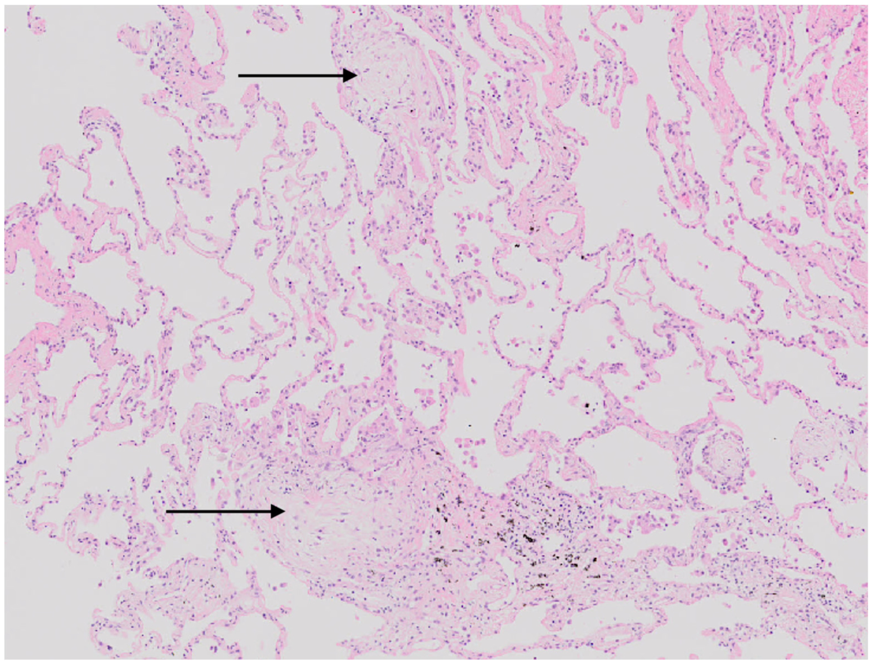
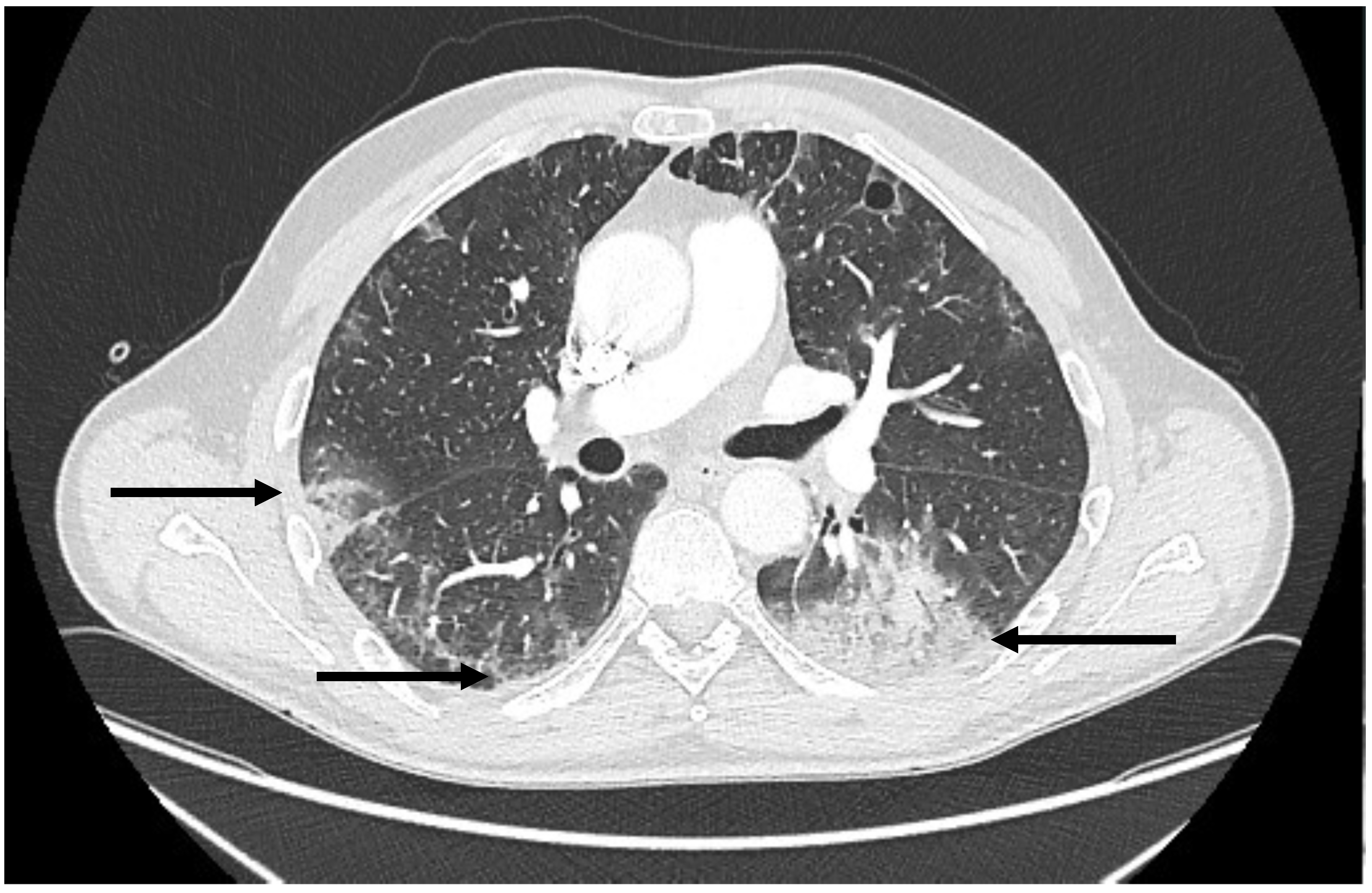
2. Case Report
3. Discussion
4. Conclusions
Author Contributions
Funding
Institutional Review Board Statement
Informed Consent Statement
Data Availability Statement
Conflicts of Interest
References
- Freeman, C.L.; Sehn, L.H. A tale of two antibodies: Obinutuzumab versus rituximab. Br. J. Haematol. 2018, 182, 29–45. [Google Scholar] [CrossRef] [PubMed] [Green Version]
- Stanojevic, S.; Kaminsky, D.A.; Miller, M.R.; Thompson, B.; Aliverti, A.; Barjaktarevic, I.; Cooper, B.G.; Culver, B.; Derom, E.; Hall, G.L.; et al. ERS/ATS technical standard on interpretive strategies for routine lung function tests. Eur. Respir. J. 2022, 60, 2101499. [Google Scholar] [CrossRef] [PubMed]
- Baang, J.H.; Smith, C.; Mirabelli, C.; Valesano, A.L.; Manthei, D.M.; Bachman, M.A.; Wobus, C.E.; Adams, M.; Washer, L.; Martin, E.T.; et al. Prolonged Severe Acute Respiratory Syndrome Coronavirus 2 Replication in an Immunocompromised Patient. J. Infect. Dis. 2021, 223, 23–27. [Google Scholar] [CrossRef] [PubMed]
- Ertesvåg, N.U.; Sakkestad, S.T.; Zhou, F.; Hoff, I.; Kristiansen, T.; Jonassen, T.M.; Follesø, E.; Brokstad, K.A.; Dyrhovden, R.; Mohn, K.G.-I. Persistent Fever and Positive PCR 90 Days Post-SARS-CoV-2 Infection in a Rituximab-Treated Patient: A Case of Late Antiviral Treatment. Viruses 2022, 14, 1757. [Google Scholar] [CrossRef] [PubMed]
- Kos, I.; Balensiefer, B.; Roth, S.; Ahlgrimm, M.; Sester, M.; Schmidt, T.; Thurner, L.; Bewarder, M.; Bals, R.; Lammert, F.; et al. Prolonged Course of COVID-19-Associated Pneumonia in a B-Cell Depleted Patient After Rituximab. Front. Oncol. 2020, 10, 1578. [Google Scholar] [CrossRef] [PubMed]
- Martínez-Barranco, P.; García-Roa, M.; Trelles-Martínez, R.; Arribalzaga, K.; Velasco, M.; Guijarro, C.; Marcos, J.; Campelo, C.; Acedo-Sanz, J.M.; Villalón, L.; et al. Management of Persistent SARS-CoV-2 Infection in Patients with Follicular Lymphoma. Acta Haematol. 2022, 145, 384–393. [Google Scholar] [CrossRef] [PubMed]
- Ueda, Y.; Asakura, S.; Wada, S.; Saito, T.; Yano, T. Prolonged COVID-19 in an Immunocompromised Patient Treated with Obinutuzumab and Bendamustine for Follicular Lymphoma. Intern. Med. 2022, 61, 2523–2526. [Google Scholar] [CrossRef] [PubMed]
- Wee, L.E.; Tan, J.Y.; Ko, K.K.-K.; Wan, W.Y.; Lai, D.C.M.; Oon, L.L.E.; Tan-Garcia, A.; Yeong, J.P.S.; Pena, A.M.T.; Lim, T.K.H.; et al. Detection of viable SARS-CoV-2 in deep respiratory specimens despite negative nasopharyngeal SARS-CoV-2 RT-PCR: Occult COVID-19 as an unsuspected cause of pulmonary infiltrates in immunocompromised patients. IDCases 2022, 30, e01611. [Google Scholar] [CrossRef] [PubMed]
- Yasuda, H.; Mori, Y.; Chiba, A.; Bai, J.; Murayama, G.; Matsushita, Y.; Miyake, S.; Komatsu, N. Resolution of One-Year Persisting COVID-19 Pneumonia and Development of Immune Thrombocytopenia in a Follicular Lymphoma Patient with Preceding Rituximab Maintenance Therapy: A follow-up Report and Literature Review of Cases With Prolonged Infections. Clin. Lymphoma Myeloma Leuk. 2021, 21, e810–e816. [Google Scholar] [CrossRef] [PubMed]
- Burgener, S.; Rochat, P.; Dollenmaier, G.; Benz, G.; Kistler, A.D.; Fulchini, R. Progression of COVID-19 in a Patient on Anti-CD20 Antibody Treatment: Case Report and Literature Review. Case Rep. Infect. Dis. 2022, 2022, 8712424. [Google Scholar] [CrossRef] [PubMed]
- Bigman-Peer, N.; Peer, E.; Pertzov, B.; Kramer, M.; Segal, G.; Eliakim-Raz, N. A Biphasic COVID-19 Clinical Course in Anti-CD20 Treated Patients: Case Series and Review of the Literature. Eur. J. Case Rep. Intern. Med. 2022, 9, 003502. [Google Scholar] [CrossRef] [PubMed]
- Seki, M.; Hashimoto, K.; Kondo, N.; Ohya, Y.; Kotajima, F.; Mitsutake, K. Sequential Treatment by Antiviral Drugs Followed by Immunosuppressive Agents for COVID-19 Patients with Hematological Malignancy. IDR 2022, 15, 7117–7124. [Google Scholar] [CrossRef] [PubMed]
- Shimizu, T.; Shirasaki, H.; Okafuji, K.; Sawazaki, A.; Iwabuchi, T.; Matubayashi, R. A case of prolonged COVID-19 treated with tixagevimab/cilgavimab. Respirol. Case Rep. 2023, 11, e01099. [Google Scholar] [CrossRef] [PubMed]
- Papanikolopoulou, A.; Thimis, V.; Antonogiannaki, E.; Rapti, V.; Tsiakos, K.; Krallis, G.; Ntouraki, S.; Kokkotis, G.; Panagiotou, E.; Sakka, V.; et al. Prolonged SARS-CoV-2 Infection with Common Features in Two Patients Receiving Anti-CD20 Therapy. In Vivo 2023, 37, 461–467. [Google Scholar] [CrossRef] [PubMed]
- Sepulcri, C.; Dentone, C.; Mikulska, M.; Bruzzone, B.; Lai, A.; Fenoglio, D.; Bozzano, F.; Bergna, A.; Parodi, A.; Altosole, T.; et al. The Longest Persistence of Viable SARS-CoV-2 With Recurrence of Viremia and Relapsing Symptomatic COVID-19 in an Immunocompromised Patient-A Case Study. Open Forum Infect. Dis. 2021, 8, ofab217. [Google Scholar] [CrossRef] [PubMed]
- Shafat, T.; Grupel, D.; Porges, T.; Levi, I.; Yagel, Y.; Nesher, L. Treatment with obinutuzumab leads to worse outcomes in haematological patients diagnosed with Omicron variant COVID-19. Br. J. Haematol. 2022, 198, 826–829. [Google Scholar] [CrossRef]
- Furlan, A.; Forner, G.; Cipriani, L.; Vian, E.; Rigoli, R.; Gherlinzoni, F.; Scotton, P. COVID-19 in B Cell-Depleted Patients After Rituximab: A Diagnostic and Therapeutic Challenge. Front. Immunol. 2021, 12, 763412. [Google Scholar] [CrossRef]
- Gaitzsch, E.; Passerini, V.; Khatamzas, E.; Strobl, C.D.; Muenchhoff, M.; Scherer, C.; Osterman, A.; Heide, M.; Reischer, A.; Subklewe, M.; et al. COVID-19 in Patients Receiving CD20-depleting Immunochemotherapy for B-cell Lymphoma. Hemasphere 2021, 5, e603. [Google Scholar] [CrossRef]
- WHO Recommendation. Available online: https://app.magicapp.org/#/guideline/nBkO1E/section/nJB6MR (accessed on 15 December 2022).
- Santenna, C.; Vidyasagar, K.; Amarneni, K.C.; Ghanta, S.N.; Sadasivam, B.; Pathan, S.; Padmavathi, R. The safety, tolerability and mortality reduction efficacy of remdesivir; based on randomized clinical trials, observational and case studies reported safety outcomes: An updated systematic review and meta-analysis. Ther. Adv. Drug Saf. 2021, 12, 20420986211042516. [Google Scholar] [CrossRef] [PubMed]
- Onishi, A.; Matsumura-Kimoto, Y.; Mizutani, S.; Tsukamoto, T.; Fujino, T.; Miyashita, A.; Nishiyama, D.; Shimura, K.; Kaneko, H.; Kawata, E.; et al. Impact of Treatment with Anti-CD20 Monoclonal Antibody on the Production of Neutralizing Antibody Against Anti–SARS-CoV-2 Vaccination in Mature B-Cell Neoplasms. IDR 2023, 16, 509–519. [Google Scholar] [CrossRef]


| Material | Test | Result: |
|---|---|---|
| Sputum | ||
| Microbiological culture | Negative | |
| Mycobacterium tuberculosis complex DNA, acid-fast bacteria (AFB) smears, and cultures | Negative | |
| Pneumocystis jiroveci oocysts and DNA | Negative | |
| NPS | ||
| The genetic material of 21 respiratory pathogens: Adenovirus: Coronavirus: HKU1, 229E, NL63, OC43; Middle East Respiratory Syndrome Coronavirus (MERS-CoV), Metapneumovirus; RSV; influenza type A subtype AH1, AH1 2009, AH3; influenza type B; parainfluenza type 1,2,3,4; Rhinovirus/Enterovirus; Bordetella pertussis; Bordetella parapertussis, Chlamydia pneumoniae, Mycoplasma pneumoniae | Negative | |
| Blood | ||
| Aspergillus fumigatus galactomannan | Negative | |
| Aspergillus fumigatus IgG | Negative | |
| Cytomegalovirus (CMV) antigen pp65 | Negative | |
| Human immunodeficiency virus (HIV) antibodies and antigen p24 | Negative | |
| Microbiological cultures | Negative | |
| Urine | ||
| Microbiological cultures | Negative | |
| Bronchoalveolar lavage fluid | ||
| Microbiological culture | Negative | |
| Mycobacterium tuberculosis complex DNA, AFB smears, and cultures | Negative | |
| Pneumocystis jiroveci oocysts and DNA | Negative |
| Parameter | Result | Z-Score |
|---|---|---|
| FEV1%FVC [%] | 81, 41 | 0.52 |
| FEV1 [L; %] | 3.06; 71 | −2.09 |
| FVC [L; %] | 3.77; 68 | −2.36 |
| TLC [L; %] | 5.98; 77 | −2.58 |
| TL, Coc Single Breath [%] | 60 | −3.21 |
| Hospitalisation | Day of Disease | Test Result | Symptoms | Suspected Diagnosis | Treatment |
|---|---|---|---|---|---|
| I | 50 | NPS antigen and PCR negative | Fever, cough, dyspnoea | Drug-related organizing pneumonia | Glucocorticosteroids |
| II | 94 | NPS antigen negative | Fever | Drug-related organizing pneumonia | Glucocorticosteroids’ dose increased |
| 103 | NPS antigen positive | Fever | SARS-CoV-2 infection | 5-day remdesivir therapy | |
| III | 123 | NPS antigen positive | Fever, dyspnoea | SARS-Cov-2 relapse | Observation |
| 131 | NPS antigen negative | Fever | Organizing pneumonia | Glucocorticosteroids’ dose increased | |
| 143 | NPS antigen positive | Fever | Ongoing SARS-CoV-2 infection and related to it organizing pneumonia | 10-day remdesivir and 1-day casirivimab with imdevimab therapy | |
| IV | 179 | NPS antigen and PCR negative | Fever | Ongoing SARS-CoV-2 infection and related to it organizing pneumonia | 1-day casirivimab with imdevimab therapy and next 10-day remdesivir therapy and next intravenous immunoglobulins |
| 187 | BALF PCR positive | Fever |
Disclaimer/Publisher’s Note: The statements, opinions and data contained in all publications are solely those of the individual author(s) and contributor(s) and not of MDPI and/or the editor(s). MDPI and/or the editor(s) disclaim responsibility for any injury to people or property resulting from any ideas, methods, instructions or products referred to in the content. |
© 2023 by the authors. Licensee MDPI, Basel, Switzerland. This article is an open access article distributed under the terms and conditions of the Creative Commons Attribution (CC BY) license (https://creativecommons.org/licenses/by/4.0/).
Share and Cite
Łyżwa, E.; Sobiecka, M.; Lewandowska, K.; Siemion-Szcześniak, I.; Barańska, I.; Klatt, M.; Langfort, R.; Szturmowicz, M.; Tomkowski, W. Prolonged SARS-CoV-2 Infection and Organizing Pneumonia in a Patient with Follicular Lymphoma, Treated with Obinutuzumab—Challenging Recognition and Treatment. Viruses 2023, 15, 693. https://doi.org/10.3390/v15030693
Łyżwa E, Sobiecka M, Lewandowska K, Siemion-Szcześniak I, Barańska I, Klatt M, Langfort R, Szturmowicz M, Tomkowski W. Prolonged SARS-CoV-2 Infection and Organizing Pneumonia in a Patient with Follicular Lymphoma, Treated with Obinutuzumab—Challenging Recognition and Treatment. Viruses. 2023; 15(3):693. https://doi.org/10.3390/v15030693
Chicago/Turabian StyleŁyżwa, E., M. Sobiecka, K. Lewandowska, I. Siemion-Szcześniak, I. Barańska, M. Klatt, R. Langfort, M. Szturmowicz, and W. Tomkowski. 2023. "Prolonged SARS-CoV-2 Infection and Organizing Pneumonia in a Patient with Follicular Lymphoma, Treated with Obinutuzumab—Challenging Recognition and Treatment" Viruses 15, no. 3: 693. https://doi.org/10.3390/v15030693