Article

Potential and Constraints on In Vitro Micropropagation of Juniperus drupacea Labill.

by
Kostas Ioannidis
1,*,
Ioanna Tomprou
1,
Danae Panayiotopoulou
2,
Stefanos Boutsios
3 and
Evangelia N. Daskalakou
3
1
Laboratory of Sylviculture, Forest Genetics and Biotechnology, Institute of Mediterranean and Forest Ecosystems, Hellenic Agricultural Organization “DIMITRA”, Ilissia, 11528 Athens, Greece
2
Library and Documentation Center, Institute of Mediterranean & Forest Ecosystems, Hellenic Agricultural Organization “DIMITRA”, Ilissia, 11528 Athens, Greece
3
Laboratory Forest Management & Forest Economics, Institute of Mediterranean & Forest Ecosystems, Hellenic Agricultural Organization “DIMITRA”, Ilissia, 11528 Athens, Greece
*
Author to whom correspondence should be addressed.
Forests 2023, 14(1), 142; https://doi.org/10.3390/f14010142
Submission received: 12 December 2022 / Revised: 30 December 2022 / Accepted: 4 January 2023 / Published: 12 January 2023
(This article belongs to the Special Issue Application of Plant Biotechnology in Forestry)

Abstract

:
Juniperus drupacea Labill. (Cupressaceae) is a species with ecological and medicinal value. In Europe, it is native only in southern Greece, and is listed as endangered. Due to its uniqueness, this study attempted, for the first time, an in vitro propagation effort of Syrian juniper. Explants of the lateral shoot tips were surface-sterilized and cultured on Murashige and Skoog (MS) medium. The cultures were subcultured on MS, woody plant medium (WPM), and Driver and Kuniyaki Walnut (DKW) supplemented with different concentrations of 6-benzylaminopurine (BA), thidiazuron (TDZ), or meta-topolin [6-(3-hy-droxybenzylamino)purine] for shoot induction. Explants derived from female trees exhibited 54.17% bud proliferation on DKW medium with 4 μM meta-topolin or 4 μM TDZ and on WPM with 4 μM meta-topolin or 4 μM BA. A total of 62.50% of the male tree derived explants produced multiple shoots on DKW with 4 μM BA. The maximum average number of shoots per explant were 1.17 per explant in both cases. The length of the shoot derived from explants of female origin was 2.94 mm compared to 2.69 mm of the in vitro shoots from the explants of male trees. Overall, the best medium and plant growth regulator combination for the explants derived from both female and male trees, for the traits under study, was proven to be DKW + 4 µM TDZ. Our experiments show that Juniperus drupacea, under in vitro conditions, shows recalcitrance in rooting, as the applications of IBA, NAA, and IAA concentrations were proven to be ineffective treatments. Although the results show low values, this avant-garde study provides a foundation for further research on the in vitro regeneration of Juniperus drupacea.

1. Introduction

Juniperus drupacea Labill., commonly known as Syrian juniper, belongs to the Cupressaceae family. It is a dioecious tree, 10–20 m in height, forming a conical crown. The needles are acicular, up to 25 mm long and 4 mm wide, with two white bands on the top, arranged in alternate whorls. The cones of Syrian juniper, being the largest among juniper species, are ovoid to globose, 20–25 mm in diameter, brownish, glaucosus, and pruniose in maturity, and have three seeds in a characteristic drupe-like strobile [1,2,3]. It is considered as a relict species with a disjunct geographical range. The location of its divergence and evolution remains unknown [3]. Rare fossil data only include remnants known from Miocene and Pliocene deposits in Europe [3,4,5,6,7] and concern the Juniperus spp. in general.
Currently, the distribution range of the species extends mainly to SE Turkey, western Syria, Israel, and Lebanon . In Europe, its natural populations are restricted only to Greece, in the SE part of the Peloponnese Peninsula [8,9,10,11,12]. Specifically, more than 95% of J. drupacea populations are found on Mt. Parnon [9] and a few have been recorded in a limited area of Mt Taygetos in small patches [10,11,12]. Syrian juniper can be used to enhance the biodiversity in oak and cedar forest restoration [13] and protect the soil from erosion [14]. It is considered as a very interesting ornamental tree because of its columnar shape, good growth rate, and resistance to frost [15].
In Greece, J. drupacea, due to its decay-resistant timber, used to be exploited for carpentry as well as fuel. However, its endangered status has prevented any extensive use today as they are included in natural habitat types of community interest whose conservation requires the designation of special areas of conservation [16]. According to the International Union for the Conservation of Nature (IUCN) [17], J. drupacea is considered worldwide as a species of Least Concern (LC). However, in Europe, it is listed as Endangered (EN) [18] under the criteria B1ab(iii) + 2ab(iii) [19]. In Greece, its ecological value has been acknowledged since 1980 when Juniperus drupacea forests has were declared as a “Natural Monument under Preservation”, according to FEK 121D/1980 [20]. In 1992, it was included in Annex I of Directive 92/43/EEC as a priority habitat type and the Mt Parnon summit and Malevi Monastery were included in Natura 2000 as Special Protected Areas (code: GR 2520006) [16].
Natural reproduction occurs through seeds and it has been proven to be a very slow procedure, especially due to its seed’s deep dormancy [21]. Juniper species have sexual reproductive capacity but their seed number varies. They mainly not only have low seed production, but also present low germination percentage, physiological dormancy, and lessened embryos viability [13,22,23]. In particular, Syrian juniper seeds present germination morphophysiological barriers and can delay natural germination for 4–5 years [24]. On the other hand, the cone flesh alone can postpone it for 1 to 2 years [25].
Several attempts have been made to in vitro regenerate Juniperus as the species shows a general recalcitrance in natural regeneration. Micropropagation by axillary shoots, among others, is considered as an effective method for the accomplishment of one of the aims of vegetative propagation, that is, the mass production of plants. Although it has shown positive results in many forest species, it presents more difficulties in conifers, particularly in the genus Juniperus, being challenging and demanding as well as arduous [14,26,27,28,29,30,31]. There are physical and chemical factors that stimulate the different conifer species to develop shoots and adventitious roots with the progress not always being triumphant. According to Ragonezi et al. [32], these factors include plant growth regulators, carbohydrates, light quality, temperature, and the rooting medium.
Gomez and Segura [33] first reported the successful application of this method for Juniperus oxycedrus L., which resulted in the induction of shoots but with very limited rooting frequency. Rooting of the in vitro regenerated microshoots is a very laborious, crawling and inefficient process in conifers [14,28,32,34]. The efficiency of shoot induction is varied among Juniperus species and depends on the types of explants [28,35]. The type of medium as well as the type of cytokinin and auxin and their concentrations were revealed to play determined roles in blastogenesis and rhizogenesis in Juniperus oxycedrus [33,36], Juniperus phoenicea [27,37], Juniperus navicularis [35], Juniperus excelsa M. Bieb, Juniperus horizontalis Moench and Juniperus chinensis L. [38], Juniperus excelsa [39], Juniperus polycarpos L. [40], and Juniperus thulifera L. [28]. Some studies have shown that the proliferation response of some juniper species increased during subsequent subcultures [27,35,38] due to overcoming the first shock after the first establishment in in vitro conditions [27].
Rizhogenesis is influenced by factors such as donor plant age and health, shoot vigor and juvenility genotype, and type of explant, auxin treatment, and environmental conditions on rooting, decreasing the mineral, sucrose, and agar concentrations in the medium [41,42,43,44,45]. In junipers, a very high rooting rate has only been reported in Juniperus oxycedrus L. and Juniperus cedrus Webb & Berthel. [26,34]. Varying rooting rates were observed in Juniperus excels M.Bieb., Juniperus horizontalis Moench and Juniperus chinensis L. [38], Juniperus navicularis Gand. [35], Juniperus thulifera L. [28], Juniperus oxycedrus L. [36], while the rooting of Juniperus polycarpos K. Koch [40] were not satisfactory. In Juniperus phoenicea L., the results were contradictory as some studies reported small to moderate rooting [27,37] and some failed in rhizogenesis [46].
In this context, the aim of the study was to investigate, for the first time, whether in vitro micropropagation would overcome the in vitro regeneration recalcitrance of Juniperus drupacea. The scientific team investigated the in vitro culture establishment, shoot proliferation as well as the potential for rooting. We incorporated three types of media and three types of plant growth regulators in several concentrations in our experiments in order to achieve blastogenesis and rhizogenesis. Although there were promising results on shooting, they lacked rooting, which, like other juniper species, was not feasible.

2. Materials and Methods

2.1. Plant Material—Explants Sterilization—Culture Establishment

Healthy mature Juniperus drupacea male and female individuals growing on Mt. Parnon were selected as explant source trees. Eight trees per sex from three different areas were selected as explant donors. The age of the selected trees ranged from 30 to 50 years. The lateral shoots of the actively growing stems of the source trees were the explant donors. These were collected during April to May, placed in damp cotton cloth, stored at 4 °C, and transferred to the laboratory until subsequent manipulations. The following day, the explants (i.e., nodal segments and apical shoot tips of 1.5–2.5 cm long) were excised from the explant donors collected during the vegetative growth stage. The explants were distinguished as female and male in relation to their tree gender.
The explant surface was successfully disinfected by successive immersions in two different aqueous solutions: the first was a solution of 70% ethanol with continuous stirring for 1 min, and the second was sodium hypochlorite (10% NaOCl, Fluka, Germany) at a concentration of 1.0% (v/v), complemented with 0.05% (v/v) Tween-20 (Fisher Bioreagents, USA) with continuous stirring for 15 min. After immersion, the explants were rinsed three times with sterile deionized water for three minutes each.
Each explant was placed in a 25 mm × 150 mm culture tube containing 20 mL of the nutrient medium. Three media were used to establish the in vitro culture: the MS of Murashige and Skoog [47] (Duchefa Biochemie, Haarlem, The Netherlands), the wood plant medium (WPM) of Lloyd and McCown [48] (Duchefa Biochemie, Haarlem, The Netherlands), and the Driver and Kuniyaki Walnut (DKW) of Driver and Kuniyuki [49] (Duchefa Biochemie, Haarlem, The Netherlands). Each medium contained 3% (w/v) sucrose (Duchefa Biochemie, Haarlem, The Netherlands) solidified with 6 g L−1 agar (Duchefa Biochemie, Haarlem, The Netherlands) and their pH was adjusted to 5.8 before agar addition and autoclaving at 121 °C and 122 kPa for 20 min. All cultures were incubated in a growth chamber at 23 ± 1 °C with a 16 h light/8 h dark photoperiod at a 50 μmol m−2 s−1 photosynthetic photon flux density (culture level) provided by cool-white fluorescent lamps.

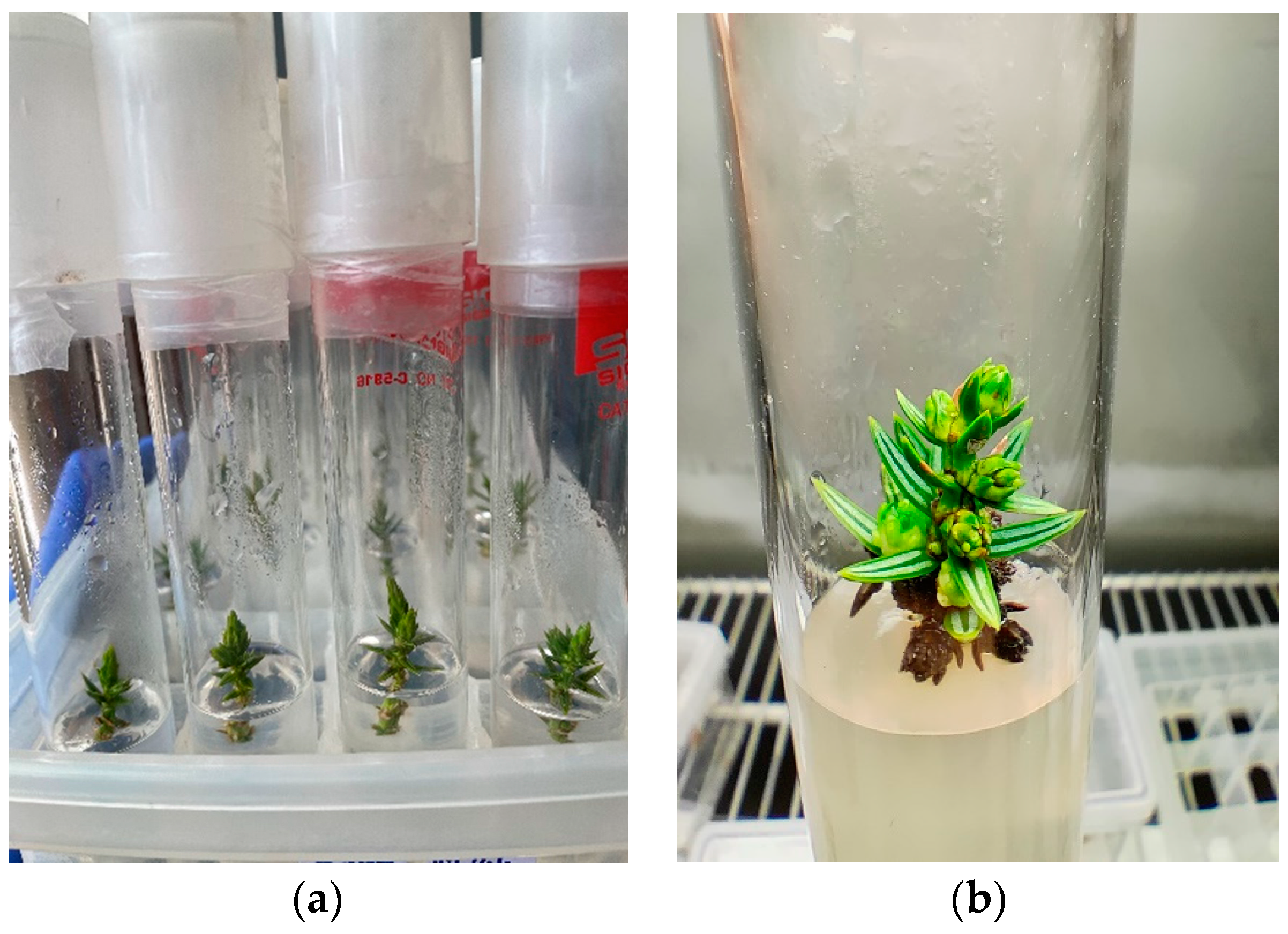
2.2. Shoot Regeneration, Multiplication, and Elongation

After 10 days, the healthy non-contaminated explants were subcultured in full-strength mediums (i.e., MS, WPM and DKW) containing 6-benzylaminopurine (BA) (Sigma Chemicals, St. Louis, MO, USA) or thidiazuron (TDZ) (Cayman Chemicals, Ann Arbor, MI, USA) or meta-topolin [6-(3-hydroxybenzylamino)purine] (Duchefa Biochemie, Haarlem, The Netherlands) at various concentrations (1.0, 2.0, 4.0 and 8.0 µM) for multiple shoot induction. Each medium contained 3% (w/v) sucrose and was solidified with 6 g L−1 agar and their pH adjusted at 5.8 before agar addition and autoclaving at 121 °C and 122 kPa for 20 min. The treatments used in the shoot regeneration experiments are shown in Table 1. The plantlets established in in vitro conditions were transferred every 2–3 weeks to new nutrient media of the same composition. After an 8-week period (three subcultures), the effect of the various concentrations that the plant growth regulators had on the average shoot formation percentage (%), average shoot number, and length per explant were evaluated. Every treatment (i.e., medium—plant growth regulator combination) included three replicates. Each replication constituted eight tubes with one explant per tube. In total, 1872 explants were incorporated in our shoot regeneration, multiplication, and elongation experiments, not counting all the explants used in the establishment of cultures. The cultures of each experiment was arranged in a completely randomized design in a growth chamber at 23 ± 1 °C with a 16 h light/8 h dark photoperiod at a 50 μmol m−2 s−1 photosynthetic photon flux density (culture level) provided by cool-white fluorescent lamps.

2.3. In Vitro Rooting of Shoots

Shoots of 2.0–2.5 cm long, derived from the shoot regeneration step, were transplanted on culture tubes containing full-strength of the same media as in the previous stage, supplemented with several auxins for rooting. Explants from each treatment of the shoot regeneration, multiplication, and elongation stage were transplanted to each rooting treatment. In order to satisfy the required number of eight explants per replicate and per treatment, where necessary, we used explants from the establishment cultures stage. Plant growth regulators were ΙΒA (indole-3-butyric acid) (Sigma Chemicals, Saint Louis, MO, USA) at concentrations of 1.0, 2.0, 4.0, 8.0, 16.0, and 32.0 μΜ; ΝAA (α-naphthalene acetic acid) (Sigma Chemicals, Saint Louis, MO, USA) at concentrations of 1.0, 2.0, 4.0, and 8.0 μΜ; and ΙAA (3-indoleacetic acid) (Sigma Chemicals, Saint Louis, MO, USA) at concentrations of 0.5, 1.0, 2.0, and 4.0 μΜ. All treatments used in the rooting experiments are shown in Table 2. The nutrient media were solidified with 7 g L−1 agar, and supplemented with 3% (w/v) sucrose. The conditions of the cultures were the same as above-mentioned. After a 4-week period, the effect of the concentrations of the plant growth regulators on the rooting percentage (%), root number, and length per shoot were evaluated. The experimental design was the same as that above-mentioned.
All cultures were incubated in a growth chamber at 23 ± 1 °C with a 16 h light/8 h dark photoperiod at a 50 μmol m−2 s−1 photosynthetic photon flux density (culture level) provided by cool-white fluorescent lamps.

2.4. Statistical Analysis

Analysis was based on individual values of the average shooting percentage, the mean number, the mean length of shoots per explant, the proportion of rooted microcuttings, the number, and the mean length of roots per explant. The following linear model was used in the analysis to specify the impact of the gender, the plant growth regulator treatment, and the interaction between the gender and the plant growth regulator treatment:
yijkl = μ + gj + mi + tk + gj*mi + gj*tk eijkl
where yijkm is the measurement for a trait of the lth explant, the kth plant growth regulator treatment, the ith nutrient medium and the jth explant gender, as dependent variables; μ is the fixed population mean of all explants; gj is the fixed effect of the jth gender; mi is the random effect of the ith nutrient medium; tk is the random effect of the kth plant growth regulator treatment; gj*tk is the interaction of the jth gender with the kth plant growth regulator treatment; gj*mi is the interaction of the jth gender with the ith nutrient medium; and eijkl is the random residual error of the lth explant, the kth plant growth regulator treatment, the ith nutrient medium, and the jth gender. The restricted maximum likelihood (REML) method was used to estimate the variance components. Moreover, a binomial logistic regression was performed to predict the probability of the explant to shoot, which was considered as the dichotomous dependent variable by using the type of the medium (categorical), type of the plant growth regulator (categorical) and its concentration (ordinal), and gender (categorical) as explanatory variables (covariates). Descriptive statistics, analysis of variance (ANOVA) as well as the Duncan’s multiple range test (MRT) based on the 0.05 level of significance were performed on the number and average shoot length per explant and the shooting proportion per treatment. Data in percentages were subjected to appropriate transformation in order to statistically analyze and were transformed back to percentages for presentation in the tables and graphs. All statistical analyses were performed using SPSS v.20 software for Windows (IBM SPSS Statistics 2011, IBM Corp., Armonk, NY, USA).

3. Results

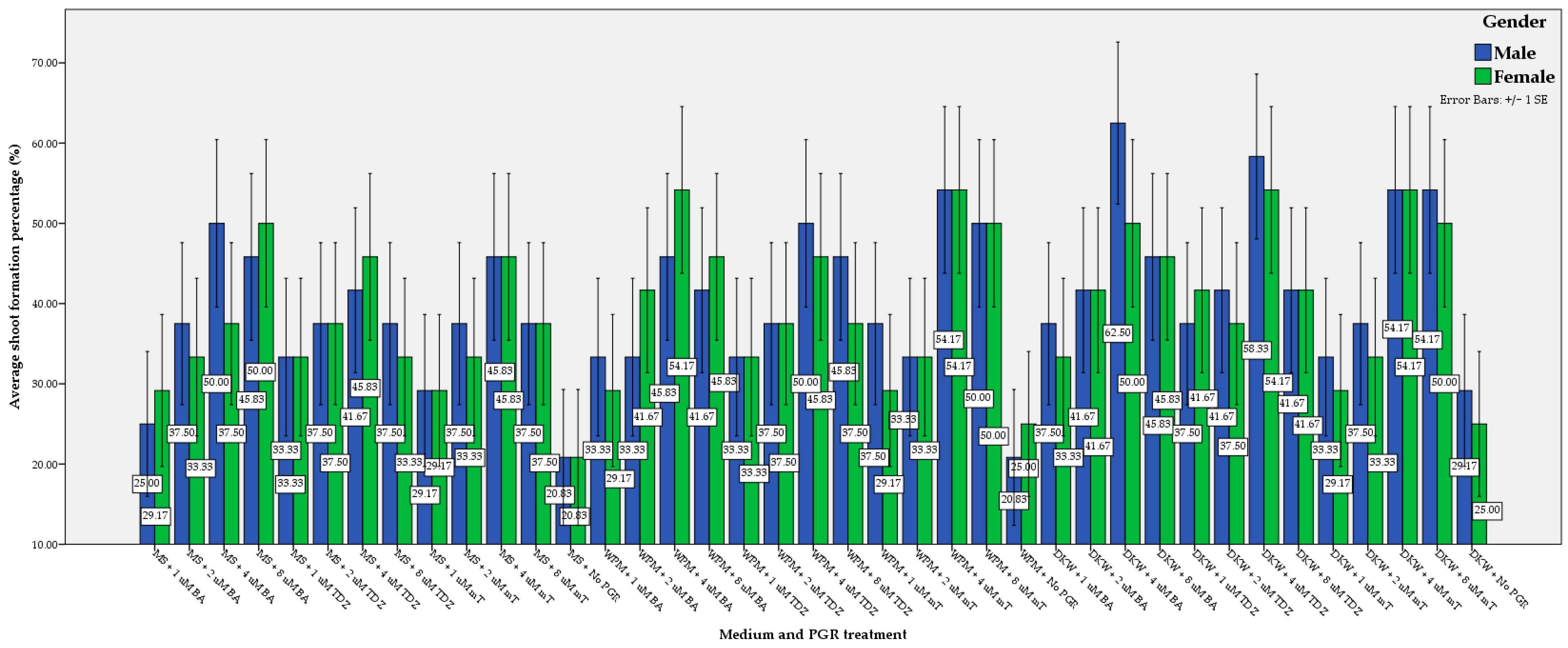
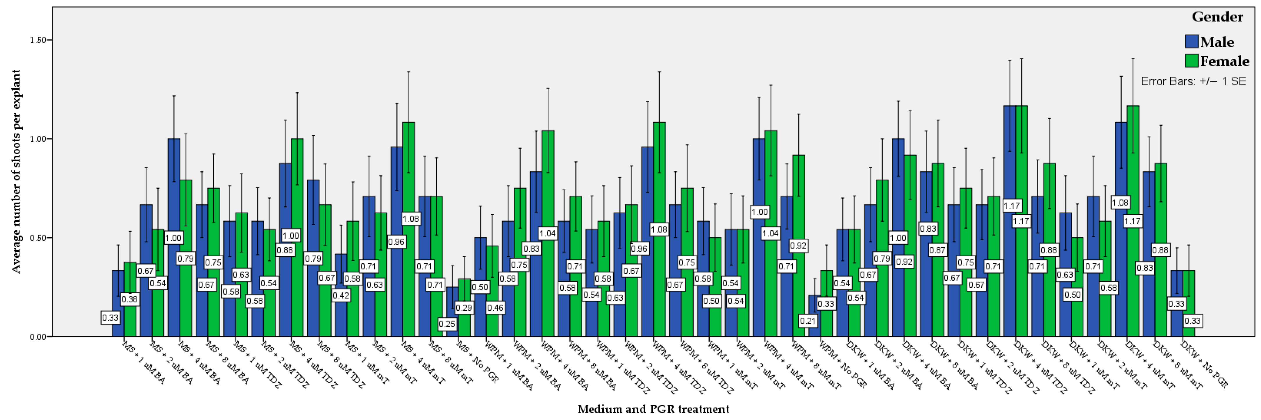
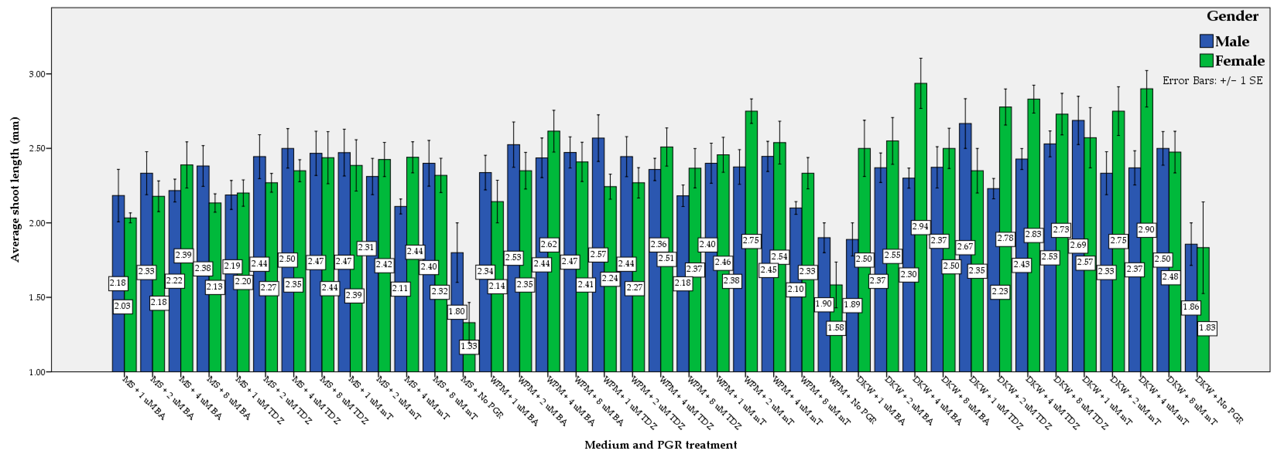
3.1. Shoot Regeneration, Multiplication, and Elongation

Shoot formation was affected by the kind and concentration of plant growth regulators and nutrient medium but not the tree gender. Throughout the experiment, no significant interactions between gender and nutrient medium and between gender and plant growth regulator treatment was observed regarding the average shoot formation percentage, the average shoot number per explant, and the average shoot length per explant. The impact of different media and plant growth regulators and their concentration in the treatments on the average number of shoots per explant, the average shoot length, and the frequency of shoot formation of female and male Juniperus drupacea explants are presented in Table 3, Supplementary Material Tables S1–S3, and Figure 1, Figure 2 and Figure 3.
Shoot induction was achieved after three weeks of culture, depending on the treatment applied. The type of nutrient medium significantly affected the average percentage of blastogenesis, the average number of shoots, and the average shoot length of the Juniperus drupacea explants (Table 3). Explants growing in DKW medium presented the best values compared to the relevant ones in WPM and MS, respectively. In contrast, MS presented the lowest values that differed statistically compared to DKW and WPM, which did not show statistically significant differences (p ≤ 0.05). Within each gender and among the different treatments, statistically significant differences (p ≤ 0.05) were observed in the mean percentage of blastogenesis (Supplementary Material Table S1), the mean number of shoots per explant (Supplementary Material Table S2), and the mean length of shoots (Supplementary Material Table S3). Τhe binomial logistic regression model adequately fit the data as the logistic regression model was statistically significant (χ2(4) = 30.005, p ≤ 0.000) and the Hosmer and Lemeshow test resulted in χ2 = 9.211 (p ≤ 0.238). The model correctly classified 59.9% of cases. The explained variation in the dependent variable based on our model ranged from 16.6 to 22.2%, depending on whether we referenced the Cox and Snell R2 or Nagelkerke R2 methods, respectively. From the analysis, we could see that the medium (p ≤ 0.030) and plant growth regulator (p ≤ 0.000) significantly enhanced the model/prediction. In contrast, gender did not contribute statistically significantly to the model.
Regarding the female explants, the maximum percentage of shooting (54.17%) was achieved in the DKW and WPM media when supplemented with 4 μM mT or 4 μM TDZ in the former and with 4 μM mT or 4 μM BA in the latter. The media with no growth regulators showed the lowest values of the percentage of blastogenesis (20.83%). Regarding the male explants, the maximum percentage of blastogenesis (62.50%) was attained in the DKW medium when it was supplemented with 4 µM BA. The media without the addition of growth regulators showed the lowest values in shooting percentage (20.83%).
The maximum average shoot number in female explants was achieved by supplementing the DKW medium with 4 µM mT or 4 µM TDZ (1.17). Similarly, regarding male explants, the maximum average shoot number was achieved using the DKW nutrient medium combined with 4 µM TDZ (1.17). The DKW, WPM, and MS media with no growth regulators presented the lowest values in the mean number of shoots (i.e., 0.33, 0.33, and 0.29, respectively) regarding the female explants. Likewise, in relation to the male explants, DKW, WPM, and MS media containing no growth regulators exhibited the lowest values in the mean number of shoots (i.e., 0.33, 0.25, and 0.21, respectively). Moreover, the treatment with MS medium supplemented with 1 µM BA also showed a very low value (0.33).
Maximum average shoot length per explant in the female explants was achieved when DKW medium was supplemented with 4 µM BA (2.94 mm) or 4 µM mT (2.90 mm). The difference between these treatments was not significant. The results were similar in the male explants, where the maximum values in average shoot length were obtained when the DKW nutrient medium was supplemented with 1 µM mT (2.69) or 1 µM TDZ (2.67). The difference between these treatments was also not significant. The DKW, WPM, and MS media with no growth regulators presented the lowest values in the average shoot length per female explant (i.e., 1.83, 1.58, and 1.33, respectively). Additionally, in relation to the male explants, all types of DKW, WPM, and MS media containing no growth regulators exhibited the lowest values in the average shoot length per explant (i.e., 1.90, 1.86, and 1.80, respectively). Moreover, the treatment of the DKW medium supplemented with 1 µM BA also showed a very low value (1.89).
In every treatment, many explants browned and showed necrosis, which eventually led to the death of many explants (Figure 4).

3.2. In Vitro Explant Rooting

Root initiation of the in vitro cultured explants was inspected immediately after they were subcultured on the rooting treatment media supplemented with auxins. At this rooting stage, all of the plant growth regulators were tested in terms of their concentrations. More specifically, ΙΒA was tested at concentrations of 1.0, 2.0, 4.0, 8.0, 16.0, and 32.0 μΜ, ΝAA at 1.0, 2.0, 4.0, and 8.0 μΜ, and finally, ΙAA at 0.5, 1.0, 2.0, and 4.0 μΜ, respectively. However, all treatments were insufficient to induce rooting. The effectiveness of the nutrient media alone, containing no auxin, was also inadequate to stimulate root formation.

4. Discussion

The shooting percentage of all cultures was moderate. A small number of new shoots was developed showing insignificant elongation. In each medium, many explants browned and showed apical and lateral necrosis, which eventually led to death. Explant discoloration and necrosis is common among Juniperus species. Salih et al. [50], after a three-week culturing period of Juniperus procera Hoechst. Ex Endl. in several media, observed the yellowing of explants, which led to their necrosis two weeks later. Khater and Benbouza [28], during in vitro culture of Juniperus thurifera L., reported that the explants’ color changed from green to yellow, and finally to brown, and necrosis reached approximately 90%. The authors attributed the necrosis to an inappropriate medium and PGR combinations. The same phenomenon of explant discoloration as well as high percentages of necrosis was observed by Momeni et al. [40] and Castro et al. [35] in the in vitro culture of Juniperus polycarpos L. and Juniperus navicularis Gand., respectively. Al-Ramamneh et al. [37] stated that microcuttings of Juniperus phoenicea L. failed to show any morphogenic response, browned, and showed progressively necrotic areas by the end of the culture. Loureiro et al. [27], in the in vitro culture of Juniperus phoenicea L., attributed the browning and the necrotic zones to the inappropriate selection of the nutrient medium they used.
In vitro regeneration depends on the composition and concentration of basal salts, growth regulators, and organic components [51]. In particular, the nitrogen content in the medium seems to influence the shoot formation of the explants [40]. Our best results concerning blastogenesis were achieved in the DKW medium, which had an intermediate concentration of nitrogen compared to the other two media used. Compared to MS, WPM also presented better results, as it was the medium with the lowest nitrogen availability. Our findings were consistent with those of Loureiro et al. [27], who reported that Juniperus phoenicea L. explants growing in DKW presented significantly better results than those growing in WPM or MS. Several studies in Juniperus thurifera L. [28], Juniperus procera Hoechst. Ex Endl. [50], Juniperus polycarpos K.Koch [52], and Juniperus phoenicea L. [37,53] have shown that the best results concerning blastogenesis were produced in media with a lower nitrogen content. The type of nutrient medium also significantly influenced the mean number of shoots formed in Juniperus oxycedrus L. [33,36].
In contrast, Bertsouklis et al. [46] faced difficulties using the poor in nitrogen MS medium in the in vitro propagation of Juniperus phoenicea. The percentage of shoot formation in MS was moderate, while the lowest values were achieved using the poorer WPM. Our results concerning shooting percentage in MS were slightly lower, while those for WPM appeared approximately twice as high in comparison. On the other hand, Bertsouklis et al. [46] reported that the use of DKW increased shoot formation, findings similar to ours. The results of Castro et al. [35] showed that WPM was less appropriate for culture. According to our study, similar results were obtained using the MS medium.
The addition of several plant growth regulators significantly affected the Juniperus drupacea explant shooting response. In our study, the addition of BA, mT, and TDZ increased the average shooting percentage, shoot number per explant, and the average shoot length. Explants of Juniperus phoenicea L. also showed the best response while using the medium supplemented with TDZ [37]. The promoting effect of TDZ was also stressed in our study and our results were in accordance with part of Al-Ramamneh et al.’s findings [53] regarding Juniperus phoenicea L. According to Salih et al. [50], the highest average shoot number and the longest average shoot length were obtained in WPM containing IAA, BAP, or IBA. Bertsouklis et al. [46] reported that the use of 2iP induced higher shooting responses in J. phoenicea while the addition of ZEA, NAA, or ΝAA in combination with ΒA presented moderate results. Khater and Benbouza [28] documented the stimulating effects of 2,4-D and BAP, alone or in combination with IBA or IAA, in the shoot development of Juniperus thurifera L. explants. In the in vitro culture of Juniperus polycarpos L., media supplementation with KIN and BA also had a significant effect [40]. Several studies on Juniperus excelsa M.Bieb., Juniperus horizontalis Moench. and Juniperus chinensis Roxb. [27], Juniperus navicularis Gand. [35], and Juniperus oxycedrus L. [33,36] have reported the shoot inducing effect of BA, NAA, and KIN.
Rooting is the primary bottleneck in the in vitro culture of most conifer species [32,35,45,54,55,56], thus restraining the establishment of commercial protocols [28]. Additionally, previous studies have documented the difficulty the Juniperus species has toward inducing adventitious roots under in vitro conditions [27,28,33,40,46,50,56,57]. In coniferous species, auxins such as NAA, IBA, and IAA are the most commonly used to achieve successful rooting under in vitro conditions. Our inability to achieve rooting is consistent with the results of several studies. Al-Ramamneh et al. [37] also did not report successful rooting in the in vitro culture. Loureiro et al. [27] reported that many treatments with IBA failed to stimulate rooting in Juniperus phoenicea L. explants. IBA, alone or in combination with NAA, was insufficient to induce rooting in Juniperus thurifera L. [28], Juniperus polycarpos K.Koch [40], and Juniperus phoenicea L. [46]. Negussie [56] found that treatment with IBA and NAA failed to stimulate shoot rooting, results that were in accordance with ours. According to Castro et al. [35], in vitro rooting experiments using NAA were also unsuccessful. Rooting of Juniperus polycarpos K.Koch was also not satisfactory [40]. Salih et al. [50] did not even try to root the in vitro cultured shoots of Juniperus procera Hoechst. Ex Endl., reporting that more research should be conducted on this species’ root regeneration.
Various explanations have been expressed concerning the recalcitrance of in vitro culture, and particularly, the rooting difficulty of Juniperus species. Micropropagation of Juniperus species depends on the specific species/ecotype [27], the age of the trees used as explant donors [27], the genotype [33,57], or even too low levels of internal auxin [58].
Further research is needed regarding blastogenesis and rooting ability of the in vitro cultures of Juniperus drupacea L. Different nutrient media, at several strengths as well as other plant growth regulators or infection with Agrobacterium rhizogenes, must be applied in order to achieve rooting and better shoot induction and elongation. The decrease in macronutrients in culture media has been found to stimulate rooting in many plants [55,59,60,61,62]; indeed, decreased concentrations of nutrients in the medium, in particular, lessening the nitrogen, seem to promote adventitious rooting [27,32,63,64]. Complementary or substitute regeneration methods could be in vitro techniques of somatic embryogenesis, which have been implemented with greater or less success in Pinus spp. [65], Juniperus communis [66], Picea abies [67,68], and Pinus nigra and Abies hybrids [69]. Moreover, ex vitro rooting systems, an alternative rooting methodology, can be used to induce rooting. Such systems are commonly used in conifers as they present positive effects in developing roots [34] compared to in vitro systems [26].

5. Conclusions

This was the first attempt to study the in vitro propagation of Juniperus drupacea L., found in Greece, where shoot induction has been achieved. The results showed that DKW was the most suitable medium regardless of any plant growth regulators used. The addition of BA, mT, and TDZ promoted the average shooting percentage, the number of shoots per explant, and the average length of shoots. However, the results of this pioneering study did not provide a protocol for the root development of in vitro cultured Juniperus drupacea L. explants, even though different auxins (i.e., IAA, IBA, and NAA) in different concentrations were used. Nevertheless, it provides a basis for further research where all alternatives of in vitro or ex vitro rooting should be examined and thoroughly analyzed, in order to successfully induce adventitious roots of in vitro cultured shoots of Juniperus drupacea L.

Supplementary Materials

The following supporting information can be downloaded at: https://www.mdpi.com/article/10.3390/f14010142/s1, Table S1: Effect of the medium and plant growth regulator types and their concentrations on the average percentage of blastogenesis (%) of Juniperus drupacea explants in relation to their gender (means followed by the same letter did not differ statistically at p ≤ 0.05, according to the Duncan test); Table S2: The effect of the medium and plant growth regulator types and their concentrations on the average number of shoots per Juniperus drupacea explants in relation to their gender (means followed by the same letter did not differ statistically at p ≤ 0.05, according to the Duncan test); Table S3: Effect of the medium and plant growth regulator types and their concentrations on the average shoot length per Juniperus drupacea explants in relation to their gender (means followed by the same letter did not differ statistically at p ≤ 0.05, according to the Duncan test).

Author Contributions

Conceptualization, K.I. and E.N.D.; Methodology, K.I.; Validation, K.I.; Formal analysis, K.I..; Investigation, K.I., D.P. and S.B.; Resources, K.I. and E.N.D.; Data curation, K.I. and I.T. Writing—original draft preparation, K.I. and D.P.; Writing—review and editing, K.I. and D.P.; Visualization, K.I. and E.N.D.; Supervision, K.I.; Project administration E.N.D.; Funding acquisition, E.N.D. All authors have read and agreed to the published version of the manuscript.

Funding

This research was co-financed by the European Regional Development Fund of the European Union and Greek National Funds (2020-2023) through the Operational Program Competitiveness, Entrepreneurship and Innovation, under the call RESEARCH—CREATE—INNOVATE (project code: T2EDK-03378).

Data Availability Statement

Not applicable.

Conflicts of Interest

The authors declare no conflict of interest.

References

  1. Vidakovic, M. Conifers. Morphology and Variation.; Grafičko Zavod Hrvatske: Zagreb, Yugoslavia, 1991; ISBN 0-85198-807-5. [Google Scholar]
  2. Adams, R.P. Junipers of the World: The Genus Juniperus; Trafford Publishing: Bloomington, IN, USA, 2014; ISBN 1-4907-2325-0. [Google Scholar]
  3. Sobierajska, K.; Boratyńska, K.; Jasińska, A.; Dering, M.; Ok, T.; Douaihy, B.; Bou Dagher-Kharrat, M.; Romo, Á.; Boratyński, A. Effect of the Aegean Sea Barrier between Europe and Asia on Differentiation in Juniperus drupacea (Cupressaceae). Bot. J. Linn. Soc. 2016, 180, 365–385. [Google Scholar] [CrossRef] [Green Version]
  4. Coode, M.; Cullen, J. Juniperus L. Flora of Turkey and the East Aegean Islands; Edinburgh University Press: Edinburgh, UK, 1965; Volume 1, pp. 78–84. [Google Scholar]
  5. Axelrod, D.I. Evolution and Biogeography of Madrean-Tethyan Sclerophyll Vegetation. Ann. Mo. Bot. Gard. 1975, 62, 280–334. [Google Scholar] [CrossRef]
  6. Kvaček, Z. A New Juniper from the Palaeogene of Central Europe. Feddes Repert. Z. Bot. Taxon. Geobot. 2002, 113, 492–502. [Google Scholar] [CrossRef]
  7. Palamarev, E. Paleobotanical Evidences of the Tertiary History and Origin of the Mediterranean Sclerophyll Dendroflora. Plant Syst. Evol. 1989, 162, 93–107. [Google Scholar] [CrossRef]
  8. Boratyński, A.; Browicz, K.; Zieliński, J. Chorology of Trees and Shrubs in Greece; Institute of Dendrology, Polish Academy of Sciences: Kornik, Poland, 1992; ISBN 83-85599-04-5. [Google Scholar]
  9. Constantinidis, T.A.; Kalpoutzakis, E. Plant Guide to Mount Parnon and Moustos Wetland Protected Area: Endemic, Rare and Threatened Species; Management Body Mount Parnon: Moustos Wetland, Greece, 2015; ISBN 960-99424-4-X. [Google Scholar]
  10. Tan, K.; Iatrou, G.; Johsen, B. Endemic Plants of Greece: The Peloponnese; Gads Forlag: Copenhagen, Denmark, 2001; Volume 1. [Google Scholar]
  11. Bergmeier, E. Plant Communities and Habitat Differentiation in the Mediterranean Coniferous Woodlands of Mt. Parnon (Greece). Folia Geobot. 2002, 37, 309–331. [Google Scholar] [CrossRef]
  12. Dimopoulos, P.; Raus, T.; Bergmeier, E.; Constantinidis, T.; Iatrou, G.; Kokkini, S.; Strid, A.; Tzanoudakis, D. Vascular Plants of Greece: An Annotated Checklist; Botanic Garden and Botanical Museum Berlin-Dahlem: Berlin, Germany, 2013. [Google Scholar]
  13. Douaihy, B.; Tarraf, P.; Stephan, J. Juniperus drupacea Labill. Stands in Jabal Moussa Biosphere Reserve, a Pilot Study for Management Guidelines. Plant Sociol. 2017, 54, 39–45. [Google Scholar]
  14. Hazubska-Przybył, T. Propagation of Juniper Species by Plant Tissue Culture: A Mini-Review. Forests 2019, 10, 1028. [Google Scholar] [CrossRef] [Green Version]
  15. Farjon, A. A Handbook of the World’s Conifers; Brill: Leiden, The Netherlands, 2010; Volume 1. [Google Scholar]
  16. Directive, H. Council Directive 92/43/EEC of 21 May 1992 on the Conservation of Natural Habitats and of Wild Fauna and Flora. Off. J. Eur. Union 1992, 206, 7–50. [Google Scholar]
  17. Gardner, M. Juniperus drupacea. The IUCN Red List of Threatened Species 2013: E. T30311A2792553. 2013. Available online: https://doi.org/10.2305/IUCN.UK.2013-1.RLTS.T30311A2792553.en (accessed on 28 October 2021).
  18. Rivers, M.; Beech, E.; Bazos, I.; Bogunić, F.; Buira, A.; Caković, D.; Carapeto, A.; Carta, A.; Cornier, B.; Fenu, G. European Red List of Trees; International Union for Conservation of Nature and Natural Resources (IUCN): Fontainebleau, France, 2019; ISBN 2-8317-1986-0. [Google Scholar]
  19. Gardner, M. Juniperus drupacea. The IUCN Red List of Threatened Species 2017: E.T30311A83932989. 2017. Available online: https://doi.org/10.2305/IUCN.UK.2013-1.RLTS.T30311A2792553.en (accessed on 1 November 2022).
  20. Greek Biotope & Wetland Centre. Available online: https://www.ekby.gr/ekby/en/EKBY_PA_en_.html (accessed on 1 August 2022).
  21. Yucedag, C.; Cetin, M.; Ozel, H.B.; Abo Aisha, A.E.S.; Alrabiti, O.B.M.; AL JAMA, A.M.O. The Impacts of Altitude and Seed Pretreatments on Seedling Emergence of Syrian Juniper (Juniperus drupacea (Labill.) Ant. et Kotschy). Ecol. Process. 2021, 10, 1–6. [Google Scholar] [CrossRef]
  22. Gaussen, H. Les Gymnospermes Actuelles et Fossiles, Fascicle 10, Les Cuppressacées; Centre National de la Recherche Scientifique, Faculté des Sciences de Toulouse: Toulouse, France, 1968. [Google Scholar]
  23. Maerki, D.; Frankis, M. Juniperus drupacea in the Peloponnese (Greece). Trip Report and Range Map, with Notes on Phenology, Phylogeny, Palaeontology, History, Types and Use. Bull. CCP 2015, 3, 3–31. [Google Scholar]
  24. Christensen, K.; Juniperus, L. Flora Hellenica; Strid, A., Tan, K., Eds.; Koeltz Scientific Books: Κönigstein, Germany, 1997; Volume 1, pp. 10–14. [Google Scholar]
  25. Gültekin, H.C.; Gültekin, Ü.G.; Divrik, A. Andız (Arceuthos drupacea (Labill.) Ant. et. Kotschy.) Tohumlarının Çimlenmesi, Diğer Tohum ve Fidan Özelliklerine Ilişkin Bazı Tespit ve Öneriler. J. Artvin Coruh Univ. For. Fac. 2004, 5, 48–54. [Google Scholar]
  26. Gómez, M.P.; Segura, J. Morphogenesis in Leaf and Single-Cell Cultures of Mature Juniperus oxycedrus. Tree Physiol. 1996, 16, 681–686. [Google Scholar] [CrossRef] [PubMed]
  27. Loureiro, J.; Capelo, A.; Brito, G.; Rodriguez, E.; Silva, S.; Pinto, G.; Santos, C. Micropropagation of Juniperus phoenicea from Adult Plant Explants and Analysis of Ploidy Stability Using Flow Cytometry. Biol. Plant. 2007, 51, 7–14. [Google Scholar] [CrossRef]
  28. Khater, N.; Benbouza, H. Preservation of Juniperus thurifera L.: A Rare Endangered Species in Algeria through in Vitro Regeneration. J. For. Res. 2019, 30, 77–86. [Google Scholar] [CrossRef]
  29. Kentelky, E. The Analysis of Rooting and Growth Peculiarities of Juniperus Species Propagated by Cuttings. Bull. UASVM Hortic 2011, 68, 380–385. [Google Scholar]
  30. Hartmann, H.T.; Kester, D.E.; Davies, F.T.; Geneve, R.L. Plant Propagation: Principles and Practices.; Prentice-Hall Inc.: Hoboken, NJ, USA, 1997; ISBN 0-13-261488-X. [Google Scholar]
  31. Berhe, D.; Negash, L. Asexual Propagation of Juniperus Procera from Ethiopia: A Contribution to the Conservation of African Pencil Cedar. For. Ecol. Manag. 1998, 112, 179–190. [Google Scholar] [CrossRef]
  32. Ragonezi, C.; Klimaszewska, K.; Castro, M.R.; Lima, M.; de Oliveira, P.; Zavattieri, M.A. Adventitious Rooting of Conifers: Influence of Physical and Chemical Factors. Trees 2010, 24, 975–992. [Google Scholar] [CrossRef] [Green Version]
  33. Gómez, M.P.; Segura, J. Axillary Shoot Proliferation in Cultures of Explants from Mature Juniperus oxycedrus Trees. Tree Physiol. 1995, 15, 625–628. [Google Scholar] [CrossRef]
  34. Harry, I.S.; Pulido, C.M.; Thorpe, T.A. Plantlet Regeneration from Mature Embryos of Juniperus cedrus. Plant Cell Tissue Organ Cult. 1995, 41, 75–78. [Google Scholar] [CrossRef]
  35. Castro, M.R.; Belo, A.F.; Afonso, A.; Zavattieri, M.A. Micropropagation of Juniperus navicularis, an Endemic and Rare Species from Portugal SW Coast. Plant Growth Regul. 2011, 65, 223–230. [Google Scholar] [CrossRef] [Green Version]
  36. Gómez, M.P.; Segura, J. Factors Controlling Adventitious Bud Induction and Plant Regeneration in Mature Juniperus oxycedrus Leaves Cultured In Vitro. Plant 1994, 30, 210–218. [Google Scholar] [CrossRef]
  37. Al-Ramamneh, E.A.; Dura, S.; Daradkeh, N. Propagation Physiology of Juniperus phoenicea L. from Jordan Using Seeds and in Vitro Culture Techniques: Baseline Information for a Conservation Perspective. Afr. J. Biotechnol. 2012, 11, 7684–7692. [Google Scholar] [CrossRef]
  38. Zaidi, M.A.; Khan, S.; Jahan, N.; Yousafzai, A.; Mansoor, A. Micropropagation and Conservation of Three Juniperus Species (Cupressaceae). Pak. J. Bot. 2012, 44, 301–304. [Google Scholar]
  39. Kashani, S.A.; Asrar, M.; Leghari, S.K. In Vitro Callus Induction and Shoot Formation of Juniperus excelsa of Ziarat, Balochistan, Pakistan. Fuuast J. Biol. 2018, 8, 203–208. [Google Scholar]
  40. Momeni, M.; Ganji-Moghadam, E.; Kazemzadeh-Beneh, H.; Asgharzadeh, A. Direct Organogenesis from Shoot Tip Explants of Juniperus Polycarpos L.: Optimizing Basal Media and Plant Growth Regulators on Proliferation and Root Formation. Plant Cell Biotechnol. Mol. Biol 2018, 19, 40–50. [Google Scholar]
  41. Fett-Neto, A.G.; Fett, J.P.; Goulart, L.W.V.; Pasquali, G.; Termignoni, R.R.; Ferreira, A.G. Distinct Effects of Auxin and Light on Adventitious Root Development in Eucalyptus Saligna and Eucalyptus Globulus. Tree Physiol. 2001, 21, 457–464. [Google Scholar] [CrossRef] [PubMed] [Green Version]
  42. Greenwood, M.S.; Cui, X.; Xu, F. Response to Auxin Changes during Maturation-related Loss of Adventitious Rooting Competence in Loblolly Pine (Pinus taeda) Stem Cuttings. Physiol. Plant. 2001, 111, 373–380. [Google Scholar] [CrossRef]
  43. Bielenin, M. Rooting and Gas Exchange of Conifer Cuttings Treated with Indolebutyric Acid. J. Fruit Ornam. Plant Res. 2003, 11, 99–106. [Google Scholar]
  44. Henrique, A.; Campinhos, E.N.; Ono, E.O.; de Pinho, S.Z. Effect of Plant Growth Regulators in the Rooting of Pinus Cuttings. Braz. Arch. Biol. Technol. 2006, 49, 189–196. [Google Scholar] [CrossRef]
  45. Harry, I.S.; Thorpe, T.A. In Vitro Culture of Forest Trees. In Plant Cell and Tissue Culture; Vasil, I.K., Thorpe, T.A., Eds.; Springer Netherlands: Dordrecht, The Netherlands, 1994; pp. 539–560. ISBN 978-94-017-2681-8. [Google Scholar]
  46. Bertsouklis, K.; Paraskevopoulou, A.; Zarkadoula, N. In Vitro Propagation of Juniperus phoenicea L. Acta Hortic. 2019, 1298, 331–334. [Google Scholar] [CrossRef]
  47. Murashige, T.; Skoog, F. A Revised Medium for Rapid Growth and Bio Assays with Tobacco Tissue Cultures. Physiol. Plant. 1962, 15, 473–497. [Google Scholar] [CrossRef]
  48. Lloyd, G.; McCown, B. Commercially-Feasible Micropropagation of Mountain Laurel, Kalmia Latifolia, by Use of Shoot-Tip Culture. Comb. Proc.-Int. Plant Propagator’s Soc. 1980, 30, 421–427. [Google Scholar]
  49. Driver, J.A.; Kuniyuki, A.H. In Vitro Propagation of Paradox Walnut Rootstock. HortScience 1984, 19, 507–509. [Google Scholar] [CrossRef]
  50. Salih, A.M.; Al-Qurainy, F.; Khan, S.; Tarroum, M.; Nadeem, M.; Shaikhaldein, H.O.; Alabdallah, N.M.; Alansi, S.; Alshameri, A. Mass Propagation of Juniperus procera Hoechst. Ex Endl. From Seedling and Screening of Bioactive Compounds in Shoot and Callus Extract. BMC Plant Biol. 2021, 21, 192. [Google Scholar] [CrossRef]
  51. Ge, X.; Chu, Z.; Lin, Y.; Wang, S. A Tissue Culture System for Different Germplasms of Indica Rice. Plant Cell Rep. 2006, 25, 392–402. [Google Scholar] [CrossRef]
  52. Thorpe, T.A.; Harry, I.S.; Kumar, P.P. Application of Micropropagation to Forestry. In Micropropagation: Technology and Application; Debergh, P.C., Zimmerman, R.H., Eds.; Springer Netherlands: Dordrecht, The Netherlands, 1991; pp. 311–336. ISBN 978-94-009-2075-0. [Google Scholar]
  53. Al-Ramamneh, E.A.-D.; Daradkeh, N.; Rababah, T.; Pacurar, D.; Al-Qudah, M. Effects of Explant, Media and Growth Regulators on in Vitro regeneration and Antioxidant Activity of Juniperus phoenicea. Aust. J. Crop Sci. 2017, 11, 828–837. [Google Scholar] [CrossRef]
  54. Oliveira, P.; Barriga, J.; Cavaleiro, C.; Peixe, A.; Potes, A.Z. Sustained in Vitro Root Development Obtained in Pinus pinea L. Inoculated with Ectomycorrhizal Fungi. For. Int. J. For. Res. 2003, 76, 579–587. [Google Scholar] [CrossRef]
  55. Renau-Morata, B.; Ollero, J.; Arrillaga, I.; Segura, J. Factors Influencing Axillary Shoot Proliferation and Adventitious Budding in Cedar. Tree Physiol. 2005, 25, 477–486. [Google Scholar] [CrossRef] [Green Version]
  56. Negussie, A. In Vitro Induction of Multiple Buds in Tissue Culture of Juniperus excelsa. For. Ecol. Manag. 1997, 98, 115–123. [Google Scholar] [CrossRef]
  57. Brito, G.M.O. Micropropagaçao de Duas Espécies Autóctones Da Ilha de Porto Santo (Olea europaea L. ssp. Maderensis Lowe e Juniperus phoenica L.) e Estudo Da Resposta de Rebentos in Vitro a Stress Osmótico. Master’s Thesis, Universidade de Aveiro, Aveiro, Portugal, 2000. [Google Scholar]
  58. Smith, N.G.; Wareing, P.F. The Rooting of Actively Growing and Dormant Leafy Cuttings in Relation to Endogenous Hormone Levels and Photoperiod. New Phytol. 1972, 71, 483–500. [Google Scholar] [CrossRef]
  59. Hyndman, S.F.; Hasegawa, P.M.; Bressan, R.A. Of Mineral Salts. HortScience 1982, 17, 82–83. [Google Scholar] [CrossRef]
  60. Sha Valli Khan, P.S.; Hausman, J.F.; Rao, K.R. Effect of Agar, MS Medium Strength, Sucrose and Polyamines on in Vitro Rooting of Syzygium Alternifolium. Biol. Plant. 1999, 42, 333–340. [Google Scholar] [CrossRef]
  61. Fadel, D. Effect of Different Strength of Medium on Organogenesis, Phenolic Accumulation and Antioxidant Activity of Spearmint (Mentha spicata L.). TOHORTJ 2010, 3, 31–35. [Google Scholar] [CrossRef]
  62. Ioannidis, K.; Dadiotis, E.; Mitsis, V.; Melliou, E.; Magiatis, P. Biotechnological Approaches on Two High CBD and CBG Cannabis sativa L. (Cannabaceae) Varieties: In Vitro Regeneration and Phytochemical Consistency Evaluation of Micropropagated Plants Using Quantitative 1H-NMR. Molecules 2020, 25, 5928. [Google Scholar] [CrossRef]
  63. Ordas, R.; Rodríguez, A.; Rodríguez, R.; Sanchez Tames, R. Desarrollo de Tecnicas de Cultivo «in Vitro» Para La Micropropagation de Variedades de Manzana Sidrera. In Anales de edafologia y agrobiologia; Consejo Superior de Investigaciones Científicas. Instituto Nacional de Edafología y Agrobiología: Madrid, Spain, 1984; Volume 43, pp. 905–917. [Google Scholar]
  64. Trewavas, A.J.; Cleland, R.E. Is Plant Development Regulated by Changes in the Concentration of Growth Substances or by Changes in the Sensitivity to Growth Substances? Trends Biochem. Sci. 1983, 8, 354–357. [Google Scholar] [CrossRef]
  65. Klimaszewska, K.; Trontin, J.-F.; Becwar, M.R.; Devillard, C.; Park, Y.-S.; Lelu-Walter, M.-A. Recent Progress in Somatic Embryogenesis of Four Pinus Spp. Tree For. Sci. Biotechnol. 2007, 1, 11–25. [Google Scholar]
  66. Helmersson, A.; von Arnold, S. Embryogenic Cell Lines of Juniperus communis; Easy Establishment and Embryo Maturation, Limited Germination. Plant Cell Tissue Organ Cult. PCTOC 2009, 96, 211–217. [Google Scholar] [CrossRef]
  67. Von Arnold, S.; Egertsdotter, U.; Ekberg, I.; Gupta, P.; Mo, H.; Nörgaard, J. Somatic Embryogenesis in Norway Spruce (Picea abies). Somat. Embryog. Woody Plants 1995, 3, 17–36. [Google Scholar]
  68. Von Arnold, S.; Clapham, D. Spruce Embryogenesis. In Plant Embryogenesis; Springer: Berlin/Heidelberg, Germany, 2008; pp. 31–47. [Google Scholar]
  69. Salaj, T.; Klubicová, K.; Matusova, R.; Salaj, J. Somatic Embryogenesis in Selected Conifer Trees Pinus nigra Arn. and Abies Hybrids. Front. Plant Sci. 2019, 10, 13. [Google Scholar] [CrossRef]
Figure 1. Effect of the medium and plant growth regulator types and their concentrations on the average percentage of blastogenesis (%) of Juniperus drupacea explants in relation to their gender.
Forests 14 00142 g001
Figure 2. Effect of the medium and plant growth regulator types and their concentrations on the average shoot number per Juniperus drupacea explant in relation to their gender.
Forests 14 00142 g002
Figure 3. Effect of the medium and plant growth regulator types and their concentrations on the average shoot length per Juniperus drupacea explant in relation to their gender.
Forests 14 00142 g003
Figure 4. Culture establishment and shoot formation: (a) explants of Juniperus drupacea on MS medium containing 4 μΜ BA after 10 days of culture; (b) shoot formation on DKW medium containing 4 μΜ BA after 4 weeks of culture. Explant discoloration (browning) and necrotic zones of the explants are common among Juniperus species. Test tube diameter = 25 mm.
Forests 14 00142 g004
Table 1. Treatments used in the shoot regeneration experiments for both the female and male explants.
Treatment Concentration
PGRMS MediumWPM MediumDKW Medium
BA or TDZ or m-TControlControlControl
1.0 μM1.0 μM1.0 μM
2.0 μM2.0 μM2.0 μM
4.0 μM4.0 μM4.0 μM
8.0 μM8.0 μM8.0 μM
MS: Murashige and Skoog medium, WPM: wood plant medium, DKW: Driver and Kuniyaki Walnut medium, BA: 6-benzylaminopurine, TDZ: thidiazuron, mT: meta-topolin.
Table 2. Treatments used in the rooting experiments for both the female and male explants.
Treatment Concentration
PGRMS WPM DKW MediaPGRMS WPM DKW MediaPGRMS WPM DKW Media
IBAControlNAAControlIAAControl
1.0 μM
2.0 μM1.0 μM0.5 μM
4.0 μM2.0 μM1.0 μM
8.0 μM4.0 μM2.0 μM
16.0 μM8.0 μM4.0 μM
32.0 μM
MS: Murashige and Skoog medium, WPM: wood plant medium, DKW: Driver and Kuniyaki Walnut, IBA: indole-3-butyric acid, NAA: α-naphthalene acetic acid, IAA: 3-indoleacetic acid.
Table 3. Effect of the medium on the average percentage of blastogenesis (%), average number of shoots, and average shoot length of Juniperus drupacea explants in relation to their gender (means followed by the same letter did not differ statistically at p ≤ 0.05 according to the Duncan test).
Average Percentage of Blastogenesis (%) Average Number of Shoots per ExplantAverage Shoot Length per Explant (mm)
Explant GenderFemaleMaleOverallFemaleMaleOverallFemaleMaleOverall
N312312624312312624312312624
Nutrient MediumDKW41.35 a44.23 a42.79 a0.78 a0.76 a0.77 a1.08 a1.04 a1.06 a
WPM39.74 ab39.74 ab39.74 ab0.72 ab0.66 ab0.68 ab0.95 ab0.95 ab0.95 ab
MS35.90 b36.86 b36.38 b0.66 b0.64 b0.66 b0.80 b0.86 b0.83 b
Disclaimer/Publisher’s Note: The statements, opinions and data contained in all publications are solely those of the individual author(s) and contributor(s) and not of MDPI and/or the editor(s). MDPI and/or the editor(s) disclaim responsibility for any injury to people or property resulting from any ideas, methods, instructions or products referred to in the content.

