
Comparison of Characteristics and Outcomes of Multisystem Inflammatory Syndrome, Kawasaki Disease and Toxic Shock Syndrome in Children

 , , ,
, , ,
Abstract
:1. Introduction
- (1)
- MIS-C without overlap with Kawasaki disease (KD) or acute COVID-19 (35% of cases)—these patients commonly present with shock, cardiac dysfunction, gastrointestinal involvement. and have significantly high levels of CRP and ferritin.
- (2)
- MIS-C overlapping with KD (35% of cases)—these patients are on average younger than in the other two groups (median age 6 years), and have Kawasaki syndrome-like symptoms—rash, mucocutaneous involvement, and less commonly shock and myocardial dysfunction. Multiple case series report that about 40–50% of children with MIS-C meet the criteria for complete or incomplete KD.
- (3)
- MIS-C overlapping with severe acute COVID-19 (30% of cases)—these patients typically present with respiratory system involvement, including pneumonia and acute respiratory distress syndrome. Most have positive SARS-CoV-2 PCR at the time of presentation; they also tend to be older and more often have comorbidities.
2. Materials and Methods
2.1. Patients
2.2. Ethics
2.3. Statistics
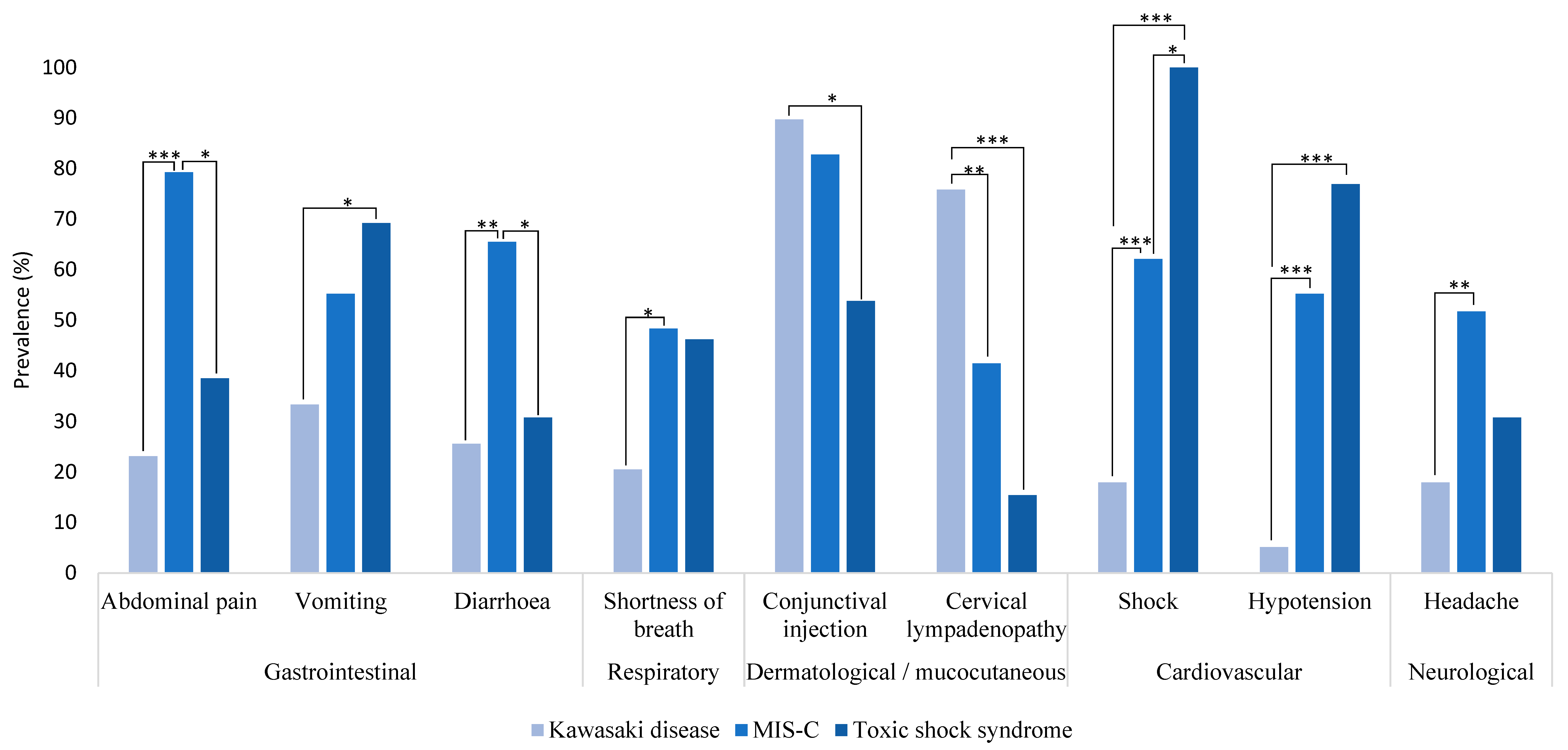
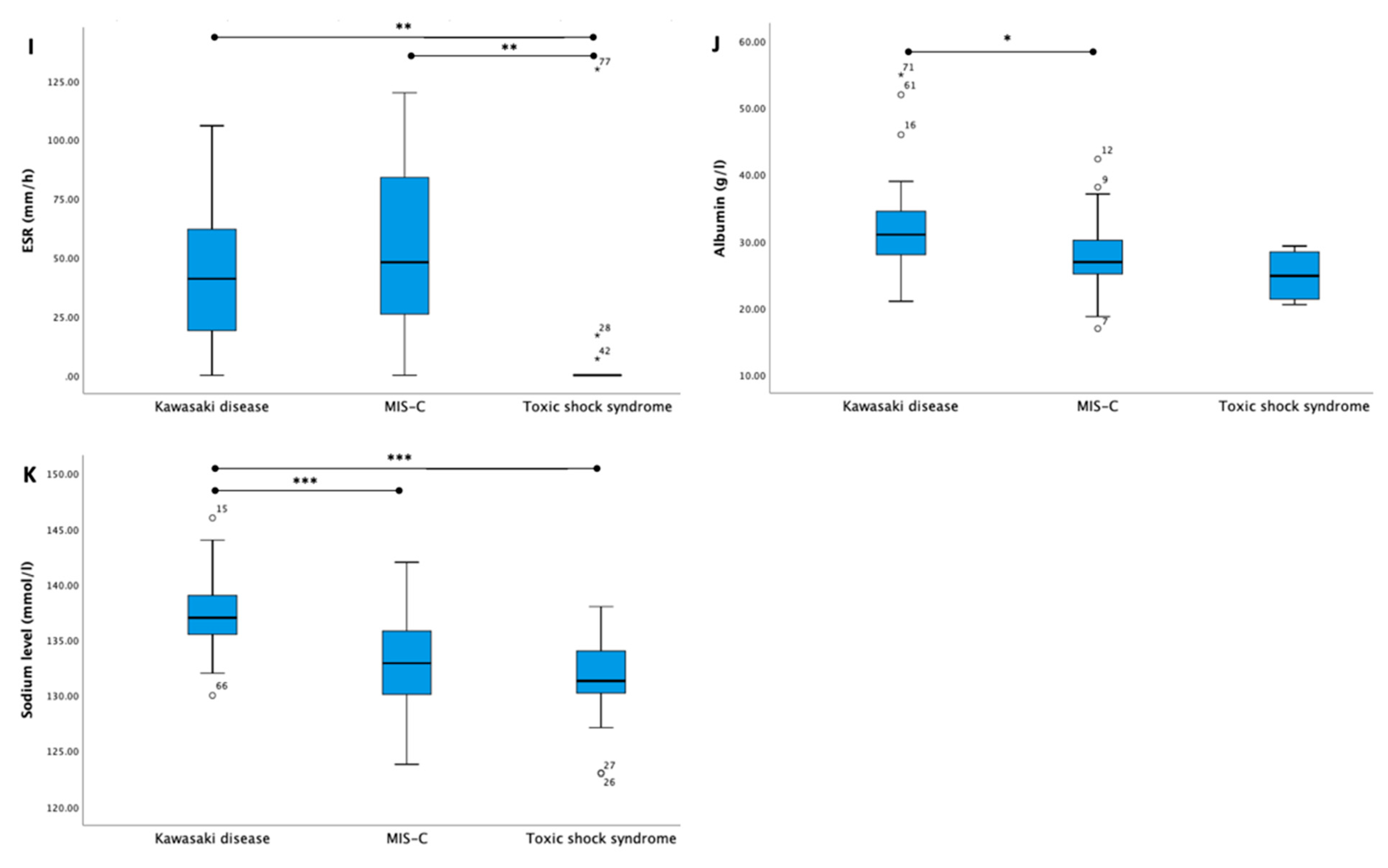
3. Results
4. Discussion
5. Conclusions
Supplementary Materials
Author Contributions
Funding
Institutional Review Board Statement
Informed Consent Statement
Data Availability Statement
Acknowledgments
Conflicts of Interest
References
- Riphagen, S.; Gomez, X.; Gonzalez-Martinez, C.; Wilkinson, N.; Theocharis, P. Hyperinflammatory shock in children during COVID-19 pandemic. Lancet 2020, 395, 1607–1608. [Google Scholar] [CrossRef] [PubMed]
- Verdoni, L.; Mazza, A.; Gervasoni, A.; Martelli, L.; Ruggeri, M.; Ciuffreda, M.; Bonanomi, E.; D’Antiga, L. An outbreak of severe Kawasaki-like disease at the Italian epicentre of the SARS-CoV-2 epidemic: An observational cohort study. Lancet 2020, 395, 1771–1778. [Google Scholar] [CrossRef]
- Dufort, E.M.; Koumans, E.H.; Chow, E.J.; Rosenthal, E.M.; Muse, A.; Rowlands, J.; Barranco, M.A.; Maxted, A.M.; Rosenberg, E.S.; Easton, D.; et al. Multisystem Inflammatory Syndrome in Children Investigation Team Multisystem inflammatory syndrome in children in New York State. N. Engl. J. Med. 2020, 383, 347–358. [Google Scholar] [CrossRef] [PubMed]
- Information for Healthcare Providers about Multisystem Inflammatory Syndrome in Children (MIS-C). Available online: https://www.cdc.gov/mis/mis-c/hcp/index.html (accessed on 16 June 2022).
- Information for Healthcare Providers about Multisystem Inflammatory Syndrome in Children (MIS-C). Available online: https://www.cdc.gov/mis/mis-c/hcp_cstecdc/index.html (accessed on 4 March 2023).
- Mohsin, S.S.; Abbas, Q.; Chowdhary, D.; Khalid, F.; Sheikh, A.S.; Khan, G.Z.A.; Aslam, N.; Bhatti, O.A.; Inam, M.; Saleem, A.F.; et al. Multisystem Inflammatory Syndrome (MIS-C) in Pakistani Children: A Description of the Phenotypes and Comparison with Historical Cohorts of Children with Kawasaki Disease and Myocarditis. Available online: https://pubmed.ncbi.nlm.nih.gov/34153080/ (accessed on 16 June 2022).
- Son, M.B.F. Friedman K, COVID-19: Multisystem Inflammatory Syndrome in Children (MIS-C) Clinical Features, Evaluation, and Diagnosis. Available online: https://www.uptodate.com/contents/covid-19-multisystem-inflammatory-syndrome-in-children-mis-c-clinical-features-evaluation-and-diagnosis#H2589496221 (accessed on 17 June 2022).
- Sapronova, K.; Kake, R.; Pavare, J.; Grāvele, D.; Šēla, I.; Ērgle, E.; Isarova, D.; Grīnberga, Z.; Zavadska, D. SARS-CoV-2 seroprevalence among children in Latvia: A cross-sectional study. Available online: https://science.rsu.lv/en/publications/sars-cov-2-seroprevalence-among-children-in-latvia-a-cross-sectio (accessed on 15 March 2023).
- Nakra, N.A.; Blumberg, D.A.; Herrera-Guerra, A.; Lakshminrusimha, S. Multi-System Inflammatory Syndrome in Children (MIS-C) Following SARS-CoV-2 Infection: Review of Clinical Presentation, Hypothetical Pathogenesis, and Proposed Management. Children 2020, 7, 69. [Google Scholar] [CrossRef] [PubMed]
- Kundu, A.; Maji, S.; Kumar, S.; Bhattacharya, S.; Chakraborty, P.; Sarkar, J. Clinical aspects and presumed etiology of multisystem inflammatory syndrome in children (MIS-C): A review. Clin. Epidemiol. Glob. Health 2022, 14, 100966. [Google Scholar] [CrossRef]
- Rivas, M.N.; Porritt, R.A.; Cheng, M.H.; Bahar, I.; Arditi, M. COVID-19–associated multisystem inflammatory syndrome in children (MIS-C): A novel disease that mimics toxic shock syndrome—The superantigen hypothesis. J. Allergy Clin. Immunol. 2021, 147, 57–59. [Google Scholar] [CrossRef] [PubMed]
- Godfred-Cato, S.D.; Abrams, J.Y.; Balachandran, N.M.; Jaggi, P.; Jones, K.M.; Rostad, C.A.; Lu, A.T.B.; Fan, L.B.; Jabbar, A.; Anderson, E.J.; et al. Distinguishing Multisystem Inflammatory Syndrome in Children from COVID-19, Kawasaki Disease and Toxic Shock Syndrome. Pediatr. Infect. Dis. J. 2022, 41, 315–323. [Google Scholar] [CrossRef]
- Whittaker, E.; Bamford, A.; Kenny, J.; Kaforou, M.; Jones, C.; Shah, P.; Ramnarayan, P.; Fraisse, A.; Miller, O.; Davies, P.; et al. Clinical Characteristics of 58 Children With a Pediatric Inflammatory Multisystem Syndrome Temporally Associated With SARS-CoV-2. JAMA 2020, 324, 259–269. [Google Scholar] [CrossRef]
- Harvey, H.; Tremoulet, A.; Chisum, P.; Burns, J. A Comparison of MIS-C, Kawasaki Disease, Toxic Shock and Septic Shock. Available online: https://journals.lww.com/ccmjournal/Fulltext/2023/01001/736__A_COMPARISON_OF_PEDIATRIC_ICU_PRESENTATION_OF.702.aspx (accessed on 19 June 2022).
- Buonsenso, D.; Riitano, F.; Valentini, P. Pediatric Inflammatory Multisystem Syndrome Temporally Related with SARS-CoV-2: Immunological Similarities with Acute Rheumatic Fever and Toxic Shock Syndrome. Front. Pediatr. 2020, 8, 574. [Google Scholar] [CrossRef]
- Centers for Disease Control and Prevention Health Alert Network (HAN) Multisystem Inflammatory Syndrome in Children (MIS-C) Associated with Coronavirus Disease 2019 (COVID-19). Available online: https://emergency.cdc.gov/han/2020/han00432.asp (accessed on 19 June 2022).
- McCrindle, B.W.; Rowley, A.H.; Newburger, J.W.; Burns, J.C.; Bolger, A.F.; Gewitz, M.; Baker, A.L.; Jackson, M.A.; Takahashi, M.; Shah, P.B.; et al. Diagnosis, Treatment, and Long-Term Management of Kawasaki Disease: A Scientific Statement for Health Professionals from the American Heart Association. Circulation 2017, 135, e927–e999. [Google Scholar] [CrossRef]
- Centers for Disease Control and Prevention. Case definitions for infectious conditions under public health surveillance. MMWR Recomm Rep. 1997, 46, 1–55. [Google Scholar]
- Centers for Disease Control (CDC). Repeat injuries in an inner city population—Philadelphia, 1987–1988. MMWR Morb. Mortal Wkly. Rep. 1990, 39, 1–3, Erratum in: MMWR Morb. Mortal Wkly. Rep. 1990, 39, 123. [Google Scholar]
- Breiman, R.F.; Davis, J.P.; Facklam, R.R.; Gray, B.M.; Hoge, C.W.; Kaplan, E.L.; Mortimer, E.A.; Schlievert, P.M.; Schwartz, B.; Stevens, D.L.; et al. Defining the group A streptococcal toxic shock syndrome. Rationale and consensus definition. The Working Group on Severe Streptococcal Infections. JAMA 1993, 269, 390–391. [Google Scholar] [CrossRef]
- Rigante, D.; Andreozzi, L.; Fastiffi, M.; Bracci, B.; Natale, M.F.; Esposito, S. Critical Overview of the Risk Scoring Systems to Predict Non-Responsiveness to Intravenous Immunoglobulin in Kawasaki Syndrome. Available online: https://www.ncbi.nlm.nih.gov/pmc/articles/PMC4813142/ (accessed on 5 March 2023).
- Park, W.Y.; Lee, S.Y.; Kim, G.B.; Song, M.K.; Kwon, H.W.; Bae, E.J.; Choi, E.H.; Park, J.D. Clinical aspects for differential diagnosis of Kawasaki disease shock syndrome: A case control study. BMC Pediatr. 2021, 21, 25. [Google Scholar] [CrossRef]
- Kostik, M.M.; Bregel, L.V.; Avrusin, I.S.; Dondurei, E.A.; Matyunova, A.E.; Efremova, O.S.; Isupova, E.A.; Kornishina, T.L.; Masalova, V.V.; Snegireva, L.S.; et al. Distinguishing between Multisystem Inflammatory Syndrome, Associated with COVID-19 in Children and the Kawasaki Disease: Development of Preliminary Criteria Based on the Data of the Retrospective Multicenter Cohort Study. Front. Pediatr. 2021, 9, 787353. [Google Scholar] [CrossRef]
- Felsenstein, S.; Willis, E.; Lythgoe, H.; McCann, L.; Cleary, A.; Mahmood, K.; Porter, D.; Jones, J.; McDonagh, J.; Chieng, A.; et al. Presentation, Treatment Response and Short-Term Outcomes in Paediatric Multisystem Inflammatory Syndrome Temporally Associated with SARS-CoV-2 (PIMS-TS). J. Clin. Med. 2020, 9, 3293. [Google Scholar] [CrossRef] [PubMed]
- Riollano-Cruz, M.; Akkoyun, E.; Briceno-Brito, E.; Kowalsky, S.; Reed, J.; Posada, R.; Sordillo, E.M.; Tosi, M.; Trachtman, R.; Paniz-Mondolfi, A. Multisystem inflammatory syndrome in children related to COVID-19: A New York City experience. J. Med. Virol. 2020, 93, 424–433. [Google Scholar] [CrossRef]
- Cook, A.; Janse, S.; Watson, J.R.; Erdem, G. Manifestations of Toxic Shock Syndrome in Children, Columbus, Ohio, USA, 2010–20171. Emerg. Infect. Dis. 2020, 26, 1077–1083. [Google Scholar] [CrossRef]
- Kawasaki, T. Kawasaki disease. Proc. Jpn. Acad. Ser. B Phys. Biol. Sci. 2006, 82, 59–71. [Google Scholar] [CrossRef] [Green Version]
- Sharma, C.; Ganigara, M.; Galeotti, C.; Burns, J.; Berganza, F.M.; Hayes, D.A.; Singh-Grewal, D.; Bharath, S.; Sajjan, S.; Bayry, J. Multisystem inflammatory syndrome in children and Kawasaki disease: A critical comparison. Nat. Rev. Rheumatol. 2021, 17, 731–748. [Google Scholar] [CrossRef]
- Henderson, L.A.; Canna, S.W.; Friedman, K.G.; Gorelik, M.; Lapidus, S.K.; Bassiri, H.; Behrens, E.M.; Ferris, A.; Kernan, K.F.; Schulert, G.S.; et al. American College of Rheumatology Clinical Guidance for Multisystem Inflammatory Syndrome in Children Associated with SARS–CoV-2 and Hyperinflammation in Pediatric COVID-19: Version 1. Arthritis Rheumatol. 2020, 72, 1791–1805. [Google Scholar] [CrossRef] [PubMed]
- Cattalini, M.; Taddio, A.; Bracaglia, C.; Cimaz, R.; Della Paolera, S.; Filocamo, G.; La Torre, F.; Lattanzi, B.; Marchesi, A.; Simonini, G.; et al. Childhood multisystem inflammatory syndrome associated with COVID-19 (MIS-C): A diagnostic and treatment guidance from the Rheumatology Study Group of the Italian Society of Pediatrics. Ital. J. Pediatr. 2021, 47, 1–6. [Google Scholar] [CrossRef] [PubMed]
- Marchesi, A.; Rigante, D.; Cimaz, R.; Ravelli, A.; de Jacobis, I.T.; Rimini, A.; Cardinale, F.; Cattalini, M.; De Zorzi, A.; Dellepiane, R.M.; et al. Revised recommendations of the Italian Society of Pediatrics about the general management of Kawasaki disease. Ital. J. Pediatr. 2021, 47, 16. [Google Scholar] [CrossRef]
- Newburger, J.W.; Takahashi, M.; Gerber, M.A.; Gewitz, M.H.; Tani, L.Y.; Burns, J.C.; Shulman, S.T.; Bolger, A.F.; Ferrieri, P.; Baltimore, R.S.; et al. Diagnosis, treatment, and long-term management of Kawasaki disease: A statement for health professionals from the Committee on Rheumatic Fever, Endocarditis and Kawasaki Disease, Council on Cardiovascular Disease in the Young, American Heart Association. Pediatrics 2004, 114, 1708–1733. [Google Scholar] [CrossRef] [Green Version]
- Lo, M.S.; Newburger, J.W. Role of intravenous immunoglobulin in the treatment of Kawasaki disease. Int. J. Rheum. Dis. 2018, 21, 64–69. [Google Scholar] [CrossRef] [PubMed] [Green Version]
- Chuang, Y.Y.; Huang, Y.C.; Lin, T.Y. Toxic shock syndrome in children: Epidemiology, pathogenesis, and management. Paediatr. Drugs 2005, 7, 11–25. [Google Scholar] [CrossRef]


| KD | MIS-C | TSS | |
|---|---|---|---|
| Prevalence of comorbidities in groups | 6 (15.4%) | 3 (10.3%) | 0 (0%) |
| Comorbidities specified |
|
|
| Variable | KD | MIS-C | TSS | Total | p-Value | KD vs. MIS-C, p-Value | MIS-C vs. TSS, p-Value | KD vs. TSS, p-Value |
|---|---|---|---|---|---|---|---|---|
| Number of patients, n (%) | 39 (48.1%) | 29 (35.8%) | 13 (16.1%) | 81 (100%) | ||||
| Age in years; mean (SD) | 3.9 (3.7) | 9.8 (4.5) | 11.3 (4.5) | 7.2 (5.2) | <0.001 | <0.001 | 0.31 | <0.001 |
| Sex | ||||||||
| Female, n (%) | 16 (41.0) | 13 (44.8) | 8 (61.5) | 37 (45.7) | 0.44 | |||
| Male, n (%) | 23 (59.0) | 16 (55.2) | 5 (38.5) | 44 (54.3) | ||||
| Comorbidities, n (%) | 6 (15.4) | 3 (10.3) | 0 | 9 (11.1) | 0.4 | |||
| Day of hospitalization since onset of symptoms, median (IQR) | 5.0 (3.0–7.0) | 5.0 (4.0–6.0) | 3.0 (2.0–5.0) | 5.0 (3.0–6.0) | 0.03 | 0.74 | 0.005 | 0.02 |
| Time from symptom onset to diagnosis, median (IQR) | 7.0 (5.0–10.0) | 6.0 (5.0–7.0) | 3.0 (2.5–5.0) | 6.0 (5.0–9.0) | <0.001 | 0.03 | <0.001 | <0.001 |
| Length of stay in hospital, days, median (IQR) | 12.0 (9.0–16.0) | 12 (11.5–16.0) | 9.0 (6.5–12.0) | 12.0 (9.5–15.0) | 0.02 | |||
| Duration of fever in days, median (IQR) | 9.0 (7.0–12.0) | 7.0 (5.0–8.0) | 4.0 (3.0–5.0) | 7.0 (5.0–9.0) | <0.001 | <0.001 | <0.001 | <0.001 |
| Fever lasting at least 5 days, n (%) | 38 (97.4) | 27 (93.1) | 4 (30.8) | 69 (85.2) | <0.001 | 0.57 | <0.001 | <0.001 |
| Unfulfilled KD criteria, n (%) | 0 | 2 (6,9) | 9 (69,2) | 11 (13,6) | <0.001 | 0.10 | 0.001 | <0.001 |
| Classic KD criteria, n (%) | 30 (76.9) | 12 (41.4) | 2 (15.4) | 44 (54.3) | 0.007 | 0.99 | 0.27 | |
| Incomplete KD criteria, n (%) | 9 (23.1) | 15 (51.7) | 2 (15.4) | 26 (32.1) | 0.53 | <0.001 | <0.001 | |
| Admission to the Pediatric Intensive Care Unit (PICU), n (%) | 6 (15.4) | 15 (51.7) | 9 (69.2) | 30 (37.0) | <0.001 | 0.001 | 0.29 | 0.001 |
| Days in PICU, median (IQR) | 4 (2.5–6) | 2 (2–3) | 3 (2–3.5) | 3 (2–4) | 0.19 | |||
| Outcome | ||||||||
| Died, n (%) | 0 | 0 | 0 | 0 | - | |||
| Variable | KD | MIS-C | TSS | Total | p-Value | KD vs. MIS-C, p-Value | MIS-C vs. TSS, p-Value | KD Vs. TSS, p-Value |
|---|---|---|---|---|---|---|---|---|
| Thoracic X-ray Pneumonia | 6 (15.4) | 10 (34.5) | 4 (30.8) | 20 (24.7) | 0.17 | |||
| Thoracic X-ray or ultrasound Pleural effusion | 9 (23.1) | 22 (75.9) | 5 (38.5) | 36 (44.4) | <0.001 | <0.001 | 0.04 | 0.30 |
| Electrocardiogram (ECG) findings | ||||||||
| Long QTc interval, n (%) | 0 | 7 (24.1) | 0 | 7 (11.9) | 0.02 | 0.01 | 0.56 | - |
| Changes in ST segment, n (%) | 5 (19.2) | 26 (89.7) | 0 | 31 (52.5) | <0.001 | <0.001 | <0.001 | 0.99 |
| Atrioventricular (AV) conduction disturbances, n (%) | 1 (3.8) | 6 (20.7) | 0 | 7 (11.9) | 0.19 | |||
| AV dissociation, n (%) | 0 | 3 (10.3) | 0 | 3 (5.1) | 0.39 | |||
| Intraventricular conduction disturbances, n (%) | 7 (26.9) | 16 (55.2) | 0 | 23 (39.0) | 0.02 | 0.03 | 0.1 | 0.55 |
| 1st degree AV block, n (%) | 1 (3.8) | 2 (6.9) | 0 | 3 (5.1) | 0.99 | |||
| 2nd degree AV block, n (%) | 0 | 0 | 0 | 0 | - | |||
| Atrial extrasystoles, n (%) | 1 (3.8) | 2 (6.9) | 0 | 3 (5.1) | 0.99 | |||
| Ventricular extrasystoles, n (%) | 1 (3.8) | 1 (3.4) | 1 (25.0) | 3 (5.1) | 0.27 | |||
| Echocardiography findings | ||||||||
| Pathological changes, n (%) | 5 (12.8) | 24 (82.8) | 0 | 29 (39.7) | <0.001 | <0.001 | 0.001 | 0.99 |
| Valvular insufficiency, n (%) | 3 (7.7) | 21 (72.4) | 0 | 24 (29.6) | <0.001 | <0.001 | <0.001 | 0.56 |
| Pericardial effusion, n (%) | 0 | 12 (41.4) | 0 | 12 (14.8) | <0.001 | <0.001 | 0.008 | - |
| Systolic disfunction, n (%) | 0 | 8 (27.6) | 0 | 8 (9.9) | <0.001 | 0.001 | 0.04 | - |
| Coronary artery changes in acute phase, n (%) | 4 (10.3) | 1 (3.4) | 0 | 5 (6.2) | 0.46 | |||
| Coronary artery changes after 1 month, n (%) | 3 (7.7) | 0 | - | 2 (3.4) | 0.53 | |||
| Coronary artery changes after 3 months, n (%) | 3 (8.1) | 0 | - | 3 (5.4) | 0.54 | |||
Disclaimer/Publisher’s Note: The statements, opinions and data contained in all publications are solely those of the individual author(s) and contributor(s) and not of MDPI and/or the editor(s). MDPI and/or the editor(s) disclaim responsibility for any injury to people or property resulting from any ideas, methods, instructions or products referred to in the content. |
© 2023 by the authors. Licensee MDPI, Basel, Switzerland. This article is an open access article distributed under the terms and conditions of the Creative Commons Attribution (CC BY) license (https://creativecommons.org/licenses/by/4.0/).
Share and Cite
Klavina, L.; Smane, L.; Kivite-Urtane, A.; Vasilevska, L.; Davidsone, Z.; Smitins, E.; Gardovska, D.; Lubaua, I.; Roge, I.; Pucuka, Z.; et al. Comparison of Characteristics and Outcomes of Multisystem Inflammatory Syndrome, Kawasaki Disease and Toxic Shock Syndrome in Children. Medicina 2023, 59, 626. https://doi.org/10.3390/medicina59030626
Klavina L, Smane L, Kivite-Urtane A, Vasilevska L, Davidsone Z, Smitins E, Gardovska D, Lubaua I, Roge I, Pucuka Z, et al. Comparison of Characteristics and Outcomes of Multisystem Inflammatory Syndrome, Kawasaki Disease and Toxic Shock Syndrome in Children. Medicina. 2023; 59(3):626. https://doi.org/10.3390/medicina59030626
Chicago/Turabian StyleKlavina, Lizete, Liene Smane, Anda Kivite-Urtane, Lauma Vasilevska, Zane Davidsone, Emils Smitins, Dace Gardovska, Inguna Lubaua, Ieva Roge, Zanda Pucuka, and et al. 2023. "Comparison of Characteristics and Outcomes of Multisystem Inflammatory Syndrome, Kawasaki Disease and Toxic Shock Syndrome in Children" Medicina 59, no. 3: 626. https://doi.org/10.3390/medicina59030626