Evaluation of the Diagnostic Value of Contrast-Enhanced Voiding Urosonography with Regard to the Further Therapy Regime and Patient Outcome—A Single-Center Experience in an Interdisciplinary Uroradiological Setting

 , ,
, ,
Abstract
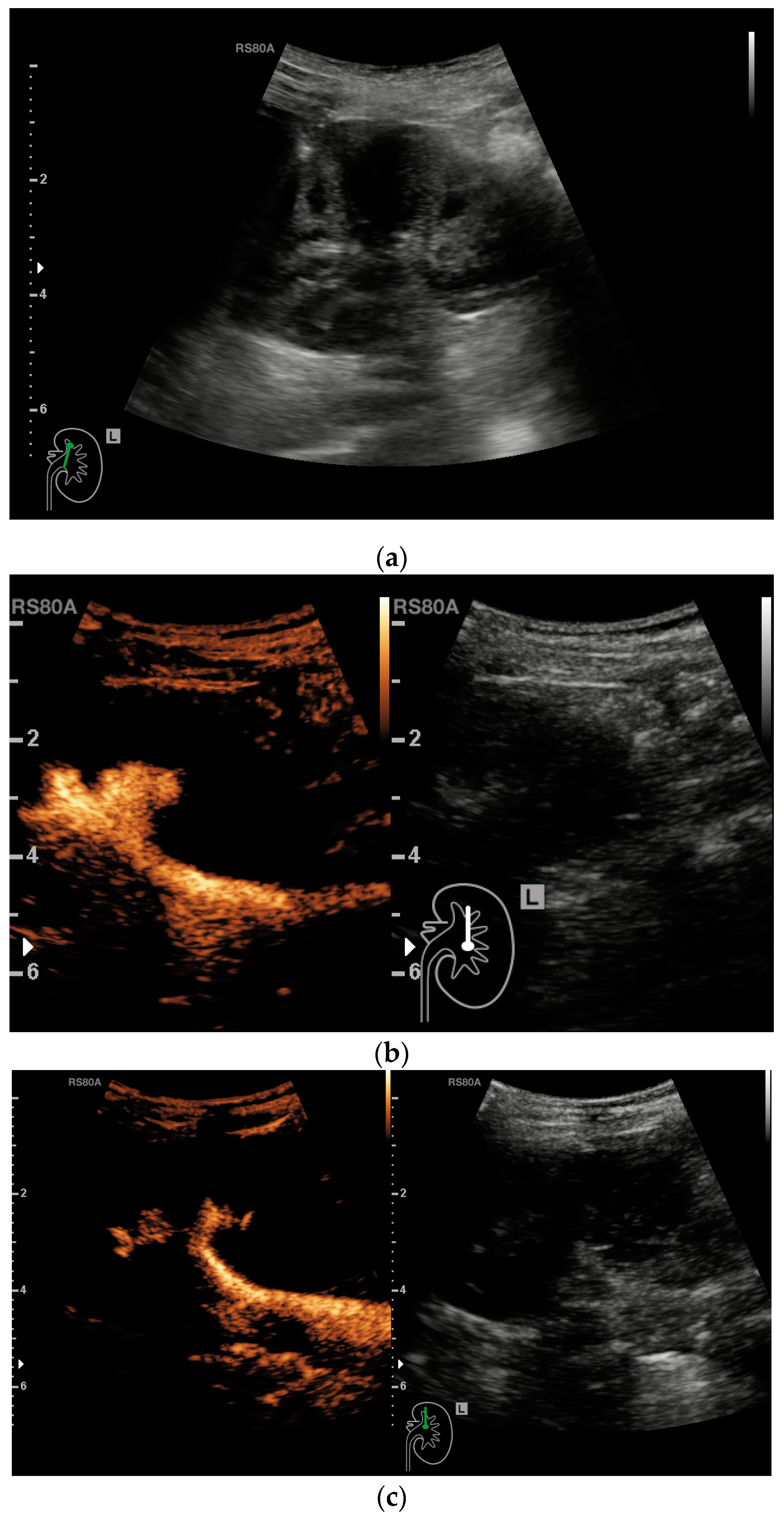
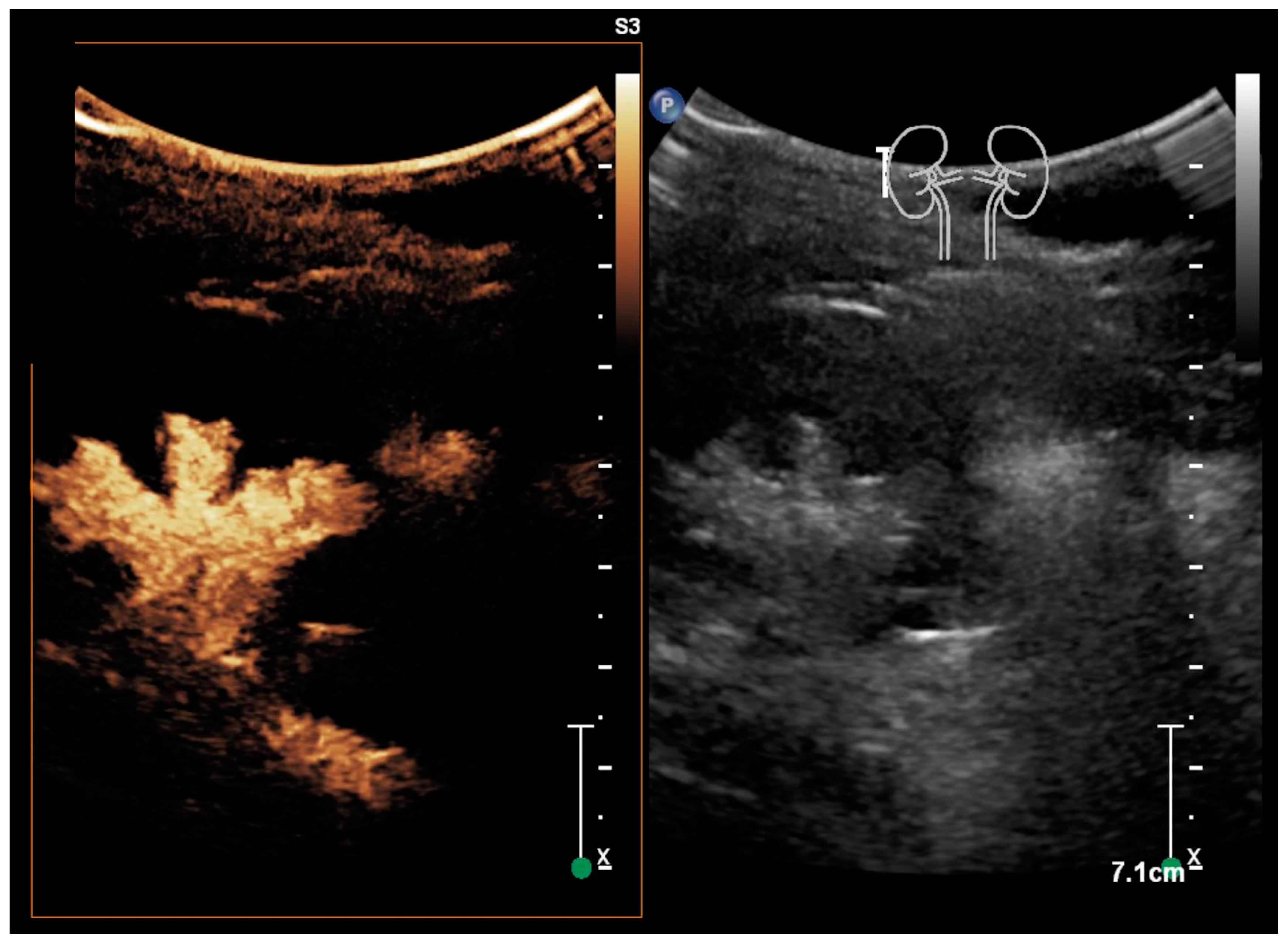
:1. Introduction
2. Materials and Methods
3. Results
4. Discussion
5. Conclusions
Author Contributions
Funding
Institutional Review Board Statement
Informed Consent Statement
Data Availability Statement
Acknowledgments
Conflicts of Interest
References
- Hajiyev, P.; Burgu, B. Contemporary Management of Vesicoureteral Reflux. Eur. Urol. Focus 2017, 3, 181–188. [Google Scholar] [CrossRef] [PubMed]
- Johnston, D.L.; Qureshi, A.H.; Irvine, R.W.; Giel, D.W.; Hains, D.S. Contemporary Management of Vesicoureteral Reflux. Curr. Treat. Options Pediatr. 2016, 2, 82–93. [Google Scholar] [CrossRef] [PubMed] [Green Version]
- Ninoa, F.; Ilaria, M.; Noviello, C.; Santoro, L.; Ratsch, I.; Martino, A.; Cobellis, G. Genetics of Vesicoureteral Reflux. Curr. Genom. 2015, 17, 70–79. [Google Scholar] [CrossRef] [PubMed] [Green Version]
- Sargent, M. Opinion. Pediatr. Radiol. 2000, 30, 587–593. [Google Scholar] [CrossRef] [PubMed]
- Straub, J.; Apfelbeck, M.; Karl, A.; Khoder, W.; Lellig, K.; Tritschler, S.; Stief, C.; Riccabona, M. Vesikoureteraler Reflux. Der Urol. 2015, 55, 27–34. [Google Scholar] [CrossRef] [PubMed]
- Faizah, M.Z.; Hamzaini, A.H.; Kanaheswari, Y.; Dayang A, A.A.; Zulfiqar, M.A. Contrast enhanced Voiding Urosonography (ce-VUS) as a radiation-free technique in the diagnosis of vesicoureteric reflux: Our early experience. Med. J. Malays. 2015, 70, 269–272. [Google Scholar]
- Hoberman, A.; Charron, M.; Hickey, R.W.; Baskin, M.; Kearney, D.H.; Wald, E.R. Imaging Studies after a First Febrile Urinary Tract Infection in Young Children. New Engl. J. Med. 2003, 348, 195–202. [Google Scholar] [CrossRef] [Green Version]
- Edwards, A.; Peters, C.A. Managing vesicoureteral reflux in children: Making sense of all the data. F1000Research 2019, 8, 29. [Google Scholar] [CrossRef] [Green Version]
- Çelebi, S.; Özaydın, S.; Baştaş, C.B.; Kuzdan, O.; Erdoğan, C.; Yazıcı, M.; Caymaz, I.; Sander, S. Reliability of the Grading System for Voiding Cystourethrograms in the Management of Vesicoureteral Reflux: An Interrater Comparison. Adv. Urol. 2016, 2016, 1–4. [Google Scholar] [CrossRef]
- Sjöström, S.; Sillén, U.; Bachelard, M.; Johansson, E.; Brandström, P.; Hellström, A.-L.; Abrahamsson, K. Bladder/bowel dysfunction in pre-school children following febrile urinary tract infection in infancy. Pediatr. Nephrol. 2020, 1–9. [Google Scholar] [CrossRef]
- Arena, S.; Iacona, R.; Impellizzeri, P.; Russo, T.; Marseglia, L.; Gitto, E.; Romeo, C. Physiopathology of vesico-ureteral reflux. Ital. J. Pediatr. 2016, 42, 103. [Google Scholar] [CrossRef] [PubMed] [Green Version]
- Skoog, S.J.; Peters, C.A.; Arant, B.S.; Copp, H.L.; Elder, J.S.; Hudson, R.G.; Khoury, A.E.; Lorenzo, A.J.; Pohl, H.G.; Shapiro, E.; et al. Pediatric Vesicoureteral Reflux Guidelines Panel Summary Report: Clinical Practice Guidelines for Screening Siblings of Children With Vesicoureteral Reflux and Neonates/Infants With Prenatal Hydronephrosis. J. Urol. 2010, 184, 1145–1151. [Google Scholar] [CrossRef] [PubMed]
- Liang, D.; McHugh, K.M.; Brophy, P.D.; Shaikh, N.; Manak, J.R.; Andrews, P.; Hakker, I.; Wang, Z.; Schwaderer, A.L.; Hains, D.S. DNA copy number variations in children with vesicoureteral reflux and urinary tract infections. PLoS ONE 2019, 14, e0220617. [Google Scholar] [CrossRef] [PubMed] [Green Version]
- Porrello, A.; Gulia, C.; Bateni, Z.H.; Zangari, A.; Gigli, S.; Briganti, V.; Tursini, S.; Koh, C.J.; Gaffi, M.; Baldassarra, S.; et al. Vesicoureteral reflux in infants: What do we know about the gender prevalence by age? Eur. Rev. Med Pharmacol. Sci. 2017, 21, 5321–5329. [Google Scholar] [CrossRef]
- The RIVUR Trial Investigators Antimicrobial Prophylaxis for Children with Vesicoureteral Reflux. New Engl. J. Med. 2014, 370, 2367–2376. [CrossRef] [PubMed] [Green Version]
- Duran, C.; Beltrán, V.P.; González, A.; Gómez, C.; Del Riego, J. Contrast-enhanced Voiding Urosonography for Vesicoureteral Reflux Diagnosis in Children. Radiographics 2017, 37, 1854–1869. [Google Scholar] [CrossRef]
- Duran, C.; Valera, A.; Alguersuari, A.; Ballesteros, E.; Riera, L.; Martin, C.; Puig, J. Voiding urosonography: The study of the urethra is no longer a limitation of the technique. Pediatr. Radiol. 2008, 39, 124–131. [Google Scholar] [CrossRef]
- Ascenti, G.; Zimbaro, G.; Mazziotti, S.; Chimenz, R.; Fede, C.; Visalli, C.; Scribano, E. Harmonic US imaging of vesicoureteric reflux in children: Usefulness of a second generation US contrast agent. Pediatr. Radiol. 2004, 34, 481–487. [Google Scholar] [CrossRef]
- Ključevšek, D.; Battelino, N.; Tomažič, M.; Levart, T.K. A comparison of echo-enhanced voiding urosonography with X-ray voiding cystourethrography in the first year of life. Acta Paediatr. 2012, 101, e235–e239. [Google Scholar] [CrossRef]
- Mane, N.; Sharma, A.; Patil, A.; Gadekar, C.; Andankar, M.; Pathak, H. Comparison of contrast-enhanced voiding urosonography with voiding cystourethrography in pediatric vesicoureteral reflux. Turk. J. Urol. 2018, 44, 261–267. [Google Scholar] [CrossRef]
- Darge, K.; Papadopoulou, F.; Ntoulia, A.; Bulas, D.I.; Coley, B.D.; Fordham, L.A.; Paltiel, H.J.; McCarville, B.; Volberg, F.M.; Cosgrove, D.O.; et al. Safety of contrast-enhanced ultrasound in children for non-cardiac applications: A review by the Society for Pediatric Radiology (SPR) and the International Contrast Ultrasound Society (ICUS). Pediatr. Radiol. 2013, 43, 1063–1073. [Google Scholar] [CrossRef] [PubMed]
- Ntoulia, A.; Anupindi, S.A.; Darge, K.; Back, S.J. Applications of contrast-enhanced ultrasound in the pediatric abdomen. Abdom. Radiol. 2017, 43, 948–959. [Google Scholar] [CrossRef] [PubMed]
- Papadopoulou, F.; Ntoulia, A.; Siomou, E.; Darge, K. Contrast-enhanced voiding urosonography with intravesical administration of a second-generation ultrasound contrast agent for diagnosis of vesicoureteral reflux: Prospective evaluation of contrast safety in 1,010 children. Pediatr. Radiol. 2014, 44, 719–728. [Google Scholar] [CrossRef] [PubMed]
- Piscaglia, F.; Bolondi, L. The safety of Sonovue® in abdominal applications: Retrospective analysis of 23188 investigations. Ultrasound Med. Biol. 2006, 32, 1369–1375. [Google Scholar] [CrossRef] [PubMed]
- Wong, L.S.; Tse, K.S.; Fan, T.W.; Kwok, K.Y.; Tsang, T.K.; Fung, H.S.; Chan, W.; Lee, K.W.; Leung, M.W.Y.; Chao, N.S.Y.; et al. Voiding urosonography with second-generation ultrasound contrast versus micturating cystourethrography in the diagnosis of vesicoureteric reflux. Eur. J. Nucl. Med. Mol. Imaging 2014, 173, 1095–1101. [Google Scholar] [CrossRef]
- Schwarze, V.; Marschner, C.; De Figueiredo, G.N.; Rübenthaler, J.; Clevert, D.-A. Single-Center Study: Evaluating the Diagnostic Performance and Safety of Contrast-Enhanced Ultrasound (CEUS) in Pregnant Women to Assess Hepatic Lesions. Ultraschall Med. Eur. J. Ultrasound 2019, 41, 29–35. [Google Scholar] [CrossRef] [PubMed]
- Radmayr, C.; Bogaert, G.; Dogan, H.S.; Kočvara, R.; Nijman, R.; Stein, R.; Tekgül, S. EAU Guidelines on Paediatric Urology 2018. In European Association of Urology Guidelines. 2018, Proceedings of the EAU Annual Congress Copenhagen, Arnhem, The Netherlands, March, 2018; European Association of Urology Guidelines Office: Arnhem, The Netherlands, 2018. [Google Scholar]
- Woźniak, M.M.; Osemlak, P.; Pawelec, A.; Brodzisz, A.; Nachulewicz, P.; Wieczorek, A.P.; Zajączkowska, M.M. Intraoperative contrast-enhanced urosonography during endoscopic treatment of vesicoureteral reflux in children. Pediatr. Radiol. 2014, 44, 1093–1100. [Google Scholar] [CrossRef] [Green Version]
- Sidhu, P.S.; Cantisani, V.; Dietrich, C.F.; Gilja, O.H.; Saftoiu, A.; Bartels, E.; Bertolotto, M.; Calliada, F.; Clevert, D.-A.; Cosgrove, D.; et al. The EFSUMB Guidelines and Recommendations for the Clinical Practice of Contrast-Enhanced Ultrasound (CEUS) in Non-Hepatic Applications: Update 2017 (Short Version). Ultraschall Med. Eur. J. Ultrasound 2018, 39, 154–180. [Google Scholar] [CrossRef] [Green Version]
- Kis, É.; Nyitrai, A.; Várkonyi, I.; Máttyus, I.; Cseprekál, O.; Reusz, G.; Szabó, A. Voiding urosonography with second-generation contrast agent versus voiding cystourethrography. Pediatr. Nephrol. 2010, 25, 2289–2293. [Google Scholar] [CrossRef]
- Papadopoulou, F.; Anthopoulou, A.; Siomou, E.; Efremidis, S.; Tsamboulas, C.; Darge, K. Harmonic voiding urosonography with a second-generation contrast agent for the diagnosis of vesicoureteral reflux. Pediatr. Radiol. 2008, 39, 239–244. [Google Scholar] [CrossRef]
- Kuzmanovska, D.; Risteski, A.; Kambovska, M.; Trpcevski, T.; Sahpazova, E.; Petrovski, M. Voiding Urosonography with Second-Generation Ultrasound Contrast Agent for Diagnosis of Vesicoureteric Reflux: First Local Pilot Study. Open Access Maced. J. Med Sci. 2017, 5, 215–221. [Google Scholar] [CrossRef] [PubMed] [Green Version]
- Davis, T.D.; Rushton, H. Managing Vesicoureteral Reflux in the Pediatric Patient: A Spectrum of Treatment Options for a Spectrum of Disease. Curr. Treat. Options Pediatr. 2016, 2, 23–34. [Google Scholar] [CrossRef] [Green Version]
- Matouschek, E. Treatment of vesicorenal reflux by transurethral teflon-injection (author’s transl). Urol. A 1981, 20, 263–264. [Google Scholar]
- Lee, T.; Park, J.M. Vesicoureteral reflux and continuous prophylactic antibiotics. Investig. Clin. Urol. 2017, 58, S32–S37. [Google Scholar] [CrossRef] [Green Version]
- Rivilla, F. Endoscopic treatment of vesicoureteral reflux in a paediatric surgery ambulatory unit. J. Minimal Access Surg. 2011, 7, 132–135. [Google Scholar] [CrossRef] [PubMed]
- Blais, A.-S.; Bolduc, S.; Moore, K. Vesicoureteral reflux: From prophylaxis to surgery. Can. Urol. Assoc. J. 2017, 11, S13–S18. [Google Scholar] [CrossRef] [Green Version]
- Baek, M.; Kim, K.D. Current Surgical Management of Vesicoureteral Reflux. Korean J. Urol. 2013, 54, 732–737. [Google Scholar] [CrossRef] [Green Version]
- Yap, T.-L.; Chen, Y.; Nah, S.A.; Ong, C.C.P.; Jacobsen, A.; Low, Y. STING versus HIT technique of endoscopic treatment for vesicoureteral reflux: A systematic review and meta-analysis. J. Pediatr. Surg. 2016, 51, 2015–2020. [Google Scholar] [CrossRef]
- Kirsch, A.J.; Perez-Brayfield, M.; Smith, E.A.; Scherz, H.C. The Modified Sting Procedure to Correct Vesicoureteral Reflux: Improved Results With Submucosal Implantation Within The Intramural Ureter. J. Urol. 2004, 171, 2413–2416. [Google Scholar] [CrossRef]
- Kirsch, A.J.; Arlen, A.M. Evaluation of new Deflux administration techniques: Intraureteric HIT and Double HIT for the endoscopic correction of vesicoureteral reflux. Expert Rev. Med. Devices 2014, 11, 439–446. [Google Scholar] [CrossRef]
- Cerwinka, W.H.; Scherz, H.C.; Kirsch, A.J. Endoscopic Treatment of Vesicoureteral Reflux with Dextranomer/Hyaluronic Acid in Children. Adv. Urol. 2008, 2008, 1–7. [Google Scholar] [CrossRef] [Green Version]
- Sung, J.; Skoog, S. Surgical management of vesicoureteral reflux in children. Pediatr. Nephrol. 2011, 27, 551–561. [Google Scholar] [CrossRef] [PubMed] [Green Version]
- Gill, I.S.; Ponsky, L.E.; Desai, M.; Kay, R.; Ross, J.H. Laparoscopic cross-trigonal Cohen ureteroneocystostomy: Novel tech-nique. J. Urol. 2001, 166, 1811–1814. [Google Scholar] [CrossRef]
- Lakshmanan, Y.; Fung, L.C. Techniques in Endourology—Laparoscopic Extravesicular Ureteral Reimplantation for Vesicoureteral Reflux: Recent Technical Advances. J. Endourol. 2000, 14, 589–594. [Google Scholar] [CrossRef] [PubMed]
- Ključevšek, D.; Riccabona, M.; Müller, L.-S.O.; Woźniak, M.M.; Franchi-Abella, S.; Darge, K.; Mentzel, H.-J.; Ntoulia, A.; Avni, F.E.; Napolitano, M.; et al. Intracavitary contrast-enhanced ultrasonography in children: Review with procedural recommendations and clinical applications from the European Society of Paediatric Radiology abdominal imaging task force. Pediatr. Radiol. 2020, 50, 596–606. [Google Scholar] [CrossRef] [PubMed]
- Montini, G.; Tullus, K.; Hewitt, I. Febrile Urinary Tract Infections in Children. New Engl. J. Med. 2011, 365, 239–250. [Google Scholar] [CrossRef] [PubMed]
- Williams, G.; Fletcher, J.T.; Alexander, S.I.; Craig, J.C. Vesicoureteral Reflux. J. Am. Soc. Nephrol. 2008, 19, 847–862. [Google Scholar] [CrossRef] [Green Version]
- Ntoulia, A.; Back, S.J.; Shellikeri, S.; Poznick, L.; Morgan, T.; Kerwood, J.; Edgar, J.C.; Bellah, R.; Reid, J.R.; Jaramillo, D.; et al. Contrast-enhanced voiding urosonography (ceVUS) with the intravesical administration of the ultrasound contrast agent Optison™ for vesicoureteral reflux detection in children: A prospective clinical trial. Pediatr. Radiol. 2017, 48, 216–226. [Google Scholar] [CrossRef]
- Chua, M.E.; Mendoza, J.S.; Ming, J.M.; Dy, J.S.; Gomez, O. Diagnostic accuracy of contrast-enhanced voiding urosonogram using second-generation contrast with harmonic imaging (CEVUS-HI) study for assessment of vesicoureteral reflux in children: A meta-analysis. World J. Urol. 2018, 37, 2245–2255. [Google Scholar] [CrossRef]
- Choi, W.; Nam, W.; Lee, C.; Han, J.H.; Shin, J.H.; Kim, K.S.; Song, S.H. Long-term Outcomes of Endoscopic Anti-reflux Surgery in Pediatric Patients with Vesicoureteral Reflux: Urinary Tract Infection, Renal Scarring, and Predictive Factors for Success. J. Korean Med. Sci. 2018, 33, e240. [Google Scholar] [CrossRef]
- Esposito, C.; Escolino, M.; Lopez, M.; Farina, A.; Cerulo, M.; Savanelli, A.; La Manna, A.; Caprio, M.G.; Settimi, A.; Varlet, F. Surgical Management of Pediatric Vesicoureteral Reflux: A Comparative Study Between Endoscopic, Laparoscopic, and Open Surgery. J. Laparoendosc. Adv. Surg. Tech. 2016, 26, 574–580. [Google Scholar] [CrossRef] [PubMed]
- Piskunowicz, M.; Swieton, D.; Rybczynska, D.; Czarniak, P.; Szarmach, A.; Kaszubowski, M.; Szurowska, E. Porównanie cystouretrografii mikcyjnej i sonocystografii mikcyjnej z użyciem ultrasonograficznego środka kontrastującego drugiej generacji w badaniu prospektywnym. J. Ultrason. 2016, 16, 339–347. [Google Scholar] [CrossRef] [PubMed]
- Tekgul, S.; Riedmiller, H.; Hoebeke, P.; Kočvara, R.; Nijman, R.J.; Radmayr, C.; Stein, R.; Dogan, H.S. EAU Guidelines on Vesicoureteral Reflux in Children. Eur. Urol. 2012, 62, 534–542. [Google Scholar] [CrossRef] [PubMed]
- Tse, K.; Wong, L.; Lau, H.; Fok, W.; Chan, Y.; Tang, K.; Chan, S.C. Paediatric vesicoureteric reflux imaging: Where are we? Novel ultrasound-based voiding urosonography. Hong Kong Med. J. 2014, 20, 437–443. [Google Scholar] [CrossRef] [PubMed]


| Number of Upper Urinary Tract Infections | Number of Patients | Percentage Distribution |
|---|---|---|
| 0 | 4 | 8.2% |
| 1 | 10 | 20.4% |
| 2 | 2 | 4.1% |
| 3 | 1 | 2.0% |
| 4 | 5 | 10.2% |
| >4 | 27 | 55.1% |
| Total number of patients | 49 | 100% |
| Number of Patients (n = 22) Depending on the Sonomorphologically Conspicuous Side | ||||
|---|---|---|---|---|
| Left Side | Right Side | Both Sides | ||
| Left | Right | |||
| Grade I | - | - | - | - |
| Grade I–II | 2 | - | 1 | - |
| Grade II | 5 | 3 | 1 | - |
| Grade II–III | 4 | 2 | - | - |
| Grade III | 3 | 3 | - | 2 |
| Total number of patients | 14 | 6 | 2 | |
| Endoscopic Treatment | Laparoscopic Surgery Procedure | Conservative Treatment | No Further Records | |
|---|---|---|---|---|
| Patients | 17 | 3 | 2 | |
| affected kidneys | 6 | 12 | 1 | 1 |
| patients < 18 years | 3 | 6 | 2 | 1 |
| patients > 18 years | 3 | 6 | 1 | 1 |
| Admission in 2018 | 4 | 1 | 1 | 1 |
| Admission in 2019 | 1 | 4 | 1 | 1 |
| Admission in 2020 | 1 | 7 | 1 | - |
| Number of Patients with ≥4 UTIs Depending on the Sonomorphologically Conspicuous Side | Number of Patients with ≥4 UTIs Depending on the Sonomorphologically Conspicuous side/Younger than 18 Years | |||
|---|---|---|---|---|
| Left Side | Right Side | Left Side | Right Sight | |
| no evidence of VUR | 17 | 9 | ||
| Grade II | 4 | 1 | 2 | 1 |
| Grade II–III | 3 | 1 | 2 | - |
| Grade III | 2 | 4 | 1 | 2 |
| Total number of patients | 32 | 17 | ||
Publisher’s Note: MDPI stays neutral with regard to jurisdictional claims in published maps and institutional affiliations. |
© 2021 by the authors. Licensee MDPI, Basel, Switzerland. This article is an open access article distributed under the terms and conditions of the Creative Commons Attribution (CC BY) license (http://creativecommons.org/licenses/by/4.0/).
Share and Cite
Marschner, C.A.; Schwarze, V.; Stredele, R.; Froelich, M.F.; Rübenthaler, J.; Geyer, T.; Clevert, D.-A. Evaluation of the Diagnostic Value of Contrast-Enhanced Voiding Urosonography with Regard to the Further Therapy Regime and Patient Outcome—A Single-Center Experience in an Interdisciplinary Uroradiological Setting. Medicina 2021, 57, 56. https://doi.org/10.3390/medicina57010056
Marschner CA, Schwarze V, Stredele R, Froelich MF, Rübenthaler J, Geyer T, Clevert D-A. Evaluation of the Diagnostic Value of Contrast-Enhanced Voiding Urosonography with Regard to the Further Therapy Regime and Patient Outcome—A Single-Center Experience in an Interdisciplinary Uroradiological Setting. Medicina. 2021; 57(1):56. https://doi.org/10.3390/medicina57010056
Chicago/Turabian StyleMarschner, Constantin A., Vincent Schwarze, Regina Stredele, Matthias F. Froelich, Johannes Rübenthaler, Thomas Geyer, and Dirk-André Clevert. 2021. "Evaluation of the Diagnostic Value of Contrast-Enhanced Voiding Urosonography with Regard to the Further Therapy Regime and Patient Outcome—A Single-Center Experience in an Interdisciplinary Uroradiological Setting" Medicina 57, no. 1: 56. https://doi.org/10.3390/medicina57010056