Systematic and MRI-Cognitive Targeted Transperineal Prostate Biopsy Accuracy in Detecting Clinically Significant Prostate Cancer after Previous Negative Biopsy and Persisting Suspicion of Malignancy

Abstract
:1. Introduction
2. Materials and Methods
2.1. Patient Population
2.2. Imaging
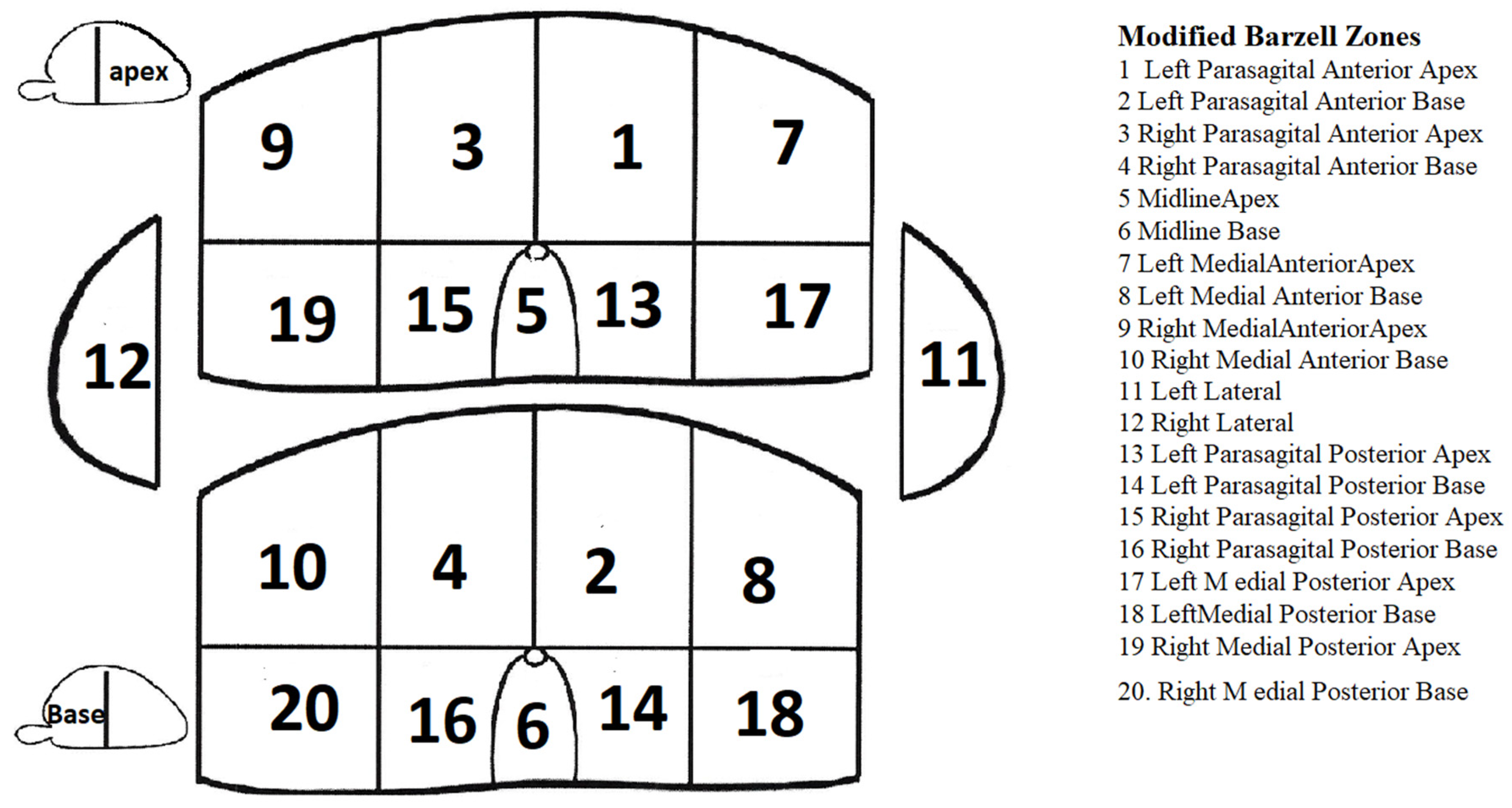
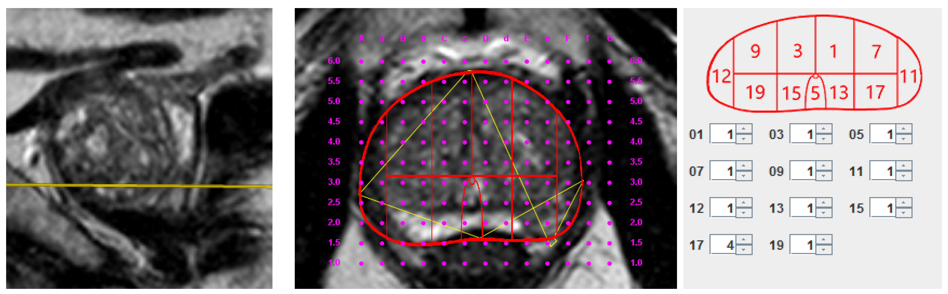
2.3. Biopsy
2.4. Histopathology
2.5. Statistics
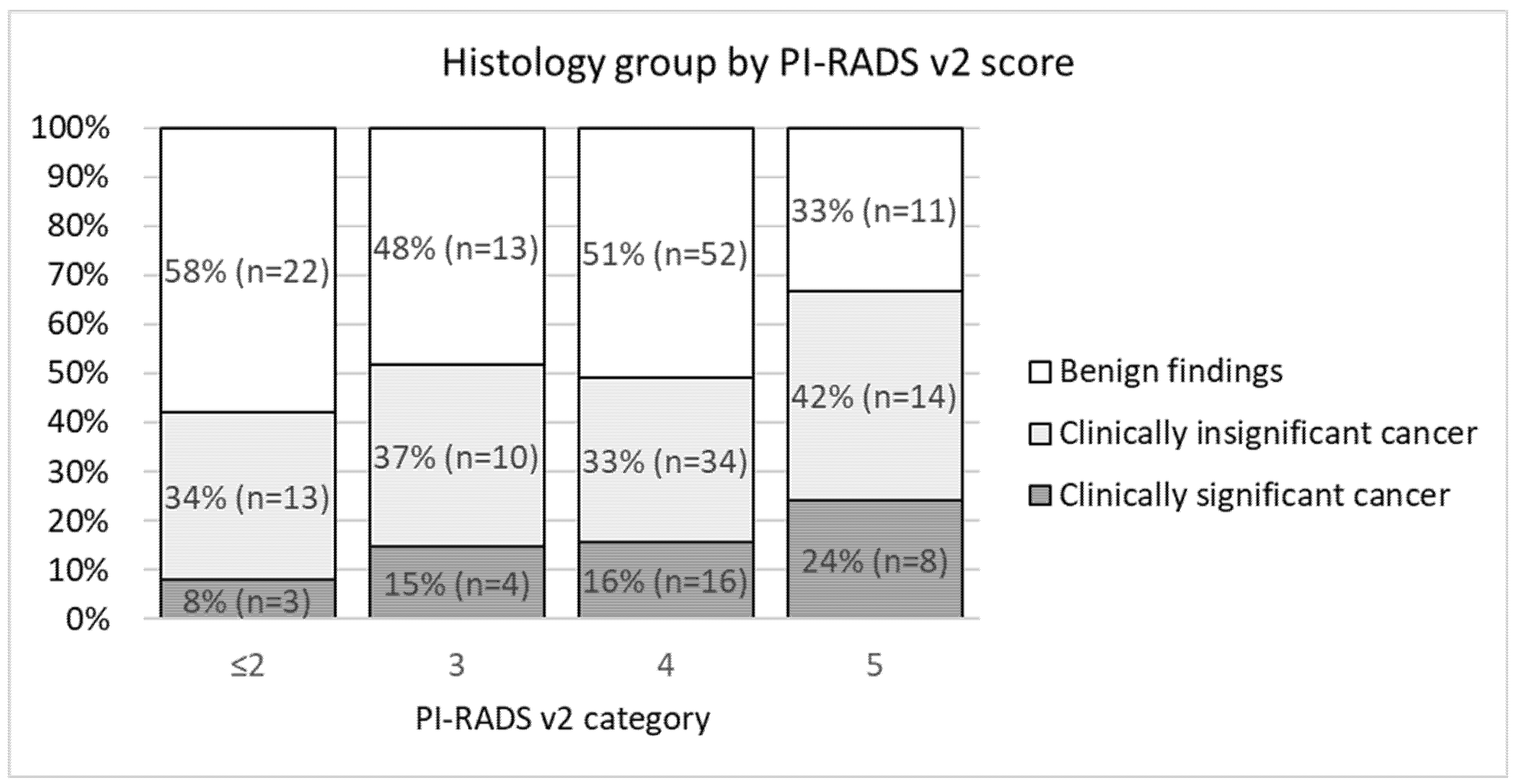
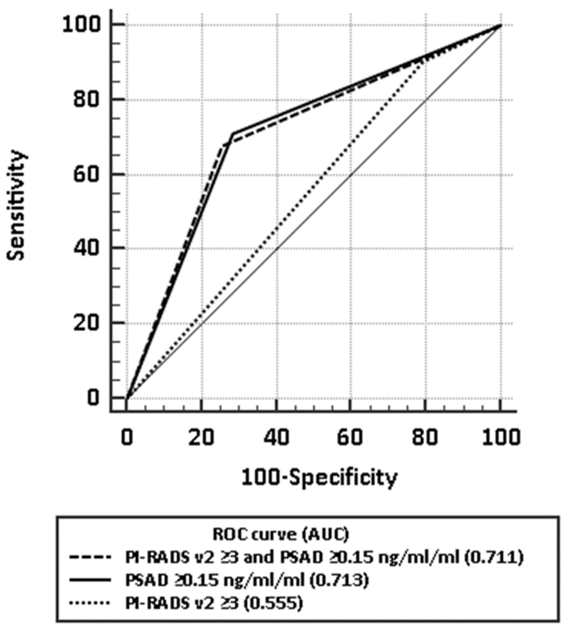
3. Results
4. Discussion
5. Conclusions
Author Contributions
Funding
Institutional Review Board Statement
Informed Consent Statement
Data Availability Statement
Conflicts of Interest
References
- Bray, F.; Ferlay, J.; Soerjomataram, I.; Siegel, R.L.; Torre, L.A.; Jemal, A. Global cancer statistics 2018: GLOBOCAN estimates of incidence and mortality worldwide for 36 cancers in 185 countries. CA Cancer J. Clin. 2018, 68, 394–424. [Google Scholar]
- Polascik, T.J.; Oesterling, J.E.; Partin, A.W. Prostate specific antigen: A decade of discovery—What we have learned and where we are going. J. Urol. 1999, 162, 293–306. [Google Scholar] [CrossRef]
- Serefoglu, E.C.; Altinova, S.; Ugras, N.S.; Akincioglu, E.; Asil, E.; Balbay, D. How reliable is 12-core prostate biopsy procedure in the detection of prostate cancer? Can. Urol. Assoc. J. 2013, 7, 293. [Google Scholar] [CrossRef] [Green Version]
- Abella, H.A. A lack of national diagnosis, care plan spurs call for action. Diagn Imaging. 2008, pp. 36–38. Available online: http://www.diagnosticimaging.com (accessed on 1 January 2008).
- National Institute for Health and Clinical Excellence. Prostate Cancer: Diagnosis and Treatment; National Institute for Health and Clinical Excellence: 2008. Available online: https://www.nice.org.uk/ (accessed on 1 January 2008).
- Bangma, C.; Roemeling, S.; Schröder, F. Overdiagnosis and overtreatment of early detected prostate cancer. World J. Urol. 2007, 25, 3–9. [Google Scholar] [CrossRef] [PubMed] [Green Version]
- Abraham, N.E.; Mendhiratta, N.; Taneja, S.S. Patterns of Repeat Prostate Biopsy in Contemporary Clinical Practice. J. Urol. 2015, 193, 1178–1184. [Google Scholar] [CrossRef] [PubMed]
- Abdulmajed, M.I.; Hughes, D.; Shergill, I.S. The role of transperineal template biopsies of the prostate in the diagnosis of prostate cancer: A review. Expert Rev. Med. Devices 2014, 12, 175–182. [Google Scholar] [CrossRef] [PubMed]
- Garcia, C.; Winter, M.; Bergersen, P.; Woo, H.; Chalasani, V. Does a transperineal prostate biopsy reduce complications compared with a transrectal biopsy? a systematic review and meta-analysis of randomised controlled trials. J. Urol. 2016, 195, e328–e329. [Google Scholar] [CrossRef] [Green Version]
- Mantica, G.; Pacchetti, A.; Aimar, R.; Cerasuolo, M.; Dotta, F.; Olivero, A.; Pini, G.; Passaretti, G.; Maffezzini, M.; Terrone, C. Developing a five-step training model for transperineal prostate biopsies in a naïve residents’ group: A prospective observational randomised study of two different techniques. World J. Urol. 2019, 37, 1845–1850. [Google Scholar] [CrossRef]
- Barzell, W.E.; Melamed, M.R.; Cathcart, P.; Moore, C.M.; Ahmed, H.U.; Emberton, M. Identifying Candidates for Active Surveillance: An Evaluation of the Repeat Biopsy Strategy for Men with Favorable Risk Prostate Cancer. J. Urol. 2012, 188, 762–768. [Google Scholar] [CrossRef]
- Bittner, N.; Merrick, G.S.; Butler, W.M.; Bennett, A.; Galbreath, R.W. Incidence and pathological features of prostate cancer detected on transperineal template guided mapping biopsy after negative transrectal ultrasound guided biopsy. J. Urol. 2013, 190, 509–514. [Google Scholar] [CrossRef] [PubMed]
- Mabjeesh, N.J.; Lidawi, G.; Chen, J.; German, L.; Matzkin, H. High detection rate of significant prostate tumours in anterior zones using a transperineal ultrasound-guided template saturation biopsy. BJU Int. 2012, 110, 993–997. [Google Scholar] [CrossRef] [PubMed]
- Zalesky, M.; Stejskal, J.; Adamcova, V.; Hrbáček, J.; Minarik, I.; Pavlicko, A.; Votrubova, J.; Babjuk, M.; Zachoval, R. Use of Prostate Specific Antigen Density Combined with Multiparametric Magnetic Resonance Imaging Improves Triage for Prostate Biopsy. Urol. Int. 2019, 103, 33–40. [Google Scholar] [CrossRef] [PubMed]
- de Rooij, M.; Hamoen, E.H.J.; Fütterer, J.J.; Barentsz, J.O.; Rovers, M.M. Accuracy of Multiparametric MRI for Prostate Cancer Detection: A Meta-Analysis. Am. J. Roentgenol. 2014, 202, 343–351. [Google Scholar] [CrossRef] [PubMed]
- Ahmed, H.U.; Bosaily, A.E.-S.; Brown, L.C.; Gabe, R.; Kaplan, R.; Parmar, M.K.; Collaco-Moraes, Y.; Ward, K.; Hindley, R.G.; Freeman, A.; et al. Diagnostic accuracy of multi-parametric MRI and TRUS biopsy in prostate cancer (PROMIS): A paired validating confirmatory study. Lancet 2017, 389, 815–822. [Google Scholar] [CrossRef] [Green Version]
- Gayet, M.; van der Aa, A.; Beerlage, H.P.; Schrier, B.P.; Mulders, P.F.A.; Wijkstra, H. The value of magnetic resonance imaging and ultrasonography (MRI/US)-fusion biopsy platforms in prostate cancer detection: A systematic review. BJU Int. 2016, 117, 392–400. [Google Scholar] [CrossRef]
- Schoots, I.G.; Roobol, M.J.; Nieboer, D.; Bangma, C.H.; Steyerberg, E.W.; Hunink, M.G.M. Magnetic Resonance Imaging–targeted Biopsy May Enhance the Diagnostic Accuracy of Significant Prostate Cancer Detection Compared to Standard Transrectal Ultrasound-guided Biopsy: A Systematic Review and Meta-analysis. Eur. Urol. 2015, 68, 438–450. [Google Scholar] [CrossRef]
- Siddiqui, M.M.; Rais-Bahrami, S.; Turkbey, B.; George, A.K.; Rothwax, J.; Shakir, N.; Okoro, C.; Raskolnikov, D.; Parnes, H.L.; Linehan, W.M.; et al. Comparison of MR/Ultrasound Fusion–Guided Biopsy With Ultrasound-Guided Biopsy for the Diagnosis of Prostate Cancer. JAMA 2015, 313, 390–397. [Google Scholar] [CrossRef]
- Radtke, J.P.; Schwab, C.; Wolf, M.B.; Freitag, M.T.; Alt, C.D.; Kesch, C.; Popeneciu, I.V.; Huettenbrink, C.; Gasch, C.; Klein, T.; et al. Multiparametric Magnetic Resonance Imaging (MRI) and MRI–Transrectal Ultrasound Fusion Biopsy for Index Tumor Detection: Correlation with Radical Prostatectomy Specimen. Eur. Urol. 2016, 70, 846–853. [Google Scholar] [CrossRef]
- Hutchinson, R.C.; Costa, D.N.; Lotan, Y. The economic effect of using magnetic resonance imaging and magnetic resonance ultrasound fusion biopsy for prostate cancer diagnosis. Urol. Oncol. 2016, 34, 296–302. [Google Scholar] [CrossRef]
- Marks, L.S.; Young, S.; Natarajan, S. MRI–ultrasound fusion for guidance of targeted prostate biopsy. Curr. Opin. Urol. 2013, 23, 43–50. [Google Scholar] [CrossRef] [PubMed] [Green Version]
- Pahwa, S.; Schiltz, N.K.; Ponsky, L.E.; Lu, Z.; Griswold, M.A.; Gulani, V. Cost-effectiveness of MR Imaging-guided Strategies for Detection of Prostate Cancer in Biopsy-Naive Men. Radiology 2017, 285, 157–166. [Google Scholar] [CrossRef] [PubMed]
- Barzell, W.E.; Melamed, M.R. Appropriate Patient Selection in the Focal Treatment of Prostate Cancer: The Role of Transperineal 3-Dimensional Pathologic Mapping of the Prostate—A 4-Year Experience. Urology 2007, 70, S27–S35. [Google Scholar] [CrossRef] [PubMed]
- Epstein, J.I.; Egevad, L.; Amin, M.B.; Delahunt, B.; Srigley, J.R.; Humphrey, P.A. Grading Committee. The 2014 International Society of Urological Pathology (ISUP) Consensus Conference on Gleason Grading of Prostatic Carcinoma: Definition of Grading Patterns and Proposal for a New Grading System. Am. J. Surg. Pathol. 2016, 40, 244–252. [Google Scholar]
- Fütterer, J.J.; Briganti, A.; Visschere, P.D.; Emberton, M.; Giannarini, G.; Kirkham, A.; Taneja, S.S.; Thoeny, H.; Villeirs, G.; Villers, A. Can Clinically Significant Prostate Cancer Be Detected with Multiparametric Magnetic Resonance Imaging? A Systematic Review of the Literature. Eur. Urol. 2015, 68, 1045–1053. [Google Scholar] [CrossRef]
- Ahmed, H.U.; Hu, Y.; Carter, T.; Arumainayagam, N.; Lecornet, E.; Freeman, A.; Hawkes, D.; Barratt, D.C.; Emberton, M. Characterizing Clinically Significant Prostate Cancer Using Template Prostate Mapping Biopsy. J. Urol. 2011, 186, 458–464. [Google Scholar] [CrossRef]
- Aminsharifi, A.; Howard, L.; Wu, Y.; de Hoedt, A.; Bailey, C.; Freedland, S.J.; Polascik, T.J. Prostate Specific Antigen Density as a Predictor of Clinically Significant Prostate Cancer When the Prostate Specific Antigen is in the Diagnostic Gray Zone: Defining the Optimum Cutoff Point Stratified by Race and Body Mass Index. J. Urol. 2018, 200, 758–766. [Google Scholar] [CrossRef]
- Nordström, T.; Akre, O.; Aly, M.; Grönberg, H.; Eklund, M. Prostate-specific antigen (PSA) density in the diagnostic algorithm of prostate cancer. Prostate Cancer Prostatic Dis. 2017, 21, 57–63. [Google Scholar] [CrossRef]
- Schoots, I.; Osses, D.F.; Drost, F.-J.H.; Verbeek, J.F.M.; Remmers, S.; Van Leenders, G.J.L.H.; Bangma, C.H.; Roobol, M.J. Reduction of MRI-targeted biopsies in men with low-risk prostate cancer on active surveillance by stratifying to PI-RADS and PSA-density, with different thresholds for significant disease. Transl. Androl. Urol. 2018, 7, 132–144. [Google Scholar] [CrossRef] [Green Version]
- Washino, S.; Okochi, T.; Saito, K.; Konishi, T.; Hirai, M.; Kobayashi, Y.; Miyagawa, T. Combination of prostate imaging reporting and data system (PI-RADS) score and prostate-specific antigen (PSA) density predicts biopsy outcome in prostate biopsy naïve patients. BJU Int. 2017, 119, 225–233. [Google Scholar] [CrossRef] [Green Version]
- Simmons, L.A.M.; Kanthabalan, A.; Arya, M.; Briggs, T.; Barratt, D.; Charman, S.C.; Freeman, A.; Gelister, J.; Hawkes, D.; Hu, Y.; et al. The PICTURE study: Diagnostic accuracy of multiparametric MRI in men requiring a repeat prostate biopsy. Br. J. Cancer 2017, 116, 1159–1165. [Google Scholar] [CrossRef] [PubMed]
- Nakai, Y.; Tanaka, N.; Anai, S.; Miyake, M.; Hori, S.; Tatsumi, Y.; Morizawa, Y.; Fujii, T.; Konishi, N.; Fujimoto, K. Transperineal template-guided saturation biopsy aimed at sampling one core for each milliliter of prostate volume: 103 cases requiring repeat prostate biopsy. BMC Urol. 2017, 17, 28. [Google Scholar] [CrossRef] [PubMed] [Green Version]
- Radtke, J.P.; Kuru, T.H.; Boxler, S.; Alt, C.D.; Popeneciu, I.V.; Huettenbrink, C.; Klein, T.; Steinemann, S.; Bergstraesser, C.; Roethke, M.; et al. Comparative Analysis of Transperineal Template Saturation Prostate Biopsy Versus Magnetic Resonance Imaging Targeted Biopsy with Magnetic Resonance Imaging-Ultrasound Fusion Guidance. J. Urol. 2015, 193, 87–94. [Google Scholar] [CrossRef] [PubMed]
- Valerio, M.; Anele, C.; Bott, S.; Charman, S.; Van Der Meulen, J.H.; El-Mahallawi, H.; Emara, A.; Freeman, A.; Jameson, C.; Hindley, R.; et al. The Prevalence of Clinically Significant Prostate Cancer According to Commonly Used Histological Thresholds in Men Undergoing Template Prostate Mapping Biopsies. J. Urol. 2016, 195, 1403–1408. [Google Scholar] [CrossRef] [PubMed]
- Kasivisvanathan, V.; Dufour, R.; Moore, C.M.; Ahmed, H.U.; Abd-Alazeez, M.; Charman, S.C.; Freeman, A.; Allen, C.; Kirkham, A.; Van Der Meulen, J.; et al. Transperineal Magnetic Resonance Image Targeted Prostate Biopsy Versus Transperineal Template Prostate Biopsy in the Detection of Clinically Significant Prostate Cancer. J. Urol. 2013, 189, 860–866. [Google Scholar] [CrossRef] [PubMed]
- European Association of Urology. Prostate Cancer guidelines 2019; European Association of Urology: Arnhem, The Netherlands, 2019. [Google Scholar]
- Drost, F.H.; Osses, D.F.; Nieboer, D.; Steyerberg, E.W.; Bangma, C.H.; Roobol, M.J.; Schoots, I.G. Prostate MRI, with or without MRI-targeted biopsy, and systematic biopsy for detecting prostate cancer. Cochrane Database Syst. Rev. 2019. [Google Scholar] [CrossRef] [PubMed]


| Patient Characteristics | N |
| Age, mean (SD), years | 62 (5.9) |
| PSA at first biopsy, median (IQR), ng/mL | 5.6 (4.8–6.17) |
| PSA at TTPM biopsy, median (IQR), ng/mL | 7.6 (5.6–10.9) |
| Prostate volume at TTPM biopsy, median (IQR), mL | 62 (46.9–87.5) |
| PSAD at TTPM biopsy, median (IQR), ng/mL/mL | 0.121 (0.084–0.187) |
| Days between the first TRUS biopsy and repeated TTPM biopsy, mean (IQR), days | 1081 (708–2064) |
| PI-RADS v2 category | N (%) |
| PI-RADS 1–2, n (%) | 38 (19%) |
| PI-RADS 3, n (%) | 27 (13.5%) |
| PI-RADS 4, n (%) | 102 (51%) |
| PI-RADS 5, n (%) | 33 (16.5%) |
| Gleason pattern | N (%) |
| Benign, n (%) | 98 (49%) |
| Gleason 3 + 3, n (%) | 78 (39%) |
| Gleason 3 + 4, n (%) | 18 (9%) |
| Gleason 4 + 3, n (%) | 6 (3%) |
| Clinically significant cancer at TTPM biopsy, n (%) | 31 (15.5%) |
| Biopsy Type on Which PC Was Found for Each Patient | PI-RADS v.2 | (PI-RADS < 3) | (PI-RADS ≥ 3) | Total | |||
|---|---|---|---|---|---|---|---|
| PC | csPC | PC | csPC | PC | csPC | ||
| Systematic, n (%) | 16 | 3 | 38 | 17 (10.5%) | 54 | 20 | |
| Targeted, n (%) | 3 | - | - | 2 | 1 (0.6%) | 9 | 8 |
| 4 | - | - | 3 | 4 (2.7%) | |||
| 5 | - | - | 4 | 3 (1.8%) | |||
| Systematic + Targeted, n (%) | 3 | - | - | 5 | - | 39 | 3 |
| 4 | - | - | 22 | 2 (1.2%) | |||
| 5 | - | - | 12 | 1 (0.6%) | |||
| No Cancer, n | 22 | 76 | 98 | ||||
| Parameter | Beta Coefficient | SE | Walds Statistics | Odds Ratio | Odds Ratio 95% CI | p Value |
|---|---|---|---|---|---|---|
| Baseline PSA | 0.13 | 0.06 | 5.31 | 1.14 | 1.02–1.27 | 0.021 |
| PSAD | 2.75 | 1.04 | 7.04 | 15.68 | 2.05–119.64 | 0.008 |
| Prostate volume | –0.04 | 0.01 | 15.18 | 0.96 | 0.94–0.98 | <0.001 |
| PiRADS ≥ 3 | 0.442 | 0.668 | 0.437 | 1.56 | 0.42–5.76 | 0.509 |
| PSA density > 0.15 | 1.759 | 0.438 | 16.162 | 5.81 | 2.46–13.69 | <0.001 |
| PiRADS ≥ 3 and PSA density > 0.15 | 1.817 | 0.423 | 18.463 | 6.15 | 2.69–14.09 | <0.001 |
Publisher’s Note: MDPI stays neutral with regard to jurisdictional claims in published maps and institutional affiliations. |
© 2021 by the authors. Licensee MDPI, Basel, Switzerland. This article is an open access article distributed under the terms and conditions of the Creative Commons Attribution (CC BY) license (http://creativecommons.org/licenses/by/4.0/).
Share and Cite
Vėželis, A.; Platkevičius, G.; Kinčius, M.; Gumbys, L.; Naruševičiūtė, I.; Briedienė, R.; Petroška, D.; Ulys, A.; Jankevičius, F. Systematic and MRI-Cognitive Targeted Transperineal Prostate Biopsy Accuracy in Detecting Clinically Significant Prostate Cancer after Previous Negative Biopsy and Persisting Suspicion of Malignancy. Medicina 2021, 57, 57. https://doi.org/10.3390/medicina57010057
Vėželis A, Platkevičius G, Kinčius M, Gumbys L, Naruševičiūtė I, Briedienė R, Petroška D, Ulys A, Jankevičius F. Systematic and MRI-Cognitive Targeted Transperineal Prostate Biopsy Accuracy in Detecting Clinically Significant Prostate Cancer after Previous Negative Biopsy and Persisting Suspicion of Malignancy. Medicina. 2021; 57(1):57. https://doi.org/10.3390/medicina57010057
Chicago/Turabian StyleVėželis, Alvydas, Gediminas Platkevičius, Marius Kinčius, Liutauras Gumbys, Ieva Naruševičiūtė, Rūta Briedienė, Donatas Petroška, Albertas Ulys, and Feliksas Jankevičius. 2021. "Systematic and MRI-Cognitive Targeted Transperineal Prostate Biopsy Accuracy in Detecting Clinically Significant Prostate Cancer after Previous Negative Biopsy and Persisting Suspicion of Malignancy" Medicina 57, no. 1: 57. https://doi.org/10.3390/medicina57010057