ACTH-dependent Hypercortisolemia in a Patient with a Pituitary Microadenoma and an Atypical Carcinoid Tumour of the Thymus

Abstract
:1. Introduction
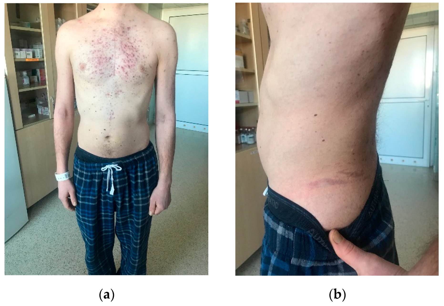
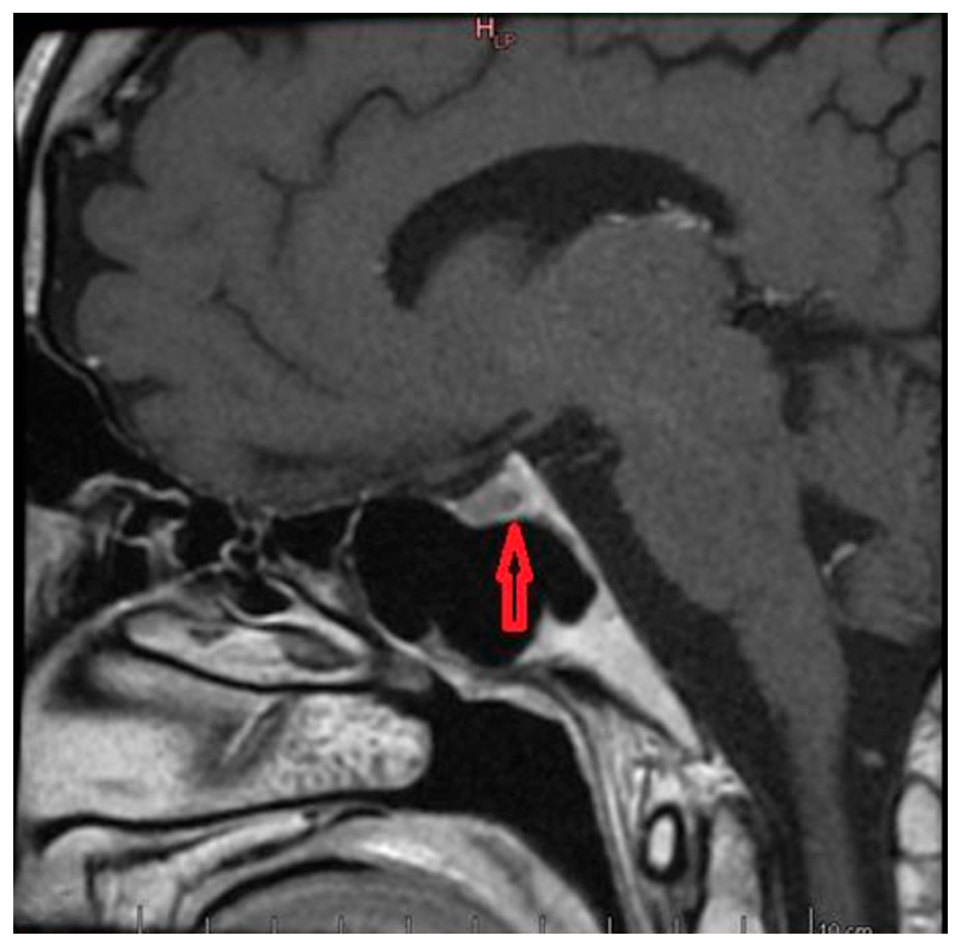
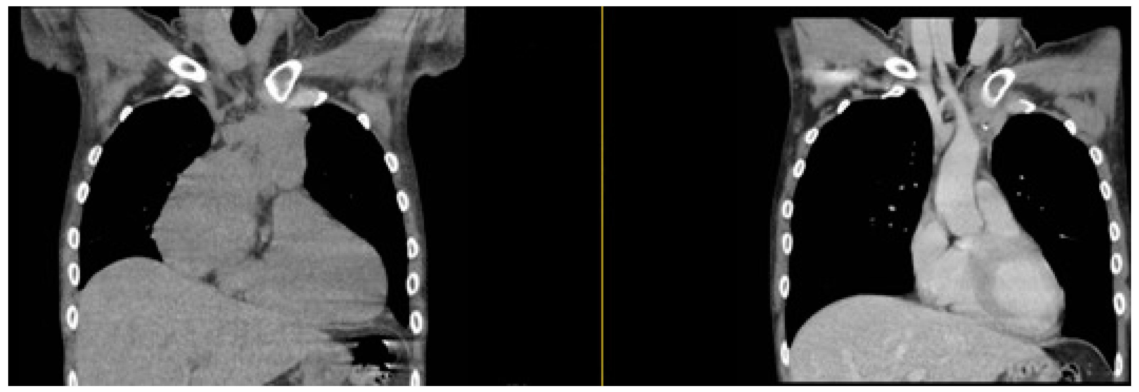
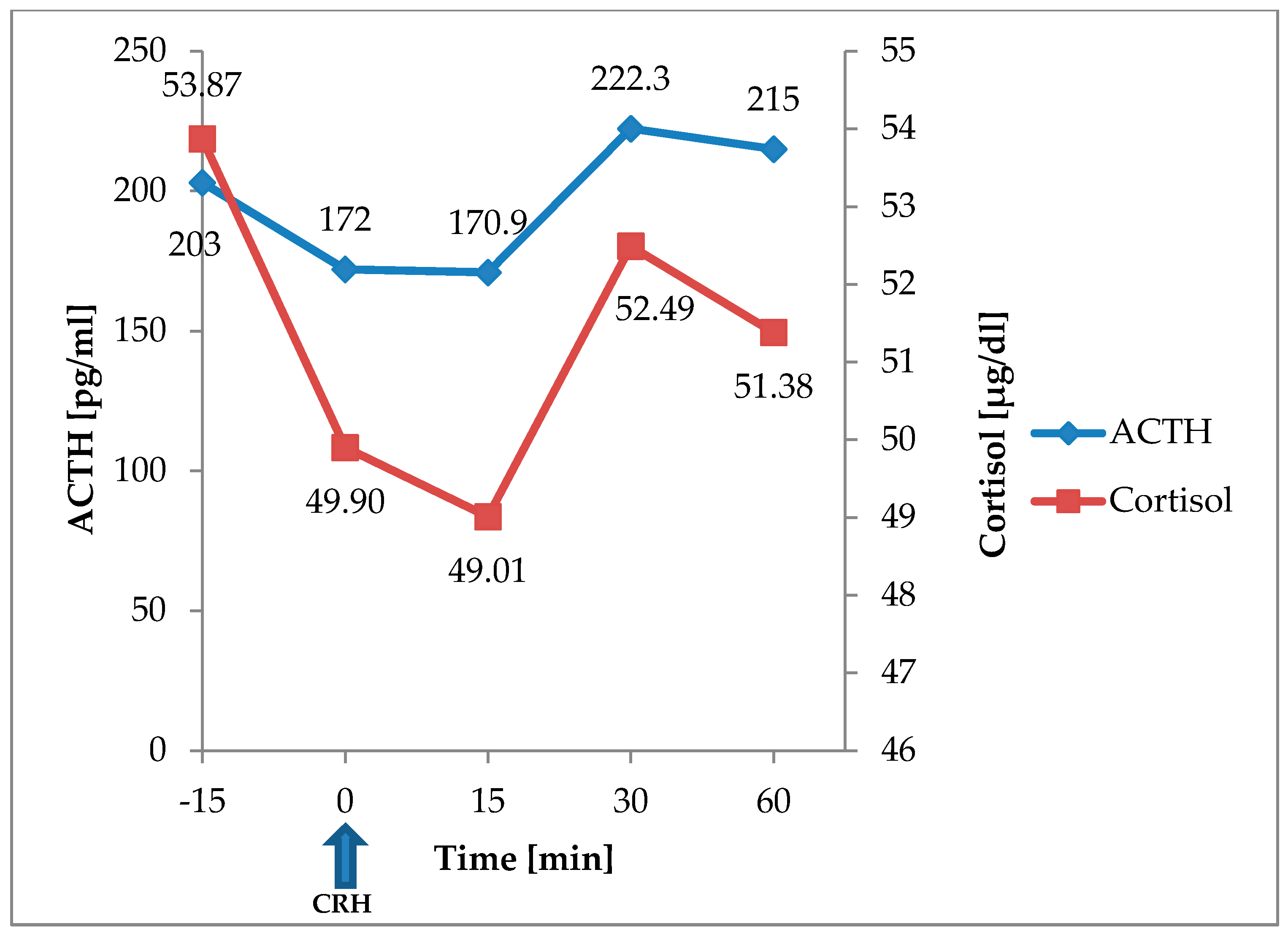
2. Case study
3. Discussion
4. Conclusions
Author Contributions
Funding
References
- Lindholm, J.; Juul, S.; Jørgensen, J.O.; Astrup, J.; Bjerre, P.; Feldt-Rasmussen, U.; Hagen, C.; Jørgensen, J.; Kosteljanetz, M.; Kristensen, L.; et al. Incidence and Late Prognosis of Cushing’s Syndrome: A Population-Based Study. J. Clin. Endocrinol. Metab. 2001, 86, 117–123. [Google Scholar] [CrossRef]
- Newell-Price, J.; Bertagna, X.; Grossman, A.B.; Nieman, L.K. Cushing’s Syndrome. Lancet Lond. Engl. 2006, 367, 1605–1617. [Google Scholar] [CrossRef]
- Ilias, I.; Torpy, D.J.; Pacak, K.; Mullen, N.; Wesley, R.A.; Nieman, L.K. Cushing’s Syndrome Due to Ectopic Corticotropin Secretion: Twenty Years’ Experience at the National Institutes of Health. J. Clin. Endocrinol. Metab. 2005, 90, 4955–4962. [Google Scholar] [CrossRef] [PubMed]
- Isidori, A.M.; Lenzi, A. Ectopic ACTH Syndrome. Arq. Bras. Endocrinol. Metabol. 2006, 51, 1217–1225. [Google Scholar] [CrossRef] [PubMed]
- Pelosof, L.C.; Gerber, D.E. Paraneoplastic Syndromes: An Approach to Diagnosis and Treatment. Mayo Clin. Proc. 2010, 85, 838–854. [Google Scholar] [CrossRef]
- Wang, X.; Li, Y.; Duan, J.; Chen, Y.; Yuan, B.; Qi, Z.; Tan, H. Capecitabine and Temozolomide as a Promising Therapy for Advanced Thymic Atypical Carcinoid. Oncologist 2019, 24, 798–802. [Google Scholar] [CrossRef]
- Ferolla, P.; Brizzi, M.P.; Meyer, T.; Mansoor, W.; Mazieres, J.; Do Cao, C.; Léna, H.; Berruti, A.; Damiano, V.; Buikhuisen, W.; et al. Efficacy and Safety of Long-Acting Pasireotide or Everolimus Alone or in Combination in Patients with Advanced Carcinoids of the Lung and Thymus (LUNA): An Open-Label, Multicentre, Randomised, Phase 2 Trial. Lancet Oncol. 2017, 18, 1652–1664. [Google Scholar] [CrossRef]
- Han, B.; Sun, J.-M.; Ahn, J.S.; Park, K.; Ahn, M.-J. Clinical Outcomes of Atypical Carcinoid Tumors of the Lung and Thymus: 7-Year Experience of a Rare Malignancy at Single Institute. Med. Oncol. 2013, 30, 479. [Google Scholar] [CrossRef]
- Ströbel, P.; Zettl, A.; Shilo, K.; Chuang, W.-Y.; Nicholson, A.G.; Matsuno, Y.; Gal, A.; Laeng, R.H.; Engel, P.; Capella, C.; et al. Tumor Genetics and Survival of Thymic Neuroendocrine Neoplasms: A Multi-Institutional Clinicopathologic Study. Genes. Chromosomes Cancer 2014, 53, 738–749. [Google Scholar] [CrossRef]
- Brown, W.H. A case of pluriglandular syndrome: “diabetes of bearded women”. Lancet 1928, 212, 1022–1023. [Google Scholar] [CrossRef]
- Liddle, G.W.; Island, D.P.; Ney, R.L.; Nicholson, W.E.; Shimizu, N. Nonpituitary Neoplasms and Cushing’s Syndrome: Ectopic Adrenocorticotropin Produced by Nonpituitary Neoplasms as a Cause of Cushing’s Syndrome. Arch. Intern. Med. 1963, 111, 471–475. [Google Scholar] [CrossRef] [PubMed]
- Wajchenberg, B.L.; Mendonça, B.; Liberman, B.; Adelaide, M.; Pereira, A.; Kirschner, M.A. Ectopic ACTH Syndrome. J. Steroid Biochem. Mol. Biol. 1995, 53, 139–151. [Google Scholar] [CrossRef]
- Filosso, P.L.; Actis Dato, G.M.; Ruffini, E.; Bretti, S.; Ozzello, F.; Mancuso, M. Multidisciplinary Treatment of Advanced Thymic Neuroendocrine Carcinoma (Carcinoid): Report of a Successful Case and Review of the Literature. J. Thorac. Cardiovasc. Surg. 2004, 127, 1215–1219. [Google Scholar] [CrossRef] [PubMed]
- Chaer, R.; Massad, M.G.; Evans, A.; Snow, N.J.; Geha, A.S. Primary Neuroendocrine Tumors of the Thymus. Ann. Thorac. Surg. 2002, 74, 1733–1740. [Google Scholar] [CrossRef]
- Gaur, P.; Leary, C.; Yao, J.C. Thymic Neuroendocrine Tumors: A SEER Database Analysis of 160 Patients. Ann. Surg. 2010, 251, 1117–1121. [Google Scholar] [CrossRef] [PubMed]
- Rosai, J.; Higa, E. Mediastinal Endocrine Neoplasm, of Probable Thymic Origin, Related to Carcinoid Tumor. Clinicopathologic Study of 8 Cases. Cancer 1972, 29, 1061–1074. [Google Scholar] [CrossRef]
- Marx, A.; Chan, J.K.C.; Coindre, J.-M.; Detterbeck, F.; Girard, N.; Harris, N.L.; Jaffe, E.S.; Kurrer, M.O.; Marom, E.M.; Moreira, A.L.; et al. The 2015 WHO Classification of Tumors of the Thymus: Continuity and Changes. J. Thorac. Oncol. 2015, 10, 1383–1395. [Google Scholar] [CrossRef]
- Filosso, P.L.; Ruffini, E.; Solidoro, P.; Roffinella, M.; Lausi, P.O.; Lyberis, P.; Oliaro, A.; Guerrera, F. Neuroendocrine Tumors of the Thymus. J. Thorac. Dis. 2017, 9, S1484. [Google Scholar] [CrossRef]
- Satta, J.; Ahonen, A.; Parkkila, S.; Leinonen, L.; Apaja-Sarkkinen, M.; Lepojärvi, M.; Juvonen, T. Multiple Endocrine Neoplastic-Associated Thymic Carcinoid Tumour in Close Relatives: Octreotide Scan as a New Diagnostic and Follow-up Modality. Two Case Reports. Scand. Cardiovasc. J. 1999, 33, 49–53. [Google Scholar]
- Gal, A.A.; Kornstein, M.J.; Cohen, C.; Duarte, I.G.; Miller, J.I.; Mansour, K.A. Neuroendocrine Tumors of the Thymus: A Clinicopathological and Prognostic Study. Ann. Thorac. Surg. 2001, 72, 1179–1182. [Google Scholar] [CrossRef]
- Lin, F.C.-F.; Lin, C.-M.; Hsieh, C.-C.; Li, W.-Y.; Wang, L.-S. Atypical Thymic Carcinoid and Malignant Somatostatinoma in Type I Multiple Endocrine Neoplasia Syndrome: Case Report. Am. J. Clin. Oncol. 2003, 26, 270–272. [Google Scholar] [CrossRef] [PubMed]
- Moran, C.A.; Suster, S. Neuroendocrine Carcinomas (Carcinoid Tumor) of the Thymus. A Clinicopathologic Analysis of 80 Cases. Am. J. Clin. Pathol. 2000, 114, 100–110. [Google Scholar] [CrossRef]
- Weissferdt, A.; Moran, C.A. Thymomas with Prominent Glandular Differentiation: A Clinicopathologic and Immunohistochemical Study of 12 Cases. Hum. Pathol. 2013, 44, 1612–1616. [Google Scholar] [CrossRef] [PubMed]
- Öberg, K.; Hellman, P.; Ferolla, P.; Papotti, M.; ESMO Guidelines Working Group. Neuroendocrine Bronchial and Thymic Tumors: ESMO Clinical Practice Guidelines for Diagnosis, Treatment and Follow-Up. Ann. Oncol. 2012, vii120–vii123. [Google Scholar] [CrossRef]
- Tiffet, O.; Nicholson, A.G.; Ladas, G.; Sheppard, M.N.; Goldstraw, P. A Clinicopathologic Study of 12 Neuroendocrine Tumors Arising in the Thymus. Chest 2003, 124, 141–146. [Google Scholar] [CrossRef] [PubMed]
- Rea, F.; Marulli, G.; Bortolotti, L.; Feltracco, P.; Zuin, A.; Sartori, F. Experience with the “Da Vinci” Robotic System for Thymectomy in Patients with Myasthenia Gravis: Report of 33 Cases. Ann. Thorac. Surg. 2006, 81, 455–459. [Google Scholar] [CrossRef]
- Weksler, B.; Dhupar, R.; Parikh, V.; Nason, K.S.; Pennathur, A.; Ferson, P.F. Thymic Carcinoma: A Multivariate Analysis of Factors Predictive of Survival in 290 Patients. Ann. Thorac. Surg. 2013, 95, 299–303. [Google Scholar] [CrossRef]
- Mei, Z.; Wang, H.; Ren, S.; Wei, J.; Gu, Y. Metastatic Thymic Carcinoid Responds to Chemoradiation and Octreotide: A Case Report. Medicine 2018, 97, e13286. [Google Scholar] [CrossRef]


| Result | Referential Values | ||
|---|---|---|---|
| Peripheral blood morphology | Leukocytes (103/uL) | 8.32 | 4.1–10.9 |
| Granulocytes (103/uL) | 7.11 | 1.5–7 | |
| Lymphocytes (103/uL) | 0.62 | 1–3.7 | |
| Haemoglobin (g/dL) | 14.2 | 14–18 | |
| Biochemical tests | Sodium (mmol/L) | 147 | 136–145 |
| Potassium (mmol/L) | 2.55 | 3.5–5.1 | |
| Fasting glucose (during antihyperglycemic therapy) (mg/dL) | 98 | 70–99 | |
| HbA1c (%) | 6.1 | ||
| C-peptide (ng/mL) | 3.12 | 0.9–4.0 | |
| Phosphorus (mg/dL) | 5.5 | 2.5–4.5 | |
| Calcium (mg/dL) | 10.2 | 8.6–10 | |
| Magnesium (mg/dl) | 1.8 | 1.6–2.6 | |
| Vitamin D (ng/mL) | 26 | 30–80 | |
| Phosphorus in 24-h urine collection (g/24 h) | 0.4 | 0.4–1.3 | |
| Calcium in 24-h urine collection (mg/24 h) | 188 | 100–300 | |
| FALK (U/L) | 86 | 40–129 | |
| Albumin (g/L) | 39.5 | 32–52 | |
| 5-hydroxyindoleacetic acid (mg/24h) | 1.4 | 2.0–9.0 | |
| Chromogranin A (ng/mL) | >1000 | <100 | |
| LDH (U/L) | 271 | 135–225 | |
| Hormonal tests | fT3 (pmol/L) | 3.1 | 3.1–6.8 |
| fT4 (pmol/L) | 16.0 | 12.0–22.0 | |
| TSH (uIU/mL) | 1.73 | 0.27–4.2 | |
| anti-TPO (IU/mL) | 9 | <34 | |
| anti-TG (IU/mL) | <10 | <115 | |
| anti-TSHR [uIU/l] | 0.91 | 0.0–1.75 | |
| DHEAS (U/L) | 284 | 80–560 | |
| Parathormone (pg/mL) | 35.8 | 14.9–56.9 | |
| ACTH (pg/mL) | 258 | 4.7–48.8 | |
| Cortisol at 8:00 (ug/dL) | 38.75 | 6.2–19.4 | |
| Cortisol at 23:00 (ug/dL) | 33.85 | 2.3–11.9 | |
| Cortisol after 2mg dexamethasone (ug/dL) | 44.72 | ||
| Free cortisol in first 24-h urine collection (ug/24 h) | 2066 | 36–137 | |
| Free cortisol in second 24-h urine collection (ug/24 h) | 1851 | 36–137 | |
| Histopathological Test | Atypical Carcinoid of the Thymus |
|---|---|
| Immunophenotype | CK 5/6 (−), CK19 (−/+), CD56 (+), CD117 (−/+), synaptophysin (−), chromogranin (+), LCA (−), TdT (−), S-100 (−), cam 5.2 (+), CK AE 1/AE 3 (+), CD 5 (−), TTF-1 (−/+). |
| Mitotic Index | 2 mitotic figures per 2 mm² |
| Proliferative Index | Ki67—up to 15% focally |
| Microscopically, foci of necrosis visible within the tumour. The tumour exceeded its capsule, focally the tumour was in the line of surgical incision. Cancer cells formed blockages in the blood vessels. | |
| Results | Referential Values | ||
|---|---|---|---|
| Peripheral blood morphology | Leukocytes (103/uL) | 7.86 | 4.1–10.9 |
| Granulocytes (103/uL) | 5.31 | 1.5–7 | |
| Lymphocytes (103/uL) | 1.7 | 1–3.7 | |
| Haemoglobin (g/dL) | 12.5 | 14–18 | |
| Biochemical tests | Sodium (mmol/L) | 142 | 136–145 |
| Potassium (mmol/L) | 3.94 | 3.5–5.1 | |
| Chlorides (mmol/L) | 101.4 | 98–107 | |
| Creatinine (mg/dL) | 0.7 | 0.7–1.2 | |
| Fasting glucose (mg/dL) | 86 | 70–99 | |
| OGTT after 2 hours (mg/dL) | 123 | ||
| HbA1c (%) | 4.5 | ||
| C-peptide (ng/mL) | 1.7 | 0.9–4.0 | |
| Phosphorus (mg/dL) | 5.5 | 2.5–4.5 | |
| Calcium (mg/dL) | 10.2 | 8.6–10 | |
| Magnesium (mg/dL) | 1.8 | 1.6–2.6 | |
| Vitamin D (ng/mL) | 26 | 30–80 | |
| Phosphorus in 24-h urine collection (g/24 h) | 0.4 | 0.4–1.3 | |
| Calcium in 24-h urine collection (mg/24 h) | 188 | 100–300 | |
| FALK (U/L) | 86 | 40–129 | |
| Albumin (g/L) | 39.5 | 32–52 | |
| 5-hydroxyindoleacetic acid (mg/24 h) | 1.4 | 2.0–9.0 | |
| Chromogranin A (ng/mL) | 75.55 | <100 | |
| Hormonal tests | Parathormone (pg/mL) | 8.8 | 14.9–56.9 |
| fT3 (pmol/L) | 6.5 | 3.1–6.8 | |
| fT4 (pmol/L) | 15.1 | 12–22 | |
| TSH (uIU/mL) | 1.66 | 0.27–4.2 | |
| FSH (mIU/mL) | 1.8 | 1.5–12.4 | |
| LH (mIU/mlL) | 2.8 | 1.7–8.6 | |
| Testosterone (ng/mL) | 2.91 | 2.8–8 | |
| Prolactin at 11:00 (uIU/mL) | 322.9 | 86–324 | |
| Prolactin at 17:00 (uIU/mL) | 496.3 | 86–325 | |
| Prolactin at 23:00 (uIU/mL) | 479.8 | 86–326 | |
| Prolactin at 3:00 (uIU/mL) | 398 | 86–327 | |
| Androstenedione (ng/mL) | <0.3 | 0.6–3.1 | |
| IGF-1 (ng/mL) | 151 | 41–246 | |
| ACTH (pg/mL) | 24.7 | <46 | |
| Cortisol at 8:00 (ug/dL) | 7.22 | 6.2–19.4 | |
| Cortisol at 23:00 (ug/dL) | 5.53 | 2.3–11.9 | |
| Dynamic test with synthetic ACTH | Cortisol 0 (ug/dL) | 13.45 | 6.2–19.4 |
| Cortisol 30 (ug/dL) | 21.31 | ||
| Cortisol 60 (ug/dL) | 30.22 | ||
| Results | Referential Values | |||
|---|---|---|---|---|
| Before Operation | After 6 Weeks | After 46 Weeks | ||
| ACTH (pg/mL) | 258 | 24.7 | 68.6 | <48.8 |
| Cortisol at 8:00 (ug/dL) | 38.75 | 7.22 | 17.93 | 6.2–19.4 |
| Cortisol at 23:00 (ug/dL) | 33.85 | 5.53 | 3.00 | 2.3–11.9 |
| 5-hydroxyindoleacetic acid (mg/24 h) | <0.9 | 1.4 | 0.56 | 2.0–6.0 |
| Chromogranin A (ng/mL) | >1000 | 75.55 | 134.82 | <100 |
| IGF-1 (ng/mL) | unknown | 151 | 320 | 41.0–246.0 |
© 2019 by the authors. Licensee MDPI, Basel, Switzerland. This article is an open access article distributed under the terms and conditions of the Creative Commons Attribution (CC BY) license (http://creativecommons.org/licenses/by/4.0/).
Share and Cite
Baranowska-Jurkun, A.; Szychlińska, M.; Matuszewski, W.; Modzelewski, R.; Bandurska-Stankiewicz, E. ACTH-dependent Hypercortisolemia in a Patient with a Pituitary Microadenoma and an Atypical Carcinoid Tumour of the Thymus. Medicina 2019, 55, 759. https://doi.org/10.3390/medicina55120759
Baranowska-Jurkun A, Szychlińska M, Matuszewski W, Modzelewski R, Bandurska-Stankiewicz E. ACTH-dependent Hypercortisolemia in a Patient with a Pituitary Microadenoma and an Atypical Carcinoid Tumour of the Thymus. Medicina. 2019; 55(12):759. https://doi.org/10.3390/medicina55120759
Chicago/Turabian StyleBaranowska-Jurkun, Angelika, Magdalena Szychlińska, Wojciech Matuszewski, Robert Modzelewski, and Elżbieta Bandurska-Stankiewicz. 2019. "ACTH-dependent Hypercortisolemia in a Patient with a Pituitary Microadenoma and an Atypical Carcinoid Tumour of the Thymus" Medicina 55, no. 12: 759. https://doi.org/10.3390/medicina55120759