
Indole-Acrylonitrile Derivatives as Potential Antitumor and Antimicrobial Agents—Synthesis, In Vitro and In Silico Studies

 , , , , , and
Abstract
:
1. Introduction
2. Results and Discussion
2.1. Chemistry
2.2. Biological Evaluation
2.2.1. In Vitro Anticancer Activity
2.2.2. Antimicrobial Activity against Reference Microbial Strains
2.2.3. Antibacterial Activity against Clinical Staphylococcus Aureus Strains
2.3. Docking Studies
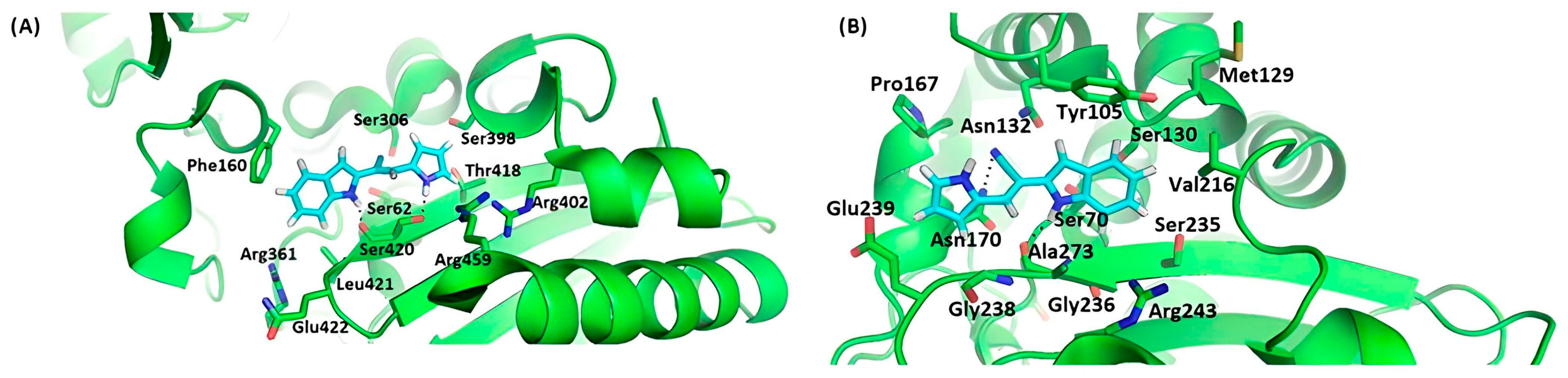
2.3.1. Docking to Anticancer Targets
2.3.2. Docking to Antibacterial Targets
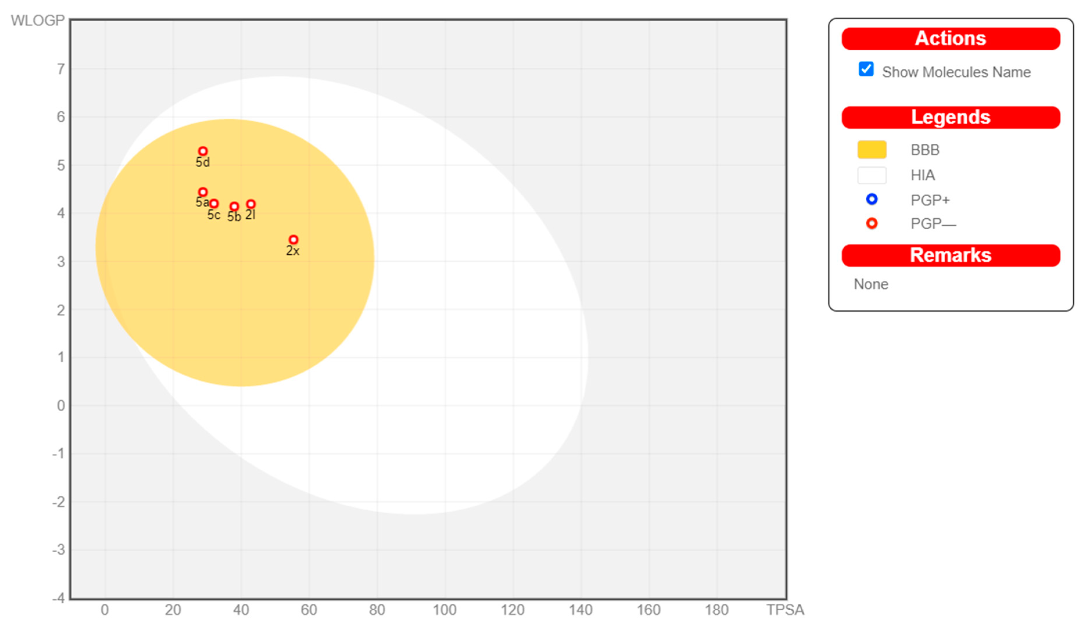
2.4. In Silico Physicochemical, Pharmacokinetic and Drug-Likeness Predictions
3. Materials and Methods
3.1. Chemistry
3.1.1. General Information
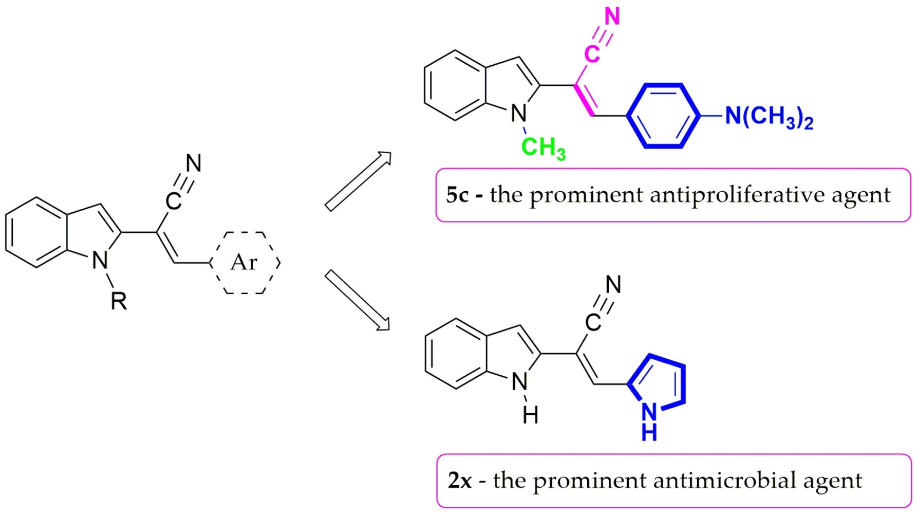
3.1.2. General Procedure for the Preparation of 2-(1H-Indol-2-yl)-3-acrylonitriles 2a–s
- 2-(1H-Indol-2-yl)-3-phenylacrylonitrile (2a). Yield: 39% (yellow solid); m.p. 197–199 °C (m.p. 173–174 °C [63]); IR νmax (KBr, cm−1): 3360, 3085, 3058, 3023, 2224, 1433, 1341, 1302, 914, 782, 726, 676; 1H NMR (300 MHz, DMSO-d6) δ (ppm): 11.74 (s, 1H, NH), 7.92 (s, 1H, CH), 7.87 (d, J = 7.1 Hz, 2H, 2 × C-Harom), 7.62–7.45 (m, 4H, 4 × C-Harom), 7.41 (d, J = 8.9 Hz, 1H, C-Harom), 7.18 (t, J = 7.1 Hz, 1H, C-Harom), 7.04 (t, J = 7.5 Hz, 1H, C-Harom), 6.80 (s, 1H, C3-H, indole); 13C NMR (75 MHz, DMSO-d6) δ (ppm): 138.6, 138.2, 134.0, 133.7, 130.9, 129.6 (two overlapping signals), 129.2 (two overlapping signals), 128.2, 123.9, 121.3, 120.4, 117.5, 111.8, 104.2, 103.7; MS (ESI) m/z: 243 [M − H]−. Anal. calcd for C17H12N2 (244.29) (%): C, 83.58; H, 4.95; N, 11.47. Found: C, 83.32; H, 5.01; N, 11.67.
- 2-(1H-Indol-2-yl)-3-(p-tolyl)acrylonitrile (2b). Yield: 62% (yellow solid); m.p. 223–224 °C (192–194 °C [63]); IR νmax (KBr, cm−1): 3356, 3045, 2918, 2223, 1611, 1435, 1302, 898, 798, 782, 730, 607; 1H NMR (400 MHz, DMSO-d6) δ (ppm): 11.72 (s, 1H, NH), 7.90 (s, 1H, CH), 7.80 (d, J = 8.2 Hz, 2H, 2 × C-Harom), 7.60 (d, J = 7.9 Hz, 1H, C-Harom), 7.43–7.37 (m, 1H, C-Harom), 7.38 (d, J = 8.0 Hz, 2H, 2 × C-Harom), 7.20 (t, J = 7.0 Hz, 1H, C-Harom), 7.05 (t, J = 7.0 Hz, 1H, C-Harom), 6.79 (s, 1H, C3-H, indole), 2.39 (s, 3H, CH3); MS (ESI) m/z: 257 [M − H]−. Anal. calcd for C18H14N2 (258.32) (%): C, 83.69; H, 5.46; N, 10.84. Found: C, 83.57; H, 5.25; N, 11.18.
- 2-(1H-Indol-2-yl)-3-(4-isopropylphenyl)acrylonitrile (2c). Yield: 21% (yellow solid); m.p. 176–178 °C; IR νmax (KBr, cm−1): 3327, 2956, 2869, 2233, 1606, 1417, 1302, 897, 827, 784, 747, 730, 613; 1H NMR (400 MHz, DMSO-d6) δ (ppm): 11.74 (s, 1H, NH), 7.91 (s, 1H, CH), 7.83 (d, J = 8.3 Hz, 2H, 2 × C-Harom), 7.60 (d, J = 7.8 Hz, 1H, C-Harom), 7.45–7.42 (m, 3H, 3 × C-Harom), 7.20 (t, J = 8.1 Hz, 1H, C-Harom), 7.06 (t, J = 7.5 Hz, 1H, C-Harom), 6.80 (s, 1H, C3-H, indole), 3.02–2.91 (m, 1H, CH), 1.24 (d, J = 6.9 Hz, 6H, 2 × CH3); 13C NMR (100 MHz, DMSO-d6) δ (ppm): 151.8, 138.6, 138.2, 133.9, 131.7, 129.4 (two overlapping signals), 128.3, 127.7 (two overlapping signals), 123.8, 121.2, 120.4, 117.7, 111.8, 103.8, 102.7, 33.9, 24.0 (two overlapping signals); MS (ESI) m/z: 285 [M − H]−. Anal. calcd for C20H18N2 (286.37) (%): C, 83.88; H, 6.34; N, 9.78. Found: C, 83.96; H, 6.29; N, 9.75.
- 2-(1H-Indol-2-yl)-3-(4-methoxyphenyl)acrylonitrile (2d). Yield: 67% (yellow solid); m.p. 210–212 °C; IR νmax (KBr, cm−1): 3352, 3045, 3009, 2949, 2927, 2831, 2222,1609, 1589, 1506, 1261, 1181, 1035, 814, 779, 729, 606; 1H NMR (400 MHz, DMSO-d6) δ (ppm): 11.67 (s, 1H, NH), 7.88 (d, J = 9.2 Hz, 3H, 2 × C-Harom + CH), 7.58 (d, J = 7.8 Hz, 1H, C-Harom), 7.41 (d, J = 8.1 Hz, 1H, C-Harom), 7.23–7.13 (m, 3H, 3 × C-Harom), 7.04 (t, J = 7.5 Hz, 1H, C-Harom), 6.75 (s, 1H, C3-H, indole), 3.86 (s, 3H, CH3); 13C NMR (100 MHz, DMSO-d6) δ (ppm): 161.5, 138.6, 138.1, 134.1, 131.1 (two overlapping signals), 128.3, 126.6, 123.6, 121.1, 120.4, 118.0, 115.2 (two overlapping signals), 111.7, 103.2, 100.7, 55.9; MS (ESI) m/z: 273 [M − H]−. Anal. calcd for C18H14N2O (274.32): C, 78.81; H, 5.14; N, 10.21. Found: C, 78.75; H, 5.27; N, 10.19.
- 3-(4-Ethoxyphenyl)-2-(1H-indol-2-yl)acrylonitrile (2e). Yield: 58% (yellow solid); m.p. 174–175 °C; IR νmax (KBr, cm−1): 3344, 3046, 2976, 2925, 2876. 2223, 1608, 1588, 1505, 1259, 1183, 1051, 779, 726; 1H NMR (400 MHz, DMSO-d6) δ (ppm): 11.67 (s, 1H, NH), 7.86–7.88 (m, 3H, C-Harom + CH), 7.58 (d, J = 7.9 Hz, 1H, C-Harom), 7.41 (d, J = 8.9 Hz, 1H, C-Harom), 7.18 (t, J = 8.1 Hz, 1H, C-Harom), 7.11 (d, J = 8.8 Hz, 2H, 2 × C-Harom), 7.04 (t, J = 7.5 Hz, 1H, C-Harom), 6.74 (s, 1H, C3-H, indole), 4.12 (q, J = 7.0 Hz, 2H, CH2), 1.36 (t, J = 7.0 Hz, 3H, CH3); 13C NMR (100 MHz, DMSO-d6) δ (ppm): 160.8, 138.6, 138.1, 134.2, 131.2 (two overlapping signals), 128.4, 126.4, 123.5, 121.1, 120.3, 118.0, 115.6 (two overlapping signals), 111.7, 103.1, 100.6, 63.9, 15.0; MS (ESI) m/z: 287 [M − H]−. Anal. calcd for C19H16N2O (288.34): C, 79.14; H, 5.59; N, 9.72. Found: C, 78.95; H, 5.49; N, 9.85.
- 3-(Benzo[d][1,3]dioxol-5-yl)-2-(1H-indol-2-yl)acrylonitrile (2f). Yield: 15% (yellow solid); m.p. 233–234 °C; IR νmax (KBr, cm−1): 3354, 3082, 3044, 2897, 2222, 1501, 1453, 1256, 1049, 782, 733, 622; 1H NMR (400 MHz, DMSO-d6) δ (ppm): 11.67 (s, 1H, NH), 7.83 (s, 1H, CH), 7.59 (d, J = 8.1 Hz, 1H, C-Harom), 7.55 (s, 1H, C-Harom), 7.41 (d, J = 8.1 Hz, 1H, C-Harom), 7.35 (d, J = 8.8 Hz, 1H, C-Harom), 7.19 (t, J = 7.0 Hz, 1H, C-Harom), 7.12 (d, J = 8.1 Hz, 1H, C-Harom), 7.05 (t, J = 7.0 Hz, 1H, C-Harom), 6.75 (s, 1H, C3-H, indole), 6.16 (s, 2H, CH2); 13C NMR (100 MHz, DMSO-d6) δ (ppm): 149.8, 148.5, 138.5, 138.2, 134.0, 128.3, 128.2, 126.0, 123.7, 121.1, 120.4, 117.9, 111.7, 109.5, 107.4, 103.4, 102.5, 101.2; MS (ESI) m/z: 287 [M − H]−. Anal. calcd for C18H12N2O2 (288.30) (%): C, 74.99; H, 4.20; N, 9.72. Found: C, 74.92; H, 4.32; N, 9.68.
- 3-(4-Fluorophenyl)-2-(1H-indol-2-yl)acrylonitrile (2g). Yield: 50% (yellow solid); m.p. 203–205 °C; IR νmax (KBr, cm−1): 3349, 3062, 2224, 1603, 1593, 1504, 1242, 1164, 892, 818, 781, 729, 605; 1H NMR (400 MHz, DMSO-d6) δ (ppm): 11.73 (s, 1H, NH), 7.96–7.93 (m, 3H, 2 × C-Harom + CH), 7.61 (d, J = 7.9 Hz, 1H, C-Harom), 7.44–7.40 (m, 3H, 3 × C-Harom), 7.21 (t, J = 7.1 Hz, 1H, C-Harom), 7.06 (t, J = 7.5 Hz, 1H, C-Harom), 6.82 (s, 1H, C3-H, indole); 13C NMR (100 MHz, DMSO-d6) δ (ppm): 163.39 (d, J(C-F) = 250.0 Hz), 138.3, 137.5, 133.6, 131.59 (d, J(C-F) = 8.0 Hz, two overlapping signals), 130.66 (d, J(C-F) = 3.0 Hz), 128.2, 123.9, 121.3, 120.5, 117.5, 116.81 (d, J(C-F) = 22.0 Hz, two overlapping signals), 111.8, 104.1, 103.6; MS (ESI) m/z: 261 [M − H]−; Anal. calcd for C17H11FN2 (262.28): C, 77.85; H, 4.23; N, 10.68. Found: C, 77.96; H, 4.25; N, 10.83.
- 3-(2-Chlorophenyl)-2-(1H-indol-2-yl)acrylonitrile (2h). Yield: 49% (yellow solid); m.p. 201–203 °C; IR νmax (KBr, cm−1): 3355, 3059, 2225, 1593, 1433, 1340, 1304, 1258, 1045, 785, 747, 731, 611; 1H NMR (300 MHz, DMSO-d6) δ (ppm): 11.94 (s, 1H, NH), 8.05 (s, 1H, CH), 8.02–7.98 (m, 1H, C-Harom), 7.65–7.59 (m, 2H, 2 × C-Harom), 7.52–7.46 (m, 2H, 2 × C-Harom), 7.42 (d, J = 8.2 Hz, 1H, C-Harom), 7.21 (t, J = 7.1 Hz, 1H, C-Harom), 7.05 (t, J = 7.1 Hz, 1H, C-Harom), 6.86 (s, 1H, C3-H, indole); 13C NMR (75 MHz, DMSO-d6) δ (ppm): 138.5, 134.6, 133.8, 133.0, 132.6, 132.1, 130.3, 130.0, 128.1, 128.0, 124.4, 121.6, 120.6, 116.8, 111.9, 107.9, 105.4; MS (ESI) m/z: 277 [M − H]−. Anal. calcd for C17H11ClN2 (278.74) (%): C, 73.25; H, 3.98; Cl, 12.72; N, 10.05. Found: C, 73.29; H, 3.82; N, 10.15.
- 3-(3-Chlorophenyl)-2-(1H-indol-2-yl)acrylonitrile (2i). Yield: 22% (yellow solid); m.p. 188–189 °C; IR νmax (KBr, cm−1): 3351, 3056, 2223, 1562, 1481, 1416, 1339, 1303, 1208, 1082, 784, 732, 674; 1H NMR (300 MHz, DMSO-d6) δ (ppm): 11.74 (s, 1H, NH), 7.89 (m, 2H, C-Harom + CH), 7.82–7.77 (m, 1H, C-Harom), 7.60–7.53 (m, 3H, 3 × C-Harom), 7.42 (d, J = 8.1 Hz, 1H, C-Harom), 7.19 (t, J = 7.6 Hz, 1H, C-Harom), 7.04 (t, J = 8.0 Hz, 1H, C-Harom), 6.83 (s, 1H, C3-H, indole); 13C NMR (75 MHz, DMSO-d6) δ (ppm): 138.4, 136.7, 136.1, 134.2, 133.4, 131.5, 130.4, 128.4, 128.2, 127.9, 124.2, 121.5, 120.5, 117.1, 111.9, 105.3, 104.8; MS (ESI) m/z: 277 [M − H]−. Anal. calcd for C17H11ClN2 (278.74) (%): C, 73.25; H, 12.72; N, 10.05. Found: C, 73.17; H, 3.86; N, 10.12.
- 3-(4-Chlorophenyl)-2-(1H-indol-2-yl)acrylonitrile (2j). Yield: 67% (yellow solid); m.p. 241–242 °C; IR νmax (KBr, cm−1): 3357, 3082, 3050, 3015, 2225, 1597, 1585, 1488, 1409, 1101, 806, 787, 732; 1H NMR (300 MHz, DMSO-d6) δ (ppm): 11.73 (s, 1H, NH), 7.89–7.85 (m, 3H, 2 × C-Harom + CH), 7.62–7.57 (m, 3H, 3 × C-Harom), 7.40 (d, J = 8.0 Hz, 1H, C-Harom), 7.19 (t, J = 7.3 Hz, 1H, C-Harom), 7.04 (t, J = 7.2 Hz, 1H, C-Harom), 6.81 (s, 1H, C3-H, indole); 13C NMR (75 MHz, DMSO-d6) δ (ppm): 138.3, 137.1, 135.3, 133.5, 132.9, 130.8 (two overlapping signals), 129.7 (two overlapping signals), 128.2, 124.1, 121.4, 120.5, 117.3, 111.8, 104.5, 104.3; MS (ESI) m/z: 274 [M − H]−. Anal. calcd for C17H11ClN2 (278.74) (%): C, 73.25; H, 3.98; N, 10.05. Found: C, 73.43; H, 3.75; N, 9.85.
- 3-(4-Bromophenyl)-2-(1H-indol-2-yl)acrylonitrile (2k). Yield: 46% (yellow solid); m.p. 239–241 °C; IR νmax (KBr, cm−1): 3358, 3082, 3056, 3019, 2225, 1581, 1485, 1406, 1078, 1010, 894, 803, 782, 733, 608; 1H NMR (400 MHz, DMSO-d6) δ (ppm): 11.76 (s, 1H, NH), 7.90 (s, 1H, CH), 7.83–7.76 (m, 4H, 4 × C-Harom), 7.61 (d, J = 7.9 Hz, 1H, C-Harom), 7.43 (d, J = 9.0 Hz, 1H, C-Harom), 7.22 (t, J = 7.6 Hz, 1H, C-Harom), 7.06 (t, J = 8.0 Hz, 1H, C-Harom), 6.84 (s, 1H, C3-H, indole); 13C NMR (100 MHz, DMSO-d6) δ (ppm): 138.3, 137.2, 133.6, 133.2, 132.7 (two overlapping signals), 131.0 (two overlapping signals), 128.2, 124.2, 124.1, 121.4, 120.5, 117.3, 111.9, 104.6, 104.4; MS (ESI) m/z: 322 [M − H]−; Anal. calcd for C17H11BrN2 (323.19): C, 63.18; H, 3.43; N, 8.67. Found: C, 62.97; H, 3.59; N, 8.53.
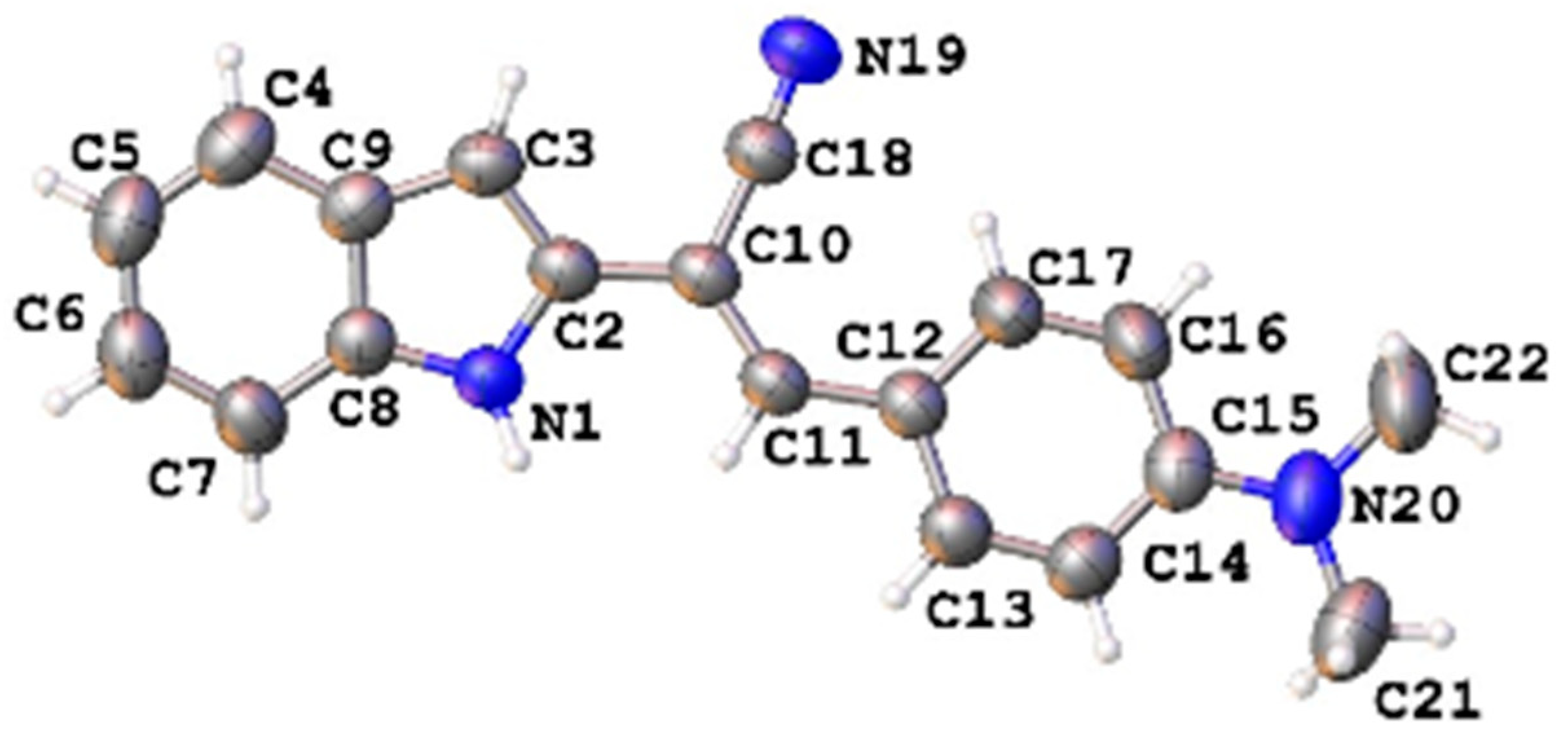
- 3-[4-(Dimethylamino)phenyl]-2-(1H-indol-2-yl)acrylonitrile (2l). Yield: 37% (brown solid); m.p. 234–236 °C; IR νmax (KBr, cm−1): 3334, 3079, 3013, 2989, 2814, 2211, 1611, 1575, 1364, 1198, 810, 788, 746; 1H NMR (400 MHz, DMSO-d6) δ (ppm): 11.55 (s, 1H, NH), 7.80 (d, J = 8.9 Hz, 2H, 2 × C-Harom), 7.75 (s, 1H, CH), 7.55 (d, J = 7.8 Hz, 1H, C-Harom), 7.39 (d, J = 8.1 Hz, 1H, C-Harom), 7.15 (t, J = 7.5 Hz, 1H, C-Harom), 7.02 (t, J = 7.4 Hz, 1H, C-Harom), 6.84 (d, J = 9.0 Hz, 2H, 2 × C-Harom), 6.64 (s, 1H, C3-H, indole), 3.03 (s, 6H, 2 × CH3); 13C NMR (100 MHz, DMSO-d6) δ (ppm): 152.1, 139.4, 138.0, 135.0, 131.1 (two overlapping signals), 128.6, 123.0, 121.2, 120.7, 120.2, 118.9, 112.3 (two overlapping signals), 111.5, 101.7, 96.3, 40.1 (two overlapping signals); MS (ESI) m/z: 286 [M − H]−. Anal. calcd for C19H17N3 (287.36) (%): C, 79.41; H, 5.96; N, 14.62. Found: C, 79.31; H, 5.87; N, 14.82.
- Crystal data for 2l: C19H17N3 (M = 287.35 g/mol), monoclinic, space group P21/n, a = 6.76580(10) Å, b = 21.9867(4) Å, c = 10.2650(2) Å, β = 93.006(2)°, V = 1524.90(5) Å3, Z = 4, μ(Cu Kα) = 0.587 mm−1, Dcalc = 1.252 g/cm3, 6428 reflections measured, 3114 unique (Rint = 0.0141, Rsigma = 0.0179), which were used in all calculations. The final R1 was 0.0412 (I > 2σ(I)) and wR2 was 0.1225 (all data).
- 3-[4-(Diethylamino)phenyl]-2-(1H-indol-2-yl)acrylonitrile (2m). Yield: 7% (brown solid); m.p. 183–185 °C; IR νmax (KBr, cm−1): 3336, 3047, 2967, 2925, 2866, 2219, 1609, 1584, 1512, 1356, 1200, 812, 799, 731; 1H NMR (400 MHz, DMSO-d6) δ (ppm): 11.54 (s, 1H, NH), 7.78 (d, J = 8.9 Hz, 2H, 2 × C-Harom), 7.72 (s, 1H, CH), 7.54 (d, J = 7.8 Hz, 1H, C-Harom), 7.38 (d, J = 8.1 Hz, 1H, C-Harom), 7.14 (t, J = 7.4 Hz, 1H, C-Harom), 7.02 (t, J = 7.4 Hz, 1H, C-Harom), 6.81 (d, J = 8.9 Hz, 2H, 2 × C-Harom), 6.62 (s, 1H, C3-H, indole), 3.44 (q, J = 6.9 Hz, 4H, 2 × CH2), 1.14 (t, J = 6.9 Hz, 6H, 2 × CH3); 13C NMR (100 MHz, DMSO-d6) δ (ppm): 149.6, 139.4, 137.9, 135.1, 131.5 (two overlapping signals), 128.6, 122.9, 120.7, 120.4, 120.2, 119.0, 111.8 (two overlapping signals), 111.5, 101.5, 95.5, 44.3 (two overlapping signals), 12.9 (two overlapping signals); MS (ESI) m/z: 314 [M − H]−; MS (ESI) m/z: 316 [M + H]+. Anal. calcd for C21H21N3 (315.41) (%): C, 79.97; H, 6.71; N, 13.32. Found: C, 79.75; H, 6.87; N, 13.38.
- 2-(1H-Indol-2-yl)-3-(4-nitrophenyl)acrylonitrile (2n). Yield: 27% (orange solid); m.p. 210–212 °C; IR νmax (KBr, cm−1): 3344, 2227, 1595, 1578, 1512, 1341, 1111, 787, 753, 684; 1H NMR (300 MHz, DMSO-d6) δ (ppm): 11.83 (s, 1H, NH), 8.36 (d, J = 8.9 Hz, 2H, C-Harom), 8.05 (d, J = 8.9 Hz, 2H, 2 × C-Harom), 8.00 (s, 1H, CH), 7.61 (d, J = 7.9 Hz, 1H, C-Harom), 7.42 (d, J = 7.7 Hz, 1H, C-Harom), 7.22 (t, J = 7.6 Hz, 1H, C-Harom), 7.05 (t, J = 7.9 Hz, 1H, C-Harom), 6.91 (s, 1H, C3-H, indole); 13C NMR (75 MHz, DMSO-d6) δ (ppm): 148.0, 140.2, 138.6, 135.6, 133.3, 130.2 (two overlapping signals), 128.1, 124.7, 124.6, 121.7, 120.7, 115.8, 114.9, 112.0, 107.2, 105.9; MS (ESI) m/z: 288 [M − H]−. Anal. calcd for C17H11N3O2 (289.29) (%): C, 70.58; H, 3.83; N, 14.53. Found: C, 70.67; H, 3.65; N, 14.87.
- 2-(1H-Indol-2-yl)-3-(naphthalen-1-yl)acrylonitrile (2o). Yield: 46% (yellow solid); m.p. 218–220 °C; IR νmax (KBr, cm−1): 3372, 3058, 3013, 2921, 2847, 2222, 1587, 1431, 1301, 882, 777, 728, 674; 1H NMR (400 MHz, DMSO-d6) δ (ppm): 11.98 (s, 1H), 8.67 (s, 1H, CH), 8.31 (d, J = 8.2 Hz, 1H, C-Harom), 8.13–8.05 (m, 3H, 3 × C-Harom), 7.71–7.64 (m, 4H, 4 × C-Harom), 7.48 (d, J = 8.2 Hz, 1H, C-Harom), 7.25 (t, J = 7.6 Hz, 1H, C-Harom), 7.09 (t, J = 7.5 Hz, 1H, C-Harom), 6.89 (s, 1H, C3-H, indole); 13C NMR (100 MHz, DMSO-d6) δ (ppm): 138.4, 136.4, 133.7, 133.7, 131.6, 131.2, 131.0, 129.3, 128.2, 127.6, 127.2, 126.9, 126.1, 124.3, 124.2, 121.5, 120.5, 117.4, 111.8, 107.1, 104.8; MS (ESI) m/z: 293 [M − H]−. Anal. calcd for C21H14N2 (294.35) (%): C, 85.69; H, 4.79; N, 9.52. Found: C, 85.58; H, 4.82; N, 9.32.
- 2-(1H-Indol-2-yl)-3-(naphthalen-2-yl)acrylonitrile (2p). Yield: 50% (yellow solid); m.p. 229–230 °C; IR νmax (KBr, cm−1): 3364, 3052, 2920, 2224, 1594, 1424, 1342, 915, 818, 769, 734, 673; 1H NMR (400 MHz, DMSO-d6) δ (ppm): 11.83 (br. s, 1H, NH), 8.35 (s, 1H, CH), 8.11–8.09 (m, 3H, 3 × C-Harom), 8.06–8.03 (m, 1H, C-Harom), 8.01–7.98 (m, 1H, C-Harom), 7.66–7.60 (m, 3H, 3 × C-Harom), 7.47–7.44 (m, 1H, C-Harom), 7.22 (t, J = 7,1 Hz, 1H, C-Harom), 7.07 (t, J = 7.1 Hz, 1H, C-Harom), 6.87 (s, 1H, C3-H, indole); 13C NMR (100 MHz, DMSO-d6) δ (ppm): 138.5, 138.4, 133.9, 133.2, 131.7, 130.2, 129.2, 129.1, 128.3, 128.24, 128.20, 127.6, 125.1, 124.0, 121.4, 120.5, 117.7, 111.9, 104.3, 103.9; MS (ESI) m/z: 293 [M − H]−. Anal. calcd for C21H14N2 (294.35) (%): C, 85.69; H, 4.79; N, 9.52. Found: C, 85.75; H, 4,67; N, 9.58.
- 2-(1H-Indol-2-yl)-3-(pyridin-2-yl)acrylonitrile (2q). Yield: 24% (yellow solid); m.p. 179–181 °C; IR νmax (KBr, cm−1): 3318, 3056, 3008, 2924, 2229, 1577, 1423, 1305, 1149, 895, 787, 723, 608; 1H NMR (300 MHz, DMSO-d6) δ (ppm): 11.81 (s, 1H, NH), 8.73 (m, 1H, C-Harom), 7.98–7.90 (m, 2H, C-Harom + CH), 7.67 (d, J = 7.9 Hz, 1H, C-Harom), 7.60 (d, J = 7.9 Hz, 1H, C-Harom), 7.45–7.40 (m, 2H, 2 × C-Harom), 7.20 (t, J = 7.6 Hz, 1H, C-Harom), 7.04 (t, J = 7.5 Hz, 1H, C-Harom), 6.90 (s, 1H, C3-H, indole); 13C NMR (75 MHz, DMSO-d6) δ (ppm): 152.0, 150.3, 138.5, 137.8, 136.4, 133.9, 128.2, 125.7, 125.0, 124.3, 121.5, 120.5, 117.0, 111.9, 106.3, 105.5; MS (ESI) m/z: 244 [M − H]−; MS (ESI) m/z: 246 [M + H]+. Anal. calcd for C16H11N3 (245.28) (%): C, 78.35; H, 4.52; N, 17.13. Found: C, 78.27; H, 4.38; N, 17.35.
- 2-(1H-Indol-2-yl)-3-(quinolin-2-yl)acrylonitrile (2r). Yield: 17% (orange solid); m.p. > 300 °C; IR νmax (KBr, cm−1): 3329, 3048, 2923, 2853, 2225, 1587, 1548, 1343, 886, 829, 796, 751, 728; 1H NMR (400 MHz, DMSO-d6) δ (ppm): 11.93 (s, 1H, NH), 8.53 (d, J = 8.4 Hz, 1H, C-Harom), 8.11 (s, 1H, CH), 8.06 (t, J = 8.6 Hz, 2H, 2 × C-Harom), 7.89–7.84 (m, 2H, 2 × C-Harom), 7.71–7.65 (m, 2H, 2 × C-Harom), 7.46 (d, J = 8.2 Hz, 1H, C-Harom), 7.25 (t, J = 7.1 Hz, 1H, C-Harom), 7.09 (t, J = 7.0 Hz, 1H, C-Harom), 7.00 (s, 1H, C3-H, indole); MS (ESI) m/z: 294 [M − H]−; MS (ESI) m/z: 296 [M + H]+. Anal. calcd for C20H13N3 (295.34) (%): C, 81.34; H, 4.44; N, 14.23. Found: C, 81.42; H, 4.32; N, 14.26.
- 2-(1H-Indol-2-yl)-3-(thiophen-2-yl)acrylonitrile (2s). Yield: 21% (yellow solid); m.p. 178–180 °C; IR νmax (KBr, cm−1): 3351 3107, 3040, 2924, 2853, 2220, 1577, 1225, 1408, 1301, 885, 779, 691, 607; 1H NMR (400 MHz, DMSO-d6) δ (ppm): 11.70 (s, 1H, NH), 8.13 (s, 1H, CH), 7.92 (d, J = 5.6 Hz, 1H, C-Harom), 7.68 (d, J = 4.6 Hz, 1H, C-Harom), 7.59 (d, J = 8.1 Hz, 1H, C-Harom), 7.41 (d, J = 9.0 Hz, 1H, C-Harom), 7.28–7.30 (m, 1H, C-Harom), 7.19 (t, J = 8.1 Hz, 1H, C-Harom), 7.05 (t, J = 7.1 Hz, 1H, C-Harom) 6.76 (s, 1H, C3-H, indole); 13C NMR (100 MHz, DMSO-d6) δ (ppm): 138.3, 137.8, 133.6, 133.4, 131.8, 131.7, 128.8, 128.4, 123.8, 121.2, 120.5, 117.7, 111.7, 103.8, 100.1; MS (ESI) m/z: 249 [M − H]−. Anal. calcd for C15H10N2S (250.32) (%): C, 71.97; H, 4.03; N, 11.19. Found: C, 71.85; H, 3.97; N, 11.27.
3.1.3. General Procedure for the Preparation of 2-(1H-Indol-2-yl)-3-acrylonitriles 2t–x
- 3-(5-Chlorothiophen-2-yl)-2-(1H-indol-2-yl)acrylonitrile (2t). Yield: 33% (green solid); m.p. 259–261 °C; IR νmax (KBr, cm−1): 3348, 3054, 2218, 1501, 1433, 1414, 1308, 1228, 786, 741, 608; 1H NMR (400 MHz, DMSO-d6) δ (ppm): 11.70 (s, 1H, NH), 8.03 (s, 1H, CH), 7.59 (d, J = 7.9 Hz, 1H, C-Harom), 7.51–7.49 (m, 1H, CHarom), 7.40 (d, J = 8.1 Hz, 1H, C-Harom), 7.34–7.32 (m, 1H, C-Harom), 7.19 (t, J = 7.6 Hz, 1H, C-Harom), 7.05 (t, J = 7.4 Hz, 1H), 6.77 (s, 1H, C3-H, indole); MS (ESI) m/z: 155 [M − H]−. Anal. calcd for C15H9ClN2S (284.76) (%): C, 63.27; H, 3.19; N, 9.84. Found: C, 63.15; H, 3.21; N, 9.72.
- 3-(Furan-2-yl)-2-(1H-indol-2-yl)acrylonitrile (2u). Yield: 53% (yellow solid); m.p. 185–186 °C; IR νmax (KBr, cm−1): 3321, 3056, 2924, 2231, 1607, 1469, 1346, 1303, 1019, 884, 781, 724; 1H NMR (400 MHz, DMSO-d6) δ (ppm): 11.71 (s, 1H, NH), 8.02 (s, 1H, CH), 7.76 (s, 1H, C-Harom), 7.58 (d, J = 7.9 Hz, 1H, C-Harom), 7.39 (d, J = 8.0 Hz, 1H, C-Harom), 7.19 (t, J = 7.6 Hz, 1H, C-Harom), 7.15 (d, J = 3.5 Hz, 1H, C-Harom), 7.05 (t, J = 7.3 Hz, 1H, C-Harom), 6.82–6.74 (m, 2H, 2 × C-Harom); 13C NMR (100 MHz, DMSO-d6) δ (ppm): 149.9, 146.7, 138.3, 133.5, 128.3, 124.7, 123.9, 121.2, 120.5, 117.3, 116.4, 113.8, 111.7, 104.0, 99.6; MS (ESI) m/z: 233 [M − H]−. Anal. calcd for C15H10N2O (234.25) (%): C, 76.91; H, 4.30; N, 11.96. Found: C, 76.83; H, 4.38; N, 11.86.
- 2-(1H-Indol-2-yl)-3-(5-methylfuran-2-yl)acrylonitrile (2v). Yield: 45% (yellow solid); m.p. 190–191 °C; IR νmax (KBr, cm−1): 3322, 3053, 2920, 2850, 2226, 1517, 1539, 1489, 1290, 1025, 781, 748; 1H NMR (400 MHz, DMSO-d6) δ (ppm): 11.64 (s, 1H, NH), 7.67 (s, 1H, CH), 7.56 (d, J = 7.9 Hz, 1H, C-Harom), 7.38 (d, J = 8.2 Hz, 1H, C-Harom), 7.17 (t, J = 7.6 Hz, 1H, C-Harom), 7.08–7.01 (m, 2H, 2 × C-Harom), 6.73 (s, 1H, C3-H, indole), 6.43 (d, J = 3.2 Hz, 1H, C-Harom), 2.41 (s, 3H, CH3); MS (ESI) m/z: 247 [M − H]−. Anal. calcd for C16H12N2O (248.28) (%): C, 77.40; H, 4.87; N, 11.28. Found: C, 77.46; H, 4.75; N, 11.32.
- 3-(5-Chlorofuran-2-yl)-2-(1H-indol-2-yl)acrylonitrile (2w). Yield: 26% (yellow solid); m.p. 223–228 °C; IR νmax (KBr, cm−1): 3333, 3045, 2227, 1519, 1469, 1345, 1020, 876, 774, 729; 1H NMR (400 MHz, DMSO-d6) δ (ppm): 11.71 (s, 1H, NH), 7.67 (s, 1H, CH), 7.59 (d, J = 7.9 Hz, 1H, C-Harom), 7.39 (d, J = 8.1 Hz, 1H, C-Harom), 7.23–7.16 (m, 2H, 2 × C-Harom), 7.05 (t, J = 7.8 Hz, 1H, C-Harom), 6.82 (d, J = 3.6 Hz, 1H, C-Harom), 6.80 (s, 1H, C3-H, indole); MS (ESI) m/z: 267 [M − H]−. Anal. calcd. for C15H9ClN2O (268.70) (%): C, 67.05; H, 3.38; N, 10.43. Found: C, 67.17; H, 3.46; N, 10.37.
- 2-(1H-Indol-2-yl)-3-(1H-pyrrol-2-yl)acrylonitrile (2x). Yield: 8% (brown, solid); m.p. 134–136 °C; IR νmax (KBr, cm−1): 3427, 3331, 3113, 3014, 2924, 2215, 1596, 1402, 1342, 1038, 786, 748, 663, 544; 1H NMR (400 MHz, DMSO-d6) δ (ppm): 11.62 (s, 1H, NH), 11.54 (s, 1H, NH), 7.64 (s, 1H, CH), 7.54 (d, J = 7.7 Hz, 1H, C-Harom), 7.38 (d, J = 8.0 Hz, 1H, C-Harom), 7.16–7.12 (m, 3H, 3 × C-Harom), 7.02 (t, J = 7.3 Hz, 1H, C-Harom), 6.62 (s, 1H, C3-H, indole), 6.36 (s, 1H, C-Harom); 13C NMR (100 MHz, DMSO-d6) δ (ppm): 138.0, 134.4, 129.4, 128.6, 127.6, 124.2, 122.9, 120.6, 120.2, 118.8, 112.9, 111.7, 111.6, 101.6, 95.2; MS (ESI) m/z: 232 [M − H]−; MS (ESI) m/z: 234 [M + H]+. Anal. calcd for C15H11N3 (233.27) (%): C, 77.23; H, 4.75; N, 18.01. Found: C, 77.31; H, 4.83; N, 17.86.
3.1.4. Synthesis of N-[4-(Dimethylamino)phenyl]-1H-indole-2-carbimidoyl cyanide (3)
3.1.5. General Procedure for the Preparation of 2-(1H-Indol-2-yl)-3-phenylpropanenitriles 4a–c
- 2-(1H-Indol-2-yl)-3-(4-methoxyphenyl)propanenitrile (4a). Yield: 73% (white solid); m.p. 151–153 °C; IR νmax (KBr, cm−1): 3397, 3044, 3009, 2935, 2839, 2242, 1612, 1512, 1242, 1177, 1032, 800, 739, 667; 1H NMR (400 MHz, DMSO-d6) δ (ppm): 11.43 (s, 1H, NH), 7.50 (d, J = 7.9 Hz, 1H, C-Harom), 7.39 (d, J = 8.9 Hz, 1H, C-Harom), 7.21 (d, J = 8.7 Hz, 2H, 2 × C-Harom), 7.11 (t, J = 7.0 Hz, 1H, C-Harom), 7.00 (t, J = 7.5 Hz, 1H, C-Harom), 6.88 (d, J = 8.7 Hz, 2H, 2 × C-Harom), 6.39 (s, 1H, C3-H, indole), 4.72–4.65 (m, 1H, CH), 3.73 (s, 3H, CH3), 3.32–3.18 (m, 2H, CH2); 13C NMR (100 MHz, DMSO-d6) δ (ppm): 158.7, 136.9, 133.2, 130.6 (two overlapping signals), 129.3, 127.8, 122.0, 120.5, 120.4, 119.7, 114.2 (two overlapping signals), 111.7, 100.8, 55.4, 37.7, 33.0; MS (ESI) m/z: 275 [M − H]−. Anal. calcd for C18H16N2O (276.33): C, 78.24; H, 5.84; N, 10.14; O, 5.79. Found: C, 78.32; H, 5.92; N, 10.06.
- 3-[4-(Dimethylamino)phenyl]-2-(1H-indol-2-yl)propanenitrile (4b). Yield: 75% (beige solid); m.p. 180–183 °C; IR νmax (KBr, cm−1): 3400, 3355, 3048, 2923, 2857, 2810, 2240, 1613, 1524, 1357, 1344, 1191, 796, 752, 743, 667; 1H NMR (400 MHz, DMSO-d6) δ (ppm): 11.42 (s, 1H, NH), 7.50 (d, J = 7.9 Hz, 1H, C-Harom), 7.40 (d, J = 8.1 Hz, 1H, C-Harom), 7.14–7.09 (m, 3H, 3 × C-Harom), 7.00 (t, J = 7.0 Hz, 1H, C-Harom), 6.67 (d, J = 8.7 Hz, 2H, 2 × C-Harom), 6.40 (s, 1H, C3-H, indole), 4.60–4.64 (m, 1H, CH), 3.28–3.14 (m, 2H, CH2), 2.86 (s, 6H, 2 × CH3); MS (ESI) m/z: 288 [M − H]−. Anal. calcd for C19H19N3 (289.37) (%): C, 78.86; H, 6.62; N, 14.52. Found: C, 78.76; H, 6.58; N, 14.66.
- 2-(1H-Indol-2-yl)-3-(naphthalen-2-yl)propanenitrile (4c). Yield: 63% (beige solid); m.p. 164–166 °C; IR νmax (KBr, cm−1): 3392, 3046, 2924, 2242, 1455, 1426, 1290, 802, 752, 743, 667; 1H NMR (300 MHz, DMSO-d6) δ (ppm): 11.50 (s, 1H, NH), 7.88–7.83 (m, 4H, 4 × C-Harom), 7.51–7.38 (m, 5H, 5 × C-Harom), 7.10 (t, J = 7.7 Hz, 1H, C-Harom), 6.98 (t, J = 7.6 Hz, 1H, C-Harom), 6.42 (s, 1H, C3-H, indole), 4.90–4.85 (m, 1H, CH), 3.58–3.41 (m, 2H, CH2); 13C NMR (75 MHz, DMSO-d6) δ (ppm): 136.9, 135.1, 133.3, 133.1, 132.5, 128.4, 128.1, 128.0, 127.9, 127.8, 127.7, 126.7, 126.3, 122.0, 120.5, 120.3, 119.7, 111.7, 100.9, 38.6, 32.6; MS (ESI) m/z: 295 [M − H]−. Anal. calcd for C21H16N2 (296.37) (%): C, 85.11; H, 5.44; N, 9.45. Found: C, 85.23; H, 5.38; N, 9.39.
3.1.6. General Procedure for the Preparation of 2-(1-Methyl-1H-indol-2-yl)-3-acrylonitriles 5a–d
- 2-(1-Methyl-1H-indol-2-yl)-3-(p-tolyl)acrylonitrile (5a). Yield: 77% (beige solid); m.p. 90–92 °C; IR νmax (KBr, cm−1): 3040, 2914, 2854, 2223, 1468, 1359, 1343, 1187, 815, 785, 750, 731; 1H NMR (400 MHz, DMSO-d6) δ (ppm): 7.89 (d, J = 8.2 Hz, 2H, 2 × C-Harom), 7.73 (s, 1H, CH), 7.61 (d, J = 7.9 Hz, 1H, C-Harom), 7.54 (d, J = 8.3 Hz, 1H, C-Harom), 7.38 (d, J = 8.1 Hz, 2H, 2 × C-Harom), 7.26 (t, J = 7.3 Hz, 1H, C-Harom), 7.11 (t, J = 7.3 Hz, 1H, C-Harom), 6.80 (s, 1H, C3-H, indole), 3.87 (s, 3H, CH3), 2.40 (s, 3H, CH3); 13C NMR (100 MHz, DMSO-d6) δ (ppm): 146.9, 141.7, 139.1, 135.4, 131.2, 130.1 (two overlapping signals), 129.7 (two overlapping signals), 127.2, 123.3, 121.1, 120.6, 118.2, 110.8, 104.0, 101.0, 31.7, 21.6. Anal. calcd for C19H16N2 (272.34) (%): C, 83.79; H, 5.92; N, 10.29. Found: C, 83.73; H, 5.86; N, 10.41.
- 3-(4-Methoxyphenyl)-2-(1-methyl-1H-indol-2-yl)acrylonitrile (5b). Yield: 96% (yellow solid); m.p. 113–114 °C; IR νmax (KBr, cm−1): 3050, 3000, 2924, 2853, 2210, 1601, 1506, 1461, 1253, 1180, 1031, 828, 798, 754, 744; 1H NMR (400 MHz, DMSO-d6) δ (ppm): 7.98 (d, J = 8.8 Hz, 2H, 2 × C-Harom), 7.69 (s, 1H, CH), 7.60 (d, J = 7.8 Hz, 1H, C-Harom), 7.52 (d, J = 8.3 Hz, 1H, C-Harom), 7.25 (t, J = 7.1 Hz, 1H, C-Harom), 7.15–7.11 (m, 3H, 3 × C-Harom), 6.76 (s, 1H, C3-H, indole), 3.86 (s, 6H, NCH3 + OCH3); 13C NMR (100 MHz, DMSO-d6) δ (ppm): 161.9, 146.8, 139.0, 135.6, 131.7 (two overlapping signals), 127.3, 126.5, 123.1, 121.0, 120.6, 118.5, 115.1 (two overlapping signals), 110.8, 103.6, 98.9, 56.0, 31.7. Anal. calcd for C19H16N2O (288.34) (%): C, 79.14; H, 5.59; N, 9.72. Found: C, 79.22; H, 5.51; N; 9.68.
- 3-[4-(Dimethylamino)phenyl]-2-(1-methyl-1H-indol-2-yl)acrylonitrile (5c). Yield: 92% (brown solid); m.p. 131–132 °C; IR νmax (KBr, cm−1): 3106, 3035, 2887, 2804, 2218, 1608, 1588, 1361, 1197, 818, 792, 732; 1H NMR (400 MHz, DMSO-d6) δ (ppm): 7.89 (d, J = 8.9 Hz, 2H, 2 × C-Harom), 7.57 (d, J = 7.8 Hz, 1H, C-Harom), 7.52–7.49 (m, 2H, C-Harom + CH), 7.22 (t, J = 7.3 Hz, 1H, C-Harom), 7.09 (t, J = 7.4 Hz, 1H, C-Harom), 6.83 (d, J = 9.0 Hz, 2H, 2 × C-Harom), 6.67 (s, 1H, C3-H, indole), 3.84 (s, 3H, CH3), 3.04 (s, 6H, 2 × CH3); 13C NMR (100 MHz, DMSO-d6) δ (ppm): 152.4, 147.6, 138.7, 136.5, 131.7 (two overlapping signals), 127.4, 122.6, 121.1, 120.7, 120.4, 119.4, 112.1 (two overlapping signals), 110.6, 102.7, 93.9, 40.1 (two overlapping signals), 31.6; MS (ESI) m/z: 302 [M + H]+. Anal. calcd. for C20H19N3 (301.38) (%): C, 79.70; H, 6.35; N, 13.94. Found: C, 79.76; H, 6.27; N, 13.97.
- 2-(1-Methyl-1H-indol-2-yl)-3-(naphthalen-2-yl)acrylonitrile (5d). Yield: 84% (yellow solid); m.p. 160–163 °C; IR νmax (KBr, cm−1): 3054, 2925, 2853, 2207, 1599, 1461, 1356, 931, 803, 791, 744; 1H NMR (400 MHz, DMSO-d6) δ (ppm): 8.46 (s, 1H, C-Harom), 8.17 (d, J = 8.7 Hz, 1H, C-Harom), 8.10 (d, J = 8.7 Hz, 1H, C-Harom), 8.02 (t, J = 6.8 Hz, 1H, C-Harom), 7.94 (s, 1H, CH), 7.69–7.60 (m, 3H, 3 × C-Harom), 7.56 (d, J = 8.3 Hz, 1H, C-Harom), 7.28 (t, J = 7.7 Hz, 1H, C-Harom), 7.13 (t, J = 7.2 Hz, 1H, C-Harom), 6.87 (s, 1H, C3-H, indole), 3.93 (s, 3H, CH3); 13C NMR (100 MHz, DMSO-d6) δ (ppm): 146.6, 139.2, 135.3, 134.2, 133.1, 131.6, 131.1, 129.2, 129.1, 128.5, 128.3, 127.6, 127.2, 125.3, 123.4, 121.2, 120.7, 118.2, 110.9, 104.3, 102.5, 31.9. Anal. calcd for C22H16N2 (308.38) (%): C, 85.69; H, 5.23; N, 9.08. Found: C, 85.75; H, 5.31; N, 8.94.
3.1.7. General Procedure for the Preparation of 2-(1-Acetyl-1H-indol-2-yl)-3-acrylonitriles 6a and 6b and 3-(4-Methoxyphenyl)-2-(1-(methylsulfonyl)-1H-indol-2-yl)acrylonitrile (7)
- 2-(1-Acetyl-1H-indol-2-yl)-3-(4-methoxyphenyl)acrylonitrile (6a). Yield: 42% (beige solid); m.p. 119–122 °C; IR νmax (KBr, cm−1): 3112, 3085, 3022, 2962, 2932, 2836, 2207, 1702, 1604, 1515, 1450, 1366, 1306, 1263, 1189, 1027, 837, 827, 740; 1H NMR (400 MHz, DMSO-d6) δ (ppm): 8.01 (d, J = 8.4 Hz, 1H, C-Harom), 7.94 (d, J = 8.9 Hz, 2H, 2 × C-Harom), 7.68 (d, J = 7.6 Hz, 1H, C-Harom), 7.60 (s, 1H, CH), 7.42 (t, J = 7.3 Hz, 1H, C-Harom), 7.33 (t, J = 7.5 Hz, 1H, C-Harom), 7.13 (d, J = 8.8 Hz, 2H, 2 × C-Harom), 7.05 (s, 1H, C3-H, indole), 3.86 (s, 3H, CH3), 2.80 (s, 3H, CH3); 13C NMR (100 MHz, DMSO-d6) δ (ppm): 170.4, 161.9, 144.8, 136.5, 135.4, 131.6 (two overlapping signals), 129.1, 126.3, 126.0, 124.0, 121.9, 118.0, 115.6, 115.1 (two overlapping signals), 113.3, 101.7, 56.0, 27.3. Anal. calcd for C20H16N2O2 (316.35) (%): C, 75.93; H, 5.10; N, 8.86. Found: C, 75.85; H, 4.88; N, 8.94.
- 2-(1-Acetyl-1H-indol-2-yl)-3-[4-(dimethylamino)phenyl]acrylonitrile (6b). Yield: 31% (yellow solid); m.p. 164–166 °C; IR νmax (KBr, cm−1): 3115, 2899, 2817, 2204, 1692, 1612, 1579, 1524, 1450, 1367, 1303, 1169, 809, 761, 742; 1H NMR (400 MHz, DMSO-d6) δ (ppm): 8.04 (d, J = 8.2 Hz, 1H, C-Harom), 7.85 (d, J = 9.0 Hz, 2H, 2 × C-Harom), 7.65 (d, J = 7.6 Hz, 1H, C-Harom), 7.47 (s, 1H, CH), 7.39 (t, J = 7.2 Hz, 1H, C-Harom), 7.31 (t, J = 7.4 Hz, 1H, C-Harom), 6.97 (s, 1H, C3-H, indole), 6.83 (d, J = 9.0 Hz, 2H, 2 × C-Harom), 3.05 (s, 6H, 2 × CH3), 2.76 (s, 3H, CH3); 13C NMR (100 MHz, DMSO-d6) δ (ppm): 170.6, 152.5, 146.0, 136.7, 136.1, 131.6 (two overlapping signals), 129.1, 125.7, 124.0, 121.5, 120.8, 115.6, 112.6, 112.1 (two overlapping signals), 112.0, 96.5, 49.1 (two overlapping signals), 27.3; MS (ESI) m/z: 330 [M + H]+. Anal. calcd for C21H19N3O (329.40) (%): C, 76.57; H, 5.81; N, 12.76. Found: C, 76.65; H, 5.75; N, 12.82.
- 3-(4-Methoxyphenyl)-2-(1-(methylsulfonyl)-1H-indol-2-yl)acrylonitrile (7). Yield: 22% (white solid); m.p. 169–171 °C; IR νmax (KBr, cm−1): 3109, 3075, 3003, 2972, 2922, 2214, 1608. 1512, 1369, 1167, 1070, 833, 773, 747, 556; 1H NMR (400 MHz, DMSO-d6) δ (ppm): 7.95–7.93 (m, 3H, 3 × C-Harom), 7.74–7.71 (m, 2H, C-Harom + CH), 7.47 (t, J = 7.2 Hz, 1H, C-Harom), 7.39 (t, J = 7.5 Hz, 1H, C-Harom), 7.16–7.14 (m, 3H, 2 × C-Harom + C3-H indole), 3.86 (s, 3H, CH3), 3.31 (s, 3H, CH3); 13C NMR (100 MHz, DMSO-d6) δ (ppm): 162.1, 147.0, 137.3, 135.7, 131.8 (two overlapping signals), 129.5, 126.3, 126.1, 124.8, 122.2, 118.1, 115.1 (two overlapping signals), 115.0, 114.0, 99.7, 56.0, 40.9. Anal. calcd for C19H16N2O3S (352.41) (%): C, 64.76; H, 4.58; N, 7.95. Found: C, 64.68; H, 4.64; N, 7.87.
3.2. Biology
3.2.1. Evaluation of In Vitro Antiproliferative Activity
3.2.2. In Vitro Antimicrobial Activity
3.3. Computational Studies
3.3.1. Preparation of Ligands and Proteins for Modeling
3.3.2. Molecular Docking
3.3.3. ADME/Drug-Likeness Calculation
4. Conclusions
Supplementary Materials
Author Contributions
Funding
Institutional Review Board Statement
Informed Consent Statement
Data Availability Statement
Acknowledgments
Conflicts of Interest
References
- Kumar, S.; Ritika. A brief review of the biological potential of indole derivatives. Future J. Pharm. Sci. 2020, 6, 121. [Google Scholar] [CrossRef]
- Sravanthi, T.V.; Manju, S.L. Indoles—A promising scaffold for drug development. Eur. J. Pharm. Sci. 2016, 91, 1–10. [Google Scholar] [CrossRef] [PubMed]
- Chadha, N.; Silakari, O. Indoles as therapeutics of interest in medicinal chemistry: Bird’s eye view. Eur. J. Med. Chem. 2017, 134, 159–184. [Google Scholar] [CrossRef]
- Kumaria, A.; Singh, R.K. Medicinal chemistry of indole derivatives: Current to future therapeutic prospectives. Bioorg. Chem. 2019, 89, 103021. [Google Scholar] [CrossRef]
- Kumar, D.; Sharma, S.; Kalra, S.; Singh, G.; Monga, V.; Kumar, B. Medicinal perspective of indole derivatives: Recent developments and structure-activity relationship studies. Curr. Drug Targets 2020, 21, 864–891. [Google Scholar]
- Ferreira, S.H.; Moncada, S.; Vane, J.R. Indomethacin and aspirin abolish prostaglandin release from the spleen. Nature 1971, 231, 237–239. [Google Scholar] [CrossRef] [PubMed]
- Mazzotta, S.; Frattaruolo, L.; Brindisi, M.; Ulivieri, C.; Vanni, F.; Brizzi, A.; Carullo, G.; Cappello, A.R.; Aiello, F. 3-Amino-alkylated indoles: Unexplored green products acting as anti-inflammatory agents. Future Med. Chem. 2020, 12, 5–17. [Google Scholar] [CrossRef]
- Jasiewicz, B.; Kozanecka Okupnik, W.; Przygodzki, M.; Warżajtis, B.; Rychlewska, U.; Pospieszny, T.; Mrówczyńska, L. Synthesis, antioxidant and cytoprotective activity evaluation of C-3 substituted indole derivatives. Sci. Rep. 2021, 11, 15425. [Google Scholar] [CrossRef]
- Gurer-Orhan, H.; Karaaslan, C.; Ozcan, S.; Firuzi, O.; Tavakkoli, M.; Saso, L.; Suzen, S. Novel indole-based melatonin analogues: Evaluation of antioxidant activity and protective effect against amyloid β-induced damage. Bioorg. Med. Chem. 2016, 24, 1658–1664. [Google Scholar] [CrossRef]
- Kerzare, D.R.K.; Menghani, S.S.; Khedekar, P.B. Synthesis, characterization, antidepressant activity and docking studies of some novel indole bearing azetidinone derivatives. Drugs 2018, 52, 110–120. [Google Scholar] [CrossRef] [Green Version]
- Gras, J.; Llenas, J.; Jansat, J.M.; Jáuregui, J.; Cabarrocas, X.; Palacios, J.M. Almotriptan, a new anti-migraine agent: A review. CNS Drug Rev. 2002, 8, 217–234. [Google Scholar] [CrossRef]
- Tan, C.; Yang, S.-J.; Zhao, D.-H.; Li, J.; Yin, L.-Q. Antihypertensive activity of indole and indazole analogues: A review. Arab. J. Chem. 2022, 15, 103756. [Google Scholar] [CrossRef]
- Muhammad Taha, M.; Alrashedy, A.S.; Almandil, N.B.; Iqbal, N.; Anouar, E.H.; Nawaz, M.; Uddin, N.; Chigurupati, S.; Wadood, A.; Rahim, F.; et al. Synthesis of indole derivatives as diabetics II inhibitors and enzymatic kinetics study of α-glucosidase and α-amylase along with their in-silico study. Int. J. Biol. Macromol. 2021, 190, 301–318. [Google Scholar] [CrossRef]
- Zhu, Y.; Zhao, J.; Luo, L.; Gao, Y.; Bao, H.; Li, P.; Zhang, H. Research progress of indole compounds with potential antidiabetic activity. Eur. J. Med. Chem. 2021, 223, 113665. [Google Scholar] [CrossRef] [PubMed]
- Dorababu, A. Indole—A promising pharmacophore in recent antiviral drug discovery. RSC Med. Chem. 2020, 11, 1335–1353. [Google Scholar] [CrossRef]
- Reddy, G.S.; Manojit Pal, M. Indole derivatives as anti-tubercular agents: An overview on their synthesis and biological activities. Curr. Med. Chem. 2021, 28, 4531–4568. [Google Scholar] [CrossRef]
- Nieto, M.J.; Lupton, H.K. Indole and indoline scaffolds in antimicrobials: Overview, synthesis and recent advances in antimicrobial research. Curr. Med. Chem. 2021, 28, 4828–4844. [Google Scholar] [CrossRef] [PubMed]
- Liu, Y.; Cui, Y.; Lu, L.; Gong, Y.; Han, W.; Piao, G. Natural indole-containing alkaloids and their antibacterial activities. Arch. Pharm. 2020, 353, e2000120. [Google Scholar] [CrossRef] [PubMed]
- Zhang, Y.; Rosado-Lugo, J.D.; Datta, P.; Sun, Y.; Cao, Y.; Banerjee, A.; Yuan, Y.; Parhi, A.K. Evaluation of a conformationally constrained indole carboxamide as a potential efflux pump inhibitor in Pseudomonas aeruginosa. Antibiotics 2022, 11, 716. [Google Scholar] [CrossRef]
- Xuecheng, C.; Liang, H.; Yanpeng, X.; Yurong, Z.; Yue, L.; Yalan, P.; Zhong, C.; Jie, Z.; Zhijian, Y.; Shiqing, H. Development of 2-alkyl-5-((phenylsulfonyl)oxy)-1H-indole-3-carboxylate derivatives as potential anti-biofilm agents. ChemistrySelect 2023, 8, e202204226. [Google Scholar] [CrossRef]
- Yuan, W.; Yu, Z.; Song, W.; Li, Y.; Fang, Z.; Zhu, B.; Li, X.; Wang, H.; Hong, W.; Sun, N. Indole-core-based novel antibacterial agent targeting FtsZ. Infect. Drug Resist. 2019, 12, 2283–2296. [Google Scholar] [CrossRef] [PubMed] [Green Version]
- Qin, H.-L.; Liu, J.; Fang, W.-Y.; Ravindar, L.; Rakesh, K.P. Indole-based derivatives as potential antibacterial activity against methicillin-resistance Staphylococcus aureus (MRSA). Eur. J. Med. Chem. 2020, 194, 112245. [Google Scholar] [CrossRef]
- Wan, Y.; Li, Y.; Yan, Y.; Yan, M.; Tang, Z. Indole: A privileged scaffold for the design of anti-cancer agents. Eur. J. Med. Chem. 2019, 183, 111691. [Google Scholar] [CrossRef]
- Dadashpour, S.; Emami, S. Indole in the target-based design of anticancer agents: A versatile scaffold with diverse mechanisms. Eur. J. Med. Chem. 2018, 150, 9–29. [Google Scholar] [CrossRef]
- Mehra, A.; Sharma, V.; Verma, A.; Venugopal, S.; Mittal, A.; Singh, G. Indole derived anticancer agents. ChemistrySelect 2022, 7, e202202361. [Google Scholar] [CrossRef]
- Almagro, L.; Fernández-Pérez, F.; Pedreño, M.A. Indole alkaloids from Catharanthus roseus: Bioproductiom and their effect on human health. Molecules 2015, 20, 2973–3000. [Google Scholar] [CrossRef] [Green Version]
- Shirley, M. Dacomitinib: First global approval. Drugs 2018, 78, 1947–1953. [Google Scholar] [CrossRef]
- Amin, S.A.; Ghosh, K.; Mondal, D.; Jha, T.; Gayen, S. Exploring indole derivatives as myeloid cell leukaemia-1 (Mcl-1) inhibitors with multi-QSAR approach: A novel hope in anti-cancer drug discovery. New J. Chem. 2020, 44, 17494–17506. [Google Scholar] [CrossRef]
- Alnabulsi, S.; Al-Hurani, E.A. Pim kinase inhibitors in cancer: Medicinal chemistry insights into their activity and selectivity. Drug Discov. 2020, 25, 2062–2069. [Google Scholar] [CrossRef]
- Panathur, N.; Gokhale, N.; Dalimba, U.; Koushik, P.V.; Yogeeswari, P.; Dharmarajan, S. New indole–isoxazolone derivatives: Synthesis, characterisation and in vitro SIRT1 inhibition studies. Bioorg. Med. Chem. Lett. 2015, 25, 2768–2772. [Google Scholar] [CrossRef] [PubMed]
- Xie, F.; Kniess, T.; Neuber, C.; Deuther-Conrad, W.; Mamat, C.; Lieberman, B.P.; Liu, B.; Mach, R.H.; Brust, P.; Steinbach, J.; et al. Novel indole-based sigma-2 receptor ligands: Synthesis, structure-affinity relationship and antiproliferative activity. Med. Chem. Commun. 2015, 6, 1093–1103. [Google Scholar] [CrossRef] [Green Version]
- Atadja, P. Development of the pan-DAC inhibitor panobinostat (LBH589): Successes and challenges. Cancer Lett. 2009, 280, 233–241. [Google Scholar] [CrossRef]
- Li, A.-L.; Hao, Y.; Wang, W.-Y.; Liu, Q.-S.; Sun, Y.; Gu, W. Design, synthesis, and anticancer evaluation of novel indole derivatives of ursolic acid as potential topoisomerase II inhibitors. Int. J. Mol. Sci. 2020, 21, 2876. [Google Scholar] [CrossRef] [Green Version]
- Tan, O.U.; Zengin, M. Insights into the chemistry and therapeutic potential of acrylonitrile derivatives. Arch. Pharm. 2022, 355, e2100383. [Google Scholar]
- Sivaramakarthikeyan, R.; Iniyaval, S.; Saravanan, V.; Lim, W.-M.; Mai, C.-W.; Ramalingan, C. Molecular hybrids integrated with benzimidazole and pyrazole structural motifs: Design, synthesis, biological evaluation, and molecular docking studies. ACS Omega 2020, 209, 10089–10098. [Google Scholar] [CrossRef]
- Ahamed, J.I.; Priya, M.; Vinothkumar, P.; Sathyamoorthy, K.; MuraliManohar, P.; Liu, J.; Valan, M.F. A combined experimental and DFT computations study of novel (E)-3-(benzofuran-2-yl)-2-(thiophen-2-yl)acrylonitrile(TACNBNF): Insight into the synthesis, single crystal XRD, NMR, vibrational spectral analysis, in vitro antioxidant and in silico molecular docking investigation with human peroxiredoxin 5 protein. J. Mol. Struct. 2020, 1202, 127241. [Google Scholar]
- Solangi, M.; Kanwal; Khan, K.M.; Saleem, F.; Hameed, S.; Iqbal, J.; Shafique, Z.; Qureshi, U.; Ul-Haq, Z.; Taha, M.; et al. Indole acrylonitriles as potential anti-hyperglycemic agents: Synthesis, α-glucosidase inhibitory activity and molecular docking studies. Bioorg. Med. Chem. 2020, 28, 115605. [Google Scholar] [CrossRef]
- Han, Y.-S.; Penthala, N.R.; Oliveira, M.; Mespléde, T.; Xu, H.; Quan, Y.; Crooks, P.A.; Wainberg, M.A. Identification of resveratrol analogs as potent anti-dengue agents using a cell-based assay. J. Med. Virol. 2017, 89, 397–407. [Google Scholar] [CrossRef] [PubMed]
- Pan, T.; He, X.; Chen, B.; Chen, H.; Geng, G.; Luo, H.; Zhang, H.; Bai, C. Developmnent of benzimidazole derivatives to inhibit HIV-1 replication through protecting APOBEC3G protein. Eur. J. Med. Chem. 2015, 95, 500–513. [Google Scholar] [CrossRef]
- Sharma, K.; Shrivastava, A.; Mehra, R.N.; Deora, G.S.; Alam, M.M.; Zaman, M.S.; Akhter, M. Synthesis of novel benzimidazole acrylonitriles for inhibition of Plasmodium falciparum growth by dual target inhibition. Arch. Pharm. Chem. Life Sci. 2017, 351, e1700251. [Google Scholar] [CrossRef]
- Sirim, M.M.; Krishna, V.S.; Sriram, D.; Tan, O.U. Novel benzimidazole-acrylonitrile hybrids and their derivatives: Design, synthesis and antimycobacterial activity. Eur. J. Med. Chem. 2020, 188, 112010. [Google Scholar] [CrossRef]
- De-la-Torre, P.; Saavedra, L.A.; Caballero, J.; Quiroga, J.; Alzate-Morales, J.H.; Cabrera, M.G.; Trilleras, J. A novel class of selective acetylcholinesterase inhibitors: Synthesis and evaluation of (E)-2-(benzo[d]thiazol-2-yl)-3-heteroarylacrylonitriles. Molecules 2012, 17, 12072–12085. [Google Scholar] [CrossRef] [Green Version]
- De-la-Torre, P.; Treuer, A.V.; Gutierrez, M.; Poblete, H.; Alzate-Morales, J.H.; Trilleras, J.; Astudillo-Saavedra, L.; Caballero, J. Synthesis and in silico analysis of the quantitative structure-activity relationship of heteroaryl-acrylonitriles as AChE inhibitors. J. Taiwan Inst. Chem. Eng. 2016, 59, 45–60. [Google Scholar] [CrossRef]
- AlNeyadi, S.; Salem, A.A.; Ghattas, M.A.; Atatrech, N.; Abdou, I. Antibacterial activity and mechanism of action of the benzazole acrylonitrile-based compounds: In vitro, spectroscopic, and docking studies. Eur. J. Med. Chem. 2017, 136, 270–282. [Google Scholar] [CrossRef] [PubMed]
- Perin, N.; Hok, L.; Beč, A.; Vanstreels, E.; Daelemans, D.; Vianello, R.; Hranjec, M. N-substituted benzimidazole acrylonitryles as in vitro tubulin polymerization inhibitors: Synthesis, biological activity and computational analysis. Eur. J. Med. Chem. 2021, 211, 113003. [Google Scholar] [CrossRef] [PubMed]
- Beč, A.; Hok, L.; Persoons, L.; Vanstreels, E.; Daelemans, D.; Vianello, R.; Hranjec, M. Synthesis, computational analysis, and antiproliferative activity of novel benzimidazole acrylonitriles as tubulin polymerization inhibitors: Part 2. Pharmaceuticals 2021, 14, 1052. [Google Scholar] [CrossRef] [PubMed]
- Riu, F.; Sanna, L.; Ibba, R.; Piras, S.; Bordono, V.; Scorciapino, M.A.; Lai, M.; Sestito, S.; Bagella, L.; Carta, A. A comprehensive assessment of a new series of 5′,6′-difluorobenzotriazole-acrylonitrile derivatives as mictrotubule targeting agents (MTAs). Eur. J. Med. Chem. 2021, 222, 113590. [Google Scholar] [CrossRef]
- Briguglio, I.; Laurini, E.; Pirisi, M.A.; Piras, S.; Corona, P.; Fermeglia, M.; Pricl, S.; Carta, A. Triazolopyridinyl-acrylonitrile derivatives as antimicrotubule agents: Synthesis, in vitro and in silico characterization of antiproliferative activity, inhibition of tubulin polymerization and binding thermodynamics. Eur. J. Med. Chem. 2017, 141, 460–472. [Google Scholar] [CrossRef] [PubMed]
- Hranjec, M.; Pavlović, G.; Marjanović, M.; Kralj, M.; Kaminska-Zamola, G. Benzimidazole derivatives related to 2,3-acrylonitriles, benzimidazo[1,2-a]quinolines and fluorenes: Synthesis, antitumor evaluation in vitro and crystal structure determination. Eur. J. Med. Chem. 2010, 45, 2405–2417. [Google Scholar] [CrossRef]
- Sączewski, F.; Reszka, P.; Gdaniec, M.; Grünert, R.; Bednarski, P.J. Synthesis, X-ray crystal structures, stabilities, an in vitro cytotoxic activities of new heteroarylacrylonitriles. J. Med. Chem. 2004, 47, 3438–3449. [Google Scholar] [CrossRef]
- Oberhuber, N.; Ghosh, H.; Nitzsche, B.; Dandawate, P.; Höpfner, M.; Schobert, R.; Biersack, B. Synthesis and anticancer evaluation of new indole-based tyrphostin derivatives and their (p-cymene)dichloridoruthenium(II) complexes. Int. J. Mol. Sci. 2023, 24, 854. [Google Scholar] [CrossRef] [PubMed]
- Tcherniuk, S.; Skoufias, D.A.; Labriere, C.; Rath, O.; Gueritte, F.; Guillou, C.; Kozielski, F. Relocation of Aurora B and surviving from centromeres to the central spindle impaired by a kinesin-specific MKLP-2 inhibitor. Angew. Chem. Int. Ed. 2010, 49, 8228–8231. [Google Scholar] [CrossRef]
- Labriere, C.; Talapatra, S.K.; Thoret, S.; Bougeret, C.; Kozielski, F.; Guillou, C. New MKLP-2 inhibitors in the paprotrain series: Design, synthesis and biological evaluations. Bioorg. Med. Chem. 2016, 24, 721–734. [Google Scholar] [CrossRef] [PubMed]
- Sączewski, F.; Stencel, A.; Bieńczak, A.M.; Langowska, K.A.; Michaelis, M.; Werel, W.; Hałasa, R.; Reszka, P.; Bednarski, P.J. Structure-activity relationships of novel heteroaryl-acrylonitriles as cytotoxic and antibacterial agents. Eur. J. Med. Chem. 2008, 43, 1847–1857. [Google Scholar] [CrossRef]
- Baciocchi, E.; Muraglia, E.; Sleiter, G. Homolytic substitution reactions of electron-rich pentatomic heteroaromatics by electrophilic carbon-centered radicals. Synthesis of α-heteroarylacetic acids. J. Org. Chem. 1992, 57, 6817–6820. [Google Scholar] [CrossRef]
- Jiang, Y.; Wang, B.; Liu, D.; Xia, D.; Liu, Z.; Li, L.; Deng, G.; Yang, X. Aryl acrylonitriles synthesis enable by palladium-catalyzed α-alkenylation of arylacetonitriles with vinyl halides/triflates. Front. Chem. 2022, 10, 1091566. [Google Scholar] [CrossRef] [PubMed]
- Treuer, A.V.; De-La-Torre, P.; Gutiérrez, M.I. Synthesis of new (E)-2-(1H-indole-3-ylcarbonyl)-3-heteroaryl-acrylonitriles via microwave-assisted Knoevenagel condensation. J. Chem. 2017, 2017, 8418930. [Google Scholar] [CrossRef] [Green Version]
- Schaller, E.; Ma, A.; Gosch, C.L.; Klefenz, A.; Schaller, D.; Goehringer, N.; Kaps, L.; Schuppan, D.; Volkamer, A.; Schobert, R.; et al. New 3-aryl-2-(2-thienyl)acrylonitriles with high activity against hepatoma cells. Int. J. Mol. Sci. 2021, 22, 2243. [Google Scholar] [CrossRef] [PubMed]
- Ying, A.; Wang, L.; Qiu, F.; Hu, H.; Yang, J. Magnetic nanoparticle supported amine: An efficient and environmental benign catalyst for versatile Knoevenagel condensation under ultrasound irradiation. Comptes Rendus Chim. 2015, 18, 223–232. [Google Scholar] [CrossRef]
- Wilkens, J.; Kühling, A.; Blechert, S. Hetero-Cope rearrangements-VI. Short and stereoselective synthesis of 2-vinylindoles by a tandem process. Tetrahedron 1987, 43, 3237–3246. [Google Scholar] [CrossRef]
- Ying, A.; Qiu, F.; Wu, C.; Hu, H.; Yang, J. Ionic tagged amine supported on magnetic nanoparticles: Synthesis and application for versatile catalytic Knoevenagel condensation in water. RSC Adv. 2014, 4, 33175–33183. [Google Scholar] [CrossRef]
- Ying, A.; Ni, Y.; Xu, S.; Liu, S.; Yang, J.; Li, R. Novel DABCO based ionic liquids: Green and efficient catalysts with dual catalytic roles for aqueous Knoevenagel condensation. Int. Eng. Chem. Res. 2014, 53, 5678–5682. [Google Scholar] [CrossRef]
- Gao, X.; Gao, C.; Gao, R. Knoevenagel condensation reaction using ionic liquid [ADPQ][CF3SO3] as green and reusable catalyst. Asian J. Chem. 2015, 27, 2145–2148. [Google Scholar] [CrossRef]
- Grever, M.R.; Schepartz, S.A.; Chabner, B.A. The National Cancer Institute: Cancer drug discovery and development program. Semin. Oncol. 1992, 19, 622–638. [Google Scholar] [PubMed]
- Boyd, M.R.; Paull, K.D. Some practical considerations and applications of the National Cancer Institute in vitro anticancer drug discovery screen. Drug Dev. Res. 1995, 34, 91–109. [Google Scholar] [CrossRef]
- Shoemaker, R.H. The NCI60 human tumour cell line anticancer drug screen. Nat. Rev. 2006, 6, 813–823. [Google Scholar] [CrossRef]
- NCI-60 Screening Methodology. Available online: https://dtp.cancer.gov/discovery_development/nci-60/methodology.htm (accessed on 27 December 2022).
- Horváth, A.; Dobay, O.; Sahin-Tóth, J.; Juhász, E.; Pongrácz, J.; Iván, M.; Fazakas, E.; Kristóf, K. Characterisation of antibiotic resistance, virulence, clonality and mortality in MRSA and MSSA bloodstream infections at a tertiary-level hospital in Hungary: A 6 year retrospective study. Ann. Clin. Microbiol. Antimicrob. 2020, 19, 17. [Google Scholar] [CrossRef]
- McLoughlin, E.C.; O’Boyle, N.M. Colchicine-binding site inhibitors from chemistry to clinic: A review. Pharmaceuticals 2020, 13, 8. [Google Scholar] [CrossRef] [Green Version]
- Sun, K.; Sun, Z.; Zhao, F.; Shan, G.; Meng, Q. Recent advances in research of colchicine binding site inhibitors and their interaction modes with tubulin. Future Med. Chem. 2021, 13, 839–858. [Google Scholar] [CrossRef]
- Carneiro, B.A.; El-Deiry, W.S. Targeting apoptosis in cancer therapy. Nat. Rev. Clin. Oncol. 2020, 17, 395–417. [Google Scholar] [CrossRef]
- Ganesan, R.; Jelakovic, S.; Mittl, P.R.E.; Caflisch, A.; Grutter, M.G. In silico identification and crystal structure validation of caspase-3 inhibitors without a P1 aspartic acid moiety. Acta Cryst. 2011, F67, 842–850. [Google Scholar]
- Chao, Y.; Shiozaki, E.N.; Srinivasula, S.M.; Rigotti, D.J.; Fairman, R.; Shi, Y. Engineering a dimeric caspase-9: A re-evaluation of the induced proximity model for caspase activation. PLoS Biol. 2005, 3, e183. [Google Scholar] [CrossRef] [Green Version]
- Ahmad, S.; Pecqueur, L.; Dreie, B.; Hamdane, H.; Aumont-Nicaise, L.; Plückthun, A.; Knossow, M.; Gigant, B. Destabilizing an interacting motif strengthens the association of adesigned ankyrin repeat protein with tubulin. Sci. Rep. 2016, 6, 28922. [Google Scholar] [CrossRef] [PubMed] [Green Version]
- Kishida, H.; Unzai, S.; Roper, D.I.I.; Lloyd, A.; Park, S.-Y.; Tame, J.R.H. Crystal Structure of Penicillin Binding Protein 4 (dacB) from Escherichia coli, both in the Native Form and Covalently Linked to Various Antibiotics. Biochemistry 2006, 45, 783–792. [Google Scholar] [CrossRef]
- Strynadka, N.C.J.; Adachi, H.; Jensen, S.E.; Johns, K.; Sielecki, A.; Betzel, C.; Sutoh, K.; James, M.N.G. Molecular structure of the acyl-enzyme intermediate in β-lactam hydrolysis at 1.7 Å resolution. Nature 1992, 359, 700–705. [Google Scholar] [CrossRef]
- Daina, A.; Michielin, O.; Zoete, V. SwissADME: A free web tool to evaluate pharmacokinetics, drug-likeness and medicinal chemistry friendliness of small molecules. Sci. Rep. 2017, 7, 42717. [Google Scholar] [CrossRef] [Green Version]
- Lipinski, C.A.; Lombardo, F.; Dominy, B.W.; Feeney, P.J. Experimental and computational approaches to estimate solubility and permeability in drug discovery and development settings. Adv. Drug Deliv. Rev. 2012, 64, 4–17. [Google Scholar] [CrossRef]
- Rigaku Oxford Diffraction. CrysAlisPro Software System; Version 1.171.38.46; Rigaku Corporation: Oxford, UK, 2015. [Google Scholar]
- Sheldrick, G.M. SHELXT-Integrated space-group and crystal-structure determination. Acta Cryst. 2015, A71, 3–8. [Google Scholar] [CrossRef] [PubMed] [Green Version]
- Sheldrick, G.M. Crystal structure refinement with SHELXL. Acta Cryst. 2015, C71, 3–8. [Google Scholar]
- Dolomanov, O.V.; Bourhis, L.J.; Gildea, R.J.; Howard, J.A.K.; Puschmann, H. OLEX2: A complete structure solution, refinement and analysis program. J. Appl. Cryst. 2009, 42, 339–341. [Google Scholar] [CrossRef]
- Clinical and Laboratory Standards Institute. Performance Standards for Antimicrobial Susceptibility Testing; Twenty-Second Informational Supplement; Document M100-S22; Clinical and Laboratory Standards Institute: Wayne, PA, USA, 2017. [Google Scholar]
- RCBS Protein Data Bank. Available online: https://www.rcsb.org/ (accessed on 3 April 2023).
- Release 4.1.2.2; OpenEye Scientific Software, Inc.: Santa Fe, NM, USA; Available online: www.eyesopen.com (accessed on 3 April 2023).
- McGann, M.R.; Almond, H.R.; Nicholls, A.; Grant, J.A.; Brown, F.K. Gaussian docking functions. Biopolymers 2003, 68, 76–90. [Google Scholar] [CrossRef] [PubMed] [Green Version]
- McGaughey, G.B.; Sheridan, R.P.; Bayly, C.I.; Culberson, J.C.; Kreatsoulas, C.; Lindsley, S.; Maiorov, V.; Truchon, J.-F.; Cornell, W.D. Comparisan of topological, shape, and docking methods in virtual screening. J. Chem. Inf. Model. 2007, 47, 1504–1519. [Google Scholar] [CrossRef] [PubMed]
- Release 4.2.0.1; OpenEye Scientific Software, Inc.: Santa Fe, NM, USA; Available online: www.eyesopen.com (accessed on 3 April 2023).
- SwissADME: A Free Web Tool to Compute Physicochemical Descriptors as Well as to Predict ADME Parameters, Pharmacokinetic Properties, Druglike Nature and Medicinal Chemistry Friendliness of One or Multiple Small Molecules to Support Drug Discovery. Available online: http://www.swissadme.ch (accessed on 16 February 2023).
- PreADMET: A Free Web Server. Available online: http://preadmet.bmdrc.kr (accessed on 1 June 2023).
- Krishnan, S.; Miller, R.M.; Tian, B.; Mullins, R.D.; Jacobson, M.P.; Taunton, J. Design of reversible, cysteine-targeted Michael acceptors guided by kinetic and computational analysis. J. Am. Chem. Soc. 2014, 136, 12624–12630. [Google Scholar] [CrossRef] [Green Version]
- Huang, F.; Han, X.; Xiao, X.; Zhou, J. Covalent warheads targeting cysteine residue: The promising approach in drug development. Molecules 2022, 27, 7728. [Google Scholar] [CrossRef] [PubMed]


| Compound | Mean Growth | Most Sensitive Cell Line | Growth Inhibition Percent (%GI) b/Lethality c |
|---|---|---|---|
| 2a | 85.56 | T-47D (breast cancer) | 59.79 |
| 2b | 49.80 | MDA-MB-435 (melanoma) | −5.10 c |
| 2c | 78.15 | K-562 (leukemia) | 72.20 |
| 2d | 30.17 | MDA-MB-435 (melanoma) | −32.56 c |
| 2e | 54.83 | MDA-MB-435 (melanoma) | −28.75 c |
| 2f | 66.94 | MDA-MB-435 (melanoma) | 87.84 |
| 2g | 93.62 | A549/ATCC (non-small cell lung cancer) | 39.97 |
| 2h | 84.56 | CCRF-CEM (leukemia) | −4.12 c |
| 2i | 84.18 | HOP-92 (non-small cell lung cancer) | 46.93 |
| 2j | 93.18 | MCF7 (breast cancer) | 36.48 |
| 2k | 91.86 | CCRF-CEM (leukemia) | −42.43 c |
| 2l | 26.05 | MDA-MB-435 (melanoma) | −25.37 c |
| 2m | 53.62 | MDA-MB-435 (melanoma) | 98.20 |
| 2n | 73.11 | K-562 (leukemia) | 71.26 |
| 2o | 85.25 | T-47D (breast cancer) | 43.76 |
| 2p | 36.99 | MDA-MB-435 (melanoma) | −18.97 c |
| 2q | 83.41 | MCF7 (breast cancer) | 79.89 |
| 2r | 88.94 | CCRF-CEM (leukemia) | 53.33 |
| 2s | 83.36 | T-47D (breast cancer) | 84.11 |
| 2t | 88.41 | SNB-75 (CNS cancer) | 56.11 |
| 2u | 92.81 | MCF7 (breast cancer) | 67.84 |
| 2v | 95.03 | SNB-75 (CNS cancer) | 46.36 |
| 2w | 91.64 | SNB-75 (CNS cancer) | 59.89 |
| 2x | 66.37 | MDA-MB-468 (breast cancer) | −3.15 c |
| 3 | 90.03 | UO-31 (renal cancer) | 45.43 |
| 4a | 87.87 | CCRF-CEM (leukemia) | −27.23 c |
| 4b | 94.94 | UO-31 (renal cancer) | 36.32 |
| 4c | 89.04 | CNB-75 (CNS cancer) | 60.83 |
| 5a | 32.33 | OVCAR-3 (ovarian cancer) | −12.57 c |
| 5b | 24.19 | MDA-MB-435 (melanoma) | −49.06 c |
| 5c | 25.80 | MDA-MB-435 (melanoma) | −26.92 c |
| 5d | 25.56 | SK-MEL-5 (melanoma) | −26.68 c |
| 6a | 81.55 | MDA-MB-435 (melanoma) | 90.31 |
| 6b | 33.80 | SNB-75 (CNS cancer) | −6.19 c |
| 7 | 82.53 | MDA-MB-435 (melanoma) | 93.95 |
| Compounds | |||||||||||
|---|---|---|---|---|---|---|---|---|---|---|---|
| 2l | 5a | 5b | 5c | 5d | |||||||
| Panel Name | Cell Name | GI50 b | TGI c | GI50 b | TGI c | GI50 b | TGI c | GI50 b | TGI c | GI50 b | TGI c |
| Leukemia | CCRF-CEM | 0.813 | 14.3 | 0.373 | >100 | 0.333 | >100 | 0.213 | >100 | 0.218 | >100 |
| HL-60(TB) | 0.455 | >100 | 0.305 | 6.64 | 0.217 | 0.582 | 0.103 | 0.555 | 0.179 | 0.703 | |
| K-562 | − | − | 0.244 | >100 | 0.216 | >100 | 0.0364 | >100 | 0.0406 | >100 | |
| MOLT-4 | 0.906 | >100 | 0.507 | >100 | 0.417 | 16.2 | 0.338 | >100 | 0.446 | >100 | |
| RPMI-8226 | 0.63 | 4.91 | 0.39 | >100 | 0.395 | >100 | 0.296 | − | 0.285 | >100 | |
| SR | 0.497 | >100 | 0.269 | >100 | 0.179 | 0.94 | 0.0357 | 1.81 | 0.0578 | >100 | |
| Non-small cell lung cancer | A549/ATCC | 1.55 | >100 | 0.438 | >100 | 0.525 | >100 | 0.159 | >100 | 0.174 | >100 |
| EKVX | 2.94 | >100 | 0.549 | >100 | 0.821 | >100 | 0.64 | >100 | 0.442 | >100 | |
| HOP-62 | 1.16 | 4.54 | 0.4 | >100 | 0.411 | >100 | 0.206 | >100 | 0.387 | >100 | |
| HOP-92 | 1.42 | 5.53 | 0.61 | >100 | 0.633 | >100 | 0.189 | 5.29 | 0.0395 | >100 | |
| NCI-H226 | 1.80 | − | 0.753 | >100 | 4.26 | >100 | 5.06 | >100 | 0.325 | >100 | |
| NCI-H23 | 3.21 | >100 | 0.536 | >100 | 0.739 | >100 | 0.734 | >100 | 0.361 | >100 | |
| NCI-H322M | 2.88 | >100 | 0.722 | >100 | 0.57 | >100 | 0.671 | >100 | − | >100 | |
| NCI-H460 | 0.476 | >100 | 0.366 | >100 | 0.397 | 21.3 | 0.307 | − | 0.182 | >100 | |
| NCI-H522 | 0.542 | >100 | 0.259 | 4.57 | 0.19 | 0.554 | 0.0244 | 0.0866 | 0.0277 | − | |
| Colon cancer | COLO 205 | 0.566 | 7.39 | 0.405 | >100 | 0.321 | 1.04 | 0.134 | 0.498 | 0.29 | >100 |
| HCC-2998 | 6.6 | >100 | 0.522 | >100 | 1.05 | 5.86 | 0.323 | >100 | 0.308 | >100 | |
| HCT-116 | 0.456 | >100 | 0.367 | >100 | 0.409 | >100 | 0.0954 | >100 | 0.042 | >100 | |
| HCT-15 | 0.509 | >100 | 0.328 | >100 | 0.359 | >100 | 0.0587 | >100 | 0.064 | >100 | |
| HT29 | 0.375 | 15.9 | 0.351 | >100 | 0.336 | 11.6 | 0.0491 | 1.43 | 0.153 | − | |
| KM12 | 0.495 | >100 | 0.431 | >100 | 0.504 | >100 | 0.285 | >100 | 0.0922 | >100 | |
| SW-620 | 0.39 | 61.9 | 0.389 | >100 | 0.366 | >100 | 0.0825 | >100 | 0.1 | >100 | |
| CNS cancer | SF-268 | 2.52 | >100 | 4.02 | >100 | 1.19 | >100 | 1.9 | >100 | − | >100 |
| SF-295 | 0.61 | 7.33 | 0.285 | 1.46 | 0.403 | 7.94 | 0.0828 | 1.28 | 0.0599 | − | |
| SF-539 | 1.97 | 35.1 | 0.313 | 1.09 | 0.283 | 0.891 | 0.17 | 0.548 | 0.0613 | 0.552 | |
| SNB-19 | 2.23 | >100 | 0.448 | >100 | 0.487 | >100 | 0.344 | >100 | 0.207 | >100 | |
| SNB-75 | 1.31 | 4.59 | 0.232 | − | 0.15 | 0.514 | 0.0193 | 0.196 | 0.0554 | 85.9 | |
| U251 | 0.913 | 18.6 | 0.389 | >100 | 0.551 | >100 | 0.299 | >100 | 0.167 | >100 | |
| Melanoma | LOX IMVI | 0.692 | >100 | 0.532 | >100 | 0.654 | >100 | 0.471 | >100 | 0.0705 | >100 |
| MALME-3M | 2.32 | >100 | − | >100 | 0.36 | >100 | 0.0725 | >100 | − | >100 | |
| M14 | 0.551 | >100 | 0.203 | − | 0.338 | >100 | 0.0855 | − | 0.0666 | >100 | |
| MDA-MB- 435 | 0.228 | 0.641 | 0.0726 | 0.254 | 0.153 | 0.403 | 0.0285 | − | 0.0318 | 0.103 | |
| SK-MEL-2 | 0.454 | >100 | − | >100 | 0.417 | >100 | 0.328 | >100 | 72.9 | >100 | |
| SK-MEL-28 | 2.63 | >100 | − | >100 | 6.96 | − | − | >100 | 16.6 | >100 | |
| SK-MEL-5 | 1.36 | 32.8 | 0.717 | >100 | 0.446 | 14.3 | 0.326 | − | 0.455 | >100 | |
| UACC-257 | 4.86 | >100 | >100 | >100 | 0.679 | >100 | 2.98 | >100 | >100 | >100 | |
| UACC-62 | 0.679 | >100 | 0.34 | >100 | 0.54 | >100 | 0.163 | >100 | 0.0436 | >100 | |
| Ovarian cancer | IGROV1 | 1.6 | >100 | 0.555 | >100 | 0.505 | >100 | 0.165 | >100 | 0.15 | >100 |
| OVCAR-3 | 0.409 | 4.06 | 0.333 | 2.24 | 0.302 | 0.955 | 0.142 | 0.663 | 0.179 | − | |
| OVCAR-4 | 3.75 | >100 | − | >100 | 0.67 | >100 | 0.521 | >100 | >100 | >100 | |
| OVCAR-5 | 3.80 | >100 | 0.607 | >100 | 0.995 | >100 | 0.428 | >100 | 0.329 | >100 | |
| OVCAR-8 | 2.35 | >100 | 0.584 | >100 | 0.577 | >100 | 0.338 | >100 | 0.392 | >100 | |
| NCI/ADR- RES | 0.49 | >100 | 0.379 | >100 | 0.317 | 8.24 | 0.0736 | >100 | 0.0607 | >100 | |
| SK-OV-3 | 2.28 | >100 | − | >100 | 0.517 | >100 | 0.377 | >100 | − | >100 | |
| Renal cancer | 786-0 | 2.52 | >100 | 0.676 | >100 | − | − | − | − | 0.306 | >100 |
| A498 | 0.391 | 6.61 | 0.235 | 0.816 | 0.277 | 0.867 | 0.0795 | 0.938 | 0.03 | 0.418 | |
| ACHN | 2.42 | >100 | 0.763 | >100 | 0.629 | >100 | 0.39 | >100 | 0.0605 | >100 | |
| CAKI-1 | 1.6 | >100 | 0.329 | >100 | 0.444 | >100 | 0.0736 | >100 | 0.0545 | >100 | |
| RXF 393 | − | − | 0.243 | − | 0.217 | 0.751 | 0.133 | 0.549 | 0.0475 | − | |
| SN12C | 2.37 | >100 | 0.69 | >100 | 0.508 | >100 | 0.392 | >100 | 0.362 | >100 | |
| TK-10 | 2.66 | >100 | >100 | >100 | 0.902 | >100 | − | >100 | >100 | >100 | |
| UO-31 | 2.01 | >100 | 0.792 | >100 | 0.514 | >100 | 0.786 | >100 | 0.0731 | >100 | |
| Prostate cancer | PC-3 | 1.15 | 31.5 | 0.431 | >100 | 0.432 | >100 | 0.166 | >100 | 0.2 | >100 |
| DU-145 | 3.41 | 35.4 | 0.469 | >100 | 0.529 | >100 | 0.488 | >100 | 0.45 | >100 | |
| Breast cancer | MCF7 | 0.416 | >100 | 0.356 | >100 | 0.317 | >100 | 0.0309 | >100 | 0.0692 | >100 |
| MDA-MB-231/ATCC | 1.63 | >100 | 0.713 | >100 | 0.615 | >100 | 0.428 | >100 | 0.406 | >100 | |
| HS 578T | 1.63 | 9.49 | 0.484 | >100 | 0.375 | >100 | 0.427 | >100 | 0.201 | >100 | |
| BT-549 | 3.86 | >100 | 0.65 | 7.12 | 0.704 | − | 0.806 | >100 | 0.251 | − | |
| T-47D | 0.585 | >100 | 0.631 | >100 | 0.31 | >100 | 0.0764 | >100 | >100 | >100 | |
| MDA-MB-468 | 0.373 | 4.2 | 0.406 | >100 | 0.236 | 0.826 | 0.0384 | 0.501 | 0.55 | >100 | |
| Subpanel Cancer Cell Line | Compounds | ||||
|---|---|---|---|---|---|
| 2l | 5a | 5b | 5c | 5d | |
| Leukemia | 0.66 | 0.35 | 0.29 | 0.17 | 0.20 |
| Non-small cell lung cancer | 1.78 | 0.51 | 0.95 | 0.89 | 0.24 |
| Colon cancer | 1.34 | 0.40 | 0.48 | 0.15 | 0.15 |
| CNS cancer | 1.59 | 0.95 | 0.51 | 0.47 | 0.11 |
| Melanoma | 1.53 | >16.98 | 1.17 | 0.56 | 23.77 |
| Ovarian cancer | 2.10 | 0.49 | 0.55 | 0.29 | 16.85 |
| Renal cancer | 2.00 | >12.97 | 0.50 | 0.31 | 12.62 |
| Prostate cancer | 2.28 | 0.45 | 0.48 | 0.33 | 0.33 |
| Breast cancer | 1.42 | 0.54 | 0.43 | 0.30 | 16.91 |
| Full panel MG-MID b | 1.63 | >3.74 | 0.60 | 0.38 | >7.91 |
| Compound | Strain | |||||||||||
|---|---|---|---|---|---|---|---|---|---|---|---|---|
| S. aureus ATCC 6538 | S. epidermidis PCM 2118 | E. faecalis ATCC 11420 | E. coli ATCC 11229 | P. aeruginosa ATCC 15442 | C. albicans ATCC 10231 | |||||||
| MIC | MBC | MIC | MBC | MIC | MBC | MIC | MBC | MIC | MBC | MIC | MFC | |
| (μg/mL) | ||||||||||||
| 2a | >256 | >256 | >256 | >256 | >256 | >256 | >256 | >256 | >256 | >256 | 128 | 256 |
| 2b | >256 | >256 | >256 | >256 | >256 | >256 | >256 | >256 | >256 | >256 | 128 | 256 |
| 2c | 128 | >256 | 128 | >256 | >256 | >256 | >256 | >256 | >256 | >256 | 128 | 128 |
| 2e | >256 | >256 | >256 | >256 | >256 | >256 | >256 | >256 | >256 | >256 | 256 | 256 |
| 2f | >256 | >256 | >256 | >256 | >256 | >256 | >256 | >256 | >256 | >256 | 256 | 256 |
| 2g | 128 | >256 | 128 | >256 | >256 | >256 | >256 | >256 | >256 | >256 | 128 | 128 |
| 2h | >256 | >256 | 128 | 256 | >256 | >256 | >256 | >256 | >256 | >256 | 128 | 256 |
| 2i | 16 | 16 | 8 | 16 | >256 | >256 | >256 | >256 | >256 | >256 | 128 | 256 |
| 2j | >256 | >256 | >256 | >256 | >256 | >256 | >256 | >256 | >256 | >256 | 256 | 256 |
| 2k | 128 | >256 | 128 | >256 | >256 | >256 | >256 | >256 | >256 | >256 | 256 | 256 |
| 2l | >256 | >256 | >256 | >256 | >256 | >256 | >256 | >256 | >256 | >256 | 128 | 256 |
| 2n | 64 | 128 | 128 | 256 | >256 | >256 | >256 | >256 | >256 | >256 | 256 | 256 |
| 2o | >256 | >256 | 128 | 256 | >256 | >256 | >256 | >256 | >256 | >256 | 256 | 256 |
| 2p | 128 | >256 | 128 | >256 | >256 | >256 | >256 | >256 | >256 | >256 | >256 | >256 |
| 2q | 8 | 8 | 16 | 16 | >256 | >256 | >256 | >256 | >256 | >256 | 128 | 256 |
| 2r | >256 | >256 | >256 | >256 | >256 | >256 | >256 | >256 | >256 | >256 | 128 | 256 |
| 2s | 16 | 32 | 32 | 32 | >256 | >256 | >256 | >256 | >256 | >256 | 16 | 128 |
| 2t | >128 | >128 | >128 | >128 | >128 | >128 | >128 | >128 | >128 | >128 | >128 | >128 |
| 2u | >128 | >128 | >128 | >128 | >128 | >128 | >128 | >128 | >128 | >128 | >128 | >128 |
| 2v | >128 | >128 | >128 | >128 | >128 | >128 | >128 | >128 | >128 | >128 | >128 | >128 |
| 2w | >128 | >128 | >128 | >128 | >128 | >128 | >128 | >128 | >128 | >128 | >128 | >128 |
| 2x | 8 | 32 | 8 | 32 | 32 | 32 | 32 | 32 | >256 | >256 | 4 | 8 |
| Compound | Strain | |||||||||||||
|---|---|---|---|---|---|---|---|---|---|---|---|---|---|---|
| 79 | 124 | 128 | 143 | 177 | 220 | 244 | ||||||||
| MIC | MBC | MIC | MBC | MIC | MBC | MIC | MBC | MIC | MBC | MIC | MBC | MIC | MBC | |
| (μg/mL) | ||||||||||||||
| 2i | >128 | >128 | >128 | >128 | >128 | >128 | >128 | >128 | >128 | >128 | >128 | >128 | >128 | >128 |
| 2q | >256 | >256 | >256 | >256 | >256 | >256 | >256 | >256 | >256 | >256 | >256 | >256 | >256 | >256 |
| 2s | >128 | >128 | >128 | >128 | >128 | >128 | >128 | >128 | >128 | >128 | >128 | >128 | >128 | >128 |
| 2x | 16 | 64 | 16 | 32 | 16 | 32 | 16 | 32 | 16 | 32 | 16 | 32 | 16 | 32 |
| Strain | MIC | MBC |
|---|---|---|
| (μg/mL) | ||
| Compound 2x | ||
| MSSA | ||
| S. aureus 1 | 16 | 64 |
| S. aureus 2 | 16 | 64 |
| S. aureus 3 | 16 | 64 |
| S. aureus 4 | 16 | 64 |
| S. aureus 5 | 16 | 64 |
| MRSA | ||
| S. aureus 6 | 16 | 64 |
| S. aureus 7 | 16 | 64 |
| S. aureus 8 | 16 | 64 |
| S. aureus 9 | 16 | 64 |
| S. aureus 10 | 16 | 64 |
| Physicochemical Properties | Lipophilicity | Water Solubility | Pharmacokinetics | Drug–Likeness | |||||||
|---|---|---|---|---|---|---|---|---|---|---|---|
| mol. wt. (g/mol) | ROTB (n) | HBA (n) | HBD (n) | TPSA | CLogP o/w | Solubility Class | GI Absorption | BBB Permeant | Lipinski Filter | BS | |
| Rule | <500 | <10 | <10 | <5 | - | <5 | - | - | - | - | - |
| 2l | 287.36 | 3 | 1 | 1 | 42.82 | 3.49 | Soluble (m) | High | Yes | Yes (0) | 0.55 |
| 2x | 233.27 | 2 | 1 | 2 | 55.37 | 2.61 | Soluble | High | Yes | Yes (0) | 0.55 |
| 5a | 272.34 | 2 | 1 | 0 | 28.72 | 3.83 | Soluble (m) | High | Yes | Yes (0) | 0.55 |
| 5b | 288.34 | 3 | 2 | 0 | 37.95 | 3.50 | Soluble (m) | High | Yes | Yes (0) | 0.55 |
| 5c | 301.38 | 3 | 1 | 0 | 31.96 | 3.51 | Soluble (m) | High | Yes | Yes (0) | 0.55 |
| 5d | 308.38 | 2 | 1 | 0 | 28.72 | 4.41 | Soluble (m) | High | Yes | Yes (0) | 0.55 |
Disclaimer/Publisher’s Note: The statements, opinions and data contained in all publications are solely those of the individual author(s) and contributor(s) and not of MDPI and/or the editor(s). MDPI and/or the editor(s) disclaim responsibility for any injury to people or property resulting from any ideas, methods, instructions or products referred to in the content. |
© 2023 by the authors. Licensee MDPI, Basel, Switzerland. This article is an open access article distributed under the terms and conditions of the Creative Commons Attribution (CC BY) license (https://creativecommons.org/licenses/by/4.0/).
Share and Cite
Kornicka, A.; Gzella, K.; Garbacz, K.; Jarosiewicz, M.; Gdaniec, M.; Fedorowicz, J.; Balewski, Ł.; Kokoszka, J.; Ordyszewska, A. Indole-Acrylonitrile Derivatives as Potential Antitumor and Antimicrobial Agents—Synthesis, In Vitro and In Silico Studies. Pharmaceuticals 2023, 16, 918. https://doi.org/10.3390/ph16070918
Kornicka A, Gzella K, Garbacz K, Jarosiewicz M, Gdaniec M, Fedorowicz J, Balewski Ł, Kokoszka J, Ordyszewska A. Indole-Acrylonitrile Derivatives as Potential Antitumor and Antimicrobial Agents—Synthesis, In Vitro and In Silico Studies. Pharmaceuticals. 2023; 16(7):918. https://doi.org/10.3390/ph16070918
Chicago/Turabian StyleKornicka, Anita, Karol Gzella, Katarzyna Garbacz, Małgorzata Jarosiewicz, Maria Gdaniec, Joanna Fedorowicz, Łukasz Balewski, Jakub Kokoszka, and Anna Ordyszewska. 2023. "Indole-Acrylonitrile Derivatives as Potential Antitumor and Antimicrobial Agents—Synthesis, In Vitro and In Silico Studies" Pharmaceuticals 16, no. 7: 918. https://doi.org/10.3390/ph16070918