Review

Therapeutic Potential of Chinese Medicine for Endogenous Neurogenesis: A Promising Candidate for Stroke Treatment

by
Lin Li
1,2,3,†,
Xiao Li
1,2,3,†,
Rui Han
1,2,3,
Meirong Wu
1,2,3,
Yaolei Ma
1,2,3,
Yuzhao Chen
1,2,3,
Han Zhang
1,2,3,* and
Yue Li
1,2,3,*
1
Institute of Traditional Chinese Medicine, Tianjin University of Traditional Chinese Medicine, Tianjin 301617, China
2
Key Laboratory of Pharmacology of Traditional Chinese Medical Formulae, Tianjin University of Traditional Chinese Medicine, Ministry of Education, Tianjin 301617, China
3
State Key Laboratory of Component-Based Chinese Medicine, Tianjin University of Traditional Chinese Medicine, Tianjin 301617, China
*
Authors to whom correspondence should be addressed.
These authors contributed equally to this work.
Pharmaceuticals 2023, 16(5), 706; https://doi.org/10.3390/ph16050706
Submission received: 20 October 2022 / Revised: 26 April 2023 / Accepted: 3 May 2023 / Published: 7 May 2023

Abstract

:
Strokes are a leading cause of morbidity and mortality in adults worldwide. Extensive preclinical studies have shown that neural-stem-cell-based treatments have great therapeutic potential for stroke. Several studies have confirmed that the effective components of traditional Chinese medicine can protect and maintain the survival, proliferation, and differentiation of endogenous neural stem cells through different targets and mechanisms. Therefore, the use of Chinese medicines to activate and promote endogenous nerve regeneration and repair is a potential treatment option for stroke patients. Here, we summarize the current knowledge regarding neural stem cell strategies for ischemic strokes and the potential effects of these Chinese medicines on neuronal regeneration.

Graphical Abstract

1. Introduction

Strokes are the leading cause of long-term disability and the second leading cause of mortality and morbidity worldwide [1]. Over the past decade, many neuroprotective drug candidates for acute ischemic stroke that showed promise in experimental animal stroke models have failed in human clinical trials [2]. Recombinant tissue plasminogen activator (rtPA) and thrombectomy are unique FDA-approved therapies for acute ischemic strokes [3]. However, these treatments have several limitations. They have a narrow temporal window (within 4.5 h of stroke onset), and the risk of hemorrhagic conversion restricts the application of rtPA [4]. Statistically, only approximately 2–4% of stroke patients can garner a benefit [3]. Although current stroke treatment strategies in the subacute and chronic phases, such as rehabilitation, the long-term management of antithrombotic therapy (antiplatelet and anticoagulation), and the treatment of traditional risk factors, have shown remarkable efficacy, the overall outcome remains poor [5,6,7,8,9].
Neurological disorders have a complex underlying molecular pathogenesis involving irreversible brain injury and neuronal repair [5]. Neurogenesis is critical for brain development and damage repair [6]. Enhanced neurogenesis has been observed in hypoxic neural stem cells (NSCs) in vitro and ischemic brains in vivo, including in neonatal mice, adult rats, and aged humans [10,11,12]. However, this category of spontaneous neurogenesis does not recover neurological function in patients who have had an ischemic stroke because most NSCs cannot survive to induce differentiation owing to tissue ischemia and reperfusion injury [13]. A continuous search for more potent alternative therapeutic approaches for neurogenesis has been conducted [14]. Recently, stem cell therapy has attracted considerable interest in regeneration therapy owing to its satisfactory preclinical and clinical results [15,16,17,18].
Among all stem-cell-based treatments for stroke, the transplantation of exogenous progenitor/stem cells and the activation/recruitment of endogenous NSCs are promising strategies for neuronal regeneration. However, many unresolved fundamental problems hamper the clinical application of NSC transplantation [19,20]. In contrast, in endogenous therapeutic strategies, NSCs in the adult brain are induced to proliferate, migrate toward the damaged area, differentiate into different cell types to contribute to functional repair, and become functionally active and integrated into the surrounding tissue to enhance neurological recovery [21]. Pharmacological interventions related to promoting adult endogenous neurogenesis may be a more practical approach to stroke treatment without the difficulties and challenges faced by exogenous stem cell transplantation therapy [22,23].
Multiple systemic factors and factors associated with pathogenesis, such as age, vascular risk, duration of therapy, and the site or extent of central nervous system (CNS) damage, may affect the recovery or repair of neurological deficits after a stroke. The development of brain circuits and recovery of neurological function after a stroke require a complex series of coordinated neurodevelopmental events [23]. Adult neurogenesis and neural regeneration after ischemic injury are not only related to NPC proliferation but also include neuronal migration, synaptic neogenesis, neural loop reconstruction, axonal regeneration, and neurovascular unit remodeling. There may be differences in the repair capacity of neurological functions in different types of cerebral ischemic injury, and such discrepancies can be attributed in part to the complexity of the CNS response after a stroke, differences in synaptic plasticity, and limitations in axonal regeneration [24]. Synaptic plasticity during the development of neural loops determines the possibility of integrating newborn neurons into the neural loops [25]. Axonal regeneration is the main requirement for the functional remodeling and restoration of the lost neural circuit, which depends on various neurobiological changes, such as the extent of neuronal impairment, myelin formation, synaptogenesis, astrocytic scars, and chondroitin sulfate proteoglycan levels [26,27]. Therefore, these differences lead to different prognoses in patients with different types of strokes.
Chinese medicines, which include single compounds, herbal extracts, and formulations, have become potential therapies for stroke treatment and are widely utilized clinically in China and other Asian countries owing to their multiple pharmacological actions, including anti-inflammatory, antioxidant, and antiapoptotic effects, thereby presenting many opportunities for the exploration of new TCM-derived drugs for this disease [28]. Furthermore, recent studies have suggested that certain Chinese medicines potentially affect endogenous neural regeneration after a stroke, implying that they may provide a prolonged window for stroke treatment [29]. In this review, we summarize the current knowledge regarding NSC therapy for ischemic brain injury and the underlying effects of these Chinese medicines on endogenous neuronal regeneration.

Endogenous Neurogenesis

Most human organs and tissues contain stem or progenitor cells. These stem cells coordinate tissue homeostasis and repair by generating new cellular units that respond to physiological or pathological conditions. However, the brain has long been considered an exception. The original idea was that the generation of newborn neurons and neurogenesis occur only during embryonic development and that neuronal death in the adult CNS could only be replaced by the proliferation of glial cells [30]. NSCs were first isolated from the striatum and hippocampus of adult mice in 1992 [31,32] and are defined as pluripotent cells in the CNS that can self-renew and differentiate into neurons, astrocytes, and oligodendrocytes [33]. Furthermore, recent extensive studies have shown that adult mammalian pluripotent stem cells in the brain also self-proliferate and differentiate into neurons and glial cells [34]. These cells not only self-renew or proliferate but also migrate [35,36].
It is currently accepted that the subgranular zone (SGZ) of the dentate gyrus (DG) of the hippocampus and the subventricular zone (SVZ) adjacent to the lateral ventricle are the main areas in which adult neurogenesis occurs [37]. The NSCs residing in these zones have the potential to enhance the regeneration of damaged tissue by replacing lost or old cells through developmental mechanisms. In addition, NSCs have been found in areas of the brain and peripheral nervous system, such as the neocortex, adult olfactory bulb, striatum, and spinal cord [38,39,40]. The necessary processes in adult neurogenesis include the proliferation, differentiation, and fate determination of NSCs and progenitor cells when they undergo symmetric and asymmetric division, migration, changes in plasticity, and the integration of newborn neurons into existing neural circuits. The distribution of endogenous NSCs in different regions of the brain and the factors that influence neurogenesis play important roles in endogenous neurogenesis (reviewed in detail in [38]).
Most endogenous NSCs in the adult brain reside in a resting state. Under normal physiological conditions, a stimulating external environment, appropriate physiological activities (e.g., running and studying), growth factors, and oxygen concentration can promote the activation, proliferation, migration, and differentiation of resting-state NSCs. In addition, specific cytokines, such as stromal cell-derived factor 1 (SDF-1) and angiopoietin-1, are produced by damaged tissues in the brains of adult animals to induce the activation of endogenous NSCs during disease states, such as cerebral ischemia, epilepsy, or trauma [41]. The aberrant activation of NSCs leads to proliferation at ectopic and orthotopic sites and migration, differentiation, and integration at the site of injury (such as the cortex and striatum) to repopulate damaged brain tissue [42]. However, spontaneous neurogenesis has been suggested to be inefficient and inadequate because of the lack of sufficient, survivable, and high-quality neurons [43]. In addition to this mechanism of NSC activation, a population of radial glial cells located in the lateral ventricular SVZ, also called B cells, are in a state of quiescence or slow proliferation [44]. B cells proliferate to form rapidly dividing neural progenitors (C cells), while C cells proliferate vigorously and differentiate into neuroblasts (also known as A cells). These neuroblasts migrate along the rostral migration stream (RMS) to the olfactory bulb to participate in the regeneration and differentiation of neurons in the middle of the olfactory nerve regeneration and differentiation neurons [45]. In contrast, endogenous NSCs localized in the SGZ migrate directionally into the stratum granulosum, mature into neurons, and integrate into hippocampal circuits [46]. As a result, new excitatory neurons are generated in the DG that play a role in learning, memory, and cognitive functions [47]. In addition, reactive astrocytes are widely distributed in the neocortex, cerebellum, striatum, amygdala, substantia nigra, hypothalamus, and spinal cord and are potential sources of progenitor cells [48]. Reactive astrocytes also exhibit NSC-like properties [49].

2. Endogenous Neurogenesis Mechanism: An Approach to Restoring Neurological Function

Stroke leads to brain tissue necrosis, nerve cell death, and a loss of order in the intricate wiring of neurons, glial cells, and the vascular system [21]. Catastrophic changes in the cellular microenvironment caused by injury to the blood–brain barrier, excitotoxicity, mitochondrial dysfunction, oxidative stress, and neuroinflammation after stroke can affect the survival, differentiation, and neurogenesis of endogenous NSCs [50]. Increasing evidence has supported that ischemic stroke induces neurogenesis in multiple brain regions [51,52]. Newborn neurons must proliferate, migrate, differentiate, and integrate to play a functional role in recovery after a stroke [53]. The basic mechanisms underlying the neuroprotective effects of NSCs in strokes have been extensively demonstrated, including nutritional support, the attenuation of inflammatory responses, immunomodulatory functions, the targeted replacement of neurons, the re-creation of neural circuits, and the restoration of neural tissues that were damaged [54,55]. Therefore, improving endogenous neurogenesis mechanisms to replace lost neurons and promote tissue repair in the CNS has long been an exciting therapeutic target for strokes. However, we must recognize that endogenous neurogenesis after a stroke is not a unitary or one-step process. It is a range of progressive events that involves the activation and proliferation of NSCs, the migration of these cells to damaged areas, the differentiation of progenitor cells into different lineages, and the integration of neurological functions (Figure 1 and Figure 2). In this process, the Wnt/β-catenin, Notch, and other signaling pathways affect endogenous neurogenesis by regulating the expression of related proteins, playing a neuroprotective role. Therefore, therapies corresponding to each of these steps may be beneficial for brain functional recovery and tissue repair after a stroke.

2.1. Activation and Proliferation

The proliferation of NSCs is greatly increased in both the SVZ and SGZ 2–5 days after a stroke, peaks on days 7–8 after ischemia, and continues for approximately 30 days [56,57]. By analyzing brain specimens from patients who had a stroke, it was also confirmed that proliferating neoblasts or new neurons were found in the penumbra area of the ischemic cortex after a stroke, some of which appeared to migrate 30 days after the stroke [51,58]. A definitive conclusion about endogenous poststroke neurogenesis is that although the proliferation period of SVZ cells appears to be short and only a few can eventually become mature neurons, this process is essential for stroke recovery [59]. The balance between the quiescence, activation, and proliferation of NSCs after a stroke determines not only the rate of neurogenesis but also the maintenance of the stem cell bank and the neurogenic capacity of the brain. This transition between the quiescent and proliferative states of NSCs is reversible and tightly regulated by multiple signaling pathways [60].
The Notch signaling pathway is a key modulator that determines the quiescent, proliferative, or differentiated states of NSCs and plays a vital role in their ability to maintain their undifferentiated state and self-renew [61]. Notch receptors are transmembrane, single-channel, heterodimeric proteins that undergo conformational changes by interacting with Notch ligands in adjacent cells to release the Notch intracellular domain (NICD). The NICD then transfers to the nucleus, which then combines with the recombination signal sequence binding protein Jκ (RBP-Jκ), a transcriptional repressor, to form a complex [62]. The NICD-RBP-Jκ complex functions as a transcriptional activator or inducer of transcription factor expression, causing the expression of various target genes, such as Hes family BHLH transcription factors 1 and 5 (Hes1 and Hes5), NeuroD, Mash1, Neurogenin1 (Ngn1), and Neurogenin2 (Ngn2) [63], and thus influencing the activation, proliferation, and differentiation of NSCs [64]. Hes genes are expressed in the SVZ, and Hes1 protein expression level oscillates in active NSCs and drives the cyclic expression of its target gene, Achaetescute homolog 1 (Ascl1), which activates cell proliferation [65].
The levels of Notch signaling and its downstream transcriptional target, Hes1, have been shown to increase in the SVZs of rats that underwent MCAO at 4 and 24 h after ischemia [66]. The inhibition of Notch1 signaling reduces the proliferation of SVZ cells in mice [67]. In contrast, an increase in the number of both proliferative cells in the SVZ and new cells expressing the immature neuronal marker Hu was observed in the rat cerebral cortex following the administration of the Notch ligand [68]. The Notch signaling pathway plays a role in the proliferation of SVZ progenitor cells in neurosphere cultures from adult rats [69]. Moreover, enhanced proliferation of NPCs in aged, ischemia-induced rats via the activation of the Notch1 signaling pathway resulted in a reduced infarct volume and improved motor deficits [70]. These findings suggest that Notch signaling mediates neuronal proliferation and adult neurogenesis in the SVZ under physiological and cerebral ischemic conditions. Targeting and modulating the Notch signaling pathway is feasible for improving neurogenesis after ischemic injury.
Shh is a pleiotropic signaling protein that modulates the activation, proliferation, and migration of NSCs [71]. The Shh signaling pathway increases the systemic division of NSCs to regulate self-renewal, which is crucial for behavioral recovery after a stroke [72,73]. On day 7 post ischemia, Shh protein levels increased significantly in mature neurons in the hippocampal region, especially in the CA3 and hilar regions [74]. In the adult hippocampus, the overexpression of Shh in neural progenitor cells within the DG via adenoviral transcription can increase cell proliferation after a stroke. In contrast, when the Shh signaling inhibitor cyclopamine was injected into the lateral ventricle and adult hippocampus, the proliferation of granule cells in the DG decreased [75,76,77]. These results suggest that Shh expression is upregulated in neurons after a stroke and that its inhibition reduces proliferation and neuronal neurogenesis.
The PI3K-Akt pathway also plays a critical role in the mechanism that regulates NSC activation and proliferation. FOXO3 is a key transcription factor that maintains the quiescent state of NSCs. FOXO3 and ASCL1 share several transcriptional target genes. FOXO3 competitively suppresses the expression of the Ascl1 target genes associated with the cell cycle to induce the resting state in NSCs [78]. The downstream mechanisms of AKT signaling that promote the activation or proliferation of NSCs include mTOR activation and FOXO3 phosphorylation and inactivation. The AKT-mTOR1 signaling cascade is an effective inducer of NSC activation and is usually considered the signaling integrator of nerves [79,80]. Several growth factors, including insulin family members and the MFGE8/integrin/ILK pathway, activate or suppress the AKT-mTOR1 signaling cascade to regulate NSC proliferation [81]. Mfge8 is secreted by the NSCs and astrocytes located in the SGZ and can bind to Itgb and activate phosphatase and tensin homolog, a dominant negative regulatory factor for PI3K activation, thus suppressing Akt-mediated mTOR activation. Hence, Mfge8 inhibits Akt-mediated mTOR activation, which is necessary for PI3K-Akt activation to induce NSC proliferation [82,83,84].

2.2. Migration

It has been well documented that after a stroke, dividing neoblasts migrate from the SVZ into the ipsilateral striatum and periinfarct area [85]. The migration of NSCs to areas of CNS injury and neurodegeneration is a critical step in the cell-mediated restoration of homeostasis in areas of injury [16,86,87,88]. During migration, these neoblasts may be remodeled and expressed as polysialylated neural cell adhesion molecules and doublecortin (DCX) [89,90].
Tropism is the natural tendency of NSCs to home in on the site of injury, in part through chemokine receptors on the NSCs [91]. The chemokine, SDF-1, and its corresponding receptor, CXCR4, are part of the most prevalent mechanisms that regulate migration. After an ischemic stroke, it was discovered that the SDF-1 receptor, CXCR4, was expressed in neural progenitors and stroke-generated neuroblasts and that the expression of SDF-1 was increased in reactive astrocytes and activated microglia in the injured area and stroke hemisphere [92]. Cortical interneurons and hippocampal dentate granule cells are two examples of cells influenced by the chemokine, SDF-1, and its corresponding receptor, CXCR-4, to migrate in a certain direction [93]. However, the inhibition of CXCR4 expression greatly attenuates neuroblast migration, suggesting that SDF-1/CXCR-4 signaling promotes the migration of neuroblasts to injured areas [20,92]. SDF-1 binding to CXCR4 can activate various signaling pathways, such as p38 MAPK, PI3K/Akt, c-Jun N-terminal kinase (JNK), and ERK1/2, to enhance stem cell migration [94]. The brain expresses osteopontin, a constitutive phosphoglycoprotein that is significant for tissue homeostasis and may exert therapeutic effects by upregulating CXCR4 expression and stimulating the migration of NSCs [95]. In addition, CC motif chemokine ligand 2 (CCL2) is one of the most popular chemokines expressed in ischemia. Studies have shown that the migration of NSCs to the infarct area and the subsequent neural repair are caused by an enhanced interaction between CCL2 and the CCL2 receptor (CCR2) [96,97].
Monocyte chemoattractant protein-1 (MCP-1) is another essential factor involved in neuroblast migration after a stroke. After a stroke, MCP-1 is upregulated in reactive astrocytes and microglia in the cortex and striatum, and the MCP-1 receptor, CCR2, is expressed in new neuroblasts. Moreover, various extracellular matrix (ECM) proteins are associated with neuroblast migration. Matrix metalloproteinases (MMPs), a family of proteases involved in ECM remodeling, are upregulated and participate in neuroblast migration after brain damage [98]. In mice that had strokes, MMP-9 in the SVZ and striatum colocalized with the neuroblast marker DCX [99]. The application of an MMP inhibitor significantly inhibited the migration of neuroblasts. In another mouse model of stroke, MMP-3 was expressed in migrating, DCX-positive neuroblasts. Endogenous MMP-3 and MMP-9 were found to promote chemokine migration using an in vitro assay system and specific siRNAs [100].

2.3. Differentiation

Neural function recovery after brain injury is marked by the completion of network reconstruction [101]. The functional differentiation of neurons is an anatomical basis for the reconstruction of neural networks in the brain [102]. The differentiation of NSCs is regulated by multiple signal interactions and is influenced by growth factors, cytokines, adhesion molecules, the extracellular matrix, and the cell microenvironment after a stroke [103].
Recent research suggests that FGF2 is an important regulatory factor for neurogenesis in the brain, and the injection of FGF2 into the lateral ventricle can increase the number of new cells formed in the hippocampal tissue of the adult brain [104]. Furthermore, conditional FGFR1-null mice showed neuronal progenitor cell proliferation and disturbances in new neuron generation in the DG region [105]. FGF-2 treatment enhances cell proliferation in the SVZ of neonatal rats with bilateral common carotid artery occlusion and accelerates the differentiation of these cells into neurons, astrocytes, and oligodendrocytes [106]. IGF-1 is a multifunctional promitotic factor that functions during development in the adult brain [107]. IGF-1 can directly stimulate hippocampal progenitor proliferation in the adult brain via MAPK kinase [108]. In parallel with promoting adult neurogenesis, IGF-1 suppresses BMP signal transduction to promote the differentiation of adult hippocampal progenitor cells into oligodendroglial cells in vitro and in vivo [109]. Vascular endothelial growth factor (VEGF) exhibits potent neurogenic activity after a stroke [110]. VEGF-A and VEGF receptor-1 were upregulated in the SVZ after a stroke [111]. A short-term VEGF-A injection into the ventricles improves cell proliferation in the SVZ and DG 28 days post stroke and is significant for functional recovery after a stroke by increasing angiogenesis and neurogenesis [24]. VEGF can directly promote the mitosis of neuron progenitor cells through an Flk-1-dependent mechanism [112]. In addition, VEGF-A enhances the differentiation of neuroglial progenitor cells into astrocytes in the SVZ. BDNF belongs to the neurotrophic factor family, which increases neurogenesis and the migration of neural progenitor cells from the SVZ, thus improving the recovery of sensorimotor function after a stroke [113]. The signal transduction of BDNF and its TrkB receptor can promote neuronal survival, dendritic arborization, and synapse formation to enhance neurogenesis [114]. BDNF also promotes gamma-aminobutyric acid release, which is important for promoting NSC differentiation and synapse formation [115].
Recent evidence has implicated the Wnt/β-catenin pathway in the proliferation and differentiation of NSCs in the adult brain and ischemic mouse models. Wnt proteins are primarily secreted by NSCs and astrocytes in neurogenic niches. The Wnt/β-catenin and Wnt/planar cell polarity pathways are the main pathways involved in NSC differentiation [116]. The genes, which code for Wnt (especially Wnt1 and Wnt3), have been shown to be upregulated in late ischemic strokes and are thus involved in recovery after ischemic injury [117]. Wnt3 is highly expressed in hippocampal DG cells and can bind to and activate the transmembrane frizzled and low-density lipoprotein receptor-related protein 5/6 receptors in target cells. Subsequently, the activated receptor complex inhibits the activation of glycogen synthase kinase-3 (GSK-3β), stabilizing β-catenin within the cell, translocating it to the nucleus, and combining it with the lymphoid enhancer factor/T-cell factor (TCF). While Wnt ligands are lacking, GSK-3β is activated, leading to the intracellular degradation of β-catenin and the promotion of target gene transcription. The target genes that are associated with neurogenesis and modulated by the Wnt signaling pathway include paired box protein 6 (Pax6), distal-less homeobox 2, and octamer-binding transcription factor 4 [63]. It has been shown that after cerebral ischemia, Wnt3a mainly enhances the expression levels of β-catenin and TCF-4 and the downstream activation of the transcription factors Pax6 and Neurogenin2 in SD rats [118]. Pax6 is expressed in the SVZ and OB and has been shown to regulate the proliferation and differentiation of NSCs in vivo and in vitro [119,120,121,122]. After inducing focal cerebral ischemia in mice using a local injection of a lentivirus expressing Wnt3a-HA into the striatum or SVZ region, a dramatic increase in the number of differentiated BrdU-positive cells in the striatum into mature and immature neurons in the SVZ was observed [123]. Moreover, after 7 days of intranasal treatment with Wnt3a, BDNF expression levels were upregulated in adult mice subjected to MCAO/R [124].

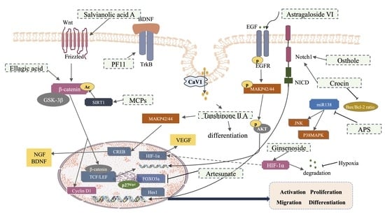
3. Effects and Mechanism of Traditional Chinese Medicine (TCM) in Promoting NSCs Involved in Neurogenesis after an Ischemic Stroke

NSCs have been shown to be a potential source of replacement for degenerated neurons in CNS diseases. Using small molecules to induce endogenous NSC neurogenesis is a potential approach for generating the desired cell types in large numbers. Thus, the effects of TCM on NSCs have excellent prospects for application. It plays a significant role in promoting the activation, proliferation, migration, differentiation, repair of neuronal loss, and functional injury in NSCs because of its multicomponent and multitarget characteristics (Graphical abstract, Table 1 and Table 2).
Momordica charantia, sometimes referred to as bitter melon, is a traditional fruit widely used in the supplementary treatment of cardiovascular disease and diabetes. M. charantia polysaccharides (MCPs) are important bioactive components with hypoglycemic, cholesterol-reducing, antioxidant, and anti-obesity properties. MCPs have been demonstrated to improve NSC proliferation in the SVZ and SGZ and restore memory and learning ability in MCAO rats [125]. In addition, it promoted C17.2 cell proliferation in response to OGD injury. The potential mechanism relies on the upregulation of SIRT1 activity and cytoplasmic β-catenin deacetylation, which promote β-catenin nuclear translocation to induce NSC proliferation [125].
Pseudoginsenoside-F11 (PF11) is a saponin extracted from the leaves of Panax pseudoginseng ssp. In tMCAO mice, PF11 significantly reduced hippocampal atrophy, cognitive impairment, sensory dysfunction, infarction, and mortality. It also promoted neuroblast migration and newborn neuron survival in the ipsilateral striatum and DG, mediated mainly through the activation of the BDNF/TrkB pathway, thereby improving long-term nerve damage and enhancing neurogenesis after a stroke, indicating its potential role in the convalescent treatment of ischemic strokes [126].
Ginsenoside, the primary active ingredient in ginseng, is widely used to treat acute ischemic strokes and ameliorates mitochondrial dysfunction by inhibiting oxidative stress, glutamate neurotoxicity, and apoptosis. Ginsenosides also support the differentiation and proliferation of NSCs. Ginsenoside can increase the optical density and area density, the number of cells positive for nestin/BrdU, nestin/vimentin, and nestin/tuj-1, and the expression levels of BrdU, tuj-1, and vimentin, suggesting that it may promote NSC proliferation and differentiation in neurons and astrocytes. In addition, ginsenosides can activate the expression of HIF-1α and VEGF proteins, indicating that neurogenesis is related to HIF-1α-VEGF pathway activation [127]. A recent study revealed that ginsenoside Rb1 promotes long-term motor function recovery and improves cortical axon regeneration after a stroke. CREB, a vital transcription factor for cell growth and development, is a phosphorylation substrate in numerous signaling pathways, including cAMP, calmodulin, and NMDA receptors [128]. Cerebral ischemia induces a massive release of glutamic acid, which activates the CREB pathway by interacting with NMDA receptors, thus inducing the expression of Bcl-2 and BDNF [129]. Research suggests that cAMP-mediated CREB phosphorylation promotes NSC proliferation and neuronal survival in the adult hippocampus. Moreover, the spontaneous knockdown of CREB in mouse hippocampal cells can impair the differentiation and maturation of newly formed hippocampal granule cells [130]. CREB signaling is essential for regulating the survival, migration, and morphological differentiation of neuroblastomas in the SVZ [128]. Interestingly, the deletion of CREB results in increased Pax6 expression, indicating that the effects of CREB signal transduction on the survival of immature neurons in the RMS may be regulated by Pax6 [131]. After 14 days of intervention, ginsenoside Rb1 increased GAP43 and BDA expression in the ipsilateral and contralateral cortices of mice with distal middle cerebral artery occlusion and improved motor function, which may have been due to the regulation of the cAMP/PKA/CREB signaling pathway [132].
Astragali radix (AR), a widely used herb in TCM, is the main ingredient of the Buyang Huanwu decoction used for the treatment of strokes. Many active ingredients extracted and isolated from AR have shown significant neuroprotective effects in a variety of experimental animal models of stroke, of which Astragaloside VI (AS-VI) has been found to promote neurogenesis. Treatment with AS-VI (2 μg/kg) for 7 days promoted spatial learning, memory, and motor function in transiently ischemic rats by inducing neurogenesis and astrogenesis in the DG, SVZ, and cortex. In addition, it enhanced the self-renewal and proliferation of NSCs in vitro. Adult NPCs express the EGF receptor (EGFR), which is upregulated under ischemic conditions to enhance their susceptibility to EGF or other EGFR ligands in response to ischemia [126]. EGF intervention promotes neuronal differentiation in the corpus striatum after the onset of ischemia in newly formed parvalbumin neurons [133]. AS-VI intervention upregulates nestin, p-EGFR, and p-MAPK protein expression and neurosphere size; however, combined treatment with the EGF receptor inhibitor, gefitinib, and ERK inhibitor, PD98059, reverses this effect [134]. AS-VI may be a therapeutic candidate for adult neurogenesis and brain repair via targeting the EGFR/MAPK signaling pathway after stroke treatment.
Several clinical and laboratory-based studies have shown that after ischemic strokes, AS-VI, another major active component of Astragali radix, confers neuroprotective effects to patients. Recent studies have indicated that in a photochemical ischemia model established in C57BL/6 mice, AS-VI intervention promoted hippocampal neurogenesis, with a significant increase in apical dendrite length and spine density after a stroke [126,135]. In addition, AS-VI reversed neuronal apoptosis, neurogenesis, and cognitive dysfunction in interleukin (IL)-17 knock-out mice. Further studies confirmed that the effects of AS-VI in promoting brain repair and improving stroke-induced cognitive impairment are mainly mediated by the downregulation of IL-17 expression through the Wnt and Akt/GSK-3β pathway signaling pathways.
Astragalus polysaccharide (APS) is an active ingredient with antitumor, antiviral, anti-inflammatory, and antioxidant effects [23]. Furthermore, the astragalus extract has been reported to have a protective effect against hippocampal neuronal damage caused by intermittent hypoxia in rats. APS pretreatment attenuates hypoxia-induced hippocampal NSC injury by increasing cell viability and reducing apoptosis. The upregulation of miR-138 expression following APS pretreatment was reversed by an miR-138 inhibitor. In addition, APS inhibited the JNK and p38MAPK pathways via miR-138. The neuroprotective effects of APS against hypoxia-induced NSC injury may be achieved by upregulating miR-138 expression and inhibiting the JNK and p38MAPK pathways.
Salvianolic acid A (SAA), one of the main water-soluble, active ingredients of Salvia miltiorrhiza Bge., has a variety of pharmacological effects, including anti-apoptosis, antioxidative stress, antiinflammation, and neurovascular protection, and is widely used in cardiovascular and cerebrovascular diseases. The long-term administration of SAA significantly reduced infarction volume, vascular embolism, neurological deficits, and pathological damage in the hippocampus and striatum. In addition, SAA significantly increased the proliferation and migration of NSPCs, promoted their differentiation into neurons, enhanced axonal regeneration, and reduced the apoptosis of neurons around the ipsilateral SVZ region, which led to the restoration of neural density in the ischemic striatum and the reconstruction of neural circuits. The underlying mechanism is related to the activation of the Wnt3a/GSK3β/β-catenin signaling pathway [136].
Tanshinone IIA (TIIA), a major lipophilic component, is extracted from Salvia miltiorrhiza Bge. (Danshen). TIIA can rescue impaired neurons after acute ischemic injury through various targets, such as attenuating oxidative stress, platelet aggregation, and blood–brain barrier disruption. TIIA activates MAPK42/44 and downstream transcription factor CREB in PC12 cells. Endogenous nerve growth factors (NGF) promote neurogenesis after a stroke. The intranasal injection of NGF has been shown to improve the survival of freshly generated neurons in the ipsilateral striatum and SVZ of rats that underwent unilateral MCAO without affecting cell proliferation [137]. TIIA upregulated NGF and BDNF expression, and the differentiation effects were partially attenuated by MEK inhibitors and antagonists of NGF and BDNF receptors, suggesting that the BDNF and NGF signals mediated by MAPK42/44 are involved in the prodifferentiation effects of TIIA. Caveolin-1 (CAV-1), the major functional protein in membrane caveolae, plays an essential role in the endocytosis of exogenous substances. Transmembrane transport of TIIA was enhanced by activating CAV-1, which initiated differentiation. The inhibition of CAV1 expression further suppresses the prodifferentiation effects of TIIA, suggesting that TIIA may partially exert neuroprotective effects in a caveolae-dependent manner through BDNF and NGF signaling mediated by MAPK42/44 [133].
Artesunate (ART) is a derivative of artemisinin that is highly effective for malaria treatment. A previous study showed that ART ameliorated blood–brain barrier damage in mice with subarachnoid hemorrhage, mainly by mediating the S1pR/PI3K signaling pathway. Moreover, studies have revealed that ART reduces ischemic brain volume and white matter lesions in MCAO mice and increases the proportion of BrdU-positive endogenous NSPCs in the ipsilateral SVZ and periinfarct cortex. However, the neurorestorative effects of ART were offset by FOXO3a overexpression, suggesting that ART can ameliorate ischemia/reperfusion damage by promoting endogenous NSPC neurogenesis and proliferation via the FOXO3a/p27Kip1 pathway [138].
Osthole (Ost), a natural coumarin derivative extracted from Cnidium monnieri (L.) Cusson, exhibits anti-inflammatory, antiapoptotic, antioxidative, and neurotrophic properties. Previous studies have demonstrated the role of Ost in promoting the proliferation of NSCs in vitro and improving neurogenesis in the hippocampus of APP/PS-1 double transgenic mice [139]. In addition, it promoted the proliferation of endogenous NSCs, improved neuronal restoration in regions of brain injury and the hippocampal DG and CA3, and improved learning and memory function in MBI mice. Ost upregulated Notch1 and Hes1 gene expression and NICD and Hes1 protein expression, which were blocked by the c-secretase inhibitor DAPT, suggesting that the neuroprotective effects of Ost are partly involved in the activation of the Notch signaling pathway [137].
Crocin, a series of ester glycosides formed by a combination of crocin acid and various sugars, is obtained from a perennial iris crocus (Crocus sativus L.) with a dry stigma. Crocin inhibits the Bax/Bcl-2 ratio in endogenous NSCs, reduces inflammatory factor release, and enhances Notch1 expression after cerebral ischemia reperfusion in the rat brain. Furthermore, the neuroprotective effects of crocin on NSC proliferation and migration were demonstrated by mediating the Notch signaling pathway in a hypoglycemic/reoxygenation model [140].
Ellagic acid (EA) is an antioxidant compound derived from pomegranate and is used to treat muscle spasms and neuropathic aches in TCM. In addition, EA has been recommended as a potential treatment for various CNS disorders, such as strokes and dementia. The administration of EA alleviated infarct volume and improved neurological function and nestin protein levels in rat ischemic penumbra. Increased cell proliferation and the upregulation of the β-catenin and cyclin D1 genes of NSCs were also observed in primary cultured NSCs, suggesting that EA can ameliorate brain injury and promote the proliferation of NSCs through the Wnt/β-catenin signaling pathway [141].

4. Discussion and Conclusions

Stroke is a leading cause of death in China with an increasing prevalence. Globally, China has the highest estimated lifetime risk of stroke at 25 years of age and beyond. The risk factors, including the aging population, the prevalence of diabetes, obesity, and hypertension, and physical inactivity, demonstrated upward trends in the 2010s. Global Burden of Disease data have reported that the age-standardized incidence and mortality rate declined from 1990 to 2019; however, a plateau or increasing trend in the incidence and mortality rates of stroke in China were noted [142].
To date, the treatment of ischemic stroke continues to be a daunting task due to the lack of effective treatment options. Neurogenesis is critical for brain development and damage repair. As the CNS has a limited repair capacity, it is clinically valuable to seek alternative methods to promote recovery after nerve injury [14]. Cellular therapy, especially stem cell therapy, is an emerging field of research in neurological disorders and has long been a promising innovative alternative to acute thrombolysis [33] and chronic rehabilitation [143,144] for many neurological diseases or injuries. Recent studies have shown that endogenous NSCs in pathological conditions such as strokes can be modulated to reverse CNS functional impairment. Overall, this endogenous treatment approach can be achieved by modulating neuroinflammatory responses, targeting neuronal replacement, improving intracerebral nutritional support, remodeling neural circuits, restoring neurological function, and modulating the paracrine signaling of nerve growth factors [54]. However, the therapeutic strategies aimed at promoting endogenous neurogenesis face numerous challenges. First, there are a series of dramatic changes in the cellular microenvironment due to damage to the blood–brain barrier, excitotoxicity, neuroinflammation, acidosis, and the production of reactive oxygen species after stroke, which affect endogenous neurogenic processes [50]. The mechanisms underlying the activation, proliferation, migration, differentiation, and integration of NSCs after a stroke are complex and require further investigation. Additionally, the functional recovery process and structural repair mechanisms of the post-stroke neural network microenvironment must be explained in detail. Second, the number and distribution of endogenous NSCs in the adult brain are limited. A stroke results in a series of cellular microenvironmental effects that usually cause endogenous NSCs to differentiate into glial cells rather than neurons, and most NSCs do not reach damaged cortical areas for functioning [145,146]. Therefore, therapeutic strategies that rely solely on the promotion of endogenous NSC activation are inadequate for promoting post-ischemic neurogenesis and repair. The development of an effective and safe endogenous NSC strategy in combination with exogenous stem cell implantation has great translational significance.
For decades, TCM has been a popular research topic for the prevention and treatment of CNS diseases, and its safety has been demonstrated through long-term clinical use. Chinese medicine has the advantages of holistic regulation and the comprehensive treatment of incurable neurological diseases, such as strokes. It is well known that the pathological environment generated after cerebral ischemic injury is not conducive to endogenous neurogenesis and repair. Chinese medicine may be an effective therapeutic option to promote the neuroprotection, antiapoptotic differentiation, and proliferation of NSCs, improve the microenvironment in the brain, and reduce neuroinflammation after a stroke. Several meta-analyses have shown that Chinese medicine can enhance clinical recovery and significantly improve neurological deficits in stroke patients, thus improving their quality of life and overall therapeutic efficacy [147]. In this review, we summarized Chinese medicines that regulate endogenous neurogenesis after a stroke and their related mechanisms (Table 1 and Table 2). However, we also noted that only some of these studies were of a high quality, the majority of the data were generated in cell culture models with short-term outcomes, supportive, robust in vivo experiments were limited, and human clinical trials and epidemiologic data were relatively inadequate, reminding researchers conducting follow-up studies to pay attention to these urgent situations. Moreover, problems with repair/regeneration in the CNS are not unique; instead, they are attributes of most terminally differentiated organs and tissues. It is worth studying whether Chinese herbal medicines exert repair/regenerative abilities in these organs and tissues. Owing to the diversity of the active ingredients of Chinese herbal medicines, the correspondence between herbs, ingredients, targets, and diseases is not easily clarified, resulting in an insufficient depth of relevant research. Therefore, the application of technologies such as high-throughput screening, small-molecule probes, label-free detection, and target identification and validation should be strengthened to thoroughly analyze network relationships and integrate regulatory processes between the signaling pathways regulated by Chinese medicine. Since no single compound will effectively treat or cure stroke, a means of integrating the use of individual substances or herbs to produce holistic treatment and illustrate the mechanism is still a challenge.
Table 1. Traditional Chinese medicine monomer and its pharmacology on endogenous neural regeneration in vivo.
SourceClassificationSpeciesDosageTreatment RouteNeurogenic
Region
ModelMechanismPhenotypeReference
Momordica charantia
(Ku Gua)
M.charantia polysaccharidesRat200 mg/kgIntragastric administrationSVZ/SGZMCAOSIRT1,
cytoplasmic, β-catenin, deacetylation
Rescue the memory and learning abilities of rats; enhance NSC proliferation [125,148]
Panax pseudoginseng subsp
(San Qi)
Pseudoginsenoside-F11Mice 16, 32 mg/kgOrally treatedDGtMCAOpro-BDNF, TrkB-T; ↑m-BDNF, TrkB-FL, p-AkT,
p-CREB
Reduce brain infarction and brain edema; attenuate the mortality, sensorimotor dysfunction, cognitive impairment, and hippocampal atrophy [126]
ginseng
(Ren Shen)
Ginsenoside Rb1Mice50 mg/kgIntraperitoneal injectionSVZ/SGZdMCAO↑cAMP, ↑PKA, ↑p-CREBImprove functional recovery; stimulate axonal regeneration and brain repair[132]
Radix Astragali
(Huang Qi)
Astragaloside VIRat 2 μg/kgIntravenous
injection
SVZ/DGMCAO,Nestin,
p-EGFR,
p-MAPK
Promote spatial learning and memory; improve impaired motor function [134]
Radix Astragali
(Huang Qi)
Astragaloside IVMice200 mg/kgIntravenous
injection
HippocampusIL-17 KO mice, Photochemical brain ischemia modelp-Akt,
p-GSK-3β, Wnt2,
β-catenin,
Nestin, IL-17, Wnt
Ameliorate stroke-induced cognitive deficits; repair spines of apical dendrites in the hippocampus; stimulate hippocampal neurogenesis; inhibite neural apoptosis; relieve anxiety after stroke[135,149]
Salvia miltiorrhiza Bge
(Dan Shen)
Salvianolic acid ARat10 mg/kgIntragastric
administration
SVZ/
Hippocampus
Electrocoagulation-induced autologous thrombus stroke modelWnt3a, p-GSK3β/GSK3β, β-catenin, TCF-4Decrease infarction volume and vascular embolism; ameliorate pathological injury; promote NSPC proliferation, migration and differentiation; enhance axonal regeneration and diminish neuronal apoptosis [136]
artemisinin
(Qing Hao Su)
ArtesunateMice150 mg/kgIntraperitoneal administrationSVZMCAOPenumbra damage, white matter injury, FOXO3a, p27Kip1;
DCX
Rescue ischemia damage; alleviate white matter injury; promote functional recovery; promote neurogenesis and proliferation of endogenous NSPCs[138]
Cnidium monnieri (L.)
(She Chuang Zi)
OstholeMice30 mg/kgIntraperitoneal administrationSVZ/SGZ/DG/
CA3
Model of stab wound injury is created to mimic the neuroendoscopy
procedure
Notch-1,
Hes-1, Nestin, NICD
Improve learning and memory function; promote the proliferation of endogenous NSCs; improve neuronal restoration; increase the number of neurons in the regions of brain injury[137]
Crocus sativus L.
(Fan Hong Hua)
CrocinRat 10, 50 mg/kgIntragastric
administration
SVZ/DGMCAO/RBax/bcl-2; Notch1Inhibit the release of inflammatory factors; reduce the apoptosis of nerve cells[140]
Pomegranates (Shi Liu)Ellagic acidRat 10, 30, 90 mg/kgIntragastric
administration
SVZ/SGZPhotothrombotic nerve injury modelnestin,
β-catenin,
Cyclin D1
Improve the rats’ nerve-related abilities; remedy infarct volumes and morphological changes in the brain[141]
Table 2. Traditional Chinese medicine monomer and its pharmacology on endogenous neural regeneration in vitro.
SourceClassificationSpeciesDosageModelMechanismPhenotypeReference
Momordica charantia
(Ku Gua)
M.charantia polysaccharidesC17.2 cells,
primary cortical neural stem cells
5 μg/mLOGD, IRISIRT1,
cytoplasmic, β-catenin, deacetylation
Change intracellular redox state; stimulate the proliferation[125,148]
Panax pseudoginseng subsp
(San Qi)
Pseudoginsenoside-F11Primary cultured NSCs100 μmOGD/Rpro-BDNF, TrkB-T; ↑m-BDNF, TrkB-FL, p-AkT,
p-CREB
Promote proliferation and differentiation[126]
ginseng
(Ren Shen)
GinsenosidePrimary cultured NSCs 1 μg/mLOGD/RHIF-1α, VEGFMaintain NSC replication; promote NSC proliferation; promote NSC differentiation into neurons and astrocytes[127]
Radix Astragali
(Huang Qi)
Astragaloside VIC17.2 cells or primary
cultured NSCs
10, 100 nMDMEM/F12 media deprived of EGF or
normal DMEM/F12 media
stimulated for 2 h
Nestin,
p-EGFR,
p-MAPK
Enhance NSCs self-renewal and proliferation without affecting NSCs [134]
Radix Astragali
(Huang Qi)
Astragaloside IVPrimary
cultured NSCs
10 nM, 100 nM, 20 μmM/p-Akt,
p-GSK-3β, Wnt2,
β-catenin,
Nestin, IL-17, Wnt
Promote
hippocampal
neurogenesis and NSC
proliferation
[135,149]
Salvia miltiorrhiza Bge
(Dan Shen)
Tanshinone II AC17.2 cells, primary culture of embryonic cortical NSCs or PC12 cells0.1–3 μMTIIA stimulated for 7 dp-MAPK42/44, p-CREB, BDNF, NGF, GAP-43Promote neuronal differentiation; facilitate endocytosis and transportation across the cell membrane[133]
Crocus sativus L.
(Fan Hong Hua)
CrocinNSCs10, 50 μM Bax/bcl-2; Notch1Promote cell
proliferation;
increase cell
migration; inhibit cell apoptosis; and promote neural
regeneration
[140]
Pomegranates (Shi Liu)Ellagic acidPrimary cultured NSCs1, 3, 9 μg/mLOGD/Rnestin,
β-catenin,
Cyclin D1
Increase
proliferation of NSCs
[141]

Author Contributions

Conceptualization. Y.L. and H.Z.; data curation, L.L., X.L. and R.H.; validation, X.L. and R.H.; resources, X.L., M.W. and Y.C.; writing—original draft preparation, L.L. and X.L. writing—review and editing, X.L., L.L., Y.L. and Y.M. All authors have read and agreed to the published version of the manuscript.

Funding

This work was supported by National Natural Science Foundation of China (Grant No. 82073832 to Y.L.; 81973792 to L.L.; 82004331 to H.Z.). Tianjin Natural Science Fund for Distinguished Young Scholars (Grant No. 20JCJQJC00070 to Y.L., China).

Institutional Review Board Statement

Not applicable.

Informed Consent Statement

Not applicable.

Data Availability Statement

Data sharing not applicable.

Conflicts of Interest

The authors declare no conflict of interest.

References

  1. Herpich, F.; Rincon, F. Management of Acute Ischemic Stroke. Crit. Care Med. 2020, 48, 1654–1663. [Google Scholar] [CrossRef]
  2. Fisher, M. New approaches to neuroprotective drug development. Stroke 2011, 42, S24–S27. [Google Scholar] [CrossRef]
  3. Mendelson, S.J.; Prabhakaran, S. Diagnosis and Management of Transient Ischemic Attack and Acute Ischemic Stroke: A Review. JAMA 2021, 325, 1088–1098. [Google Scholar] [CrossRef]
  4. Li, L.; Fan, X.; Zhang, X.-T.; Yue, S.-Q.; Sun, Z.-Y.; Zhu, J.-Q.; Zhang, J.-H.; Gao, X.-M.; Zhang, H. The effects of Chinese medicines on cAMP/PKA signaling in central nervous system dysfunction. Brain Res. Bull. 2017, 132, 109–117. [Google Scholar] [CrossRef] [PubMed]
  5. Wan, W.; Cao, L.; Kalionis, B.; Xia, S.; Tai, X. Applications of Induced Pluripotent Stem Cells in Studying the Neurodegenerative Diseases. Stem Cells Int. 2015, 2015, 382530. [Google Scholar] [CrossRef]
  6. Verheyden, G.; Nieuwboer, A.; De Wit, L.; Thijs, V.; Dobbelaere, J.; Devos, H.; Severijns, D.; Vanbeveren, S.; De Weerdt, W. Time course of trunk, arm, leg, and functional recovery after ischemic stroke. Neurorehabilit. Neural Repair 2008, 22, 173–179. [Google Scholar] [CrossRef]
  7. Campbell, B.C.; Mitchell, P.J.; Kleinig, T.J.; Dewey, H.M.; Churilov, L.; Yassi, N.; Yan, B.; Dowling, R.J.; Parsons, M.W.; Oxley, T.J.; et al. Endovascular therapy for ischemic stroke with perfusion-imaging selection. N. Engl. J. Med. 2015, 372, 1009–1018. [Google Scholar] [CrossRef] [PubMed]
  8. Berkhemer, O.A.; Fransen, P.S.S.; Beumer, D.; Berg, L.A.V.D.; Lingsma, H.F.; Yoo, A.J.; Schonewille, W.J.; Vos, J.A.; Nederkoorn, P.J.; Wermer, M.J.H.; et al. A randomized trial of intraarterial treatment for acute ischemic stroke. N. Engl. J. Med. 2015, 372, 11–20. [Google Scholar] [CrossRef]
  9. Feliciano, D.; Zhang, S.; Nasrallah, C.M.; Lisgo, S.; Bordey, A. Embryonic cerebrospinal fluid nanovesicles carry evolutionarily conserved molecules and promote neural stem cell amplification. PLoS ONE 2014, 9, e88810. [Google Scholar] [CrossRef] [PubMed]
  10. Thored, P.; Wood, J.; Arvidsson, A.; Cammenga, J.; Kokaia, Z.; Lindvall, O. Long-Term Neuroblast Migration along Blood Vessels in an Area with Transient Angiogenesis and Increased Vascularization after Stroke. Stroke 2007, 38, 3032–3039. [Google Scholar] [CrossRef]
  11. Macas, J.; Nern, C.; Plate, K.H.; Momma, S. Increased Generation of Neuronal Progenitors after Ischemic Injury in the Aged Adult Human Forebrain. J. Neurosci. 2006, 26, 13114–13119. [Google Scholar] [CrossRef]
  12. Picard-Riera, N.; Nait-Oumesmar, B.; Evercooren, A.B.-V. Endogenous adult neural stem cells: Limits and potential to repair the injured central nervous system. J. Neurosci. Res. 2004, 76, 223–231. [Google Scholar] [CrossRef]
  13. Zhang, Z.; Chopp, M. Neural Stem Cells and Ischemic Brain. J. Stroke 2016, 18, 267–272. [Google Scholar] [CrossRef]
  14. Barker, R.A.; Götz, M.; Parmar, M. New approaches for brain repair—From rescue to reprogramming. Nature 2018, 557, 329–334. [Google Scholar] [CrossRef]
  15. Baker, E.W.; Platt, S.R.; Lau, V.W.; Grace, H.E.; Holmes, S.P.; Wang, L.; Duberstein, K.J.; Howerth, E.W.; Kinder, H.A.; Stice, S.L.; et al. Induced Pluripotent Stem Cell-Derived Neural Stem Cell Therapy Enhances Recovery in an Ischemic Stroke Pig Model. Sci. Rep. 2017, 7, 10075. [Google Scholar] [CrossRef]
  16. Eckert, A.; Huang, L.; Gonzalez, R.; Kim, H.-S.; Hamblin, M.H.; Lee, J.-P. Bystander Effect Fuels Human Induced Pluripotent Stem Cell-Derived Neural Stem Cells to Quickly Attenuate Early Stage Neurological Deficits after Stroke. Stem. Cells Transl. Med. 2015, 4, 841–851. [Google Scholar] [CrossRef]
  17. Tornero, D.; Wattananit, S.; Madsen, M.G.; Koch, P.; Wood, J.; Tatarishvili, J.; Mine, Y.; Ge, R.; Monni, E.; Devaraju, K.; et al. Human induced pluripotent stem cell-derived cortical neurons integrate in stroke-injured cortex and improve functional recovery. Brain 2013, 136, 3561–3577. [Google Scholar] [CrossRef]
  18. Kawabori, M.; Shichinohe, H.; Kuroda, S.; Houkin, K. Clinical Trials of Stem Cell Therapy for Cerebral Ischemic Stroke. Int. J. Mol. Sci. 2020, 21, 7380. [Google Scholar] [CrossRef]
  19. Sinden, J.D.; Hicks, C.; Stroemer, P.; Vishnubhatla, I.; Corteling, R. Human Neural Stem Cell Therapy for Chronic Ischemic Stroke: Charting Progress from Laboratory to Patients. Stem Cells Dev. 2017, 26, 933–947. [Google Scholar] [CrossRef]
  20. Trounson, A.; McDonald, C. Stem Cell Therapies in Clinical Trials: Progress and Challenges. Cell Stem Cell 2015, 17, 11–22. [Google Scholar] [CrossRef]
  21. Dibajnia, P.; Morshead, C.M. Role of neural precursor cells in promoting repair following stroke. Acta Pharmacol. Sin. 2013, 34, 78–90. [Google Scholar] [CrossRef]
  22. Ahmed, M.E.; Tucker, D.; Dong, Y.; Lu, Y.; Zhao, N.; Wang, R.; Zhang, Q. Methylene Blue promotes cortical neurogenesis and ameliorates behavioral deficit after photothrombotic stroke in rats. Neuroscience 2016, 336, 39–48. [Google Scholar] [CrossRef]
  23. Zhao, Y.; Guan, Y.-F.; Zhou, X.-M.; Li, G.-Q.; Li, Z.-Y.; Zhou, C.-C.; Wang, P.; Miao, C.-Y. Regenerative Neurogenesis After Ischemic Stroke Promoted by Nicotinamide Phosphoribosyltransferase–Nicotinamide Adenine Dinucleotide Cascade. Stroke 2015, 46, 1966–1974. [Google Scholar] [CrossRef]
  24. Huang, L.; Zhang, L. Neural stem cell therapies and hypoxic-ischemic brain injury. Prog. Neurobiol. 2019, 173, 1–17. [Google Scholar] [CrossRef]
  25. Bassi, M.S.; Iezzi, E.; Gilio, L.; Centonze, D.; Buttari, F. Synaptic Plasticity Shapes Brain Connectivity: Implications for Network Topology. Int. J. Mol. Sci. 2019, 20, 6193. [Google Scholar] [CrossRef]
  26. Farah, M.; Marshall, K. Axonal regeneration and sprouting as a potential therapeutic target for nervous system disorders. Neural Regen. Res. 2021, 16, 1901–1910. [Google Scholar] [CrossRef]
  27. Li, F.; Sami, A.; Noristani, H.N.; Slattery, K.; Qiu, J.; Groves, T.; Wang, S.; Veerasammy, K.; Chen, Y.X.; Morales, J.; et al. Glial Metabolic Rewiring Promotes Axon Regeneration and Functional Recovery in the Central Nervous System. Cell Metab. 2020, 32, 767–785.e7. [Google Scholar] [CrossRef]
  28. Liu, T.; Ding, Y.; Wen, A. Traditional Chinese medicine for ischaemic stroke. Lancet Neurol. 2018, 17, 745. [Google Scholar] [CrossRef]
  29. Wang, J.; Hu, J.; Chen, X.; Lei, X.; Feng, H.; Wan, F.; Tan, L. Traditional Chinese Medicine Monomers: Novel Strategy for Endogenous Neural Stem Cells Activation after Stroke. Front. Cell Neurosci. 2021, 15, 628115. [Google Scholar] [CrossRef]
  30. Haas, S.; Weidner, N.; Winkler, J. Adult stem cell therapy in stroke. Curr. Opin. Neurol. 2005, 18, 59–64. [Google Scholar] [CrossRef]
  31. Richards, L.J.; Kilpatrick, T.J.; Bartlett, P.F. De novo generation of neuronal cells from the adult mouse brain. Proc. Natl. Acad. Sci. USA 1992, 89, 8591–8595. [Google Scholar] [CrossRef]
  32. Reynolds, B.A.; Weiss, S. Generation of Neurons and Astrocytes from Isolated Cells of the Adult Mammalian Central Nervous System. Science 1992, 255, 1707–1710. [Google Scholar] [CrossRef]
  33. Zhang, P.; Liu, C.; Zheng, X.; Wu, L.; Liu, Z.; Liao, B.; Shi, Y.; Li, X.; Xu, J.; Chen, S. Full-Length Multi-Barcoding: DNA Barcoding from Single Ingredient to Complex Mixtures. Genes 2019, 10, 343. [Google Scholar] [CrossRef]
  34. Temple, S. The development of neural stem cells. Nature 2001, 414, 112–117. [Google Scholar] [CrossRef]
  35. Breunig, J.J.; Haydar, T.F.; Rakic, P. Neural stem cells: Historical perspective and future prospects. Neuron 2011, 70, 614–625. [Google Scholar] [CrossRef]
  36. Yadirgi, G.; Marino, S. Adult neural stem cells and their role in brain pathology. J. Pathol. 2009, 217, 242–253. [Google Scholar] [CrossRef]
  37. Li, Y.; Guo, W. Neural Stem Cell Niche and Adult Neurogenesis. The Neuroscientist: A Review Journal Bringing Neurobiology. Neurol. Psychiatry 2021, 27, 235–245. [Google Scholar]
  38. Shihabuddin, L.S.; Horner, P.J.; Ray, J.; Gage, F.H. Adult Spinal Cord Stem Cells Generate Neurons after Transplantation in the Adult Dentate Gyrus. J. Neurosci. 2000, 20, 8727–8735. [Google Scholar] [CrossRef]
  39. Mehler, M.F.; Gokhan, S. Postnatal Cerebral Cortical Multipotent Progenitors: Regulatory Mechanisms and Potential Role in the Development of Novel Neural Regenerative Strategies. Brain Pathol. 1999, 9, 515–526. [Google Scholar] [CrossRef]
  40. Huang, L.; Wang, G. The Effects of Different Factors on the Behavior of Neural Stem Cells. Stem Cells Int. 2017, 2017, 1–16. [Google Scholar] [CrossRef]
  41. Sun, Y.; Li, J.; Georgi, R.; Kolberg, B.; Yang, L. Effects of acupuncture on angiogenesis-associated factor expression in ischemic brain tissue following cerebral infarction in rats. Acupunct. Herb. Med. 2023, 3, 46–54. [Google Scholar] [CrossRef]
  42. Song, M.; Yu, S.P.; Mohamad, O.; Cao, W.; Wei, Z.Z.; Gu, X.; Jiang, M.Q.; Wei, L. Optogenetic stimulation of glutamatergic neuronal activity in the striatum enhances neurogenesis in the subventricular zone of normal and stroke mice. Neurobiol. Dis. 2017, 98, 9–24. [Google Scholar] [CrossRef]
  43. Kaneko, N.; Sawada, M.; Sawamoto, K. Mechanisms of neuronal migration in the adult brain. J. Neurochem. 2017, 141, 835–847. [Google Scholar] [CrossRef]
  44. Ihrie, R.A.; Alvarez-Buylla, A. Cells in the astroglial lineage are neural stem cells. Cell Tissue Res. 2008, 331, 179–191. [Google Scholar] [CrossRef]
  45. Li, W.L.; Chu, M.W.; Wu, A.; Suzuki, Y.; Imayoshi, I.; Komiyama, T. Adult-born neurons facilitate olfactory bulb pattern separation during task engagement. eLife 2018, 7, e33006. [Google Scholar] [CrossRef]
  46. Cameron, H.A.; Mckay, R.D. Adult neurogenesis produces a large pool of new granule cells in the dentate gyrus. J. Comp. Neurol. 2001, 435, 406–417. [Google Scholar] [CrossRef]
  47. Ming, G.-L.; Song, H. Adult Neurogenesis in the Mammalian Brain: Significant Answers and Significant Questions. Neuron 2011, 70, 687–702. [Google Scholar] [CrossRef]
  48. Robel, S.; Berninger, B.; Götz, M. The stem cell potential of glia: Lessons from reactive gliosis. Nat. Rev. Neurosci. 2011, 12, 88–104. [Google Scholar] [CrossRef]
  49. Kleiderman, S.; Gutbier, S.; Tufekci, K.U.; Ortega, F.; Sá, J.V.; Teixeira, A.P.; Brito, C.; Glaab, E.; Berninger, B.; Alves, P.M.; et al. Conversion of Nonproliferating Astrocytes into Neurogenic Neural Stem Cells: Control by FGF2 and Interferon-γ. Stem Cells 2016, 34, 2861–2874. [Google Scholar] [CrossRef]
  50. Tan, L.; Zhong, J.; Li, R.-W.; Wang, J.; Wang, Y.; Ge, H.-F.; Xian, J.-S.; Feng, H. Neuroprotection by cattle encephalon glycoside and ignotin beyond the time window of thrombolysis in ischemic stroke. Neural Regen. Res. 2021, 16, 312. [Google Scholar] [CrossRef]
  51. Nakayama, D.; Matsuyama, T.; Ishibashi-Ueda, H.; Nakagomi, T.; Kasahara, Y.; Hirose, H.; Kikuchi-Taura, A.; Stern, D.M.; Mori, H.; Taguchi, A. Injury-induced neural stem/progenitor cells in post-stroke human cerebral cortex. Eur. J. Neurosci. 2010, 31, 90–98. [Google Scholar] [CrossRef]
  52. Marti-Fabregas, J.; Romaguera-Ros, M.; Gomez-Pinedo, U.; Martinez-Ramirez, S.; Jimenez-Xarrie, E.; Marin, R.; Marti-Vilalta, J.L.; Garcia-Verdugo, J.M. Proliferation in the human ipsilateral subventricular zone after ischemic stroke. Neurology 2010, 74, 357–365. [Google Scholar] [CrossRef]
  53. Quiroz, E.N.; Quiroz, R.N.; Ahmad, M.; Escorcia, L.G.; Villarreal, J.L.; Ponce, C.F.; Martinez, G.A. Cell Signaling in Neuronal Stem Cells. Cells 2018, 7, 75. [Google Scholar] [CrossRef]
  54. Bain, J.M.; Moore, L.; Ren, Z.; Simonishvili, S.; Levison, S.W. Vascular Endothelial Growth Factors A and C are Induced in the SVZ following Neonatal Hypoxia–Ischemia and Exert Different Effects on Neonatal Glial Progenitors. Transl. Stroke Res. 2013, 4, 158–170. [Google Scholar] [CrossRef]
  55. Yu, J.H.; Seo, J.-H.; Lee, J.Y.; Lee, M.-Y.; Cho, S.-R. Induction of Neurorestoration from Endogenous Stem Cells. Cell Transplant. 2016, 25, 863–882. [Google Scholar] [CrossRef]
  56. Yagita, Y.; Kitagawa, K.; Ohtsuki, T.; Takasawa, K.-I.; Miyata, T.; Okano, H.; Hori, M.; Matsumoto, M. Neurogenesis by Progenitor Cells in the Ischemic Adult Rat Hippocampus. Stroke 2001, 32, 1890–1896. [Google Scholar] [CrossRef]
  57. Zhang, R.; Zhang, Z.; Zhang, L.; Chopp, M. Proliferation and differentiation of progenitor cells in the cortex and the subventricular zone in the adult rat after focal cerebral ischemia. Neuroscience 2001, 105, 33–41. [Google Scholar] [CrossRef]
  58. Jin, K.; Wang, X.; Xie, L.; Mao, X.O.; Zhu, W.; Wang, Y.; Shen, J.; Mao, Y.; Banwait, S.; Greenberg, D.A. Evidence for stroke-induced neurogenesis in the human brain. Proc. Natl. Acad. Sci. USA 2006, 103, 13198–13202. [Google Scholar] [CrossRef]
  59. Jin, K.; Wang, X.; Xie, L.; Mao, X.O.; Greenberg, D.A. Transgenic ablation of doublecortin-expressing cells suppresses adult neurogenesis and worsens stroke outcome in mice. Proc. Natl. Acad. Sci. USA 2010, 107, 7993–7998. [Google Scholar] [CrossRef]
  60. Kobayashi, T.; Kageyama, R. Lysosomes and signaling pathways for maintenance of quiescence in adult neural stem cells. FEBS J. 2021, 288, 3082–3093. [Google Scholar] [CrossRef]
  61. Grandbarbe, L.; Bouissac, J.; Rand, M.; de Angelis, M.H.; Artavanis-Tsakonas, S.; Mohier, E. Delta-Notch signaling controls the generation of neurons/glia from neural stem cells in a stepwise process. Development 2003, 130, 1391–1402. [Google Scholar] [CrossRef]
  62. Bray, S.; Bernard, F. Notch Targets and Their Regulation. In Current Topics in Developmental Biology; Elsevier: Amsterdam, The Netherlands, 2010; pp. 253–275. [Google Scholar]
  63. Qin, W.; Chen, S.; Yang, S.; Xu, Q.; Xu, C.; Cai, J. The Effect of Traditional Chinese Medicine on Neural Stem Cell Proliferation and Differentiation. Aging Dis. 2017, 8, 792–811. [Google Scholar] [CrossRef]
  64. Sivakumar, K.C.; Dhanesh, S.B.; Shobana, S.; James, J.; Mundayoor, S.; Song, K.; Ge, D.; Guan, S.; Sun, C.; Liu, T.; et al. A Systems Biology Approach to Model Neural Stem Cell Regulation by Notch, Shh, Wnt, and EGF Signaling Pathways. OMICS: A J. Integr. Biol. 2011, 15, 729–737. [Google Scholar] [CrossRef]
  65. Alexson, T.O.; Hitoshi, S.; Coles, B.L.; Bernstein, A.; van der Kooy, D. Notch signaling is required to maintain all neural stem cell populations—Irrespective of spatial or temporal niche. Dev. Neurosci. 2006, 28, 34–48. [Google Scholar] [CrossRef]
  66. Wang, X.; Mao, X.; Xie, L.; Greenberg, D.A.; Jin, K. Involvement of notch1 signaling in neurogenesis in the subventricular zone of normal and ischemic rat brain in vivo. J. Cereb. Blood Flow Metab. 2009, 29, 1644–1654. [Google Scholar] [CrossRef]
  67. Nyfeler, Y.; Kirch, R.D.; Mantei, N.; Leone, D.P.; Radtke, F.; Suter, U.; Taylor, V. Jagged1 signals in the postnatal subventricular zone are required for neural stem cell self-renewal. EMBO J. 2005, 24, 3504–3515. [Google Scholar] [CrossRef]
  68. Androutsellis-Theotokis, A.; Leker, R.R.; Soldner, F.; Hoeppner, D.J.; Ravin, R.; Poser, S.W.; Rueger, M.A.; Bae, S.-K.; Kittappa, R.; McKay, R.D.G. Notch signalling regulates stem cell numbers in vitro and in vivo. Nature 2006, 442, 823–826. [Google Scholar] [CrossRef]
  69. Wang, L.; Chopp, M.; Zhang, R.; Zhang, L.; LeTourneau, Y.; Feng, Y.; Jiang, A.; Morris, D.; Zhang, Z. The Notch pathway mediates expansion of a progenitor pool and neuronal differentiation in adult neural progenitor cells after stroke. Neuroscience 2009, 158, 1356–1363. [Google Scholar] [CrossRef]
  70. Sun, F.; Mao, X.; Xie, L.; Ding, M.; Shao, B.; Jin, K. Notch1 signaling modulates neuronal progenitor activity in the subventricular zone in response to aging and focal ischemia. Aging Cell 2013, 12, 978–987. [Google Scholar] [CrossRef]
  71. Jessell, T.M. Neuronal specification in the spinal cord: Inductive signals and transcriptional codes. Nat. Rev. Genet. 2000, 1, 20–29. [Google Scholar] [CrossRef]
  72. Ferent, J.; Cochard, L.; Faure, H.; Taddei, M.; Hahn, H.; Ruat, M.; Traiffort, E. Genetic activation of Hedgehog signaling unbalances the rate of neural stem cell renewal by increasing symmetric divisions. Stem Cell Rep. 2014, 3, 312–323. [Google Scholar] [CrossRef]
  73. Gao, Z.Y.; Guo, C.Y.; Shi, D.Z. Effect of shengmai injection on the fatality rate of patients with acute myocardial in-farction: A systematic review. Zhongguo Zhong Xi Yi Jie He Za Zhi 2008, 28, 1069–1073. [Google Scholar]
  74. Merson, T.D.; Bourne, J.A. Endogenous neurogenesis following ischaemic brain injury: Insights for therapeutic strategies. Int. J. Biochem. Cell Biol. 2014, 56, 4–19. [Google Scholar] [CrossRef]
  75. Banerjee, S.B.; Rajendran, R.; Dias, B.G.; Ladiwala, U.; Tole, S.; Vaidya, V.A. Recruitment of the Sonic hedgehog signalling cascade in electroconvulsive seizure-mediated regulation of adult rat hippocampal neurogenesis. Eur. J. Neurosci. 2005, 22, 1570–1580. [Google Scholar] [CrossRef]
  76. Machold, R.; Hayashi, S.; Rutlin, M.; Muzumdar, M.D.; Nery, S.; Corbin, J.G.; Gritli-Linde, A.; Dellovade, T.; Porter, J.A.; Rubin, L.L.; et al. Sonic Hedgehog Is Required for Progenitor Cell Maintenance in Telencephalic Stem Cell Niches. Neuron 2003, 39, 937–950. [Google Scholar] [CrossRef]
  77. Lai, K.; Kaspar, B.K.; Gage, F.H.; Schaffer, D.V. Sonic hedgehog regulates adult neural progenitor proliferation in vitro and in vivo. Nat. Neurosci. 2003, 6, 21–27. [Google Scholar] [CrossRef]
  78. Webb, A.E.; Pollina, E.A.; Vierbuchen, T.; Urbán, N.; Ucar, D.; Leeman, D.S.; Martynoga, B.; Sewak, M.; Rando, T.A.; Guillemot, F.; et al. FOXO3 Shares Common Targets with ASCL1 Genome-wide and Inhibits ASCL1-Dependent Neurogenesis. Cell Rep. 2013, 4, 477–491. [Google Scholar] [CrossRef]
  79. Bonaguidi, M.A.; Wheeler, M.A.; Shapiro, J.S.; Stadel, R.P.; Sun, G.J.; Ming, G.-L.; Song, H. In Vivo Clonal Analysis Reveals Self-Renewing and Multipotent Adult Neural Stem Cell Characteristics. Cell 2011, 145, 1142–1155. [Google Scholar] [CrossRef]
  80. Pilz, G.A.; Bottes, S.; Betizeau, M.; Jörg, D.J.; Carta, S.; April, S.; Simons, B.D.; Helmchen, F.; Jessberger, S. Live imaging of neurogenesis in the adult mouse hippocampus. Science 2018, 359, 658–662. [Google Scholar] [CrossRef]
  81. Khalifeh-Soltani, A.; Ha, A.; Podolsky, M.J.; McCarthy, D.A.; McKleroy, W.; Azary, S.; Sakuma, S.; Tharp, K.M.; Wu, N.; Yokosaki, Y.; et al. α8β1 integrin regulates nutrient absorption through an Mfge8-PTEN dependent mechanism. Elife 2016, 5, e13063. [Google Scholar] [CrossRef]
  82. Paik, J.-H.; Ding, Z.; Narurkar, R.; Ramkissoon, S.; Muller, F.; Kamoun, W.S.; Chae, S.-S.; Zheng, H.; Ying, H.; Mahoney, J.; et al. FoxOs Cooperatively Regulate Diverse Pathways Governing Neural Stem Cell Homeostasis. Cell Stem Cell 2009, 5, 540–553. [Google Scholar] [CrossRef]
  83. Schäffner, I.; Minakaki, G.; Khan, M.A.; Balta, E.-A.; Schlötzer-Schrehardt, U.; Schwarz, T.J.; Beckervordersandforth, R.; Winner, B.; Webb, A.E.; DePinho, R.A.; et al. FoxO Function Is Essential for Maintenance of Autophagic Flux and Neuronal Morphogenesis in Adult Neurogenesis. Neuron 2018, 99, 1188–1203.e6. [Google Scholar] [CrossRef]
  84. Renault, V.M.; Rafalski, V.A.; Morgan, A.A.; Salih, D.A.; Brett, J.O.; Webb, A.E.; Villeda, S.A.; Thekkat, P.U.; Guillerey, C.; Denko, N.C.; et al. FoxO3 regulates neural stem cell homeostasis. Cell Stem Cell 2009, 5, 527–539. [Google Scholar] [CrossRef]
  85. Arvidsson, A.; Collin, T.; Kirik, D.; Kokaia, Z.; Lindvall, O. Neuronal replacement from endogenous precursors in the adult brain after stroke. Nat. Med. 2002, 8, 963–970. [Google Scholar] [CrossRef]
  86. Lee, J.-P.; Jeyakumar, M.; Gonzalez, R.; Takahashi, H.; Lee, P.-J.; Baek, R.C.; Clark, D.; Rose, H.; Fu, G.; Clarke, J.; et al. Stem cells act through multiple mechanisms to benefit mice with neurodegenerative metabolic disease. Nat. Med. 2007, 13, 439–447. [Google Scholar] [CrossRef]
  87. Lee, J.-P.; Zhang, R.; Yan, M.; Duggineni, S.; Wakeman, D.R.; Niles, W.L.; Feng, Y.; Chen, J.; Hamblin, M.H.; Han, E.B.; et al. Chemical mutagenesis of a GPCR ligand: Detoxifying “inflammo-attraction” to direct therapeutic stem cell migration. Proc. Natl. Acad. Sci. USA 2020, 117, 31177–31188. [Google Scholar] [CrossRef]
  88. Boese, A.C.; Eckert, A.; Hamblin, M.H.; Lee, J.-P. Human neural stem cells improve early stage stroke outcome in delayed tissue plasminogen activator-treated aged stroke brains. Exp. Neurol. 2020, 329, 113275. [Google Scholar] [CrossRef]
  89. Coviello, S.; Benedetti, B.; Jakubecova, D.; Belles, M.; Klimczak, P.; Gramuntell, Y.; Couillard-Despres, S.; Nacher, J. PSA Depletion Induces the Differentiation of Immature Neurons in the Piriform Cortex of Adult Mice. Int. J. Mol. Sci. 2021, 22, 5733. [Google Scholar] [CrossRef]
  90. Cipriani, S.; Ferrer, I.; Aronica, E.; Kovacs, G.G.; Verney, C.; Nardelli, J.; Khung, S.; Delezoide, A.-L.; Milenkovic, I.; Rasika, S.; et al. Hippocampal Radial Glial Subtypes and Their Neurogenic Potential in Human Fetuses and Healthy and Alzheimer’s Disease Adults. Cereb. Cortex 2018, 28, 2458–2478. [Google Scholar] [CrossRef]
  91. Imitola, J.; Comabella, M.; Chandraker, A.K.; Dangond, F.; Sayegh, M.H.; Snyder, E.Y.; Khoury, S.J. Neural Stem/Progenitor Cells Express Costimulatory Molecules That Are Differentially Regulated by Inflammatory and Apoptotic Stimuli. Am. J. Pathol. 2004, 164, 1615–1625. [Google Scholar] [CrossRef]
  92. Robin, A.M.; Zhang, Z.G.; Wang, L.; Zhang, R.L.; Katakowski, M.; Zhang, L.; Wang, Y.; Zhang, C.; Chopp, M. Stromal Cell-Derived Factor 1α Mediates Neural Progenitor Cell Motility after Focal Cerebral Ischemia. J. Cereb. Blood Flow Metab. 2006, 26, 125–134. [Google Scholar] [CrossRef] [PubMed]
  93. Tiveron, M.C.; Rossel, M.; Moepps, B.; Zhang, Y.L.; Seidenfaden, R.; Favor, J.; König, N.; Cremer, H. Molecular Interaction between Projection Neuron Precursors and Invading Interneurons via Stromal-Derived Factor 1 (CXCL12)/CXCR4 Signaling in the Cortical Subventricular Zone/Intermediate Zone. J. Neurosci. 2006, 26, 13273–13278. [Google Scholar] [CrossRef]
  94. Zhou, G.; Wang, Y.; Gao, S.; Fu, X.; Cao, Y.; Peng, Y.; Zhuang, J.; Hu, J.; Shao, A.; Wang, L. Potential Mechanisms and Perspectives in Ischemic Stroke Treatment Using Stem Cell Therapies. Front. Cell Dev. Biol. 2021, 9, 646927. [Google Scholar] [CrossRef]
  95. Rabenstein, M.; Hucklenbroich, J.; Willuweit, A.; Ladwig, A.; Fink, G.R.; Schroeter, M.; Langen, K.-J.; Rueger, M.A. Osteopontin mediates survival, proliferation and migration of neural stem cells through the chemokine receptor CXCR4. Stem Cell Res. Ther. 2015, 6, 99. [Google Scholar] [CrossRef]
  96. Huang, Y.; Wang, J.; Cai, J.; Qiu, Y.; Zheng, H.; Lai, X.; Sui, X.; Wang, Y.; Lu, Q.; Zhang, Y.; et al. Targeted homing of CCR2-overexpressing mesenchymal stromal cells to ischemic brain enhances post-stroke recovery partially through PRDX4-mediated blood-brain barrier preservation. Theranostics 2018, 8, 5929–5944. [Google Scholar] [CrossRef]
  97. Lee, S.; Kim, O.J.; Lee, K.O.; Jung, H.; Oh, S.-H.; Kim, N.K. Enhancing the Therapeutic Potential of CCL2-Overexpressing Mesenchymal Stem Cells in Acute Stroke. Int. J. Mol. Sci. 2020, 21, 7795. [Google Scholar] [CrossRef]
  98. Lee, S.-R.; Kim, H.-Y.; Rogowska, J.; Zhao, B.-Q.; Bhide, P.; Parent, J.M.; Lo, E.H. Involvement of Matrix Metalloproteinase in Neuroblast Cell Migration from the Subventricular Zone after Stroke. J. Neurosci. 2006, 26, 3491–3495. [Google Scholar] [CrossRef]
  99. Wang, L.; Zhang, Z.G.; Zhang, R.L.; Gregg, S.R.; Hozeska-Solgot, A.; LeTourneau, Y.; Wang, Y.; Chopp, M. Matrix Metalloproteinase 2 (MMP2) and MMP9 Secreted by Erythropoietin-Activated Endothelial Cells Promote Neural Progenitor Cell Migration. J. Neurosci. 2006, 26, 5996–6003. [Google Scholar] [CrossRef]
  100. Lindvall, O.; Kokaia, Z. Neurogenesis following Stroke Affecting the Adult Brain. Cold Spring Harb. Perspect. Biol. 2015, 7, a019034. [Google Scholar] [CrossRef]
  101. Navlakha, S.; Bar-Joseph, Z.; Barth, A.L. Network Design and the Brain. Trends Cogn. Sci. 2018, 22, 64–78. [Google Scholar] [CrossRef]
  102. Park, H.-J.; Friston, K. Structural and Functional Brain Networks: From Connections to Cognition. Science 2013, 342, 1238411. [Google Scholar] [CrossRef] [PubMed]
  103. Greenberg, D.A.; Jin, K. Growth factors and stroke. Neurorx 2006, 3, 458–465. [Google Scholar] [CrossRef] [PubMed]
  104. Kirby, E.D.; Muroy, S.E.; Sun, W.G.; Covarrubias, D.; Leong, M.J.; Barchas, L.A.; Kaufer, D. Acute stress enhances adult rat hippocampal neurogenesis and activation of newborn neurons via secreted astrocytic FGF2. eLife 2013, 2, e00362. [Google Scholar] [CrossRef] [PubMed]
  105. Zhao, M.; Li, D.; Shimazu, K.; Zhou, Y.-X.; Lu, B.; Deng, C.-X. Fibroblast Growth Factor Receptor-1 is Required for Long-Term Potentiation, Memory Consolidation, and Neurogenesis. Biol. Psychiatry 2007, 62, 381–390. [Google Scholar] [CrossRef]
  106. Sun, J.-Q.; Sha, B.; Zhou, W.-H.; Yang, Y. Basic fibroblast growth factor stimulates the proliferation and differentiation of neural stem cells in neonatal rats after ischemic brain injury. Brain Dev. 2009, 31, 331–340. [Google Scholar] [CrossRef]
  107. Talbot, K.; Wang, H.Y.; Kazi, H.; Han, L.Y.; Bakshi, K.P.; Stucky, A.; Fuino, R.L.; Kawaguchi, K.R.; Samoyedny, A.J.; Wilson, R.S.; et al. Demonstrated brain insulin resistance in Alzheimer’s disease patients is associated with IGF-1 resistance, IRS-1 dysregulation, and cognitive decline. J. Clin. Investig. 2012, 122, 1316–1338. [Google Scholar] [CrossRef]
  108. Åberg, M.A.; Åberg, N.D.; Palmer, T.D.; Alborn, A.M.; Carlsson-Skwirut, C.; Bang, P.; Rosengren, L.E.; Olsson, T.; Gage, F.H.; Eriksson, P.S. IGF-I has a direct proliferative effect in adult hippocampal progenitor cells. Mol. Cell Neurosci. 2003, 24, 23–40. [Google Scholar] [CrossRef]
  109. Hsieh, J.; Aimone, J.; Kaspar, B.K.; Kuwabara, T.; Nakashima, K.; Gage, F.H. IGF-I instructs multipotent adult neural progenitor cells to become oligodendrocytes. J. Cell Biol. 2004, 164, 111–122. [Google Scholar] [CrossRef]
  110. Greenberg, D.A.; Jin, K. Vascular endothelial growth factors (VEGFs) and stroke. Cell Mol. Life Sci. 2013, 70, 1753–1761. [Google Scholar] [CrossRef]
  111. Sun, J.; Zhou, W.; Sha, B.; Yang, Y. Ischemia induced neural stem cell proliferation and differentiation in neonatal rat involved vascular endothelial growth factor and transforming growth factor-beta pathways. Brain Dev. 2010, 32, 191–200. [Google Scholar] [CrossRef]
  112. Takahashi, T.; Shibuya, M. The 230 kDa mature form of KDR/Flk-1 (VEGF receptor-2) activates the PLC-gamma pathway and partially induces mitotic signals in NIH3T3 fibroblasts. Oncogene 1997, 14, 2079–2089. [Google Scholar] [CrossRef] [PubMed]
  113. Tran, P.B.; Ren, D.; Veldhouse, T.J.; Miller, R.J. Chemokine receptors are expressed widely by embryonic and adult neural progenitor cells. J. Neurosci. Res. 2004, 76, 20–34. [Google Scholar] [CrossRef] [PubMed]
  114. Gao, X.; Chen, J. Conditional knockout of brain-derived neurotrophic factor in the hippocampus increases death of adult-born immature neurons following traumatic brain injury. J. Neurotrauma 2009, 26, 1325–1335. [Google Scholar] [CrossRef]
  115. Waterhouse, E.G.; An, J.J.; Orefice, L.L.; Baydyuk, M.; Liao, G.-Y.; Zheng, K.; Lu, B.; Xu, B. BDNF Promotes Differentiation and Maturation of Adult-born Neurons through GABAergic Transmission. J. Neurosci. 2012, 32, 14318–14330. [Google Scholar] [CrossRef] [PubMed]
  116. Hirota, Y.; Sawada, M.; Huang, S.-H.; Ogino, T.; Ohata, S.; Kubo, A.; Sawamoto, K. Roles of Wnt Signaling in the Neurogenic Niche of the Adult Mouse Ventricular–Subventricular Zone. Neurochem. Res. 2016, 41, 222–230. [Google Scholar] [CrossRef]
  117. Lambert, C.; Cisternas, P.; Inestrosa, N.C. Role of Wnt Signaling in Central Nervous System Injury. Mol. Neurobiol. 2016, 53, 2297–2311. [Google Scholar] [CrossRef] [PubMed]
  118. Sun, F.-L.; Wang, W.; Zuo, W.; Xue, J.-L.; Xu, J.-D.; Ai, H.-X.; Zhang, L.; Wang, X.-M.; Ji, X.-M. Promoting neurogenesis via Wnt/β-catenin signaling pathway accounts for the neurorestorative effects of morroniside against cerebral ischemia injury. Eur. J. Pharmacol. 2014, 738, 214–221. [Google Scholar] [CrossRef]
  119. Martínez-Cerdeño, V.; Cunningham, C.L.; Camacho, J.; Keiter, J.A.; Ariza, J.; Lovern, M.; Noctor, S.C. Evolutionary origin of Tbr2-expressing precursor cells and the subventricular zone in the developing cortex: Tbr2-expressing precursor cells. J. Comp. Neurol. 2016, 524, 433–447. [Google Scholar] [CrossRef]
  120. Tanaka, T.; Abe, H.; Kimura, M.; Onda, N.; Mizukami, S.; Yoshida, T.; Shibutani, M. Developmental exposure to T-2 toxin reversibly affects postnatal hippocampal neurogenesis and reduces neural stem cells and progenitor cells in mice. Arch. Toxicol. 2016, 90, 2009–2024. [Google Scholar] [CrossRef]
  121. Jami, A.; Gadi, J.; Lee, M.J.; Kim, E.J.; Lee, M.J.; Jung, H.-S.; Kim, H.-H.; Lim, S.-K. Pax6 expressed in osteocytes inhibits canonical Wnt signaling. Mol. Cells 2013, 35, 305–312. [Google Scholar] [CrossRef]
  122. Singh, S.; Mishra, A.; Srivastava, N.; Shukla, S. MK-801 (Dizocilpine) Regulates Multiple Steps of Adult Hippocampal Neurogenesis and Alters Psychological Symptoms via Wnt/β-Catenin Signaling in Parkinsonian Rats. ACS Chem. Neurosci. 2017, 8, 592–605. [Google Scholar] [CrossRef] [PubMed]
  123. Shruster, A.; Ben-Zur, T.; Melamed, E.; Offen, D. Wnt signaling enhances neurogenesis and improves neurological function after focal ischemic injury. PLoS ONE 2012, 7, e40843. [Google Scholar] [CrossRef] [PubMed]
  124. Wei, Z.Z.; Zhang, J.Y.; Taylor, T.M.; Gu, X.; Zhao, Y.; Wei, L. Neuroprotective and regenerative roles of intranasal Wnt-3a administration after focal ischemic stroke in mice. J. Cereb. Blood Flow Metab. 2018, 38, 404–421. [Google Scholar] [CrossRef] [PubMed]
  125. Ma, J.; Fan, H.; Cai, H.; Hu, Z.; Zhou, X.; Li, F.; Chen, H.; Shen, J.; Qi, S. Promotion of Momordica Charantia polysaccharides on neural stem cell proliferation by increasing SIRT1 activity after cerebral ischemia/reperfusion in rats. Brain Res. Bull. 2021, 170, 254–263. [Google Scholar] [CrossRef]
  126. Yuan, L.; Sun, S.; Pan, X.; Zheng, L.; Li, Y.; Yang, J.; Wu, C. Pseudoginsenoside-F11 improves long-term neurological function and promotes neurogenesis after transient cerebral ischemia in mice. Neurochem. Int. 2020, 133, 104586. [Google Scholar] [CrossRef] [PubMed]
  127. Gao, J.; Bai, H.; Li, Q.; Li, J.; Wan, F.; Tian, M.; Li, Y.; Song, Y.; Zhang, J.; Si, Y. In vitro investigation of the mechanism underlying the effect of ginsenoside on the proliferation and differentiation of neural stem cells subjected to oxygen-glucose deprivation/reperfusion. Int. J. Mol. Med. 2018, 41, 353–363. [Google Scholar] [CrossRef]
  128. Giachino, C.; De Marchis, S.; Giampietro, C.; Parlato, R.; Perroteau, I.; Schütz, G.; Fasolo, A.; Peretto, P. cAMP response element-binding protein regulates differentiation and survival of newborn neurons in the olfactory bulb. J. Neurosci. 2005, 25, 10105–10118. [Google Scholar] [CrossRef]
  129. Hong, H.; Lu, X.; Lu, Q.; Huang, C.; Cui, Z. Potential therapeutic effects and pharmacological evidence of sinomenine in central nervous system disorders. Front. Pharmacol. 2022, 13, 1015035. [Google Scholar] [CrossRef]
  130. Egeland, M.; Zunszain, P.; Pariante, C.M. Molecular mechanisms in the regulation of adult neurogenesis during stress. Nat. Rev. Neurosci. 2015, 16, 189–200. [Google Scholar] [CrossRef]
  131. Herold, S.; Jagasia, R.; Merz, K.; Wassmer, K.; Lie, D. CREB signalling regulates early survival, neuronal gene expression and morphological development in adult subventricular zone neurogenesis. Mol. Cell Neurosci. 2011, 46, 79–88. [Google Scholar] [CrossRef]
  132. Gao, X.; Zhang, X.; Cui, L.; Chen, R.; Zhang, C.; Xue, J.; Zhang, L.; He, W.; Li, J.; Wei, S.; et al. Ginsenoside Rb1 Promotes Motor Functional Recovery and Axonal Regeneration in Post-stroke Mice through cAMP/PKA/CREB Signaling Pathway. Brain Res. Bull. 2020, 154, 51–60. [Google Scholar] [CrossRef]
  133. Zhao, Y.; Xu, P.; Hu, S.; Du, L.; Xu, Z.; Zhang, H.; Cui, W.; Mak, S.; Xu, D.; Shen, J.; et al. Tanshinone II A, a multiple target neuroprotectant, promotes caveolae-dependent neuronal differentiation. Eur. J. Pharmacol. 2015, 765, 437–446. [Google Scholar] [CrossRef]
  134. Chen, X.; Wu, H.; Chen, H.; Wang, Q.; Xie, X.-J.; Shen, J. Astragaloside VI Promotes Neural Stem Cell Proliferation and Enhances Neurological Function Recovery in Transient Cerebral Ischemic Injury via Activating EGFR/MAPK Signaling Cascades. Mol. Neurobiol. 2019, 56, 3053–3067. [Google Scholar] [CrossRef] [PubMed]
  135. Sun, L.; Zhang, H.; Wang, W.; Chen, Z.; Wang, S.; Li, J.; Li, G.; Gao, C.; Sun, X. Astragaloside IV Exerts Cognitive Benefits and Promotes Hippocampal Neurogenesis in Stroke Mice by Downregulating Interleukin-17 Expression via Wnt Pathway. Front. Pharmacol. 2020, 11, 421. [Google Scholar] [CrossRef] [PubMed]
  136. Zhang, S.; Kong, D.W.; Ma, G.D.; Liu, C.D.; Yang, Y.J.; Liu, S.; Jiang, N.; Pan, Z.R.; Zhang, W.; Kong, L.L.; et al. Long-term administration of salvianolic acid A promotes endogenous neurogenesis in ischemic stroke rats through activating Wnt3a/GSK3beta/beta-catenin signaling pathway. Acta Pharmacol. Sin. 2022, 43, 2212–2225. [Google Scholar] [CrossRef]
  137. Yan, Y.; Kong, L.; Xia, Y.; Liang, W.; Wang, L.; Song, J.; Yao, Y.; Lin, Y.; Yang, J. Osthole promotes endogenous neural stem cell proliferation and improved neurological function through Notch signaling pathway in mice acute mechanical brain injury. Brain, Behav. Immun. 2018, 67, 118–129. [Google Scholar] [CrossRef]
  138. Zhang, K.; Yang, Y.; Ge, H.; Wang, J.; Lei, X.; Chen, X.; Wan, F.; Feng, H.; Tan, L. Neurogenesis and Proliferation of Neural Stem/Progenitor Cells Conferred by Artesunate via FOXO3a/p27Kip1 Axis in Mouse Stroke Model. Mol. Neurobiol. 2022, 59, 4718–4729. [Google Scholar] [CrossRef]
  139. Kong, L.; Hu, Y.; Yao, Y.; Jiao, Y.; Li, S.; Yang, J. The Coumarin Derivative Osthole Stimulates Adult Neural Stem Cells, Promotes Neurogenesis in the Hippocampus, and Ameliorates Cognitive Impairment in APP/PS1 Transgenic Mice. Biol. Pharm. Bull. 2015, 38, 1290–1301. [Google Scholar] [CrossRef] [PubMed]
  140. An, B.; Ma, Y.; Xu, Y.; Liu, X.; Zhang, X.; Zhang, J.; Yang, C. Crocin regulates the proliferation and migration of neural stem cells after cerebral ischemia by activating the Notch1 pathway. Folia Neuropathol. 2020, 58, 201–212. [Google Scholar] [CrossRef] [PubMed]
  141. Liu, Q.-S.; Li, S.-R.; Li, K.; Li, X.; Yin, X.; Pang, Z. Ellagic acid improves endogenous neural stem cells proliferation and neurorestoration through Wnt/β-catenin signaling in vivo and in vitro. Mol. Nutr. Food Res. 2017, 61, 1600587. [Google Scholar] [CrossRef]
  142. Tu, W.-J.; Zhao, Z.; Yin, P.; Cao, L.; Zeng, J.; Chen, H.; Fan, D.; Fang, Q.; Gao, P.; Gu, Y.; et al. Estimated Burden of Stroke in China in 2020. JAMA Netw. Open 2023, 6, e231455. [Google Scholar] [CrossRef] [PubMed]
  143. Kalladka, D.; Sinden, J.; Pollock, K.; Haig, C.; McLean, J.; Smith, W.; McConnachie, A.; Santosh, C.; Bath, P.M.; Dunn, L.; et al. Human neural stem cells in patients with chronic ischaemic stroke (PISCES): A phase 1, first-in-man study. Lancet 2016, 388, 787–796. [Google Scholar] [CrossRef] [PubMed]
  144. Boese, A.C.; Le, Q.-S.E.; Pham, D.; Hamblin, M.H.; Lee, J.-P. Neural stem cell therapy for subacute and chronic ischemic stroke. Stem Cell Res. Ther. 2018, 9, 154. [Google Scholar] [CrossRef] [PubMed]
  145. Parent, J.M.; Vexler, Z.S.; Gong, C.; Ma, N.D.; Ferriero, D.M. Rat forebrain neurogenesis and striatal neuron replacement after focal stroke. Ann. Neurol. 2002, 52, 802–813. [Google Scholar] [CrossRef] [PubMed]
  146. Kreuzberg, M.; Kanov, E.; Timofeev, O.; Schwaninger, M.; Monyer, H.; Khodosevich, K. Increased subventricular zone-derived cortical neurogenesis after ischemic lesion. Exp. Neurol. 2010, 226, 90–99. [Google Scholar] [CrossRef]
  147. Zhang, G.; Cunningham, M.; Zhang, H.; Dai, Y.; Zhang, P.; Ge, G.; Wang, B.; Bai, M.; Hazel, T.; Johe, K.; et al. First Human Trial of Stem Cell Transplantation in Complex Arrays for Stroke Patients Using the Intracerebral Microinjection Instrument. Neurosurg. 2020, 18, 503–510. [Google Scholar] [CrossRef]
  148. Hu, Z.; Li, F.; Zhou, X.; Zhang, F.; Huang, L.; Gu, B.; Shen, J.; Qi, S. Momordica charantia polysaccharides modulate the differentiation of neural stem cells via SIRT1/Beta-catenin axis in cerebral ischemia/reperfusion. Stem. Cell Res. Ther. 2020, 11, 485. [Google Scholar] [CrossRef] [PubMed]
  149. Sun, L.; Han, R.; Guo, F.; Chen, H.; Wang, W.; Chen, Z.; Liu, W.; Sun, X.; Gao, C. Antagonistic effects of IL-17 and Astragaloside IV on cortical neurogenesis and cognitive behavior after stroke in adult mice through Akt/GSK-3beta pathway. Cell Death Discov. 2020, 6, 74. [Google Scholar] [CrossRef]
Figure 1. The general process of endogenous neurogenesis.
Pharmaceuticals 16 00706 g001
Figure 2. Molecular mechanisms in the regulation of endogenous neurogenesis after stroke.
Pharmaceuticals 16 00706 g002
Disclaimer/Publisher’s Note: The statements, opinions and data contained in all publications are solely those of the individual author(s) and contributor(s) and not of MDPI and/or the editor(s). MDPI and/or the editor(s) disclaim responsibility for any injury to people or property resulting from any ideas, methods, instructions or products referred to in the content.

