Article

Development of Novel Small-Molecule Activators of Pyruvate Kinase Muscle Isozyme 2, PKM2, to Reduce Photoreceptor Apoptosis

1
Department of Ophthalmology and Visual Sciences, Kellogg Eye Center, University of Michigan, Ann Arbor, MI 48105, USA
2
Department of Internal Medicine, Hematology and Oncology, Michigan Center for Therapeutic Innovation, University of Michigan, Ann Arbor, MI 48109, USA
3
Departments of Biological Chemistry and Biophysics, Center for Structural Biology, University of Michigan, Ann Arbor, MI 48109, USA
*
Authors to whom correspondence should be addressed.
Pharmaceuticals 2023, 16(5), 705; https://doi.org/10.3390/ph16050705
Submission received: 13 April 2023 / Revised: 1 May 2023 / Accepted: 4 May 2023 / Published: 6 May 2023

Abstract

:
Treatment options are lacking to prevent photoreceptor death and subsequent vision loss. Previously, we demonstrated that reprogramming metabolism via the pharmacologic activation of PKM2 is a novel photoreceptor neuroprotective strategy. However, the features of the tool compound used in those studies, ML-265, preclude its advancement as an intraocular, clinical candidate. This study sought to develop the next generation of small-molecule PKM2 activators, aimed specifically for delivery into the eye. Compounds were developed that replaced the thienopyrrolopyridazinone core of ML-265 and modified the aniline and methyl sulfoxide functional groups. Compound 2 demonstrated that structural changes to the ML-265 scaffold are tolerated from a potency and efficacy standpoint, allow for a similar binding mode to the target, and circumvent apoptosis in models of outer retinal stress. To overcome the low solubility and problematic functional groups of ML-265, compound 2’s efficacious and versatile core structure for the incorporation of diverse functional groups was then utilized to develop novel PKM2 activators with improved solubility, lack of structural alerts, and retained potency. No other molecules are in the pharmaceutical pipeline for the metabolic reprogramming of photoreceptors. Thus, this study is the first to cultivate the next generation of novel, structurally diverse, small-molecule PKM2 activators for delivery into the eye.

1. Introduction

Photoreceptor cell death underlies vision loss in many retinal disorders, such as macular degeneration, inherited retinal diseases, and retinal detachment. Presently, there is a scarcity of treatment options that prevent photoreceptor death, which leaves an urgent unmet need for neuroprotective modalities that improve photoreceptor survival and ultimately prevent vision loss. Recent studies have suggested that the dysregulation of metabolism is a unifying mechanism in photoreceptor cell death and that the metabolic reprogramming of these cells is a potential novel therapeutic strategy to prolong their survival in retinal degenerations [1,2,3,4,5,6,7,8,9].
Photoreceptors have biosynthetic requirements that are among the highest in the body, and to meet these requirements, photoreceptors maintain the ability to perform aerobic glycolysis [10]. This form of glycolysis, which converts glucose to lactate despite the presence of oxygen, allows cells to budget their bioenergetic needs and is critical to photoreceptor function and survival [3,5,11,12]. Pyruvate kinase muscle isoform 2 (PKM2) is a crucial regulatory enzyme of aerobic glycolysis and catalyzes the penultimate step of glycolysis, producing pyruvate and adenosine triphosphate (ATP) from phosphoenolpyruvate (PEP) and adenosine diphosphate (ADP) [13]. In contrast to the isozyme PKM1, which localizes to the inner retina and has constitutively high activity [14], PKM2 is under complex regulation and is principally expressed in photoreceptors in the retina [8,15,16,17,18]. Tetrameric PKM2 is the high catalytic activity state associated with ATP production and catabolism. Nutrient stress, serine, fructose-1,6-bisphosphate, and SAICAR (succinylaminoimidazolecarboxamide ribose-5′-phsophate) promote this state. On the other hand, its non-tetrameric or dimer/monomer state has low catalytic activity; is associated with anabolism; and is favored by high nutrient availability, certain growth factor signaling, and a multitude of post-translational modifications [15].
We previously characterized a murine model that selectively deleted Pkm2 in photoreceptors [8]. This deletion led to a compensatory upregulation of the PKM1 isoform in photoreceptors as well as increased total PK activity in the retina [8]. Furthermore, under acute outer retinal stress induced by experimental retinal detachment, this conditional knockout mouse exhibited decreased photoreceptor apoptosis and increased survival [8]. From these data, we hypothesized that increasing PKM2 activity pharmacologically would show similar effects on photoreceptor survival. To this end, we published that ML-265, a small-molecule activator of PKM2 [19,20], increased PK activity in vivo in the rat retina and reduced entrance into apoptosis and improved photoreceptor viability in both in vitro and in vivo outer retinal stress models, suggesting that pharmacologically activating PKM2 to reprogram photoreceptor metabolism is a novel therapeutic approach for photoreceptor neuroprotection [9].
While ML-265 is an ideal tool compound to examine this novel neuroprotective strategy, its lack of aqueous solubility and methyl sulfoxide and aniline functional groups preclude its advancement as an ideal intraocular clinical candidate (Figure 1) [21]. The lack of aqueous solubility associated with ML-265 requires formulation with organic solvents (e.g., DMSO) in amounts that prevent safe intraocular delivery due to the known ocular toxicity of these solvents [9]. The aniline moiety of ML-265 is associated with chemically reactive metabolites [22], and the inclusion of racemic functionality, such as the methyl sulfoxide, complicates and increases the cost of the drug development process.
Therefore, in the present study, we sought to develop the next generation of small-molecule PKM2 activators to lay the foundation for future translational and pharmaceutical efforts in the eye. A series of compounds were developed that replaced the thienopyrrolopyridazinone core of ML-265 with a pyridazinoindolone core, which has expanded synthetic accessibility and regiochemical and functional group flexibility, to ultimately replace the aniline and sulfoxide functional groups and incorporate solubilizing functionality. Compound 2 demonstrated that changes to the core scaffold of the tool compound are tolerated from a potency and efficacy standpoint, allow for a similar binding mode to the target, and continue to circumvent apoptosis in in vitro and in vivo models of photoreceptor stress. To overcome the low solubility and problematic functional groups of ML-265, compound 2’s versatile core structure for the incorporation of diverse functional groups was then utilized to develop novel PKM2 activators with improved solubility, lack of structural alerts, and retained potency and demonstrable activity after intravitreal delivery.

2. Results

2.1. Synthesis of PKM2 Activators for Intraocular Delivery

Initial medicinal chemistry efforts to transform ML-265 from a pharmacologic tool to an intraocular lead compound sought to replace the thienopyrrolopyridazinone core of ML-265 with a pyridazinoindolone core, which has expanded synthetic accessibility and regiochemical flexibility for solubilizing functional groups, and sought to assess if the aniline and/or methyl sulfoxide functional groups are required for PKM2 activation. The PKM2 activators developed in these initial medicinal chemistry efforts are shown in Table 1 and were generated via Scheme 1, Scheme 2 and Scheme 3 with additional experimental methods and the NMR spectra of each compound presented in the Supporting Information, File S1.
Starting from commercially available methyl 6-methyl-1h-indole-2-carboxylate (6) and methyl 6-(methylsulfanyl)-1H-indole-2-carboxylate (7), the formation of the core tricyclic ring system commenced with aldehyde installation via standard Vilsmeier–Haack reaction conditions [23] with 79% and 83% yields, respectively (Scheme 1). Treatment with potassium carbonate and methyliodide (MeI) in dimethylformamide (DMF) resulted in the methylation of the indoles, providing desired products 8 and 9 in good yield (62% and 87% yield, respectively). Subsequent treatment with hydrazine (N2H4) in ethoxyethanol at an elevated temperature resulted in the formation of 10 and 11 in 58% and 63% yield, respectively.
Compound 1 was generated from 5,7-dimethyl-3,5-dihydro-4H-pyridazino[4,5-b]indol-4-one (10) through treatment with benzyl bromide and potassium tert-butoxide (KOtBu) in 61% yield (Scheme 2). In a similar fashion, compound 5 was generated through alkylation with tert-butyl 3-(bromomethyl)phenylcarbamate and KOtBu to provide 12 in 66% yield. The removal of the tert-butylcarbamate protecting group (Boc) under acid-mediated conditions provided compound 5. Compounds 2, 3, and 4 were generated from 5-methyl-7-(methylthio)-3,5-dihydro-4H-pyridazino[4,5-b]indol-4-one (11), as shown in Scheme 3. The installation of the benzyl functional group was accomplished via treatment with benzyl bromide and KOtBu to produce 13 in 72% yield. The oxidation of thioether 13 with meta-chloroperoxybenzoic acid (mCPBA) provided compound 3 in 53% yield. Compound 14 was generated in 96% yield via the treatment of 11 with tert-butyl 3-(bromomethyl)phenylcarbamate and KOtBu. The treatment of 14 with HCl resulted in the loss of the Boc protecting group and the generation of compound 4 in 63% yield. The oxidation of 14 to generate 15 occurred in the presence of mCPBA with a 57% yield, and the subsequent treatment of 15 with acid resulted in the cleavage of the Boc moiety and the generation of compound 2 in 75% yield.

2.2. Small Molecules Activate Recombinant PKM2 and Increase PK Activity In Vitro and In Vivo

The potency and efficacy of each compound, 1 through 5, was assessed with recombinant human PKM2 using an enzyme-coupled assay that uses lactate dehydrogenase and measures the reduction in NADH via absorbance at 340 nm. The tool compound, ML-265, displayed a maximum activation of 292 ± 21% (best-fit value ± std. error) and a half-maximum activating concentration (AC50) of 70 ± 17 nM when tested with the human recombinant enzyme (Table 1). These values are similar to our previous study, validating this assay and demonstrating the consistency in our methods [9]. All the compounds developed in this study demonstrated 200% or above maximum activation and nanomolar potency when tested with recombinant human PKM2 (Table 1). To assess the ability of this pyridazinoindolone series of compounds to cross cellular membranes and increase PK activity, we utilized 661W cells, which are immortalized photoreceptor-like cells that express cone markers [24,25,26]. Similar to the results obtained with the recombinant enzyme, this pyridazinoindolone series of compounds demonstrated nanomolar potency when tested in vitro, with compound 2 providing the greatest PK activation (355 ± 26%, Table 1).
The intravitreal injection of medications has revolutionized how numerous retinal diseases are treated and are ever-increasing in ophthalmic clinical practice [27,28]. Therefore, to assess the ability of these small molecules to activate PK in vivo in the rat retina, we utilized this well-accepted drug delivery method. Two microliters of each respective compound at 850 μM or DMSO were intravitreally injected into rat eyes. The vitreous volume of the rat eye was assumed to be 15 μL, giving a final intravitreal concentration of approximately 100 μM for each compound after injection [29,30]. Retinas were harvested four hours after injection, lysed, and assayed for PK activity as previously described [9]. The eyes treated with DMSO served as the control upon which PK activation was based [9]. Since only a single concentration of each compound was tested in vivo, PK activation was reported as the fold change compared to ML-265 (Table 1). Compound 1 did not increase PK activity appreciably above the baseline PK activity noted in those eyes treated with DMSO. Compounds 35 increased PK activity in the rat retina after intravitreal injection to a greater extent than compound 1, but it was still less than half of that observed with the tool compound, ML-265. Compound 2, though, increased PK activity in the rat retina to a similar degree to ML-265, suggesting it may also have a comparable in vivo AC50 (Table 1). Correspondingly, compound 2 had the greatest aqueous solubility (99.8 μM) of the pyridazinoindolone series of small-molecule PKM2 activators (Table 1). To confirm that compound 2, which was a direct comparator to ML-265 with only core scaffold changes, had comparable in vivo potency and activation, 2 μL of increasing concentrations of compound 2, ML-265, or DMSO was intravitreally injected into rat eyes with the retinas harvested 4 h later, and the PK activity was assayed in the retinal lysate (Figure 2). ML-265 increased PK activity 202 ± 8% with an AC50 = 866 ± 176 nM, while compound 2 activated PK up to 183 ± 4% in vivo with an AC50 = 299 ± 42 nM.

2.3. X-ray Structure of PKM2 in Complex with Compound 2

Considering the comparable potency and target activation achieved with compound 2, the crystal structure of PKM2 in complex with compound 2 was sought to gain insight into its binding mode and assess if changes to the core scaffold resulted in fundamental differences in the target–ligand interactions. The crystal structure of this complex was solved in the I222 space group to 1.84 Å resolution via molecular replacement using a monomer of the activator-bound form of PKM2 (PDB ID: 3ME3). The structure was refined to an R-factor of 0.163 and an Rfree equal to 0.190 (Table 2). The asymmetric unit contained one monomer. The monomer within the asymmetric unit contained a single compound 2 molecule, and each tetrameric PKM2 contained two compound 2 molecules with the small-molecule activator spanning the A-A′ interface (Figure 3a), as previously observed in the co-crystal structure of PKM2 and ML-265 [20]. The PKM2:compound 2 monomer also co-purified with an oxalate molecule, which is an analog of PEP, in the active site.
The binding pocket for compound 2 on PKM2 is distinct from that of the active site and the endogenous activator fructose-1,6-bisphosphate, which is in proximity to the C-C’ interface [20]. As a two-fold axis exists at the A-A′ interface, the binding pocket for compound 2 contains equivalent residues from both protomers that make up this dimer, and the small-molecule activator can alternate between orientations based on the two-fold symmetry axis (Figure 3b). In the binding pocket, the pyridazinoindolone core of compound 2 was observed to be sandwiched between two Phe26 side chains with the aniline moiety forming an edge-to-face π-π interaction with a Phe26 side chain. This aniline moiety also forms a direct hydrogen bond to the carbonyl group of Asp354 and a water-mediated hydrogen bond network with the sidechain of Asn318 and carbonyl group of Asp354. The sulfoxide of compound 2 forms a direct hydrogen bond with the main chain nitrogen of Tyr390. Additionally, the carbonyl group of the pyridazinoindolone core is involved in a water-mediated hydrogen bond network with the carbonyl group of Leu353 and the side chain of Asp354 (Figure 3b). Similar key interactions with the protein were previously noted for the tool compound, ML-265 [20].

2.4. Compound 2 Treatment Reduces Apoptosis and Prevents Cell Death In Vitro

The above experiments demonstrated that replacing the thienopyrrolopyridazinone core of ML-265 with a pyridazinoindolone core is tolerated from a potency and target activation standpoint and also allows for a similar binding mode to the target. To be confident the pyridazinoindolone core could be utilized in future medicinal chemistry efforts, its photoreceptor neuroprotective effect was assessed. We previously showed that ML-265 decreases photoreceptor apoptosis and improves viability in vitro with 661W photoreceptor-like cells and in vivo in an experimental retinal detachment model [9]. As compound 2 is a direct comparator to ML-265 with its pyridazinoindolone core and aniline and methyl sulfoxide functionality and demonstrated similar activation of and binding to PKM2 as ML-265, we first sought to investigate the ability of compound 2 to reduce apoptosis and cell death using a disease-relevant in vitro model. Fas signaling has a critical role in caspase activation and photoreceptor apoptosis after experimental retinal detachment [33,34]. 661W cells die via caspase-mediated mechanisms [24,26,35], and the Fas-ligand (FasL) treatment of these cells leads to the activation of caspases and subsequent death, analogous to the in vivo experimental retinal detachment model [8,36]. Furthermore, similar to photoreceptors, 661W cells express PKM2 (Figure 4a), and these cells have been used to garner information on the role of metabolism in photoreceptor degeneration [37]. As such, this is a suitable in vitro model of photoreceptor stress that can be utilized to assess the consequences of reprogramming metabolism by pharmacologically activating PKM2 [9]. 661W cells were treated with FasL and either compound 2 or vehicle (DMSO). Compound 2 treatment at a concentration near its AC50 in cells (Table 1) or at a concentration more than 10-fold greater than its AC50 had no significant effect on caspase activity or cell viability, comparable to what we previously reported with ML-265 (Figure 4b–d) [9]. Analogous to previous reports, FasL treatment alone resulted in a statistically significant increase in caspase 8 and 3 and 7 activation and a decrease in cell viability as compared to DMSO or compound 2 treatment alone (Figure 4b–d) [9,36,38]. Compound 2 treatment significantly decreased caspase 8 activation in the presence of FasL at all concentrations tested and significantly decreased caspase 3 and 7 activation at the highest concentration tested (Figure 4b,c). In accordance with this decrease in caspase activation, compound 2 treatment at a concentration of 200 nM significantly improved cell viability as compared to vehicle in the presence of FasL and trended towards improved cell viability at a concentration of 2 μM as well (Figure 4d). Hence, these results demonstrate that compound 2 and its pyridazinoindolone core can circumvent the Fas proapoptotic pathway and promote cell survival in this in vitro model of photoreceptor cell stress, similar to our previous observations with ML-265 [9].

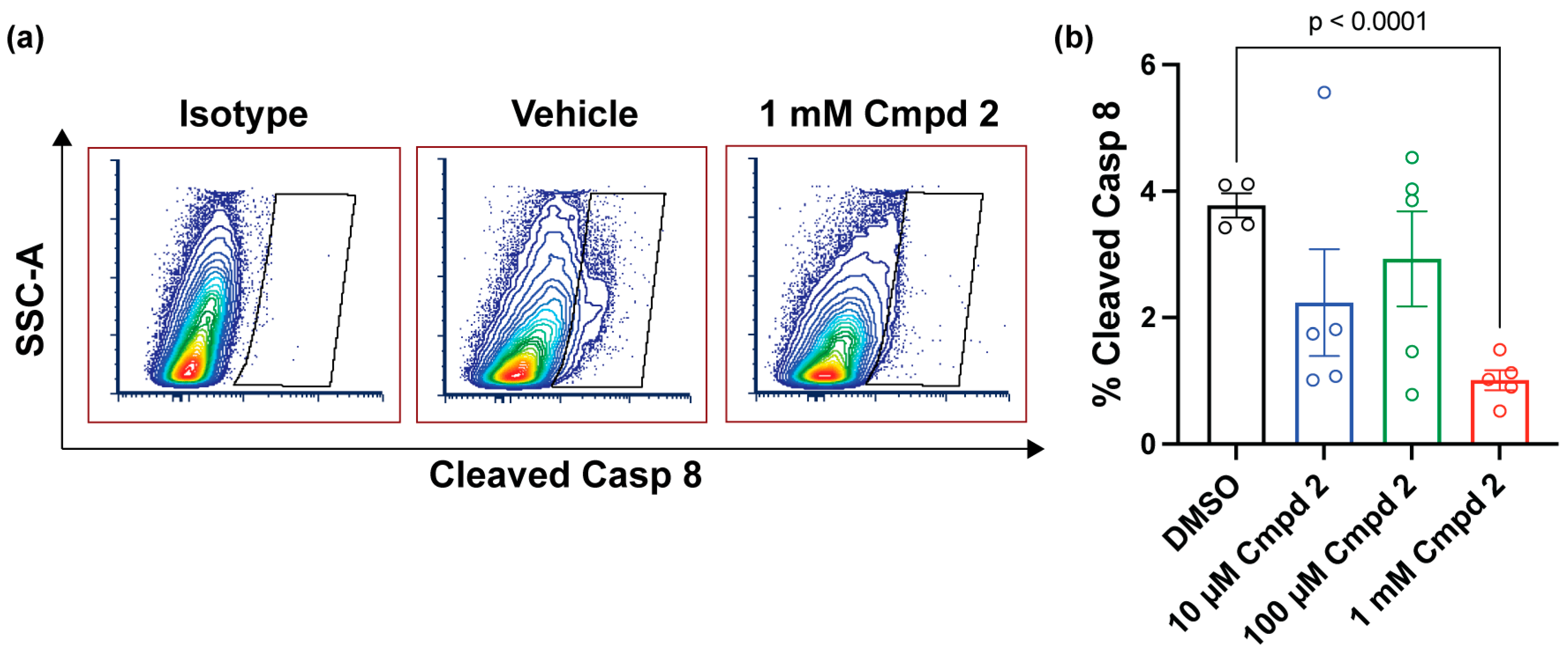
2.5. Compound 2 Reduces Apoptosis after Experimental Retinal Detachment

Considering compound 2 produced in vivo PK activation that was on par with ML-265 and demonstrated efficacy in circumventing apoptosis in vitro, we next sought to assess the ability of compound 2, with its changes to the core scaffold, to reduce apoptosis in an experimental model of retinal detachment in rats. Caspase activation, including caspase 8, increases 3 days status post experimental retinal detachment and has been validated to be a marker of photoreceptor apoptosis and the extent of retinal-detachment-induced photoreceptor cell death [8,9,33,36,39]. Additionally, this caspase activation coincides with the peak of the terminal deoxynucleotidyl transferase dUTP nick end labeling (TUNEL) staining of photoreceptors, specifically after experimental retinal detachment in rodents [9,36,40]. So, the detection and quantification of cleaved caspase 8 can be used as a marker of photoreceptor death after experimental retinal detachment and to assess the efficacy of neuroprotective agents [36]. As such, different concentrations of compound 2 or its vehicle, DMSO, were injected at the time of retinal detachment creation in rats, and the retinas were harvested 3 days later to assess caspase 8 activation. Considering their similarities, the concentrations of compound 2 evaluated were chosen to match those previously tested with the tool compound, ML-265 [9]. Compound 2 statistically significantly reduced the percentage of cleaved caspase-8-positive cells in the retina at the highest dose tested as compared to DMSO (Figure 5).

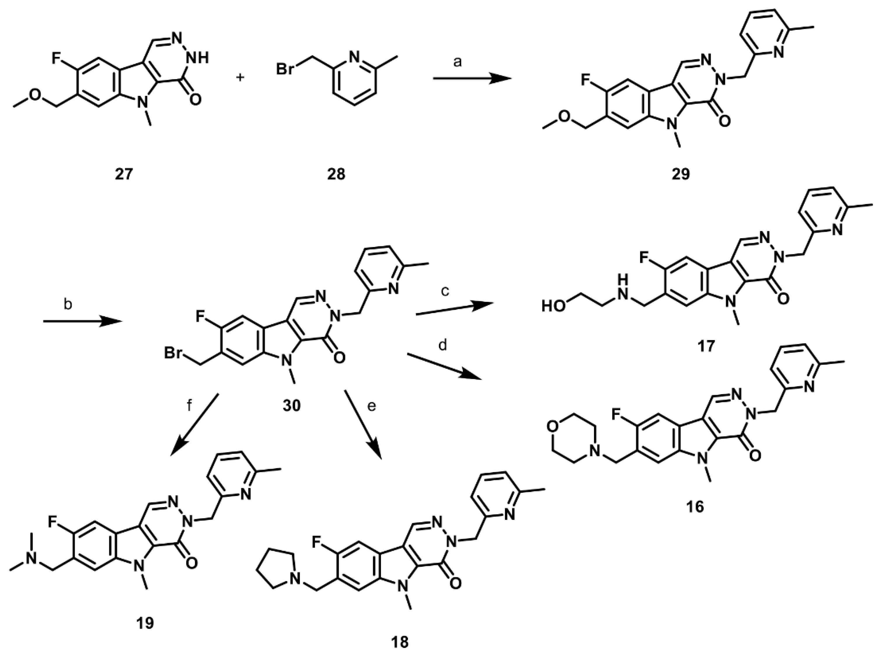
2.6. Synthesis of Structurally Diverse PKM2 Activators with Improved Solubility and Retained Potency

Compound 2 demonstrated that structural changes to the ML-265 scaffold are tolerated from a potency and efficacy standpoint (Figure 2), allowed for a similar binding mode to the target (Figure 3), and circumvented apoptosis in in vitro and in vivo models of photoreceptor stress (Figure 4 and Figure 5). It did not solve the issues of solubility or problematic functional groups but rather provided a more versatile template from which to build structurally diverse PKM2 activators to address these critical drug development issues. So, the next phase of medicinal chemistry efforts focused on replacing the aniline and sulfoxide moieties as well as adding solubilizing functional groups. Importantly, the aniline and sulfoxide moieties were shown, via crystallography (Figure 3b), to participate in hydrogen bonding interactions with PKM2 but were determined to not be critical for the activation of PKM2 as the elimination of these functionality in compound 1, for example, did not demonstrate a decrease in potency (Table 1). Due to the apparent flexibility and functional group tolerance of these positions, we elected to focus on the incorporation of small, basic functionality known to increase the solubility and formulatability with the goal of generating a homogenous aqueous dosing solution. Additionally, with the results in Table 1 showing a correlation between compound solubility and the in vivo activation of the target, the next series of compounds was designed to not only be more soluble than ML-265 and compound 2 but cover a range of solubility levels with the intention of identifying optimal compound properties. The synthetic routes described in Scheme 4, Scheme 5 and Scheme 6 were utilized to generate this next series of PKM2 activators (Table 3) along with the additional experimental methods presented in the Supporting Information, File S1.
Commercially available (5-amino-2-fluorophenyl)methanol (22) was treated with sodium hydride (NaH) and MeI in dimethylformamide (DMF) to install the desired methyl ether (Scheme 4). Subsequent treatment with N-iodosuccinamide (NIS) in acetic acid provided 23 in 68% yield over the two chemical steps. The treatment of 23 with tosyl chloride (TsCl) in the presence of pyridine in dichloromethane resulted in the formation of the sulfonamide (24) in 71% yield. Treating compound 24 with tetrakis(triphenylphosphine)palladium (Pd(PPh3)4), ethyl propiolate, zinc bromide (ZnBr2), and diisopropylethylamine (DIPEA) in tetrahydrofuran (THF) at 80 °C allowed for a palladium-mediated cyclization to form the indole, 25, in 65% yield. The treatment of 25 with potassium hydroxide (KOH) in THF removed the tosylate moiety, and subsequent treatment with MeI in the presence of potassium carbonate (K2CO3) in DMF provided the desired N-methylindole, 26, in 92% yield. The treatment of 26 with the Vilsmeier–Haack reagent (generated upon adding phosphorous oxychloride to DMF) installed an aldehyde at the 3-position of the indole in 73% yield [23]. The resulting aldehyde was subjected to hydrazine (N2H4) in ethoxyethanol at elevated temperatures to provide intermediate 27 in 89% yield.
The alkylation of intermediate 27 with 2-(bromomethyl)-6-methylpyridine, 28, in the presence of cesium carbonate, Cs2CO3, in DMF provided pyridine 29 in 77% yield (Scheme 5). The treatment of 29 with boron tribromide (BBr3) in dichloromethane provided bromide 30, which was critical for the placement of the structurally diverse functional groups of compounds 1619 (Table 3). Bromide 30 was utilized without purification and treated individually with ethanolamine, morpholine, pyrrolidine, or dimethylamine hydrochloride in the presence of triethylamine in dichloroethane to produce compounds 16, 17, 18, and 19 in 62%, 90%, 59%, and 78% yields, respectively (Scheme 5).
The synthesis of compounds 20 and 21 (Table 3) commenced with the alkylation of intermediate 27 with 2-fluorobenzyl bromide, 31, in the presence of KOtBu in DMF to provide compound 32 in 81% yield (Scheme 6). Compound 32 was then treated with boron tribromide in dichloromethane, resulting in bromide 33. Without purifying it, bromide 33 was subjected to N-Boc-piperazine and triethylamine in dichloroethane to synthesize intermediate 34 with 56% yield. The removal of the tert-butyl carbamate protecting group on intermediate 34 under acid-mediated conditions (4 M HCl in dichloromethane) provided compound 21 in 92% yield. Exposing compound 33 to ethanolamine in the presence of triethylamine in dichloroethane resulted in the formation of compound 20 in 65% yield.
This structurally diverse series of compounds (1621) that resulted from the above medicinal chemistry schemes (Scheme 4, Scheme 5 and Scheme 6) demonstrated nanomolar potency and greater than 200% activation of recombinant human PKM2 (Table 3), similar to ML-265 (Table 1). All compounds except compound 17 showed nanomolar potency and greater than 200% PK activation in 661W cells. Importantly, compounds 1621 were determined to have greater aqueous solubility (Table 3) as compared to ML-265 when tested at pH 7.4 (Table 1). The ability to activate the target in vivo was then assessed for compounds 1821, which span the range of aqueous solubility identified and demonstrate similar potencies to ML-265 in vitro. Two microliters of each respective compound at 8.5 μM (final vitreous concentration of 1 μM) or DMSO was intravitreally injected into rat eyes, and the PK activity was assessed four hours later. All compounds increased PK activity above the baseline observed in eyes treated with DMSO (Figure 6a). Compound 20 increased PK activity in the rat retina to a similar degree to ML-265 after intravitreal injection. Compounds 18 and 19 increased PK activity appreciably in the rat retina after intravitreal injection but approximately 20–30% less than that observed with the tool compound, ML-265. Compound 21 demonstrated approximately a third of the retinal PK activation as that achieved with ML-265. Additionally, compound 19, chosen as a representative of this more soluble class of PKM2 activators, was selective for PKM2 and did not activate recombinant PKM1 (Figure 6b), retaining a key aspect of the tool compound [20]. Moreover, compound 19 demonstrated similar potency and activation of the target in cellular assays, regardless of whether it was dissolved in an organic solvent (DMSO) or an aqueous-based formulation at neutral pH and 320 mOsm/kg (Figure 6c).

3. Discussion

In this study, we synthesized a series of pyridazinoindolone-based PKM2 activators that were capable of activating the recombinant human enzyme with nanomolar AC50 and increasing the total PK activity in vitro in 661W cells and in vivo in rat retinas. Compound 2 in this series of activators, with its pyridazinoindolone core and aniline and methyl sulfoxide functionality, circumvented photoreceptor apoptosis in oft-utilized in vitro and in vivo pre-clinical models of outer retinal stress. Building on this versatile, efficacious pyridazinoindolone core structure, a series of novel, structurally diverse PKM2 activators (compounds 1621) devoid of problematic functional groups and more soluble than the tool compound was then developed with retained potency and activity with the recombinant enzyme and in vitro, as well as with demonstrable activity in vivo after intravitreal injection. These next-generation, small-molecule PKM2 activators with improved solubility, formulate-ability, and retained potency will lay the foundation for future translational and pharmaceutical efforts in photoreceptor neuroprotection.
The potency, selectivity, and commercial availability of ML-265 [19,20] made it an excellent tool compound to demonstrate that PKM2 activation is neuroprotective to photoreceptors [9]. However, its low aqueous solubility, which necessitates organic solvents with known ocular toxicity [9], prevented it from being an ideal intraocular clinical candidate and prompted the exploration of alternative chemotypes to the thienopyrrolopyridazinone heterocycle of ML-265 with the intention of identifying a more versatile and synthetically accessible class of PKM2 activators that can be built upon to circumvent the issues of the tool compound. The pyridazinoindolone core was chosen as it provides expanded synthetic accessibility, functional group compatibility, and greater regiochemical flexibility for solubilizing functional group incorporation.
Compound 2, which has a pyridazinoindolone core with methyl sulfoxide and aniline functional groups, is a direct comparator to ML-265 and demonstrates that changes to the core heterocycle are tolerated, as compound 2 retained potency with the recombinant enzyme and in 661W cells, improved upon in vivo potency after intravitreal injection in rat eyes (Table 1 and Figure 2), and, importantly, circumvented apoptosis in in vitro and in vivo models of photoreceptor stress (Figure 4 and Figure 5), similar to ML-265 [9]. The comparable activity and pre-clinical efficacy of compound 2 and ML-265 is also consistent with the similar binding mode and molecular interactions observed in the X-ray crystal structures of PKM2 in complex with compound 2 (Figure 3) or ML-265 [20].
Compounds 1 and 35 demonstrated that the aniline and methyl sulfoxide functional groups are not required for PKM2 activation. Replacing the methyl sulfoxide group with a methyl group while keeping the aniline moiety in place (compound 5), replacing the aniline with a benzyl group while keeping the methyl sulfoxide in place (compound 3), or modifying both the methyl sulfoxide with a methyl group and the aniline with a benzyl group (compound 1) resulted in compounds that demonstrated similar potency and activation of PKM2 (Table 1). These results are similar to the previously published structure–activity studies for ML-265 [21]. Taken together, the methyl sulfoxide and aniline groups, which pose issues in drug development, do not appear to be necessary for PKM2 activation. Furthermore, these structure–activity relationships, in combination with the similar nanomolar potencies observed for compounds 1621 despite their differing functional groups, suggest the importance of the π-π stacking interactions between the core heterocycle and the binding pocket phenylalanine residues (Figure 3b) for PKM2 binding and activation by this series of small-molecule activators.
Removing one or both solubilizing methyl sulfoxide and/or aniline functional groups (compounds 1 and 35) resulted in a significant decrease in solubility as well as a significant rightward shift in in vivo potency as compared to compound 2 and ML-265 (Table 1). This finding may suggest that intravitreal injection into the vitreous humor, which is a predominantly aqueous fluid with a water content of at least 98% [41], results in the precipitation or crystallization of the small-molecule activator, preventing significant concentrations of the compound from reaching photoreceptors where PKM2 is predominantly expressed in the retina [8,17]. Even though the aqueous solubility of compound 2 and ML-265 provides for the increased activation of PKM2 in the retina after intravitreal injection as compared to compounds 1 and 35, the solubility is still relatively low and requires the inclusion of organic solvents (e.g., DMSO) in amounts that prevent safe intraocular delivery due to the ocular toxicity of these solvents [9]. Hence, we took advantage of the pyridazinoindolone core’s expanded synthetic accessibility and versatility with respect to functional group incorporation and the regiochemical placement of such moieties to next develop novel, structurally diverse PKM2 activators with improved aqueous solubilities and retained potencies (Table 3).
To enhance the solubility, the methyl sulfoxide of compound 2 was replaced with a variety of methylene-linked solubilizing functionalities (Table 3). The morpholine (compound 16), ethanolamines (compounds 17 and 20), pyrrolidine (compound 18), dimethylamine (compound 19), and piperazine (compound 21) are all functional groups known to reduce the lipophilicity and enhance the solubility of organic compounds. However, the reduced lipophilicity imparted by these functional groups can attenuate cell permeability. To mitigate any loss in cell permeability via the incorporation of these hydrophilic functionalities, we added an adjacent fluoro group to help mask the polar nature of these functional groups. Gratifyingly, compounds 1621, with 2- to 7-fold greater aqueous solubilities than ML-265, retained nanomolar potency with the recombinant target enzyme. Additionally, all except compound 17 demonstrated comparable activity levels in vitro in 661W cells to ML-265, and the greater aqueous solubilities of these compounds did not prevent retinal PK activation when delivered intravitreally (Figure 6). The pendent aniline in ML-265 and compounds 2 and 4 was also replaced with 6-methylpyridine (compounds 1619) and 2-fluorophenyl (compounds 20 and 21) with no loss of potency, likely due to their ability to participate in similar types of interactions with the protein.
In conclusion, this study is the first to cultivate the next generation of novel, structurally diverse small-molecule PKM2 activators for delivery into the eye. With the improved solubility, lack of structural alerts, and retained in vivo activity of compounds 1621, organic solvents can be avoided, aniline-derived toxicity can be mitigated, and the drug development process can be streamlined to take metabolic reprogramming from the bench to the bedside for photoreceptor neuroprotection. A scarcity of treatment options currently exists to prevent photoreceptor death and ultimately prevent vision loss. While the ability of these novel, more soluble PKM2 activators to improve photoreceptor survival in pre-clinical models of retinal degeneration needs to be ascertained in future studies, the work herein provides the basis for new therapeutic agents that could impact millions of patients by combating currently untreatable retinal degenerative diseases.

4. Materials and Methods

4.1. Materials

Animals. All animals were treated according to the Association for Research in Vision and Ophthalmology (ARVO) Statement for the Use of Animals in Ophthalmic and Vision Research. The University Committee on Use and Care of Animals of the University of Michigan approved the protocol (Protocol numbers: PRO00011133 and PRO00011135). Adult Brown-Norway rats of both sexes that had been retired from breeding were housed with a 12 h light and 12 h dark cycle and utilized for all in vivo studies.

4.2. Cell Culture

The 661W photoreceptor-like cell line was utilized for all in vitro studies and was provided by Dr. Muayyad al-Ubaidi (Department of Cell Biology, University of Oklahoma Health Sciences Center, Oklahoma City, OK, USA) [26]. Dulbecco’s modified Eagle’s medium (DMEM, Thermo Fisher Scientific, Waltham, MA, USA, Cat No. 11995065) supplemented with 10% fetal bovine serum (FBS), 90 units/mL penicillin, 0.09 mg/mL streptomycin, 32 mg/L putrescine, 40 µL/L of β-mercaptoethanol, and 40 µg/L of both hydrocortisone 21-hemisuccinate and progesterone was utilized. Cells were grown at 37 °C in 5% CO2 and 95% air.
Chemicals. Reagents of analytical grade were purchased from Sigma (St. Louis, MO, USA) or Combi Blocks Inc. (San Diego, CA, USA). ML-265 was obtained through Cayman Chemical (Ann Arbor, MI, USA; CAS 1221186-53-3).

4.3. Pyruvate Kinase Activity Enzyme Assay

Recombinant Enzyme. An enzyme-coupled assay that measures the depletion of NADH via absorbance at 340 nm and uses lactate dehydrogenase (LDH) was employed to determine the activity of pyruvate kinase (PK) [8,9]. To determine the AC50 (concentration of activator necessary to achieve half-maximal activation) of the different small molecules with PKM2, assays were conducted in a 96-well format using 200 μL/well assay volume with 10 nM human recombinant PKM2 (Sigma, St. Louis, MO, USA, Cat No. SAE0021) or PKM1 (Sigma, Waltham, MA, USA, Cat No. SRP0415), differing concentrations of activator, 0.5 mM PEP, 1 mM ADP, 0.2 mM NADH, and 8 U of LDH in a buffer of 50 mM Tris-HCl (pH 7.4), 100 mM KCl, and 5 mM MgCl2 as previously described [8,9,17]. A SPECTROstar Omega plate reader (BMG LABTECH Inc., Cary, NC, USA) monitored absorbance at 340 nm. The MARS software suite was utilized to determine initial velocities. Data were normalized to DMSO (dimethyl sulfoxide)-treated enzyme activity, as previously described [9].
Cell culture. Next, 661W cells were incubated with either DMSO or different concentrations of small-molecule PKM2 activator for 2 h, lysed and homogenized in RIPA Lysis and Extraction Buffer (Thermo Fisher, Waltham, MA, USA, Cat No. 89900) with protease inhibitors (Complete-Mini, Roche Diagnostics, Indianapolis, IN, USA), and centrifuged at 10,000 rpm for 10 min to remove cellular debris. The supernatant (4–8 μL) was then used to assess the pyruvate kinase activity as described above. The total protein content was determined using the Pierce BCA protein assay kit (Thermo Fisher, Waltham, MA, USA, Cat No. 23225) with the Pierce™ Bovine Serum Albumin Standard Pre-Diluted Set (Thermo Fisher, Waltham, MA, USA, Cat No. 23208) and used to normalize activity as described previously [19].
Animals. Intravitreal injections of various small-molecule PKM2 activators or vehicle (DMSO) were performed in Brown-Norway adult rats. The rats were anesthetized with ketamine (90 mg/mL) and xylazine (10 mg/mL) and their pupils dilated with topical phenylephrine (2.5%) and tropicamide (1%) as described previously [9]. A sclerotomy was created with a 25-gauge microvitreoretinal blade (Walcott Rx Products, Ocean View, NJ, USA) just posterior to the limbus. A blunt 35-gauge cannula was inserted through this sclerotomy into the vitreous cavity to deliver 2 μL of different concentrations of small-molecule activator or DMSO. This injection volume produces minimal reflux with adequate reproducibility [29,30]. Fifteen microliters of vitreous volume was assumed in rats to calculate the intravitreal concentrations of the compound after injection [29,30]. As we have previously described, left eyes were treated with small-molecular activator, while right eyes were treated with DMSO, which served as the control upon which pyruvate kinase activation was based [9]. Four hours later, the retina from each rat eye was dissected from the RPE-choroid, homogenized, and lysed in RIPA Lysis and Extraction Buffer with protease inhibitors. The pyruvate kinase (PK) activity assay, as described above, was performed using 4 to 8 microliters of rat retinal lysate, similar to what we and others have previously described [8,9,17]. Analogous to the cell culture assays, the total protein of the lysate was used to normalize pyruvate kinase activity.

4.4. Aqueous Solubility of Small Molecule PKM2 Activators

Compounds 15, as well as ML-265, underwent aqueous solubility testing at Eurofins Panlabs (St. Charles, MO, USA), a full-service drug discovery contract research organization under contract with the University of Michigan. In brief, each compound was tested at a concentration of 200 μM in PBS, pH 7.4, and room temperature. After a 24-h incubation, the aqueous solubility (μM) was determined using high-performance liquid chromatography (HPLC) with an ultraviolet/visible light (UV/Vis) detector. The area of the principal peak in a calibration standard for each compound (200 μM) in organic solvent (60/40 v/v% methanol: water) was compared to the area of the corresponding peak in the PBS sample [42]. Compounds 1621 underwent solubility testing at Analiza (Cleveland, OH, USA), a contract research organization. Five hundred microliters of PBS, pH 7.4 was added to each amount of compound provided. The samples were vortexed briefly and then placed on a rotary shaker at 200 RPM for 24 h at ambient temperature. Thereafter, the samples were vacuum filtered through a 0.45-micron membrane, and the filtrates were injected into a nitrogen detector for quantification. The nitrogen detector was calibrated using standards, and the filtrates were quantitated based on the calibration curve.

4.5. PKM2 Protein Expression and Purification

A pET28a-LIC expression vector containing PKM2 (residues 1–531) with an N-terminal, non-cleavable His6 tag (MGSSHHHHHHSSGLVPRGS) was purchased from Addgene (Watertown, MA, USA, Addgene plasmid # 25360). The plasmid was transformed into Rosetta2 cells. The cells were grown in Terrific Broth at 37 °C to an O.D.600 of 1.0, induced with 0.5 mM Isopropyl β-D-1-thiogalactopyranoside, and the protein was expressed overnight at 18 °C. The cells were pelleted via centrifugation and stored at −80 °C. The frozen cells were thawed and lysed using sonication in buffer containing 10 mM HEPES, pH 7.5, 300 mM KCl, 5 mM imidazole, 5 mM MgCl2, 5% glycerol, 2.5 mM TCEP, 0.5% CHAPS, and protease inhibitors. Cellular debris was removed via centrifugation, and cleared supernatant was batch bound to Ni-NTA resin (Qiagen, Germantown, MD, USA) pre-equilibrated in wash buffer (10 mM HEPES, pH 7.5; 300 mM KCl, 5 mM imidazole, 5 mM MgCl2, 5% glycerol, and 2.5 mM TCEP) for 1 h at 4 °C. The resin was washed with 15 column volumes of wash buffer and then applied to a column where protein was eluted with 5 column volumes of wash buffer containing 300 mM imidazole. The eluted protein was then incubated for 30 min at 4 °C with 5 molar equivalents of ADP in 5 mM TCEP and 5 mM MgCl2, concentrated, and applied to a Supderdex 200 pre-equilibrated with 10 mM HEPES pH 7.5, 150 mM KCl, 5 mM TCEP, 5 mM MgCl2, and 5% glycerol. The protein was eluted as a tetramer and was judged to be >95% pure via SDS-PAGE. The purified tetramer was further concentrated to 28 mg/mL and stored at −80 °C.

4.6. PKM2 Crystallization and Structure Determination

Prior to crystallization, purified PKM2 was incubated with 10 molar excess of compound 2 overnight at room temperature. The following day, the PKM2:compound 2 complex was set up for crystallization at 20 °C against wells containing 200 mM ammonium sulfate and 20% polyethylene glycol 3350. Crystals appeared within 24 h in drops containing 0.5 μL of protein complex and 0.5 μL of well solution. The crystals continued to grow for 7 days and were harvested and cryoprotected in well solution containing 25% ethylene glycol. Diffraction data were collected at the Advanced Photon Source on the LS-CAT 21-ID-D beamline equipped with an Eiger 9M detector. The data were processed with HKL2000 [43], and the structure was solved via molecular replacement [44] using a monomer of the activator-bound form of PKM2 (PDB ID: 3ME3) as the search model. The structure was solved in space group I222 with one monomer in the asymmetric unit. Coordinates and restraints for compound 2 were created using Grade [45]. The protein complex was iteratively refined and fit to electron density maps using Buster [45] and Coot [46], respectively. The resulting structure showed the activator bound in two orientations; one orientation was supplied by a symmetry mate as compound 2 resides on a symmetry axis. In addition to compound 2, the protein co-purified with an oxalate in the active site. The N-terminal His-tag along with the first 12 residues of the protein were disordered in the structure. The data collection and refinement statistics for the structure are shown in Table 2.

4.7. Western Blot

The 661W cells were plated in 6-well plates overnight in DMEM with 10% FBS. Subsequently, media were replaced with DMEM, no glucose (Thermo Fisher Scientific, Waltham, MA, USA, Cat No. 11966025), supplemented with either 5.5 mM or 25 mM glucose and 10% dialyzed serum. Forty-eight hours later, the cells were harvested and lysed in RIPA Lysis and Extraction Buffer containing protease and phosphatase inhibitors (Cell Signaling, Cat No. 5872). The protein concentration was estimated using the Pierce BCA protein assay kit. Twenty-five micrograms of protein was diluted in Laemmli buffer (Bio-Rad, Hercules, CA, USA, Cat No. 1610747) complemented with β-mercaptoethanol (Millipore-Sigma, Burlington, MA, USA, Cat No. M6250) and run on a 4–20% Mini-PROTEAN® TGX™ Precast Gel (Bio-Rad, Hercules, CA, USA, Cat No. 4561093EDU). The blots were processed and developed as we have previously described [47]. The antibodies that were utilized were as follows: PKM2-specific monoclonal antibody (Proteintech, Rosemont, IL, USA, Cat No. 60268-1-lg) and anti-α-tubulin antibody, mouse monoclonal (Sigma-Aldrich, St. Louis, MO, USA, Cat No. T6199).

4.8. Caspase Activity Assay

Caspase 8 and caspase 3/7 activity was measured using luminescent assay kits (Caspase-Glo 8 and 3/7 Assay Systems, Promega, Madison, WI, USA, Cat Nos. G8200 and G8090, respectively). Twenty-four hours prior to treatment, 661W cells were seeded in white-walled 96-well plates at 2500 cells/well. Cells were treated for 2 h with compound 2 or DMSO prior to the addition of 500 ng/mL FasL (Recombinant Mouse Fas Ligand/TNFSF6 Protein, R&D Systems Inc., Minneapolis, MN, USA, Cat No. 6128-SA-025) and 250 ng/mL HA (Hemagglutinin/HA Peptide Antibody, R&D Systems Inc., Minneapolis, MN, USA, Cat No. MAB060) [9]. As previously published, caspase activity was measured 8 h after treatment by incubating cells with substrate for 1 h as per the manufacturer’s instructions. A plate reader luminometer (BMG Labtech, Inc., Cary, NC, USA) assessed luminescence [9,38].

4.9. Cell Viability

A luminescent assay kit (RealTime-Glo MT Cell Viability Assay, Promega, Madison, WI, USA, Cat No. G9711) was utilized to assess cell viability. As above, 661W cells were seeded in 96-well plates (Nunc, Rochester, NY, USA) at 2500 cells/well 1 day before treatment [9,38]. The 661w cells were also pre-treated with compound 2 or DMSO for 2 h before adding 500 ng/mL FasL and 250 ng/mL HA. Viability was measured 48 h after the addition of these latter reagents [9]. As with the caspase assays, luminescence was monitored via a plate reader luminometer (BMG Labtech, Inc., Cary, NC, USA).

4.10. Experimental Model of Retinal Detachment

Detachments were created in rats as previously outlined [8,33,36,39,48]. In brief, rats were anesthetized with ketamine (100 mg/mL) and xylazine (20 mg/mL), and their pupils were dilated as above. A sclerotomy was created 1–2 mm posterior to the limbus to avoid damaging the lens. Two microliters of varying concentrations of compound 2 (85 μM, 850 μM, or 8.5 mM) or equal-volume DMSO was injected into the vitreous cavity through this sclerotomy at the time of retinal detachment. To create the experimental retinal detachment, a subretinal injector with a 35-gauge beveled cannula was inserted through the sclerotomy and then through a peripheral retinotomy into the subretinal space. Eight microliters of sodium hyaluronate (10 mg/mL) (Abbott Medical Optics, Irvine, CA, USA, Healon OVD) was injected to detach the neurosensory retina from the retinal pigment epithelium (RPE).

4.11. Flow Cytometric Assessment of Cleaved Caspase 8 after Experimental Retinal Detachment

PKM2 activator and DMSO-treated detached rat retinas were collected after 3 days to assess the percent of cells positive for cleaved caspase 8 via flow cytometry. The retinas were dissociated in a 0.25% Trypsin solution (Thermo Fisher, Waltham, MA, USA, Cat No. 25200) containing 400 ug of deoxyribose nuclease (Worthington Biochemical, Lakewood, NJ, USA, Cat No. LS002425) and incubated at 37 °C for 20 min with occasional agitation. Dissociated samples were collected via 35 μm nylon mesh, strainer cap flow tubes. The cell suspension was centrifuged at 300× g for 5 min at room temperature.
After washing the cells in ice-cold PBS, the cells were fixed with 4% paraformaldehyde for 20 min at room temperature followed by two PBS washes via centrifugation at 300× g for 10 min at 4 °C. The cells were then permeabilized using 70% ice-cold methanol at −20 °C overnight. The rinsed cell pellets were incubated with cleaved caspase 8 (Asp387) (D5B2) rabbit monoclonal antibody (Cell Signaling, Danvers, MA, USA, Cat No. 14071) at 1:50 dilution or concentration-matched Rabbit IgG as an isotype control for 1 h at room temperature. The cells were washed with FACS buffer (1% BSA in PBS) and subjected to flow cytometry using an LSR II from BD Biosciences. To exclude events that may represent clumped cells or debris, events were gated based on forward and side scatter. Data were analyzed with FCS Express software (De Novo Software, Ontario, CA, USA).

4.12. Statistical Analysis

The results observed in this report are represented as mean ± SEM. Data were analyzed using one-way ANOVA for in vitro and in vivo efficacy data in Prism 9.0 (GraphPad Software, San Diego, CA, USA). The initial velocity data obtained from the steady state kinetic assay described above were analyzed using nonlinear regression curve fitting with the agonist versus response model in Prism 9.0.

Supplementary Materials

The following supporting information can be downloaded at: https://www.mdpi.com/article/10.3390/ph16050705/s1, File S1. Experimental details and NMR spectra of PKM2 activators.

Author Contributions

Conceptualization, T.J.W., J.C.R. and C.G.B.; methodology, T.J.W., E.W., J.A.S., J.C.R. and C.G.B.; validation, T.J.W., E.W., J.C.R., M.G. and C.G.B.; formal analysis, T.J.W., E.W. and J.A.S.; investigation, S.C., B.T.W., J.A.S., E.W., R.F., M.G. and M.P.; resources, T.J.W., E.W., R.F., J.A.S. and C.G.B.; data curation, T.J.W., J.C.R., E.W., M.G., J.A.S. and C.G.B.; writing—original draft preparation, T.J.W.; writing—review and editing, T.J.W., E.W., J.C.R. and C.G.B.; visualization, T.J.W. and J.C.R.; supervision, J.C.R. and C.G.B.; project administration, T.J.W., J.C.R. and C.G.B.; funding acquisition, T.J.W., J.C.R. and C.G.B. All authors have read and agreed to the published version of the manuscript.

Funding

The study was supported by grants from Michigan Center for Therapeutic Innovation and Michigan Translation and Commercialization (MTRAC) for Life Sciences Hub (T.W., J.R., and C.B.) and Paul R. Lichter M.D., Vision Research Discovery Fund (T.W.). Additional support was provided from the National Eye Institute (NEI) (R01EY029675) and an unrestricted grant from the Research to Prevent Blindness within the Department of Ophthalmology and Visual Sciences at the University of Michigan. This work also utilized the Vision Research Core funded by the NEI P30 EY007003. The funders had no role in study design, data collection and analysis, decision to publish, or preparation of the manuscript.

Institutional Review Board Statement

The University Committee on Use and Care of Animals of the University of Michigan approved the animal protocols (protocol numbers: PRO00011133 and PRO00011135).

Informed Consent Statement

Not applicable.

Data Availability Statement

The data generated and analyzed during this study are available from the corresponding author upon reasonable request. The coordinates and structure factors for the X-ray crystallography protein structure reported herein have been deposited in the PDB under accession code 8G2E. The NMR spectra for each compound is presented in Supplementary Material, File S1.

Acknowledgments

The authors thank Heather Hager for her technical expertise in handling the animals utilized in this report. The research reported in this publication was supported by the University of Michigan Center for Structural Biology (CSB). The CSB is grateful for support from the U-M Life Sciences Institute, the U-M Rogel Cancer Center, the U-M Medical School Endowment for Basic Sciences, and grants from the National Institute of Health. This research used resources of the Advanced Photon Source, a U.S. Department of Energy (DOE) Office of Science User Facility operated for the DOE Office of Science by Argonne National Laboratory under Contract No. DE-AC02-06CH11357. Use of the LS-CAT Sector 21 was supported by the Michigan Economic Development Corporation and the Michigan Technology Tri-Corridor (Grant 085P1000817). Parts of the graphical abstract were created with BioRender.com.

Conflicts of Interest

The authors report the following conflict of interest: T.W.—equity and royalty, Ocutheia; J.R. equity and royalty, Ocutheia; C.B.—equity and royalty, Ocutheia; royalty, ONL Therapeutics; employment, Janssen Research & Development. All other authors declare that no competing interests exist.

References

  1. Punzo, C.; Kornacker, K.; Cepko, C.L. Stimulation of the Insulin/MTOR Pathway Delays Cone Death in a Mouse Model of Retinitis Pigmentosa. Nat. Neurosci. 2009, 12, 44–52. [Google Scholar] [CrossRef]
  2. Venkatesh, A.; Ma, S.; Le, Y.Z.; Hall, M.N.; Rüegg, M.A.; Punzo, C. Activated MTORC1 Promotes Long-Term Cone Survival in Retinitis Pigmentosa Mice. J. Clin. Investig. 2015, 125, 1446–1458. [Google Scholar] [CrossRef]
  3. Aït-Ali, N.; Fridlich, R.; Millet-Puel, G.; Clérin, E.; Delalande, F.; Jaillard, C.; Blond, F.; Perrocheau, L.; Reichman, S.; Byrne, L.C.; et al. Rod-Derived Cone Viability Factor Promotes Cone Survival by Stimulating Aerobic Glycolysis. Cell 2015, 161, 817–832. [Google Scholar] [CrossRef] [PubMed]
  4. Kanow, M.A.; Giarmarco, M.M.; Jankowski, C.S.; Tsantilas, K.; Engel, A.L.; Du, J.; Linton, J.D.; Farnsworth, C.C.; Sloat, S.R.; Rountree, A.; et al. Biochemical Adaptations of the Retina and Retinal Pigment Epithelium Support a Metabolic Ecosystem in the Vertebrate Eye. eLife 2017, 6, e28899. [Google Scholar] [CrossRef]
  5. Petit, L.; Ma, S.; Cipi, J.; Cheng, S.-Y.; Zieger, M.; Hay, N.; Punzo, C. Aerobic Glycolysis Is Essential for Normal Rod Function and Controls Secondary Cone Death in Retinitis Pigmentosa. Cell Rep. 2018, 23, 2629–2642. [Google Scholar] [CrossRef] [PubMed]
  6. Zhang, L.; Du, J.; Justus, S.; Hsu, C.-W.; Bonet-Ponce, L.; Wu, W.-H.; Tsai, Y.-T.; Wu, W.-P.; Jia, Y.; Duong, J.K.; et al. Reprogramming Metabolism by Targeting Sirtuin 6 Attenuates Retinal Degeneration. J. Clin. Investig. 2016, 126, 4659–4673. [Google Scholar] [CrossRef]
  7. Chinchore, Y.; Begaj, T.; Wu, D.; Drokhlyansky, E.; Cepko, C.L. Glycolytic Reliance Promotes Anabolism in Photoreceptors. eLife 2017, 6, e25946. [Google Scholar] [CrossRef]
  8. Wubben, T.J.; Pawar, M.; Smith, A.; Toolan, K.; Hager, H.; Besirli, C.G. Photoreceptor Metabolic Reprogramming Provides Survival Advantage in Acute Stress While Causing Chronic Degeneration. Sci. Rep. 2017, 7, 17863. [Google Scholar] [CrossRef] [PubMed]
  9. Wubben, T.J.; Pawar, M.; Weh, E.; Smith, A.; Sajjakulnukit, P.; Zhang, L.; Dai, L.; Hager, H.; Pai, M.P.; Lyssiotis, C.A.; et al. Small Molecule Activation of Metabolic Enzyme Pyruvate Kinase Muscle Isozyme 2, PKM2, Circumvents Photoreceptor Apoptosis. Sci. Rep. 2020, 10, 2990. [Google Scholar] [CrossRef]
  10. Casson, R.J.; Chidlow, G.; Han, G.; Wood, J.P.M. An Explanation for the Warburg Effect in the Adult Mammalian Retina. Clin. Experiment. Ophthalmol. 2013, 41, 517. [Google Scholar] [CrossRef]
  11. Chertov, A.O.; Holzhausen, L.; Kuok, I.T.; Couron, D.; Parker, E.; Linton, J.D.; Sadilek, M.; Sweet, I.R.; Hurley, J.B. Roles of Glucose in Photoreceptor Survival. J. Biol. Chem. 2011, 286, 34700–34711. [Google Scholar] [CrossRef]
  12. Ng, S.K.; Wood, J.P.M.; Chidlow, G.; Han, G.; Kittipassorn, T.; Peet, D.J.; Casson, R.J. Cancer-like Metabolism of the Mammalian Retina. Clin. Exp. Ophthalmol. 2015, 43, 367–376. [Google Scholar] [CrossRef]
  13. Wong, N.; Ojo, D.; Yan, J.; Tang, D. PKM2 Contributes to Cancer Metabolism. Cancer Lett. 2015, 356, 184–191. [Google Scholar] [CrossRef] [PubMed]
  14. Casson, R.J.; Wood, J.P.M.; Han, G.; Kittipassorn, T.; Peet, D.J.; Chidlow, G. M-Type Pyruvate Kinase Isoforms and Lactate Dehydrogenase A in the Mammalian Retina: Metabolic Implications. Investig. Ophthalmol. Vis. Sci. 2016, 57, 66–80. [Google Scholar] [CrossRef]
  15. Gui, D.Y.; Lewis, C.A.; Vander Heiden, M.G. Allosteric Regulation of PKM2 Allows Cellular Adaptation to Different Physiological States. Sci. Signal. 2013, 6, pe7. [Google Scholar] [CrossRef] [PubMed]
  16. Lindsay, K.J.; Du, J.; Sloat, S.R.; Contreras, L.; Linton, J.D.; Turner, S.J.; Sadilek, M.; Satrústegui, J.; Hurley, J.B. Pyruvate Kinase and Aspartate-Glutamate Carrier Distributions Reveal Key Metabolic Links between Neurons and Glia in Retina. Proc. Natl. Acad. Sci. USA 2014, 111, 15579–15584. [Google Scholar] [CrossRef] [PubMed]
  17. Rajala, R.V.S.; Rajala, A.; Kooker, C.; Wang, Y.; Anderson, R.E. The Warburg Effect Mediator Pyruvate Kinase M2 Expression and Regulation in the Retina. Sci. Rep. 2016, 6, 37727. [Google Scholar] [CrossRef] [PubMed]
  18. Yang, W.; Lu, Z. Pyruvate Kinase M2 at a Glance. J. Cell Sci. 2015, 128, 1655–1660. [Google Scholar] [CrossRef] [PubMed]
  19. Jiang, J.; Walsh, M.J.; Brimacombe, K.R.; Anastasiou, D.; Yu, Y.; Israelsen, W.J.; Hong, B.-S.; Tempel, W.; Dimov, S.; Veith, H.; et al. ML265: A Potent PKM2 Activator Induces Tetramerization and Reduces Tumor Formation and Size in a Mouse Xenograft Model. In Probe Reports from the NIH Molecular Libraries Program; National Center for Biotechnology Information (US): Bethesda, MD, USA, 2010. [Google Scholar]
  20. Anastasiou, D.; Yu, Y.; Israelsen, W.J.; Jiang, J.-K.; Boxer, M.B.; Hong, B.S.; Tempel, W.; Dimov, S.; Shen, M.; Jha, A.; et al. Pyruvate Kinase M2 Activators Promote Tetramer Formation and Suppress Tumorigenesis. Nat. Chem. Biol. 2012, 8, 839–847. [Google Scholar] [CrossRef] [PubMed]
  21. Jiang, J.; Boxer, M.B.; Vander Heiden, M.G.; Shen, M.; Skoumbourdis, A.P.; Southall, N.; Veith, H.; Leister, W.; Austin, C.P.; Park, H.W.; et al. Evaluation of Thieno[3,2-b]Pyrrole[3,2-d]Pyridazinones as Activators of the Tumor Cell Specific M2 Isoform of Pyruvate Kinase. Bioorg. Med. Chem. Lett. 2010, 20, 3387–3393. [Google Scholar] [CrossRef]
  22. Sodano, T.M.; Combee, L.A.; Stephenson, C.R.J. Recent Advances and Outlook for the Isosteric Replacement of Anilines. ACS Med. Chem. Lett. 2020, 11, 1785–1788. [Google Scholar] [CrossRef]
  23. Xue, J.; Zhang, Y.-S.; Huan, Z.; Yang, J.-D.; Cheng, J.-P. Catalytic Vilsmeier-Haack Reactions for C1-Deuterated Formylation of Indoles. J. Org. Chem. 2022, 87, 15539–15546. [Google Scholar] [CrossRef]
  24. Tan, E.; Ding, X.-Q.; Saadi, A.; Agarwal, N.; Naash, M.I.; Al-Ubaidi, M.R. Expression of Cone-Photoreceptor-Specific Antigens in a Cell Line Derived from Retinal Tumors in Transgenic Mice. Investig. Ophthalmol. Vis. Sci. 2004, 45, 764–768. [Google Scholar] [CrossRef] [PubMed]
  25. Al-Ubaidi, M.R.; Matsumoto, H.; Kurono, S.; Singh, A. Proteomics Profiling of the Cone Photoreceptor Cell Line, 661W. Adv. Exp. Med. Biol. 2008, 613, 301–311. [Google Scholar] [CrossRef] [PubMed]
  26. al-Ubaidi, M.R.; Font, R.L.; Quiambao, A.B.; Keener, M.J.; Liou, G.I.; Overbeek, P.A.; Baehr, W. Bilateral Retinal and Brain Tumors in Transgenic Mice Expressing Simian Virus 40 Large T Antigen under Control of the Human Interphotoreceptor Retinoid-Binding Protein Promoter. J. Cell Biol. 1992, 119, 1681–1687. [Google Scholar] [CrossRef]
  27. Parikh, R.; Ross, J.S.; Sangaralingham, L.R.; Adelman, R.A.; Shah, N.D.; Barkmeier, A.J. Trends of Anti-Vascular Endothelial Growth Factor Use in Ophthalmology Among Privately Insured and Medicare Advantage Patients. Ophthalmology 2017, 124, 352–358. [Google Scholar] [CrossRef]
  28. Patel, S. Medicare Spending on Anti-Vascular Endothelial Growth Factor Medications. Ophthalmol. Retin. 2018, 2, 785–791. [Google Scholar] [CrossRef]
  29. Vezina, M.; Bussieres, M.; Glazier, G.; Gagnon, M.-P.; Martel, D. Determination of Injectable Intravitreous Volumes in Rats. Investig. Ophthalmol. Vis. Sci. 2011, 52, 3219. [Google Scholar]
  30. Dureau, P.; Bonnel, S.; Menasche, M.; Dufier, J.L.; Abitbol, M. Quantitative Analysis of Intravitreal Injections in the Rat. Curr. Eye Res. 2001, 22, 74–77. [Google Scholar] [CrossRef]
  31. Chen, V.B.; Arendall, W.B.; Headd, J.J.; Keedy, D.A.; Immormino, R.M.; Kapral, G.J.; Murray, L.W.; Richardson, J.S.; Richardson, D.C. MolProbity: All-Atom Structure Validation for Macromolecular Crystallography. Acta Crystallogr. D Biol. Crystallogr. 2010, 66, 12–21. [Google Scholar] [CrossRef]
  32. Smart, O.S.; Horský, V.; Gore, S.; Svobodová Vařeková, R.; Bendová, V.; Kleywegt, G.J.; Velankar, S. Validation of Ligands in Macromolecular Structures Determined by X-Ray Crystallography. Acta Crystallogr. Sect. Struct. Biol. 2018, 74, 228–236. [Google Scholar] [CrossRef] [PubMed]
  33. Zacks, D.N.; Zheng, Q.-D.; Han, Y.; Bakhru, R.; Miller, J.W. FAS-Mediated Apoptosis and Its Relation to Intrinsic Pathway Activation in an Experimental Model of Retinal Detachment. Investig. Ophthalmol. Vis. Sci. 2004, 45, 4563–4569. [Google Scholar] [CrossRef]
  34. Zacks, D.N.; Boehlke, C.; Richards, A.-L.; Zheng, Q.-D. Role of the Fas-Signaling Pathway in Photoreceptor Neuroprotection. Arch. Ophthalmol. Chic. Ill 1960 2007, 125, 1389–1395. [Google Scholar] [CrossRef]
  35. Kanan, Y.; Moiseyev, G.; Agarwal, N.; Ma, J.-X.; Al-Ubaidi, M.R. Light Induces Programmed Cell Death by Activating Multiple Independent Proteases in a Cone Photoreceptor Cell Line. Investig. Ophthalmol. Vis. Sci. 2007, 48, 40–51. [Google Scholar] [CrossRef] [PubMed]
  36. Besirli, C.G.; Chinskey, N.D.; Zheng, Q.-D.; Zacks, D.N. Inhibition of Retinal Detachment-Induced Apoptosis in Photoreceptors by a Small Peptide Inhibitor of the Fas Receptor. Investig. Ophthalmol. Vis. Sci. 2010, 51, 2177–2184. [Google Scholar] [CrossRef]
  37. Lin, J.B.; Kubota, S.; Ban, N.; Yoshida, M.; Santeford, A.; Sene, A.; Nakamura, R.; Zapata, N.; Kubota, M.; Tsubota, K.; et al. NAMPT-Mediated NAD(+) Biosynthesis Is Essential for Vision In Mice. Cell Rep. 2016, 17, 69–85. [Google Scholar] [CrossRef] [PubMed]
  38. Besirli, C.G.; Zheng, Q.-D.; Reed, D.M.; Zacks, D.N. ERK-Mediated Activation of Fas Apoptotic Inhibitory Molecule 2 (Faim2) Prevents Apoptosis of 661W Cells in a Model of Detachment-Induced Photoreceptor Cell Death. PLoS ONE 2012, 7, e46664. [Google Scholar] [CrossRef] [PubMed]
  39. Zacks, D.N.; Hänninen, V.; Pantcheva, M.; Ezra, E.; Grosskreutz, C.; Miller, J.W. Caspase Activation in an Experimental Model of Retinal Detachment. Investig. Ophthalmol. Vis. Sci. 2003, 44, 1262–1267. [Google Scholar] [CrossRef]
  40. Hisatomi, T.; Sakamoto, T.; Goto, Y.; Yamanaka, I.; Oshima, Y.; Hata, Y.; Ishibashi, T.; Inomata, H.; Susin, S.A.; Kroemer, G. Critical Role of Photoreceptor Apoptosis in Functional Damage after Retinal Detachment. Curr. Eye Res. 2002, 24, 161–172. [Google Scholar] [CrossRef]
  41. Le Goff, M.M.; Bishop, P.N. Adult Vitreous Structure and Postnatal Changes. Eye 2008, 22, 1214–1222. [Google Scholar] [CrossRef]
  42. Lipinski, C.A.; Lombardo, F.; Dominy, B.W.; Feeney, P.J. Experimental and Computational Approaches to Estimate Solubility and Permeability in Drug Discovery and Development Settings. Adv. Drug Deliv. Rev. 2001, 46, 3–26. [Google Scholar] [CrossRef]
  43. Otwinowski, Z.; Minor, W. Processing of X-Ray Diffraction Data Collected in Oscillation Mode. Methods Enzymol. 1997, 276, 307–326. [Google Scholar] [PubMed]
  44. Vagin, A.; Teplyakov, A. Molecular Replacement with MOLREP. Acta Crystallogr. D Biol. Crystallogr. 2010, 66, 22–25. [Google Scholar] [CrossRef] [PubMed]
  45. Roversi, P.; Sharff, A.; Smart, O.S.; Vonrhein, C.; Womack, T.O. BUSTER Version 2.10.3; Global Phasing Ltd.: Cambridge, UK, 2011. [Google Scholar]
  46. Emsley, P.; Cowtan, K. Coot: Model-Building Tools for Molecular Graphics. Acta Crystallogr. D Biol. Crystallogr. 2004, 60, 2126–2132. [Google Scholar] [CrossRef] [PubMed]
  47. Weh, E.; Lutrzykowska, Z.; Smith, A.; Hager, H.; Pawar, M.; Wubben, T.J.; Besirli, C.G. Hexokinase 2 Is Dispensable for Photoreceptor Development but Is Required for Survival during Aging and Outer Retinal Stress. Cell Death Dis. 2020, 11, 422. [Google Scholar] [CrossRef] [PubMed]
  48. Besirli, C.G.; Chinskey, N.D.; Zheng, Q.-D.; Zacks, D.N. Autophagy Activation in the Injured Photoreceptor Inhibits Fas-Mediated Apoptosis. Investig. Ophthalmol. Vis. Sci. 2011, 52, 4193–4199. [Google Scholar] [CrossRef]
Figure 1. Structure of PKM2 activator tool compound, ML-265.
Pharmaceuticals 16 00705 g001
Scheme 1. Synthesis of PKM2 activators. a. POCl3, DMF, 79% and 83% yield, respectively, b. MeI, K2CO3, DMF, 62% (8) and 87% (9) yield, c. N2H4, ethoxyethanol, 135 °C, 12 h, 58% (10) and 63% (11) yield.
Pharmaceuticals 16 00705 sch001
Scheme 2. Synthesis of PKM2 activators. a. Benzyl bromide, KOtBu, DMF, 61% yield, b. tert-Butyl 3-(bromomethyl)phenylcarbamate, KOtBu, DMF, 66% yield, c. 4 M HCl in dioxane, dichloromethane, 60% yield.
Pharmaceuticals 16 00705 sch002
Scheme 3. Synthesis of PKM2 activators. a. Benzyl bromide, KOtBu, DMF, 72% yield, b. tert-Butyl 3-(bromomethyl)phenylcarbamate, KOtBu, DMF, 96% yield, c. mCPBA, dichloromethane, 53% (3) and 57% (15) yield, d. 4 M HCl in dioxane, dichloromethane, 63% (4) and 75% (2) yield.
Pharmaceuticals 16 00705 sch003
Figure 2. Pharmacologic activation of PKM2 with compound 2 is comparable to ML-265 in vivo. Pyruvate kinase (PK) activity with increasing concentrations of either compound 2 or ML-265 in rat retinas after intravitreal injection of the respective compound (n = 3 animals per concentration). Data were normalized to PK activity in rat retinas treated with DMSO. Mean ± SEM.
Pharmaceuticals 16 00705 g002
Figure 3. PKM2 in complex with compound 2. (a) Tetrameric PKM2 in complex with compound 2. Each protomer of the tetramer is depicted in cartoon mode and colored differently. Compound 2 is bound across the A-A’ interface and depicted as red spheres. An oxalate molecule (gray spheres) is bound in the active site of each PKM2 protomer along with a magnesium ion (pink sphere). Sodium ions are depicted as magenta spheres. (b) Representation of the residues and water molecules in contact with compound 2. The binding site consists of equivalent residues from each PKM2 protomer that makes up the A-A’ dimer interface. The 2FoFc map is contoured at 1.0σ. Residues from the different protomers are colored in blue and cyan, respectively. Hydrogen bonds are depicted as dashed yellow lines. Water molecules are depicted as red spheres. For clarity, only the bound form of compound 2 from one asymmetric unit is shown with dimeric form of the protein. The symmetry-related conformation of compound 2 is related by a two-fold symmetry about the z-axis of the figure.
Pharmaceuticals 16 00705 g003
Figure 4. Compound 2 treatment reduces caspase activation and improves 661W cell survival in an in vitro model of apoptosis. (a) 661W cells express PKM2 under 5.5 mM or 25 mM glucose concentration, the latter of which was and has been [9,36,38] used in the caspase and cell viability experiments that follow. Caspase 8 activation (b), caspase 3 and 7 activation (c), and cell viability (d) assayed in cells treated with DMSO or different concentrations of compound 2 (Cmpd 2) under standard tissue culture conditions or in the presence of 500 ng/mL FasL and 250 ng/mL hemagglutinin. Two hours before treatment with FasL, 661W cells were pre-treated with compound 2 or DMSO, and caspase activation and viability were measured 8 and 48 h after, respectively. n = 6; Mean ± SEM.
Pharmaceuticals 16 00705 g004
Figure 5. Compound 2 decreases apoptosis in experimental retinal detachment. Detached retinas treated with increasing concentrations of compound 2 (Cmpd 2) or vehicle (DMSO) at the time of experimental retinal detachment were harvested after 3 days. The percent of cells positive for cleaved caspase 8 was utilized as a marker of apoptotic activity in the retina. Compound 2 concentrations are the final vitreous concentrations after injection. (a) Representative contour plots depict a decrease in the number of cells positive for cleaved caspase 8 three days post experimental retinal detachment after treatment with 1 mM compound 2. (b) The graph shows a statistically significant change in the percent of cells positive for cleaved caspase 8 at the highest concentration of compound 2 tested. n = 4–5; Mean ± SEM.
Pharmaceuticals 16 00705 g005
Scheme 4. Synthesis of PKM2 activators. a. MeI, NaH, DMF, b. NIS, acetic acid, 68% yield across both steps (a and b), c. TsCl, pyridine, dichloromethane, 71%, d. Pd(PPh3)4, ethyl propiolate, ZnBr2, DIPEA, THF, 80 °C, 65% yield, e. KOH, THF, 78%, f. MeI, K2CO3, DMF, 92% yield, g. POCl3, DMF, 73%, h. N2H4, ethoxyethanol, 135 °C, 89% yield.
Pharmaceuticals 16 00705 sch004
Scheme 5. Synthesis of PKM2 activators. a. Cs2CO3, DMF, 77% b. BBr3, dichloromethane, c. ethanolamine, triethylamine, dichloroethane, 62% yield, d. morpholine, triethylamine, dichloroethane, 90% yield, e. pyrrolidine, triethylamine, dichloroethane, 59% yield, f. dimethylamine hydrochloride, triethylamine, dichloroethane, 78% yield.
Pharmaceuticals 16 00705 sch005
Scheme 6. Synthesis of PKM2 activators. a. KOtBu, DMF, 81% yield, b. BBr3, dichloromethane, c. N-Boc-piperazine, triethylamine, dichloroethane, 56% yield after steps b and c, d. 4 M HCl in dichloromethane, 92% yield, e. ethanolamine, triethylamine, dichloroethane, 65% yield after steps b and e.
Pharmaceuticals 16 00705 sch006
Figure 6. Soluble, structurally diverse, small-molecule PKM2 activators increase PK activity in the retina after intravitreal injection, remain selective for PKM2, and circumvent the need for organic solvents. (a) Pyruvate kinase (PK) activation determined at a final vitreous concentration of 1 μM of each compound after intravitreal injection in rats. The values achieved with ML-265 were normalized to 1, and the data are expressed as fold changes. n = 3–7; Mean ± SEM. (b) Pyruvate kinase (PK) activity with recombinant PKM2 or PKM1 in the presence of increasing concentrations of compound 19. n = 3; Mean ± SEM. (c) PK activity in 661W cell lysates after being incubated in increasing concentrations of compound 19 dissolved in either DMSO or an aqueous-based formulation. n = 3; Mean ± SEM.
Pharmaceuticals 16 00705 g006
Table 1. Structure–activity relationships for early-stage pyridazinoindolone series of PKM2 activators.
Pharmaceuticals 16 00705 i001
Recombinant PKM2 661W Cells In Vivo
Cmpd IDXR1R2AC50 (nM)Max Activation (%)AC50 (nM)Max Activation (%)PK Activation, Fold Change &Aqueous Solubility (M) *
ML-265Pharmaceuticals 16 00705 i002Pharmaceuticals 16 00705 i003Pharmaceuticals 16 00705 i00470 ± 17292 ± 21108 ± 32370 ± 211.00 ± 0.06153.4
1Pharmaceuticals 16 00705 i005Pharmaceuticals 16 00705 i006Pharmaceuticals 16 00705 i00723 ± 4224 ± 9175 ± 64219 ± 160.04 ± 0.09<0.01
2Pharmaceuticals 16 00705 i008Pharmaceuticals 16 00705 i009Pharmaceuticals 16 00705 i01050 ± 12292 ± 20120 ± 45355 ± 260.95 ± 0.1399.8
3Pharmaceuticals 16 00705 i011Pharmaceuticals 16 00705 i012Pharmaceuticals 16 00705 i01347 ± 10279 ± 16104 ± 32280 ± 170.50 ± 0.1612.1
4Pharmaceuticals 16 00705 i014Pharmaceuticals 16 00705 i015Pharmaceuticals 16 00705 i01648 ± 10216 ± 1266 ± 19256 ± 130.20 ± 0.09<0.01
5Pharmaceuticals 16 00705 i017Pharmaceuticals 16 00705 i018Pharmaceuticals 16 00705 i01961 ± 12249 ± 14132 ± 47236 ± 160.13 ± 0.0612.7
 n = 3 for each compound for both the recombinant PKM2 and 661W cell experiments. & Pyruvate kinase (PK) activation determined at a final vitreous concentration of 100 μM of each compound after intravitreal injection in rats (n = 3–4 eyes per compound). The values achieved with ML-265 were normalized to 1, and the data are expressed as fold change. * Solubility determined in PBS at pH 7.4.
Table 2. Crystallography data collection and refinement statistics.
PKM2-Compound 2
Data Collection
Space groupI222
Unit cell dimensions
a, b, c (Å)84.81, 115.97, 131.84
α, β, γ (°)90.0, 90.0, 90.0
Resolution (Å) &1.84 (1.87–1.84)
Rsym #0.038 (0.137)
<I/I> *29.5 (10.9)
% Completeness $94.4 (95.3)
Redundancy3.9 (3.8)
Refinement
Resolution (Å)1.84
No. of unique reflections53,262
R-factor +0.163
Rfree ##0.190
RMSD !
Bond lengths (Å)0.008
Bond angles (°)0.89
MolProbity Score %$0.91
Clash Score %$1.61
RSCC **0.96
RSR **0.14
& Statistics for highest-resolution bin of reflections in parentheses. # Rsym = ∑hj|Ihj − <Ih>|/∑hjIhj, where Ihj is the intensity of observation j of reflection h, and <Ih> is the mean intensity for multiple recorded reflections. * Intensity signal-to-noise ratio. $ Completeness of the unique diffraction data. + R-factor = ∑h|IFoI − IFcI|/∑h|Fo|, where Fo and Fc are the observed and calculated structure factor amplitudes for reflection h. ## Rfree is calculated against a 5% random sampling of the reflections that were removed before structure refinement. ! Root mean square deviation of bond lengths and bond angles. %$ Reference [31]. ** RSR—real-space R value; RSCC—real-space correlation coefficient; reference [32].
Table 3. Structure–activity relationships for late-stage pyridazinoindolone series of PKM2 activators.
Recombinant PKM2 661W Cells
Cmpd IDRAC50 (nM)Max Activation (%)AC50 (nM)Max Activation (%)Aqueous Solubility (M) *Chemical Scaffold
16Pharmaceuticals 16 00705 i02065 ± 8224 ± 8162 ± 46268 ± 15396.6Pharmaceuticals 16 00705 i021
17Pharmaceuticals 16 00705 i02258 ± 5213 ± 61616 ± 428430 ± 331116.5
18Pharmaceuticals 16 00705 i02364 ± 4245 ± 5106 ± 27257 ± 12858.1
19Pharmaceuticals 16 00705 i02483 ± 9266 ± 9222 ± 81297 ± 221013.8
20Pharmaceuticals 16 00705 i02562 ± 11239 ± 1293 ± 29288 ± 17462.2Pharmaceuticals 16 00705 i026
21Pharmaceuticals 16 00705 i02766 ± 10339 ± 1672 ± 24299 ± 18333.7
 n = 3 for each compound for both the recombinant PKM2 and 661W cell experiments. * Solubility determined at pH 7.4.
Disclaimer/Publisher’s Note: The statements, opinions and data contained in all publications are solely those of the individual author(s) and contributor(s) and not of MDPI and/or the editor(s). MDPI and/or the editor(s) disclaim responsibility for any injury to people or property resulting from any ideas, methods, instructions or products referred to in the content.

