Article

Ancient DNA Contradicts the Presence of Social Voles (Genus Microtus, Subgenus Sumeriomys) in the Late Pleistocene of Western Europe

1
Institute of Systematics and Evolution of Animals, Polish Academy of Sciences, Sławkowska 17, 31-016 Kraków, Poland
2
CNRS, UMR 7401 ArScAn, Archéologies Environnementales, Maison de l’Archéologie et de l’Ethnologie R. Ginouvès, 21 Allée de l’Université, FR-92023 Nanterre CEDEX, France
3
Centre of New Technologies, University of Warsaw, S. Banacha 2c, 02-097 Warszawa, Poland
*
Authors to whom correspondence should be addressed.
Diversity 2023, 15(4), 538; https://doi.org/10.3390/d15040538
Submission received: 31 January 2023 / Revised: 20 March 2023 / Accepted: 29 March 2023 / Published: 7 April 2023
(This article belongs to the Special Issue The Environment and Climate during Pleistocene and Holocene)

Abstract

:
Taxonomic decisions made by palaeontologists are often based on a few morphological features preserved in the fossil material. This practice may sometimes lead to the description of new species based on single specimens, which are, in fact, extreme or aberrant morphological variants of known taxa. Ancient DNA (aDNA) analysis of the Late Pleistocene specimens from the archaeological site Petits Guinards (Creuzier-la-Vieux, Allier, France), described as a new vole Microtus (Sumeriomys) bifrons, did not confirm the species distinctness of the studied population. The genetically examined specimens belonged to Stenocranius anglicus and/or Microtus arvalis, the dominant species at the site. Our findings show that it is risky to describe new fossil taxa on the basis of phenotypic outliers or morphologically aberrant, rare specimens that do not fall within the previously known population variability. We also highlight the importance of ancient DNA in resolving taxonomic and nomenclature problems and classifying fossil mammals of the Late Pleistocene age.

1. Introduction

Rodents belonging to the subfamily Arvicolinae (voles and lemmings) are the second most diverse subfamily of cricetids (Cricetidae) [1], with species distributed in the Palearctic and Nearctic and a rapid rate of speciation [2]. Therefore, there is ongoing progress in the systematics and taxonomy of extant fauna of this group of mammals, resulting in the description of new species or verification of the taxonomic status of some of them [3]. The central and most speciose genus of Arvicolinae is Microtus Schrank, 1798 with ca. 60 species when one narrowly defined this taxon (excluding Lasiopodomys Lataste, 1887, Neodon Horsfield, 1951 and Alexandromys Ognev, 1914) [1]. The genus was also common and diversified in Europe during the Late Pleistocene.
The Late Pleistocene species of Microtus are relatively well known as most of them represent extant taxa. However, still new species are occasionally described based on morphological evidence. An example is a new species of pine vole Microtus (Terricola) grafi Brunet-Lecomte, Nadachowski et Chaline, 1992 from Bacho Kiro in Bulgaria. It has been distinguished on the basis of the morphometric features of the first lower molar (m1), which show intermediate metric parameters between Microtus (Terricola) subterraneus (Sélys, 1836) and Microtus (Terricola) multiplex (Fatio, 1905) [4]. A second example is a new species from the group of social voles, Microtus (Sumeriomys) bifrons Jeannet et Fontana, 2015 described from the Les Petits Guinards site in southern France, which was distinguished on the basis of m1 measurements and morphology [5]. Morphological analyses have recently been supplemented with studies of ancient mitochondrial DNA (aDNA), which significantly expanded the interpretation possibilities of the paleontological materials. Molecular tools have been successfully used to study the demographic history of small mammals from the Late Pleistocene and both morphological and molecular data should be coupled whenever possible. Recent studies of ancient mitochondrial DNA (mtDNA) allowed revision of the taxonomic status of the Late Pleistocene European narrow-headed vole and ranking it as Stenocranius anglicus (Hinton, 1910), a species distinct from the Asiatic Stenocranius gregalis (Pallas, 1779) [6,7]. In another case, among Late Pleistocene grey voles (subgenus Microtus) from southeastern Europe, a new cryptic species was discovered representing a divergent lineage of the Tien Shan vole (Microtus ilaeus Thomas, 1912) [8].
The scope of this study Is to review the taxonomic status of Microtus bifrons based on ancient DNA and explore its taxonomic relationship with other species of Microtus.

2. Material and Methods

2.1. Ancient Sample and Morphological Methods

A new vole species (Microtus bifrons) was described from Petits Guinards (Creuzier-la-Vieux, Allier, France), a rockshelter discovered in 1981 and excavated in subsequent years [9]. The site yielded numerous lithic and bone industries and fauna of large and small mammals (altogether about 125,000 remains), dated to the period between ca. 19,500 and 13,230 uncal. years BP [5,10,11]. Isolated first lower molars (m1) of five specimens from Petits Guinards classified as Microtus bifrons nov. sp. were chosen for DNA analyses. Voles of the genus Microtus can be recognised in the fossil record based on morphology of the m1. Tooth morphologies were studied following Nadachowski [12]. The nomenclature of dental pattern followed guidelines in Hibbard [13] and van der Meulen [14]. The total length of the tooth was measured following guidelines in van der Maulen [14]. We investigated teeth of various morphology of the anteroconid complex (ACC) and different degrees of development of BSA 4, BSA 5, LSA 5, and LSA6, as well as T6–T9 (explanations of acronyms are shown in Figure 1A) to preserve intraspecific morphological variation. Among the examined specimens there occurred paratype (MI2869), distinguished by Jeannet and Fontana [5] (Figures 4/14 and 16b, CVPG 47-957). Taxonomy and nomenclature followed Kryštufek and Shenbrot [3] with some modifications. For example, narrow-headed voles have been ranked as a genus Stenocranius Kashchenko, 1901 and not as a subgenus of Lasiopodomys. The names of the morphotypes (e.g., “extratriangulatus”) were assumed after Nadachowski [15,16].

2.2. DNA Extraction, Enrichment and Sequencing

DNA extraction and pre-PCR library preparation steps were performed in the dedicated ancient DNA laboratory at the Centre of New Technologies at the University of Warsaw. Each tooth was washed with 500 µL of ultrapure water in a 2 mL tube and crushed with a pipette tip. DNA was extracted following the protocol optimised for short DNA molecules [17]. To control possible contamination, a negative control without biological material was processed alongside each batch of 15 samples. For each specimen we prepared two libraries: a double-stranded one following Meyer and Kircher [18] and a single-stranded one following Gansuage et al. [19], each from 20 μL of DNA extract.
The libraries were enriched for vole mtDNA using the in-solution target enrichment protocol described by Horn [20]. A hybridisation bait set was produced from amplified mtDNA of various arvicoline species including the common vole Microtus arvalis (Pallas, 1779), field vole Microtus agrestis (Linnaeus, 1761), root vole Alexandromys oeconomus (Pallas, 1776), bank vole Clethrionomys glareolus (Schreber, 1780), and narrow-headed vole Stenocranius gregalis (Pallas, 1779). This bait set was efficient in enriching libraries in mtDNA molecules of species divergent from the ones used to build this set, such as the collared lemming Dicrostonyx torquatus (Pallas, 1779) [21]. Up to five libraries were pooled for enrichment reaction. Two rounds of hybridisation were performed at 65 °C for 22–24 h each. After each round enriched libraries were amplified in tree replicates for 10–15 cycles. The enriched pools were combined, quantified using Qubit 4 Fluorometer (ThermoFisher, Waltham, MA, USA), and sequenced on the Illumina NextSeq550 platform (San Diego, CA, USA) using MID output, 2 × 75 bp kit (see Supplementary Information for more details).

2.3. Sequencing Data Processing

The raw sequencing reads were demultiplexed using bcl2fastq v. 2.19 (Illumina). Overlapping reads were collapsed, and adaptor and quality trimmed using AdapterRemoval v. 2.2.2 [22]. The reads were mapped using the MEM algorithm in BWA v. 0.7.17 [23] to the mtDNA genomes of three species: Microtus guentheri (Danford et Alston, 1880), a representative of the subgenus Sumeriomys Argyropulo, 1933; M. arvalis; and S. anglicus. The two latter are the most common species identified at Les Petits Guinards. Each reference was combined with a human mtDNA sequence. The competitive mapping approach eliminates possible contamination with human DNA [24]. In each case, duplicates, as well as short (<30 bp) and low mapping quality (MAPQ < 30) reads, were filtered out using samtools v. 1.7 [25]. Variants and consensus sequences were called using bcftools v. 1.7 and ivar v. 1.3 [26]. The bam alignment files were inspected in Tablet v. 1.21 [27]. Positions with a coverage lower than 3 were masked, and bases supported by <75% of the reads were coded with the appropriate IUPAC symbol. MapDamage v.2.08 [28] was used to assess the damage patterns and length distribution of the DNA molecules. In the case of specimens identified as S. anglicus we called the entire mtDNA genome, while in the case of M. arvalis we called only 4.2 kb fragment as in Baca et al. [29] (see Supplementary Information for more details).

2.4. Phylogenetic Analyses

We preliminarily assigned specimens to species based on the number of reads mapping to specific mtDNA genome and further verified based on the percentage of divergence of mapped reads from the reference calculated by Tablet software. To further confirm taxonomic assignment of samples we reconstructed a phylogeny based on a dataset of 24 species representing main genera of Arvicolinae. We used IQ-Tree2 [30] to reconstruct Maximum Likelihood phylogeny based on 3 kb mtDNA fragment spanning nad5, nad6, and Cytb genes and some tRNAs. We used this fragment because it was the only one available for the specimen identified as M. arvalis. We used previously published intraspecific mitogenomic datasets on the common vole (Microtus arvalis) [29] and the European narrow-headed vole (Stenocranius anglicus) [7] to estimate the phylogenetic position and age of the samples using the tip-dating approach [31]. The common vole dataset consisted of 199 sequences of 4.2 kb fragment of mtDNA. Fifty-one sequences were from modern specimens whereas 148 were from ancient ones. The specimens originated from multiple localities across Europe and their ages ranged from ca. 55 ka to modern times. Twenty sequences came from directly radiocarbon-dated specimens. The Stenocranius anglicus dataset consisted of 145 partial mitogenome sequences of which six came from modern and 139 from ancient specimens originating from localities across Europe and Western Asia. Ten sequences came from directly radiocarbon-dated specimens. We performed phylogenetic analyses using BEAST 1.10.4 [32] and parameters established previously for each dataset. First the age of each specimen was estimated in separate analyses using sequences of modern and radiocarbon-dated specimens to calibrate the molecular clock before we ran joint analysis using the whole dataset. In this analysis we fixed ages of not dated specimens to the values estimated previously [7,29] and we set lognormal priors on the ages of the specimens from Les Petits Guinards based on the results of individual age estimations (see Supplementary Information for more details).

3. Results

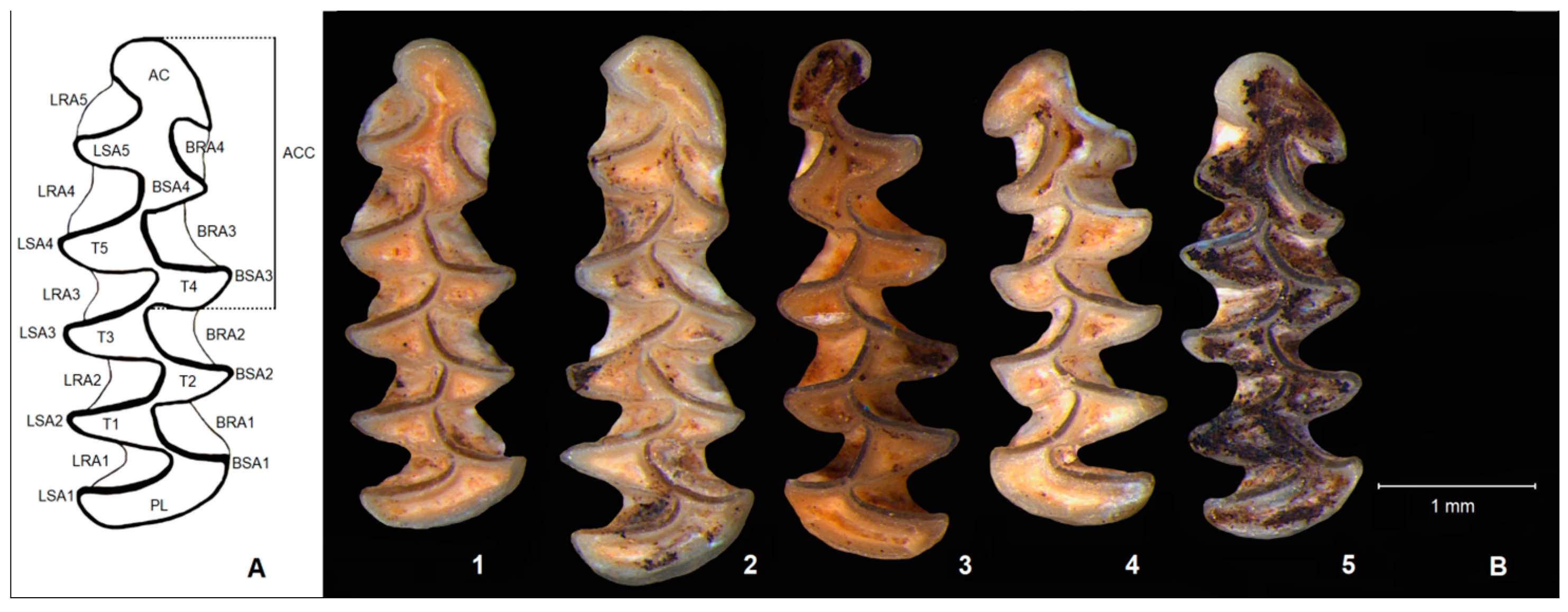
3.1. Morphological Characteristics of M. bifrons

A new species was described due to the peculiar morphology of the anteroconid complex of m1 relative to that of other Microtus species found in Petits Guinards. Species of the genus Microtus s.l. (understood by the authors broadly, including Stenocranius, Alexandromys, and Chionomys Miller, 1908) were dominant in the assemblages of the small mammals at this site (Figure 2 in [5]). Among the 1776 m1 belonging to Microtus arvalis (687), Microtus agrestis (79), Stenocranius gregalis (919), Alexandromys oeconomus (87), and Chionomys nivalis (Martins, 1842) (4), Jeannet and Fontana [5] selected 16 m1 of unusual morphology (Figure 4 in [5]) and described them as Microtus (Sumeriomys) bifrons nov. sp. They defined the new species as a large-sized vole with six or seven dental triangles (T6–T7), sometimes five (T5), and a highly dissymmetrical anterior cap with voluminous spur-shaped BSA 5. In most specimens the anterior part of the tooth (AC + T7 + T8) is wider and flatter than that of M. arvalis and M. agrestis, and is characterised by a pronounced development of BSA 5 and LSA 6. However, if someone carefully looks at the specimens classified as M. bifrons (Figure 4 in [5]), each individual differs from the others in morphological details, and it is difficult to identify features common to all specimens. The holotype (CVPG 57-1248a, Figure 16a in [5]) and the paratype (CVPG 47-957, Figure 16b in [5]) are clearly larger than the other teeth (in both L = 3.45 mm), with the almost identical anteroconid complex, probably representing one individual.
The morphology of the five genetically studied individuals is characterized as follows (Figure 1):
  • MI2868 (CVPG 18-371) (right m1, L = 3.04 mm): five triangles (T1–T5); T6 and T7 broadly confluent; BSA 4 less pronounced in comparison with LSA 5; and anterior cap (AC) wide with spur-shaped BSA 5 (Figure 1/1);
  • MI2869 (CVPG 47-957) (right m1, L = 3.45 mm, paratype): seven dental triangles (T1–T7); T6 and T7 separated; BSA 4 and LSA 5 almost of the same width; and anterior cap (AC) wide, with spur-shaped BSA 5 (Figure 1/2);
  • MI2870 (CVPG 59-1309, left m1, L = 3.22 mm): five dental triangles (T1–T5); T6 and T7 confluent; BSA 4 much less pronounced in comparison with LSA 5; and anterior cap (AC) separated from T6–T7 with small BSA 5 (Figure 1/3);
  • MI2871 (CVPG 56-1213, left m1, L = 2.95 mm): five dental triangles (T1–T5); T6 and T7 broadly confluent; BSA 4 pronounced as much as LSA 5; and anterior cap (AC) not fully separated from T6–T7 with pronounced BSA 5 and very small LSA 6 (Figure 1/4);
  • MI2872 (CVPG 51-1065, right m1, L = 2.98 mm): five dental triangles (T1–T5); T6 and T7 broadly confluent; BSA 4 pronounced as much as LSA 5; and anterior cap (AC) wide with spur-shaped BSA 5 (Figure 1/5).
The remaining specimens presented in Figure 4 [5] are mostly smaller (L ranges from 2.80 to 3.22 mm), have a variable anteroconid complex, and, in most of them, the anterior cap is morphologically different from the holotype and paratype. Therefore, the opinion of homogeneity of the sample distinguished as a new species cannot be maintained. They might represent aberrant or phenotypic outliers of M. arvalis, M. agrestis, and/or S. anglicus.

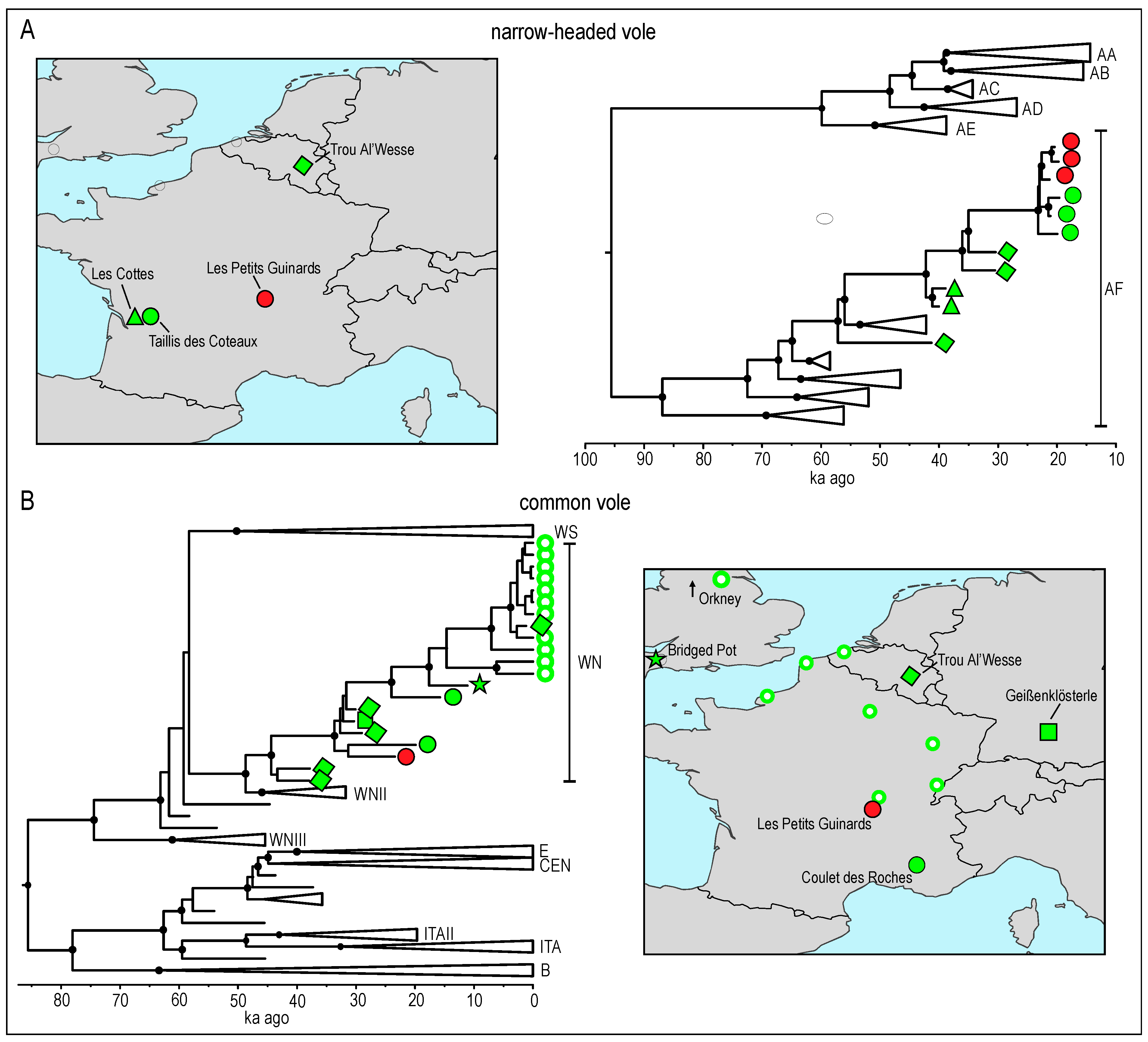
3.2. Phylogenetic Analysis and Molecular Dating

We tentatively assigned the specimens from Les Petit Guinards to species based on the number of reads mapping to mtDNA genomes of three vole species: common vole, narrow-headed vole, and Gunther’s vole (Table 1), and the divergence of mapped reads from the respective reference sequence calculated by the Tablet software.
Three of the specimens were assigned to S. anglicus and two to M. arvalis. The results from two libraries produced from the DNA extracted from each specimen were consistent. In each library the mapped DNA molecules exhibit the deamination pattern characteristic for ancient DNA (Supplementary Figures S1 and S2). For the three specimens identified as S. anglicus and one as M. arvalis the number of reads uniquely mapped to the respective mtDNA reference was sufficient to reconstruct partial mtDNA genomes (Table 1). We used the resulting mtDNA sequences to confirm the taxonomic assignment and to estimate the phylogenetic position and the age of these specimens.
The reconstructed Maximum Likelihood phylogeny of Arvicolinae showed topology identical to those obtained in previous studies [6,33] and confirmed taxonomic assignment of samples from Petit Guinards as S. anglicus and M. arvalis (Figure 2).
The reconstructed intraspecific phylogeny of narrow-headed voles was similar to the previous ones [7] and revealed three main lineages corresponding to the Asiatic S. gregalis and S. raddei (Polyakov, 1881) and the European S. anglicus (Figure 3A). Six lineages were distinguished (AA–AF) within the S. anglicus clade. The three S. anglicus specimens from Petit Guinards were placed in the lineage AF and yielded ages between 20.8 and 19.3 ka ago (Table 1, Figure 3A). The AF lineage was widespread in Europe until ca. 40 ka ago, but later it became confined to southwestern France [7].
The three investigated specimens were closely associated with specimens of similar age from the Taillis des Coteaux site in Central Western France.
The reconstructed phylogeny of the common vole revealed a topology similar to that obtained in a previous study [29]. Six main lineages were detected. The studied specimen belonged to the WN lineage which occurred, starting at least 30 ka ago, in Western Europe except for the Iberian Peninsula. It was the most closely related to the specimen from the Coulet des Roches site from southern France and yielded an age of 23.4 ka.
The estimated age and phylogenetic position of the four specimens are highly consistent with the dating and geographic location of the site. This further strengthens the reliability of molecular analyses.

4. Discussion

4.1. Taxonomic Attribution Revealed from Morphological Characteristics and Genetic Research

From the whole set of available specimens Jeannet and Fontana [5] selected a few individuals of peculiar anteroconid complex, which did not fit well into the observed variation of M. arvalis, M. agrestis and/or Stenocranius. The specimens designated as M. bifrons have an additional triangle (T6) or two supernumerary (T6 and T7) triangles within the ACC of m1. Almost half of the Petits Guinards population (7 specimens, 43.7%) exhibits these morphological features (Figures 4/1, 3, 5, 9, 10, 14 and 16 in [5]). An equally important feature (10 specimens, 56.2%) is the very wide anterior cap (AC), highly dissymmetrical with voluminous spur-shaped T8 (Figures 4/1, 7–9 and 11–16 in [5]). In light of findings of previous studies [34,35], as well as morphometric analyzes (Figures 8 and 9 in [5]), Jeannet and Fontana [5] concluded that the aberrant teeth may belong to one of the representatives of the social voles, subgenus Sumeriomys. Although most of the previous studies paid little attention to the detailed morphology of m1, some noted the presence of extra triangles and/or very wide anterior cap in the first lower molars of extant and subfossil Sumeriomys [35,36,37], which coincide with the features of Microtus (Sumeriomys) bifrons nov. sp. Currently, social voles consist of at least eight nominal, partly cryptic species, primarily diversified in Turkey and Western Iran with marginal occurrence from southern Balkans to Central Asia and North Africa (Libya) [3].
Genetic identification of the five specimens attributed to M. bifrons revealed that three belonged to S. anglicus and two to M. arvalis, but this identification was in some cases inconsistent with morphological examination results and required further discussion. Morphology of the two first specimens (MI2868, MI2869), which according to genetic data belong to the European narrow-headed vole (Stenocranius anglicus), contradict their attribution to this species (Figure 1/1–2). The genus Stenocranius is traditionally characterised by a complete absence or a strong reduction of T6 and BSA 5 in m1 and a relatively small anterior cap. These are the most important features distinguishing Stenocranius from Microtus species in the fossil record. In contrast, the two specimens have an extensively built anteroconid complex with a fully developed T6 and a wide anterior cap with pronounced BSA 5 and LSA 6. Such occlusal morphology of m1 has never been described earlier in both extant and fossil populations of Stenocranius [12,38,39,40,41,42,43,44,45,46,47,48,49,50].
The third specimen (MI2870) genetically assigned to Stenocranius has a reduced T6, has not pronounced BSA 5 and LSA 6 (Figure 1/3), seems to be a morphologically advanced S. anglicus, and is known as a most complex morphotype in extant populations of S. gregalis [40,47]. It has also been described from a number of sites in the Late Pleistocene of Eurasia [12,38,42,46,49,51].
The last two genetically studied specimens (MI2871 and MI2872) belong to Microtus arvalis (Figure 1/4–5). The first of them has an aberrantly shaped anterior cap, a not developed LSA 6, and a strongly marked BSA 5, which causes a large asymmetry of AC. Such a morphology is very rare in extant and subfossil populations of M. arvalis (Figure 15 D3 in [12], Figure 6 in [52]) and also occurs in some other species, e.g., in M. transcaspicus Satunin, 1905 (Figure 307b′ in [3]) and M. irani Thomas, 1921 (Figure 317a′ in [3]). The last genetically determined m1 is similar to that of the holotype and paratype of M. bifrons with a very wide anterior cap, but differs from them by having confluent T6 and T7.

4.2. Supernumerary Triangles

The genus Microtus s. l., as treated by Jeannet and Fontana [5], is characterised by the presence of five closed triangles (T1–T5) in m1 and the differently developed anteroconid part of the tooth where diagnostic features of individual species can be identified. The presence of additional closed triangles (T6 and T7) was the basis for the description of the new species [5], in agreement with Ognev (Figure 163 in [34]) who used the presence of extra triangle in m1 to distinguish one of the species of social voles—Microtus (Sumeriomys) socialis (Pallas, 1773).
However, the morphotype with one extra developed triangle (T6), sometimes called “extratriangulatus” (Figures 20 and 21 in [15]), is found with a low or very low frequency in almost all extant gray voles (genus Microtus) and narrow-headed voles (genus Stenocranius). In social voles (subgenus Sumeriomys) this morphotype is rarely illustrated in publications, e.g., in M. socialis and M. dogramacii Kefelioğlu et Kryštufek, 1999 (Figures 313a′ and 326d′,f′, respectively, in [3]), without specifying the frequency of its occurrence in various Sumeriomys species. The frequency of the “extratriangulatus” morphotype can vary greatly in M. agrestis and range from ca. 5–6% (Figure 1d,f,g, Table 1 in [53]), through 7% (Figure 4f in [54]) to ca. 17% (Figure 20/49–50 in [16] and Table 1, Abb. 1e,f in [55]). This morphotype is present in populations from the Urals and Western Siberia (Figure 43 in [46]) and was also recorded in Microtus lavernedii (Crespon, 1844) (Figure 253f′ in [3]), a close relative of M. agrestis. Specimens with an extra triangle have already been found in Microtus nivaloides Major, 1902, a putative direct ancestor of M. agrestis [16,56,57,58]. The “extratriangulatus” morphotype is present in geographically distant sites dated to the end of the Early Pleistocene from the Kozarnika Cave in Bulgaria (Figure 6/10 in [59]) and Chigirin, Ukraine (Figure 12–13/13 in [60]), as well as to the early Middle Pleistocene (from MIS 17 to MIS 11), for example, in West Runton in the UK (Figure 65/3 in [61] and Figure 19/50–51 in [16]), Kozi Grzbiet in Poland (Figures 20–22 in [15]), Za Hájovnou Cave in Czechia (listed as Microtus aff. “coronensis”) (Text-Figure 2/D in [62]), Semybalka 1 (Figure 13/11 in [57]) and Morozivka 2 (Figure 3/19 in [63]) in the Ukraine, and in Gornopravdinsk 2, West Siberia, Russian Federation (Figure 3 in [58]). This morphotype probably extended its distribution and gradually increased its frequency in the late Middle Pleistocene, as it is quite common, for instance, in Microtus agrestis from Saint-Estève-Janson in France (Figures 32/14, 15, 16 and 22 in [38]). The morphotype “extratriangulatus” was also recorded in the Late Pleistocene population of M. agrestis in Teixoneres Cave, Spain (Figure 3/5–6 in [64]), Porlyuk Cave and Istállóskő Cave, Hungary (Figures 4/M and 4/P, respectively, in [65]), as well as Baranica Cave, Serbia (Figure 5e in [66]). Nadachowski [12] estimates the frequency of “extratriangulatus” in the Late Pleistocene populations of Poland at 7–24% (Figure 19 in [12]), depending on the studied population and site.
In extant populations of M. arvalis, the “extratriangulatus” morphotype is much less common, although Rörig and Börner [67] present a drawing of such a tooth (Taf. V, Figure 106 in [67]). This morphotype does not occur in most of the studied extant populations; however, its frequency in some populations ranges from 0.8% (Figure 4b in [54]) to 4.0% (Figure 3/7, Table 5 in [68]). In fossil populations of M. arvalis from the Middle Pleistocene, teeth with an additional T6 triangle have been observed from Bulgarian locality Morovitsa Cave (Figure 5/17 in [69]), some Late Pleistocene samples from France (Figure 31/8 in [38]), and Holocene assemblages from Poland (Figure 15/J in [12]). Although the morphotype “extratriangulatus” was absent in extant species belonging to the M. arvalis group, subfossil specimens with an extra triangle have been described from Bacho Kiro Cave (Figure 9/I in [70]) and Cave 16 in Bulgaria (Figure 22/8 in [71]), which most likely belong not only to M. arvalis but also to M. ilaeus [8].
Publications describing the variability of m1 in Stenocranius consider the presence of the “extratriangulatus” morphotype, although its frequency is always very low (Figure 23/H3 in [12], Figure 24/VI-P in [37], Figure 38/5 in [42], and Figure 38/5 in [46]). A tooth with an additional triangle was illustrated for Stenocranius gregalis from Zabaykalsky Krai, Russian Federation (Figure 239 a′ in [3]).
Whereas one supernumerary triangle (T6) is relatively common in some populations of Microtus species, two supernumerary triangles are reported in the literature only occasionally. In extant populations, such specimens have been documented in Microtus arvalis from Croatia (Figure 297c′ in [3]) and M. agrestis from Germany (Figure 1e in [53]), as well as in the subfossil population of M. arvalis from Bacho Kiro Cave in Bulgaria (Figure 9/J in [70]). A morphotype with two closed triangles in M. agrestis has also been published from Khotylevo 2, Ukraine (Figure 33/18 in [60]) and from Western Siberia (Figure 44/2 in [46]).
This study shows that the claim that additional triangles/prisms are present only in the Sumeriomys subgenus and cannot be further maintained.

4.3. Wide and Spur-Shaped Anterior Cap

Another distinguishing feature of Microtus bifrons is a wide anterior cap with a specific spur-shape morphology on the buccal side, developed independently of the number of closed triangles [5]. The wide or very wide anterior cap occurs primarily in species of the subgenus Sumeriomys, e.g., in M. guentheri (Figure 2 in [35] and Figure 5A,B in [37]) and M. hartingi Barrett-Hamilton, 1903 (Figure 5A in [72]) from Turkey and Bulgaria, as well as subfossil populations of M. guentheri from Israel (Figure 66a–c in [36]). This type of morphology also occurs in Microtus arvalis from Croatia (Figure 297c′ in [3]) and M. mystacinus (Filippi, 1865) from Iran (Figure 302c′ in [3]). A wide anterior cap is rare in subfossil populations of M. arvalis and M. agrestis from Poland (Figures 15/G4 and 18/H, respectively, in [12]). However, in all these cases the spur-shape buccal triangle is not as distinct as in M. bifrons.

4.4. Reasons for Incorrect Species Determination Based on Morphological Criteria

In the evolution of Arvicolinae rodents, complication of molars by adding new elements to the crown is a general trend that reflects adaptation to the low-calorie diet of vegetative plant parts, especially grasses [73]. Voles and lemmings developed several structural adaptations in dentition such as high crown check teeth with cusps transformed to prisms and flat occlusal surfaces with enamel cutting edges to increase and improve the efficiency of grating [61]. The crown complexity and occlusal regularity of Arvicolinae teeth are characters closely associated with fitness [74,75]. The frequencies of extra elements of occlusal surface in natural populations is a variable trait that tends to exhibit high frequencies in small populations during the periods of relatively low species abundance [76]. Therefore, the contribution of naturally occurring bottlenecks to phenotypic evolution under natural selection is the most plausible explanation.
A comparison of laboratory colonies of Dicrostonyx and Microtus with wild populations suggests that dental aberrations typical of small and inbred populations are also present in the wild and are not eliminated by large-scale population dynamics [76,77]. The emergence of atypical, aberrant individuals in small and geographically isolated populations is probably a relatively common phenomenon. Aberrant specimens may increase when a population is spatially restricted, and inhabits an isolated niche in a mosaic landscape. It was hypothesise that the signs of the intensified genetic drift might be associated with the patchy distribution of the species in heterogeneous landscapes [75]. The occurrence of peculiar morphology in the relic population of Dicrostonyx from the Chaleux site (Belgium), probably shortly before the extinction of this species in Europe, might be an example of this phenomenon [78]. Extremely rare dental traits related to close breeding in extant species were documented in M. arvalis [79] and extant populations of D. torquatus [76]. For example, an unusual specimen with seven triangles was identified in the population of M. arvalis from Cetinsko polje, Croatia (Figure 297c′ in [3]). The population at this site is isolated and inhabits a karstic field surrounded by eroded and frequently barren slopes (B. Kryštufek–pers.info.). The probability of trapping individuals with a rare dental trait in extant populations is higher during the periods of low species abundance when the species is confined to source habitats or live in ephemeral populations formed after major outbreaks [76].
When the characters related to small population sizes occur simultaneously in different species in one fossil assemblage, we can attribute them to the impact of important changes in the environment [76]. The appearance of the extra wide anterior cap with spur-shaped buccal salient angle might be explained as an adaptation to local food resources in the vicinity of the Petits Guinads site. Ecomorphological adaptation is supported by the fact that almost identical anterior caps developed in the specimens genetically confirmed as S. anglicus (Figure 1/1–2) and M. arvalis (Figure 1/5).
It seems that the morphological variability of molar teeth within one species is unexpectedly high. The nature of this variability is complex and depends on many factors, which are sometimes difficult to identify [80]. Therefore, one should be very cautious with species identification based exclusively on extraordinary morphological criteria.

5. Conclusions

The most unexpected finding of this study was the discrepancy between the conclusions based on analyses of aDNA and those based on morphological characters. In light of this genetic study, the morphology-based taxonomic attribution of the studied specimens cannot be maintained. Literature on m1 morphological variation in extant species of Microtus and Stenocranius, as well as fossil populations of these genera, showed that the criteria proposed to distinguish a new taxon based solely on m1 variation are not sufficient for taxonomic decisions at the species level. Specimens described as Microtus bifrons are just extreme, very rare morphological variants of the most common Petits Guinards species and should be treated as outliers.
The frequency of rare morphotypes keeps below 1% or almost zero, but their manifestation is sometimes spectacular. Such phenotypic outliers fall outside the expected species-specific range of variability, can also be found in related taxa, and their presence in subfossil populations depends on the size of the sample; the greater it is, the higher is the chance to find aberrant specimens.

Supplementary Materials

The following supporting information can be downloaded at: https://www.mdpi.com/article/10.3390/d15040538/s1, Figure S1: DNA damage patterns produced by mapDamage software for double-stranded libraries produced in this study. The double-stranded libraries were not USER treated; Figure S2: DNA damage patterns obtained using mapDamage software for single-stranded (SS) libraries produced in this study; Table S1: Primers used to generate mitogenomes from various vole species; Supplementary Information S1: Supplementary materials and methods. References [81,82,83] are cited in the supplementary materials.

Author Contributions

Conceptualization, A.N. and M.B.; methodology, A.N., M.B. and D.P.; investigation, A.N., M.B., A.L., D.P., M.G. and B.B.; resources, L.F.; writing—original draft preparation, A.N. and M.B.; writing—review and editing, A.N., M.B., A.L., D.P. and L.F.; funding acquisition, M.B. All authors have read and agreed to the published version of the manuscript.

Funding

This research was supported by the Polish National Science Centre grants no: 2015/19/D/NZ8/03878 to M.B. and 2017/25/B/NZ8/02005 to A.N.

Data Availability Statement

DNA sequences were deposited in GenBank under accession no. OQ473582-OQ473584, OQ473587. The raw sequencing reads generated for each specimen were deposited in the European Nucleotide Archive under project PRJEB60165. The alignment of mtDNA sequences used for taxonomic identification of studied specimens was deposited in RepOD repository (DOI 10.18150/20WKE0).

Acknowledgments

We thank Aurélien Royer, Biogéosciences, UMR 6282, CNRS, EPHE, Université Bourgogne Franche-Comté, Dijon, France, for assistance in obtaining material for research. The authors are grateful to Boris Kryštufek, Slovenian Museum of Natural History, Ljubljana, Slovenia, Evgenia Markova, Institute of Plant and Animal Ecology, Ural Branch of Russian Academy of Sciences, Ekaterinburg, Russian Federation and Lilia Popova, Schmalhausen Institute of Zoology of National Academy of Sciences of the Ukraine, Kyiv, the Ukraine, for consulting and discussions on morphology of vole molars.

Conflicts of Interest

The authors declare no conflict of interest.

References

  1. Pardiñas, U.F.J.; Myers, P.; León-Paniagua, L.; Ordóñez Garza, N.; Cook, J.; Kryštufek, B.; Haslauer, R.; Bradley, R.; Shenbrot, G.; Patton, J. Family Cricetidae (true hamsters, voles, lemmings and new world rats and mice). In Handbook of the Mammals of the World, Rodents II; Wilson, D.E., Lacher, T.E., Mittermeier, R.A., Eds.; Lynx Edicions: Barcelona, Spain, 2017; Volume 7, pp. 204–279. [Google Scholar]
  2. Jaarola, M.; Martínková, N.; Gündüz, İ.; Brunhoff, C.; Zima, J.; Nadachowski, A.; Amori, G.; Bulatova, N.S.; Chondropoulos, B.; Fraguedakis-Tsolis, S.; et al. Molecular phylogeny of the speciose vole genus Microtus (Arvicolinae, Rodentia) inferred from mitochondrial DNA sequences. Mol. Phylogenet. Evol. 2004, 33, 647–663. [Google Scholar] [CrossRef] [PubMed]
  3. Kryštufek, B.; Shenbrot, G.I. Voles and Lemmings (Arvicolinae) of the Palaearctic Region; University of Maribor Press: Maribor, Slovenia, 2022; p. 436. [Google Scholar]
  4. Brunet-Lecomte, P.; Nadachowski, A.; Chaline, J. Microtus (Terricola) grafi nov. sp. du pléistocène supérieur de la grotte de Bacho Kiro (Bulgarie). Geobios 1992, 25, 505–509. [Google Scholar] [CrossRef]
  5. Jeannet, M.; Fontana, L. Microtus (Sumeriomys) bifrons nov. sp. (Rodentia, Mammalia), a new vole in the French Upper Pleistocene identified at the Petits Guinards site (Creuzier-le-Vieux, Allier, France). PALEO Rev. Archéol. Préhist. 2015, 26, 59–77. [Google Scholar] [CrossRef]
  6. Baca, M.; Popović, D.; Lemanik, A.; Baca, K.; Horáček, I.; Nadachowski, A. Highly divergent lineage of narrow-headed vole from the Late Pleistocene Europe. Sci. Rep. 2019, 9, 17799. [Google Scholar] [CrossRef] [Green Version]
  7. Baca, M.; Popović, D.; Agadzhanyan, A.K.; Baca, K.; Conard, N.J.; Fewlass, H.; Filek, T.; Golubiński, M.; Horáček, I.; Knul, M.V.; et al. Ancient DNA of narrow-headed voles reveals common features of the Late Pleistocene population dynamics in cold-adapted small mammals. Proc. R. Soc. B 2023, 290, 20222238. [Google Scholar] [CrossRef] [PubMed]
  8. Baca, M.; Popović, D.; Lemanik, A.; Fewlass, H.; Talamo, S.; Zima, J.; Ridush, B.; Popov, V.; Nadachowski, A. The Tien Shan vole (Microtus ilaeus; Rodentia: Cricetidae) as a new species in the Late Pleistocene of Europe. Ecol. Evol. 2021, 11, 16113–16125. [Google Scholar] [CrossRef]
  9. Fontana, L.; Lang, L.; Chauvière, F.-X.; Jeannet, M.; Mourer-Chauviré, C.; Magoga, L. Paléolithique supérieur récent du nord du Massif Central: Des données inattendues sur le site des Petits Guinards à Creuzier-le-Vieux (Allier, France). Bull. Préhist. Sud Quest. 2003, 10, 77–93. [Google Scholar]
  10. Chauvière, F.-X.; Fontana, L.; Land, L.; Bonani, G.; Hajdas, I. Une préhampe magdalénienne en bois du renne aux Petits Guinards (Allier, France). Comptes Rendus Palevol 2006, 5, 725–733. [Google Scholar] [CrossRef]
  11. Fontana, L.; Chauvière, F.-X. The total exploitation of reindeer at the site of Les Petits Guinards: What’s new about the annual cycle of Magdalenian groups in the French Massif Central? In Search of Total Animal Exploitation: Case Studies from the Upper Palaeolithic and Mesolithic; Fontana, L., Chauvière, F.-X., Bridault, A., Eds.; British Archaeological Reports, International Series 2040; John and Erica Hedges Ltd.: Oxford, UK, 2009; pp. 101–111. [Google Scholar]
  12. Nadachowski, A. Late Quaternary Rodents of Poland with Special Reference to Morphotype Dentition Analysis of Voles; Państwowe Wydawnictwo Naukowe: Warsaw, Poland, 1982; p. 109. [Google Scholar]
  13. Hibbard, C.W. Mammals from the Rexroad Formation from Fox Canyon, Kansas; University of Michigan: Ann Arbor, MI, USA, 1950; Volume 8, pp. 113–192. [Google Scholar]
  14. Van der Meulen, A.J. Middle Pleistocene smaller mammals from the Monte Peglia (Orveto, Italy) with special reference to the phylogeny of Microtus (Arvicolidae, Rodentia). Quaternaria 1973, 17, 1–144. [Google Scholar]
  15. Nadachowski, A. Biharian voles (Arvicolidae, Rodentia, Mammalia) from Kozi Grzbiet (Central Poland). Acta Zool. Crac. 1985, 29, 13–28. [Google Scholar]
  16. Nadachowski, A. Systematics, geographic variation, and evolution of snow voles (Chionomys) based on dental characters. Acta Theriol. 1991, 36, 1–45. [Google Scholar] [CrossRef] [Green Version]
  17. Rohland, N.; Glocke, I.; Aximu-Petri, A.; Meyer, M. Extraction of highly degraded DNA from ancient bones, teeth and sediments for high-throughput sequencing. Nat. Protoc. 2018, 13, 2447–2461. [Google Scholar] [CrossRef]
  18. Meyer, M.; Kircher, M. Illumina sequencing library preparation for highly multiplexed target capture and sequencing. Cold Spring Harb. Protoc. 2010, 5, 5448. [Google Scholar] [CrossRef]
  19. Gansauge, M.T.; Aximu-Petri, A.; Nagel, S.; Meyer, M. Manual and automated preparation of single-stranded DNA libraries for the sequencing of DNA from ancient biological remains and other sources of highly degraded DNA. Nat. Protoc. 2020, 15, 2279–2300. [Google Scholar] [CrossRef] [PubMed]
  20. Horn, S. Case study: Enrichment of ancient mitochondrial DNA by hybridization capture. Methods Mol. Biol. 2012, 840, 189–195. [Google Scholar] [PubMed]
  21. Lord, E.; Marangoni, A.; Baca, M.; Popović, D.; Goropashnaya, A.V.; Stewart, J.R.; Knul, M.V.; Noiret, P.; Germonpré, M.; Jimenez, E.-L.; et al. Population dynamics and demographic history of Eurasian collared lemmings. BMC Ecol. Evol. 2022, 22, 126. [Google Scholar] [CrossRef]
  22. Schubert, M.; Lindgreen, S.; Orlando, L. AdapterRemoval v2: Rapid Adapter Trimming, identification, and read merging. BMC Res Notes 2016, 9, 88. [Google Scholar] [CrossRef] [PubMed] [Green Version]
  23. Li, H. Aligning sequence reads, clone sequences and assembly contigs with BWA-MEM. arXiv 2013, arXiv:1303.3997v2. [Google Scholar]
  24. Feuerborn, T.R.; Palkopoulou, E.; van der Valk, T.; von Seth, J.; Munters, A.R.; Pečnerová, P.; Dehasque, M.; Ureña, I.; Ersmark, E.; Lagerholm, V.K.; et al. Competitive mapping allows for the identification and exclusion of human DNA contamination in ancient faunal genomic datasets. BMC Genom. 2020, 21, 844. [Google Scholar] [CrossRef]
  25. Danecek, P.; Bonfield, J.K.; Liddle, J.; Marshall, J.; Ohan, V.; Pollard, M.O.; Whitwham, A.; Keane, T.; McCarthy, S.A.; Davies, R.M.; et al. Twelve Years of SAMtools and BCFtools. Gigascience 2021, 10, giab008. [Google Scholar] [CrossRef]
  26. Grubaugh, N.D.; Gangavarapu, K.; Quick, J.; Matteson, N.L.; de Jesus, J.G.; Main, B.J.; Tan, A.L.; Paul, L.M.; Brackney, D.E.; Grewal, S.; et al. An amplicon-based sequencing framework for accurately measuring intrahost virus diversity using PrimalSeq and IVar. Genome Biol. 2019, 20, 8. [Google Scholar] [CrossRef] [PubMed] [Green Version]
  27. Milne, I.; Stephen, G.; Bayer, M.; Cock, P.J.A.; Pritchard, L.; Cardle, L.; Shaw, P.D.; Marshall, D. Using tablet for visual exploration of second-generation sequencing data. Brief Bioinform. 2013, 14, 193–202. [Google Scholar] [CrossRef] [PubMed]
  28. Jónsson, H.; Ginolhac, A.; Schubert, M.; Johnson, P.L.F.; Orlando, L. MapDamage2.0: Fast approximate Bayesian estimates of ancient DNA damage parameters. Bioinformatics 2013, 29, 1682–1684. [Google Scholar] [CrossRef] [PubMed]
  29. Baca, M.; Popović, D.; Lemanik, A.; Bañuls-Cardona, S.; Conard, N.J.; Cuenca-Bescós, G.; Desclaux, E.; Fewlass, H.; Garcia, J.T.; Hadravova, T.; et al. Ancient DNA reveals interstadials as a driver of common vole population dynamics during the last glacial period. J. Biogeogr. 2023, 50, 183–196. [Google Scholar] [CrossRef]
  30. Minh, B.Q.; Schmidt, H.A.; Chernomor, O.; Schrempf, D.; Woodhams, M.D.; von Haeseler, A.; Lanfear, R. IQ-TREE 2: New models and efficient methods for phylogenetic inference in the genomic era. Mol. Biol. Evol. 2020, 37, 1530–1534. [Google Scholar] [CrossRef] [Green Version]
  31. Shapiro, B.; Ho, S.Y.W.; Drummond, A.J.; Suchard, M.A.; Pybus, O.G.; Rambaut, A.A. Bayesian Phylogenetic Method to estimate unknown sequence ages. Mol. Biol. Evol. 2011, 28, 879–887. [Google Scholar] [CrossRef] [Green Version]
  32. Suchard, M.A.; Lemey, P.; Baele, G.; Ayres, D.L.; Drummond, A.J.; Rambaut, A. Bayesian phylogenetic and phylodynamic data integration using BEAST 1.10. Virus Evol. 2018, 4, vey016. [Google Scholar] [CrossRef] [Green Version]
  33. Abramson, N.I.; Bodrov, S.Y.; Bondareva, O.V.; Genelt-Yanovskiy, E.A.; Petrova, T.V. A mitochondrial genome phylogeny of voles and lemmings (Rodentia: Arvolinae): Evolutionary and taxonomic implications. PLoS ONE 2021, 16, e0248198. [Google Scholar] [CrossRef]
  34. Ognev, S.I. Mammals of the USSR and Adjacent Countries; Izd. AN SSSR, Moskva: Leningrad, Russia, 1950; Volume 7, p. 706. (In Russian) [Google Scholar]
  35. Golenishchev, F.N.; Sablina, O.V.; Borodin, P.M.; Gerasimov, S. Taxonomy of voles of the subgenus Sumeriomys Argyropulo, 1933 (Rodentia, Arvicolinae, Microtus). Russ. J. Theriol. 2002, 1, 43–55. [Google Scholar] [CrossRef]
  36. Tchernov, E. Succession of Rodents Fauna during the Upper Pleistocene of Israel; Verlag Paul Parey: Hamburg, Germany; Berlin, Germany, 1968; p. 152. [Google Scholar]
  37. Aşan Baymedir, N.; Duman, L. Molar pattern in Microtus guentheri (Danford and Alston, 1880) (Mammalia: Rodentia) from Kirikkale Province. J. Appl. Biol. Sci. 2009, 3, 47–53. [Google Scholar]
  38. Chaline, J. Les Rongeurs du Pléistocène Moyen et Supérieur de France: (Systématique, Biostratigraphie, Paléoclimatologie); Cahiers Paléontologie: Paris, France, 1972; p. 410. [Google Scholar]
  39. Rekovets, L.I. New subspecies of narrow-headed vole (Microtus gregalis) from the Upper Pleistocene deposits of Ukraine. Dokl. AN USSR. Ser. Biol. 1978, 6, 559–563. (In Ukrainian) [Google Scholar]
  40. Bolshakov, V.N.; Vasilyeva, I.A.; Maleeva, A.G. Morphotypical Variability of Voles’ Dentition; Nauka Publisher: Moscow, Russia, 1980; p. 140. (In Russian) [Google Scholar]
  41. Kochev, V.A. Species criteria for M1 molars of Microtus agrestis, M. arvalis, M. oeconomus, M. gregalis, M. middendorfi and M. hyperboreus. Vestn. Zool. 1986, 3, 40–45. (In Russian) [Google Scholar]
  42. Smirnov, N.G.; Bolshakov, V.N.; Borodin, A.V. Pleistocene Rodents of Northern Western Siberia; Nauka Publishers: Moscow, Russia, 1986; p. 144. (In Russian) [Google Scholar]
  43. Rekovets, L.I.; Nadachowski, A. Pleistocene voles (Arvicolidae) of the Ukraine. Paleontol. Evol. 1995, 28, 145–245. [Google Scholar]
  44. Válóczi, T. A Vaskapu-barlang (Bükk hegység) felső-pleisztocén faunájának vizsgálata. Folia Hist. Nat. Musei Matra. 1998, 23, 79–96. (In Hungarian) [Google Scholar]
  45. Dupal, T.A. Geografic variability and sytematics of the subspecies in the narrow-skulled vole Microtus (Stenocranius) gregalis (Rodentia, Cricetidae). Zool. Zhurnal 2000, 79, 851–858. (In Russian) [Google Scholar]
  46. Borodin, A.W. Identification Guide for Vole’s Teeth of the Urals and Western Siberia (Late Pleistocene—Recent); Ural Branch of RAS: Yekaterinburg, Russia, 2009; p. 100. (In Russian) [Google Scholar]
  47. Dupal, T.A.; Abramov, S.A. Intrapopulation morphological varation of the narrow-skulled vole (Microtus gregalis, Rodentia, Arvicolinae). Zool. Zhurnal 2010, 80, 850–861. (In Russian) [Google Scholar]
  48. Markova, E.A.; Smirnov, N.G.; Kourova, T.P.; Kropacheva, Y.E. Ontogenetic variation in occlusal shape of evergrowing molars in voles: An intravital study in Microtus gregalis (Arvicolidae, Rodentia). Mamm. Biol. 2013, 78, 251–257. [Google Scholar] [CrossRef]
  49. Ponomarev, D.; Puzachenko, A. Changes in the morphology and morphological diversity of the first lower molar of narrow-headed voles (Microtus gregalis, Arvicolinae, Rodentia) from northeastern European Russia since the Late Pleistocene. Quat. Int. 2017, 436, 239–252. [Google Scholar] [CrossRef]
  50. Fadeeva, T.V.; Kosintsev, P.A.; Gimranov, D.O.; Yakovlev, A.G. The finding of molars of the archaic vole Lasiopodomys (Stenocranius) gregaloides (Hinton, 1923) (Mammalia, Rodentia, Cricetidae) in the Late Pleistocene of the Southrn Urals. Dokl. Biol. Sci. 2022, 505, 105–108. [Google Scholar] [CrossRef]
  51. Klimowicz, M.; Nadachowski, A.; Lemanik, A.; Socha, P. Is enamel differentiation quotient (SDQ) of the narrow-headed vole (Microtus gregalis) useful for the Pleistocene biostratigraphy? Quat. Inter. 2016, 420, 348–356. [Google Scholar] [CrossRef]
  52. Jánossy, D.; Schmidt, E. Extreme Varianten des M1 der Feldmaus (Microtus arvalis Pallas) in Ungarn. II. Z. Säugetierkd. 1975, 40, 34–36. [Google Scholar]
  53. Kapischke, H.-J.; Kraft, R.; Jentzsch, M.; Hiermeier, M. Variation and complexity of the enamel pattern in the first lower molar of the Field vole, Microtus agrestis (L., 1761) (Mammalia: Rodentia: Arvicolinae). Vertebr. Zool. 2009, 59, 191–195. [Google Scholar] [CrossRef]
  54. Dienske, H. Notes on differences between sime external and skull characters of Microtus arvalis (Pallas, 1778) and of Microtus agrestis (Linnaeus, 1761) from the Netherlands. Zool. Meded. 1969, 44, 83–108. [Google Scholar]
  55. Jentzsch, M. Zur Variabilität der Molarenmuster einer Population von Erdmäusen Microtus agrestis (L., 1761) aus dem Norden Sachsen-Anhalts (Mammalia: Rodentia: Arvicolidae). Zool. Abh. 2006, 55, 191–198. [Google Scholar]
  56. Nadachowski, A. Comments on variation, evolution and phylogeny of Chionomys (Arvicolidae). In Proceedings of the International Symposium on Evolution, Phylogeny and Biostratygraphy of Arvicolids (Rodentia, Mammalia), Rohanov, Czechoslovakia, May 1987; Fejfar, O., Heinrich, W.-D., Eds.; Geological Survey: Prague, Czech Republic, 1990; pp. 353–368. [Google Scholar]
  57. Popova, L.V.; Nezdolyii, Y.S.; Syniavska, I.; Rekovets, L.; Krokhmal, O.; Mironchuk, T.; Dzeverin, I. Spatial and temeporal patterns of species replacement in the Middle Pleistocene: A case study of Microtus nivaloides Major, 1902 and morphologically related species of the Northern Black Sea and Azov areas. J. Quat. Sci. 2022, 37, 1229–1245. [Google Scholar] [CrossRef]
  58. Markova, E.; Borodin, A. An advanced form of Microtus nivaloides Forsyth Major, 1902 (Arvicolinae, Rodentia) in the late Middle Pleistocene of West Siberia: Facts and hypotheses. Hist. Biol. 2022, 1–17. [Google Scholar] [CrossRef]
  59. Popov, V.V.; Marinska, M. An almost one million year long (Early to Late Pleistocene) small mammal succession from the archaeological layers of Kozarnika Cave in Northern Bulgaria. Cour. Forsch. Senckenberg 2007, 259, 79–92. [Google Scholar]
  60. Markova, A.K. Pleistocene Rodents of the Russian Plain (Their Paleogeographic and Stratigraphic Implications); Nauka Publisher: Moscow, Russia, 1982; p. 186. (In Russian) [Google Scholar]
  61. Hinton, M.A.C. Monograph of the Voles and Lemmings (Microtinae) Living and Extinct; Richard Clay & Sons: Bungay, UK, 1926; p. 488. [Google Scholar]
  62. Ivanov, M.; Vöröš, D. Middle Pleistocene voles and lemmings (Rodentia: Arvicolinae) from Za Hájovnou Cave (Javoříčko Karst). Acta Mus. Nat. Pragae Ser. B Hist. Nat. 2014, 70, 43–54. [Google Scholar] [CrossRef]
  63. Popova, L.V.; Nezdolyii, Y.S.; Krokhmal, O.I.; Rekovets, L.I. Appearance of Microtus agrestis in the territory of Ukraine in the Middle Pleistocene. Geo&Bio 2021, 20, 102–116. (In Ukrainian) [Google Scholar]
  64. Luzi, E.; López-García, J.M.; Blasco, R.; Rivals, F. Variation in Microtus arvalis and Microtus agrestis (Arvicolinae, Rodentia) dental morphologies in an archaeological context: The case of Teixoneres Cave (Late Pleistocene, North-Eastern Iberia). J. Mamm. Evol. 2017, 24, 495–503. [Google Scholar] [CrossRef]
  65. Luzi, E.; Pazonyi, P.; López-García, J.M. The influence of climate on morphometric traits of fossil populations of Microtus arvalis and M. agrestis from the Carpathian Basin, northern Hungary. Lethaia 2019, 52, 123–132. [Google Scholar] [CrossRef] [Green Version]
  66. Bogićević, K.; Nenadić, D.; Mihailović, D. Late Pleistocene voles (Arvicolinae, Rodentia) from the Baranica Cave (Serbia). Geol. Carpathica 2012, 63, 83–94. [Google Scholar] [CrossRef] [Green Version]
  67. Rörig, G.; Börner, C. Studien über das Gebiss mitteleuropäischer Mäuse. Arb. Kaiserl. Biol. Anst. Land-Forst-Wirtsch. Berl. 1905, 5, 33–89. [Google Scholar]
  68. Uhlíková, J. Epigenetic and dental variation of the common vole, Microtus arvalis (Mammalia: Rodentia) in the Czech Republic. Folia Zool. 2004, 53, 157–170. [Google Scholar]
  69. Popov, V.V. Middle Pleistocene small mammals (Insectivora, Lagomorpha, Rodentia) from Morovitsa Cave (North Bulgaria). Acta Zool. Crac. 1989, 32, 561–588. [Google Scholar]
  70. Nadachowski, A. Morphomertic variability of dentition of the Late Pleistocene voles (Arvicolidae, Rodentia) from Bacho Kiro Cave (Bulgaria). Acta Zool. Crac. 1984, 27, 149–176. [Google Scholar]
  71. Popov, V.V. The small mammals (Mammalia: Insectivora, Chiroptera, Lagomorpha, Rodentia) from Cave 16 and the paleoenvironmental changes during the Late Pleistocene. In Temnata Cave: Excavations in Karlukovo Karst Area, Bulgaria; Ginter, B., Kozłowski, J.K., Laville, K., Eds.; Jagiellonian University Press: Cracow, Poland, 2000; Volume 2, pp. 159–240. [Google Scholar]
  72. Golenishchev, F.N.; Zorenko, T.A.; Petrova, T.V.; Voyta, L.L.; Kryuchkova, L.Y.; Atanasov, N. Evaluation of the “bottleneck” effect in an isolated population of Microtus hartingi (Rodentia, Arvicolinae) from the Eastern Rhodopes (Bulgaria) by methods of integrative analysis. Diversity 2022, 14, 709. [Google Scholar] [CrossRef]
  73. Guthrie, R.D. Factors regulating the evolution of Microtine tooth complexity. Z. Sägetierk. 1971, 36, 37–54. [Google Scholar]
  74. Markova, E.A. Assessment of tooth complexity in Arvicolines (Rodentia): A morphotype ranking approach. Zool. Zhurnal 2013, 92, 968–980. (In Russian) [Google Scholar] [CrossRef]
  75. Markova, E.A.; Sibiryakov, P.A.; Kartavtseva, I.V.; Lapin, A.S.; Morozkina, A.V.; Petukhov, V.A.; Tiunov, M.P.; Starikov, V.P. What can an invasive species tell us about evolution? A study of dental variation in disjunctive populations of Microtus rossiaemeridionalis (Arvicolinae, Rodentia). J. Mamm. Evol. 2019, 26, 267–282. [Google Scholar] [CrossRef]
  76. Markova, E.; Bobretsov, A.; Borodin, A.; Rakitin, S.; Sibiriakov, P.; Smirnov, N.; Yalkovskaya, L.; Zykov, S. The effects of population bottlenecks on dental phenotype in extant arvicoline rodents: Implications for studies of the Quaternary fossil record. Quat. Sci. Rev. 2020, 228, 106045. [Google Scholar] [CrossRef]
  77. Markova, E.; Smirnov, N. Phenotypic diversity arising from a limited number of founders: A study of dental variation in laboratory colonies of collared lemmings, Dicrostonyx (Rodentia: Arvicolinae). Biol. J. Linn. Soc. 2018, 125, 777–793. [Google Scholar] [CrossRef]
  78. Montuire, S.; Royer, A.; Lemanik, A.; Gilg, O.; Sokolova, N.; Sokolov, A.; Desclaux, E.; Nadachowski, A.; Navarro, N. Molar shape differentiation during range expansions of the collared lemming (Dicrostonyx torquatus) related to past climate changes. Quat. Sci. Rev. 2019, 221, 105886. [Google Scholar] [CrossRef]
  79. Markova, E.; Malygin, V.; Montuire, S.; Nadachowski, A.; Quéré, J.-P.; Ochman, K. Dental variation in sibling species Microtus arvalis and M. rossiaemeridionalis (Arvicolinae, Rodentia): Between-species comparisons and geography of morphotype dental patterns. J. Mamm. Evol. 2010, 17, 121–139. [Google Scholar] [CrossRef]
  80. Polly, P.D.; Killick, L.; Ruddy, M. Using left-right asummetry to estimate non-genetic variation in vole teeth (Arvicolinae, Muridae, Rodentia). Palaeont. Electr. 2011, 14, 12. [Google Scholar]
  81. Maricic, T.; Whitten, M.; Pääbo, S. Multiplexed DNA sequence capture of mitochondrial genomes using PCR products. PLoS ONE 2010, 5, 9–13. [Google Scholar] [CrossRef] [PubMed]
  82. Li, H.; Handsaker, B.; Wysoker, A.; Fennell, T.; Ruan, J.; Homer, N.; Marth, G.; Abecasis, G.R.; Durbin, R.; Subgroup. 1000 genome project data processing the sequence alignment/map format and SAMtools. Bioinformatics 2009, 25, 2078–2079. [Google Scholar] [CrossRef] [PubMed] [Green Version]
  83. Quinlan, A.R.; Hall, I.M. BEDTools: A flexible suite of utilities for comparing genomic features. Bioinformatics 2010, 26, 841–842. [Google Scholar] [CrossRef] [PubMed] [Green Version]
Figure 1. (A) dental nomenclature of Arvicolinae first lower molars (m1) (after Hibbard [13] and van der Meulen [14]); AC—anterior cap, T—dental triangle, ACC—anteroconid complex, LRA–lingual re-entrant angle, LSA—lingual salient angle, BRA—buccal re-entrant angle, BSA—buccal salient angle, PL—posterior lobe; (B) 1–5—Microtus bifrons from Les Petits Guinards, occlusal surface of first lower molars (m1); 1—MI2868 (original collection number: CVPG 18-357, Figure 4/15 in [5]), 2—MI2869 (CVPG 47-957, Figure 4/14), 3—MI2870 (CVPG 59-1309, Figure 4/2), 4—MI2871 (CVPG 56-1213, Figure 4/8), 5—MI2872 (CVPG 51-1065, Figure 4/13).
Diversity 15 00538 g001
Figure 2. Maximum Likelihood phylogeny of Arvicolinae rodents based on 3 kb fragment of mtDNA. Black circles at nodes indicate ultrafast bootstrap support higher than 90. Tree is rooted with the sequence of Arvicola amphibius (not shown). Red circles denote samples analyzed in this study. Generic names are given above the branches and follow the nomenclature of Abramson et al. [33] and Kryštufek and Shenbrot [3].
Diversity 15 00538 g002
Figure 3. Genetic analyses of Les Petits Guinards specimens. (A) The Bayesian phylogeny of European narrow-headed voles (Stenocranius anglicus) based on mtDNA genome sequences. The Asiatic lineages (S. gregalis and S. raddei) are not shown. (B) The Bayesian phylogeny of European common voles (Microtus arvalis) based on 4.2 kb fragment of mtDNA. On both panels, the majority of the branches were collapsed and only a fragment containing sequences originating from Les Petits Guinards is presented. Symbols at tips denote localities, as presented on the accompanying maps. The filled symbols represent Late Pleistocene and empty symbols represent modern localities. Black dots indicate nodes with posterior probability above 0.95.
Diversity 15 00538 g003
Table 1. Genetic assignment of Les Petit Guinards samples. Lib: DNA library type; DS–double stranded; SS–single stranded. For each sequencing library the highest number of mapping reads is given in bold.
SampleLibReadsUnique Mapped ReadsAssigned SpeciesMean mtDNA CoverageFraction of mtDNA Genome Recovered *Estimated Age (Cal BP)
M. arvalisS. anglicusM. guentheri Median 95% HPD
MI2868SS990,9162171439345S. anglicus8.40.8319,290[22, 212, 16, 004]
DS593,8274031133489
MI2869SS4,776,6564752440629S. anglicus17.50.9520,819[23, 474, 16, 858]
DS2,436,718104825001102
MI2870SS701,333110608161S. anglicus5.10.6919,735[22, 575, 15, 943]
DS808,330316874352
MI2871SS86,90623875123M. arvalis2.90.46n.a.n.a.
DS280,844521316431
MI2872SS3,510,503833928374884M. arvalis27.80.9623,410[27, 369, 19, 290]
DS113,370862416639
*—in the case of M. arvalis, fraction of the analyzed 4.2 kb fragment is given.
Disclaimer/Publisher’s Note: The statements, opinions and data contained in all publications are solely those of the individual author(s) and contributor(s) and not of MDPI and/or the editor(s). MDPI and/or the editor(s) disclaim responsibility for any injury to people or property resulting from any ideas, methods, instructions or products referred to in the content.

