Communication

Effect of Plant Growth Promoting Microorganisms on Pepper Plants Infected with Tomato Brown Rugose Fruit Virus

1
Faculty of Horticulture, Department of Vegetable Sciences and Floriculture, Mendel University in Brno, 613 00 Brno, Czech Republic
2
Faculty of Horticulture, Mendeleum-Institute of Genetics, Mendel University in Brno, 613 00 Brno, Czech Republic
*
Author to whom correspondence should be addressed.
Diversity 2022, 14(8), 635; https://doi.org/10.3390/d14080635
Submission received: 23 June 2022 / Revised: 3 August 2022 / Accepted: 8 August 2022 / Published: 10 August 2022
(This article belongs to the Special Issue Recent Advances in Plant-Pathogen Interactions)

Abstract

:
Symbiotic interaction between plants and microorganisms in the rhizosphere is an important factor affecting plant growth and fitness. Arbuscular mycorrhiza fungi symbiosis increases resistance of the plants to stress factors, including pathogens. Tomato brown rugose fruit virus (ToBRFV) is an important destructive virus damaging tomatoes and peppers with losses that can reach 100%. It is listed on the list of current quarantine organisms in the Czech Republic. The aim of this study was to evaluate influence of root colonization with Funneliformis mosseae or/and Azospirillum brasilense on ToBRFV symptoms and viral titre reduction. Plants treated with arbuscular mycorhizal fungi (AMF) had lower symptom emergence after 14 dpi, however there was no difference in symptoms emergence after 21 dpi within all treatments. The highest colonization intensity by Funneliformis mosseae was detected in ToBRFV negative plants treated with both AMF and Azospirillum (AZO) and the lowest in ToBRFV positive plants with the same treatment (AMF + AZO). Colonization intensity of Azospirillum brasilense in all treated variants went from 20% to 41%. Results suggest that the combination of those two beneficial microorganisms in ToBRFV-infected plants negatively affected AMF colonization.

1. Introduction

Pepper is a worldwide economic important agricultural commodity with a total production over 38 million tons per year. In the Czech Republic, production reaches over 11 thousand tons per year [1]. Tomato brown rugose fruit virus (ToBRFV), a single stranded RNA virus belonging to genus Tobamovirus infects tomatoes (Solanum lycopersicum L.) and peppers (Capsicum spp.). Under controlled laboratory conditions, symptoms were observed as well on eggplant (Solanum nigrum L.), goosefoot (Chaenopodium quinoa Willd., Chaenopodium murale L.) and tobacco (Nicotiana tabacum L.) [2]. Recently, it has been reported in many countries worldwide; in Jordan [3], Israel [4], Italy [5], Germany [6], Mexico [7], Palestine [8], the United States [9], China [10], Spain [11], Florida [12] and Saudi Arabia [13]. Symptoms of ToBRFV infection varies from mild foliar symptoms such as chlorosis, mosaic with dark bulges and mottling and deformation to chlorotic spotting and marbling on fruits [6,14]. Damaged fruits are not marketable, and the loss of yield can reach 100%. This serious disease has been detected in 2021 in the Czech Republic as well [2]. The virus is spread by many ways including infected seeds or seedlings, mechanically on tools and clothes of workers in production, irrigation systems, soil, plant debris and pollinators used in greenhouse production. Currently, in the Czech Republic, it is listed on the list of quarantine organisms and its presence in tomato and pepper production has been monitored since 2020 [2].
In agricultural practices, application of arbuscular mycorrhizal fungi (AMF) is well-known as highly effective in terms of improvement of crop growth, health, yield and general fitness of plants [15,16,17]. Plant growth promoting microorganisms beside mycorrhizal fungi include many bacteria species (e.g., Azotobacter spp., Azospirillum spp.). According to a study of Fukami et al. [18] Azospirillum brasilense improves the capacity to fix nitrogen, synthetize phytohormones and plant regulators and increase tolerance to abiotic and biotic stress.
Symbiotic interactions with arbuscular mycorrhizal fungi (AMF) are able to promote plant nutrition and prime systemic plant defenses against pathogen attack, so-called mycorrhiza-induced resistance (MIR) [19]. However, at present, studies on the effect of AMF on viral infections are limited and the interactions are not fully understood [20]. Several authors reported reduced symptoms and viral titre of various plant viruses (e.g., Tomato yellow leaf curl Sardinia virus, Beet curly top virus, Potato virus Y, Tobacco mosaic virus, Cucumber green mottle mosaic virus) in plants colonized with AMF compared to non-mycorrhizal plants [21,22,23,24]. On the other hand, the detrimental effects on plant defenses against viruses including increased virus multiplication and/or symptom severity in infected mycorrhizal plants have been reported as well [25,26].
The aim of this study was to assess beneficial and/or detrimental effects, respectively, of two beneficial microorganisms; Funneliformis mosseae (Fungi: Glomeraceae) and Azospirillum brasilense (Bacteria: Azospirillaceae) and their combination on ToBRFV-infected pepper plants.

2. Materials and Methods

2.1. Plant Material and Inoculation by Beneficial Microorganisms

Pepper plants (Capsicum annuum L. cv. Corno di Toro) were seeded to a sterilized peat substrate and cultivated under temperature 25 °C/20 °C (day/night). Temperature was decreased after germination to 22 °C/18 °C. After four weeks (germination and growth) seedlings were transplanted to individual pots with sterilized peat substrate that was inoculated by (a) 15 g/L of Funneliformis mosseae BEG25 inoculum, containing a minimum of 145 spores/g (Symbiom, Ltd., Lanškroun, Czech Republic), (b) 10 mL per plant of 106 CFU/mL suspension of culture Azospirillum brasilense Tarrand et al. 1979 (CCM 3862) (Czech Collection of Microorganisms, Masaryk University, Brno, Czech Republic) and (c) a combination of both in the same amounts with concentration as described above. Non-inoculated sterilized substrate was used for control plants. Culture of Azospirillum brasilense was grown on Luria Bertani agar medium (HiMedia Laboratories Pvt. Ltd., Mumbai, India) for 5 days. Suspension was prepared with sterile 1× phosphate-buffered saline solution, concentration estimated as optical density (OD) at 600 nm and adjusted to final concentration 106 CFU/mL according to Pii et al. [27]. Plants were watered according to actual need approximately twice a week. All treatments including control plants (non-inoculated) were performed in 20 replicates. From each treatment one half (10 plants) was used for virus inoculation later.

2.2. Virus Inoculum Preparation, Inoculation on Pepper Plants and Symptoms Evaluation

Virus inoculation was performed four weeks after inoculation by beneficial microorganisms to ensure successful root colonization by arbuscular mycorrhiza and Azospirillum bacteria as described by Miozzi et al. [20]. Natural isolate of ToBRFV, multiplied on tobacco plants and collected by CISTA (Central Institute for Supervising and Testing in Agriculture, Czech Republic) from tomato plants in 2021 (Czech Republic), was provided for this experiment. The virus inoculum was prepared by grinding the symptomatic virus-positive leaves of tobacco (Nicotiana tabacum) in mortar. A total of 25 mg of ground leaves were mixed with 0.5 mL 1× phosphate-buffered saline solution (1 × PBS), pH 7.2, mixed with sterile carborundum powder (400-grit, Polpur, Turnov, Czech Republic) and gently thrust by hand into the three youngest developed leaves of pepper plants [28]. All treatments including control (virus-negative) plants were performed in ten replicates.
Symptoms were observed and evaluated two and three weeks after virus inoculation. Evaluated symptoms included leaf mosaic and mottling, deformations, stunting, necrotic spots or shoestring. The six-level scale of evaluation was used (Table 1) [28,29].

2.3. Plant Sampling, Detection of Virus by Real-Time qPCR

For the molecular evaluation of ToBRFV presence, five out of ten plants from each treatment were randomly selected. In case of negative controls, the pooled leaf samples were used. The total RNA was isolated by SpectrumTM Plant Total RNA Kit (Sigma-Aldrich, St. Louis, MO, USA) according to the manufacturer instructions and transcribed to the cDNA using the protocol of Eichmeier et al. [30]. To evaluate ToBRFV quantity in pepper tissues, relative quantification by the 2−ΔΔCT method [31] was used. The amplification according to Menzel and Winter [32] was used for ToBRFV detection, whereas the 18S rRNA gene from the plant [33] was used for a normalization of the ToBRFV titre between tested samples. All reactions were carried out in triplicate in the final volume of 20 μL on the qTOWER3 instrument (Analytic Jena, Jena, Germany). For the analyses of the results, qPCRsoft (Analytic Jena, Jena, Germany) was used.

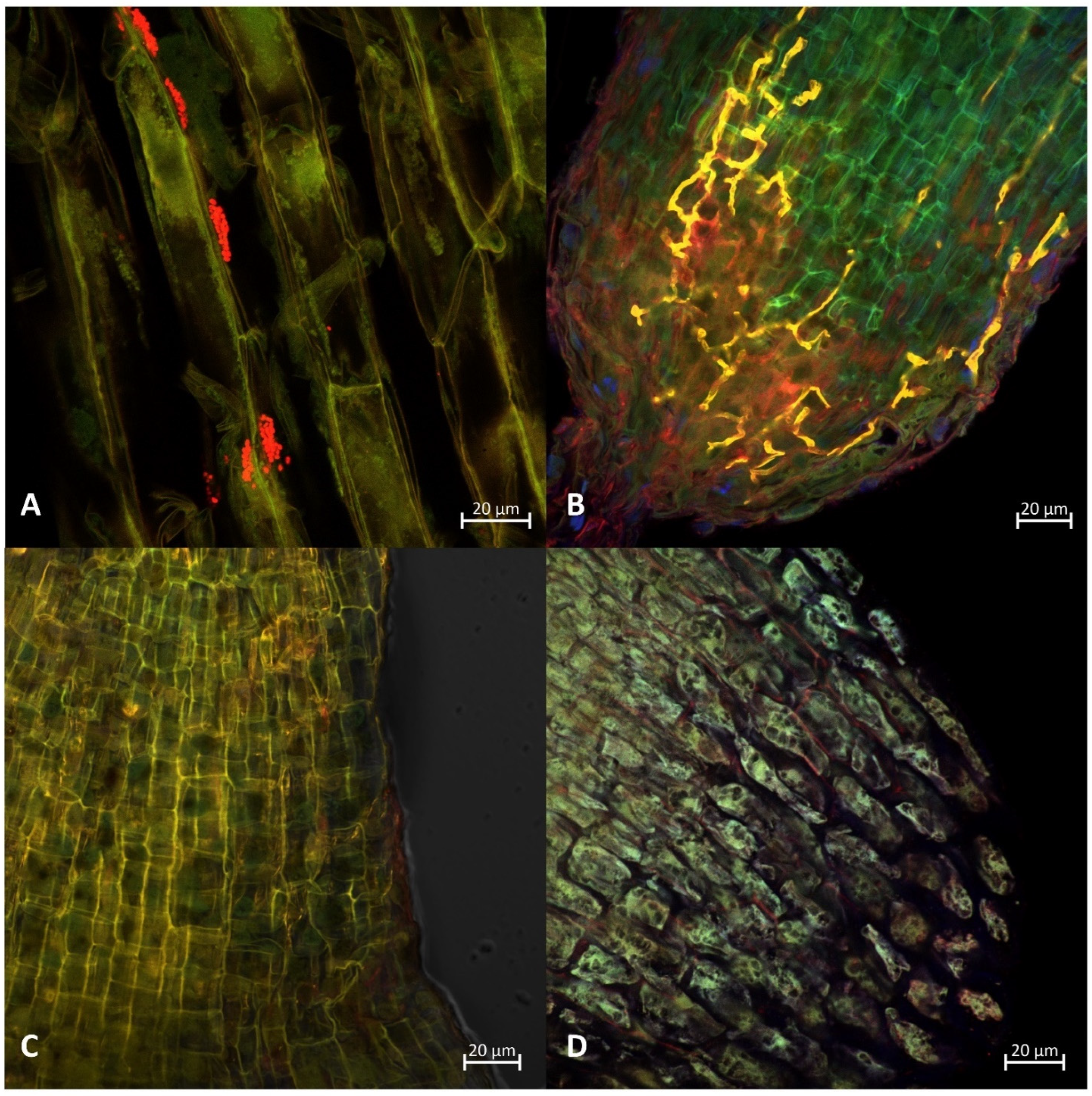
2.4. Staining of Roots, Fluorescence In Situ Hybridization and Microscopy Detection and Evaluation of Colonization by AMF and Azospirillum Bacterium

Roots were gently washed from substrate and fixed in a formalin-aceto-alcohol (FAA) fixative [34]. After fixation, roots were rinsed in distilled water, then cleared in 2% KOH solution (1 h at 50 °C) and afterwards washed in distilled water (4 × 3 min) and neutralized by 3% HCl solution. Roots were stained in a tube with a staining mixture consisting of wheat germ agglutinin Alexa Fluor 594 conjugate (WGA AF 594) (InvitrogenTM, Waltham, Massachusetts, USA) (50 μg·mL−1), concanavalin Alexa Fluor 647 conjugate (ConA AF 647) (InvitrogenTM, Waltham, Massachusetts USA) (50 μg·mL−1) and acid fuchsine (3%) at a ratio of 1:1:1, for 4–5 h at room temperature. After staining, roots were rinsed in 1 × PBS (4 × 3 min) and incubated for 12 h in 1 × PBS to remove all excess stains. Before mounting on the slide, a few drops of Hoechst stain were added to the slides with roots [35].
Roots intended for fluorescence in situ hybridization were, after fixation with FAA, dehydrated in a graded ethanol series (50, 75, 96% ethanol; 5 min each). Hybridization of the small pieces of the roots was performed in a tube with hybridization buffer containing 0.9 M sodium chloride, 0.01% sodium dodecyl sulfate (SDS), 10 mM TrisHCl, 50% of formamide (FA) and 5 ng·µL−1 of specific probe targeting 16S, 440–457 position labeled at ’5 end by Cy5 (AZO440a+; GTCATCATCGTCGCGTGC) at 46 °C for overnight [36]. Post-hybridization treatment was performed after removal of the hybridization buffer followed by adding washing buffer containing 20 mM TrisHCl, 0.01% SDS, 0.028 mol·L−1 NaCl and 10 µL·mL−1 of 0.5 M EDTA at 48 °C for 20 min. Afterwards the roots were rinsed in sterile distilled water and air dried for 24 h [36,37]. Hybridized samples were placed on microscopy slides with a drop of Hoechst solution, covered and observed under the microscope. Colonization of beneficial organisms was observed and evaluated on five randomly selected roots from three randomly selected plants (15 replicates).
Confocal microscopy was completed using the LSM800 (Carl Zeiss, Jena, Germany) microscope at 590/617 nm (excitation max./emission max.) for WGA AF 594, 650/668 nm for ConA AF 647, 350/461 nm for Hoechst stain and 649/666 for Cy5 labelled FISH probe. Lens 20×/0.8 NA, and 40×/1.2 NA were used. Processing of pictures was conducted in Zen Blue 2.6 (Carl Zeiss, Jena, Germany).
The intensity of AM colonization (%M) in each root segment was scored based on the presence of the fungus in the entire fragment using values from 0 to 5 (Table 2).
The intensity of colonization (M%) was estimated by the following equation:
M% = (95n5 + 70n4 + 30n3 + 5n2 + n1)/N
where n5, n4, n3, n2 and n1 are the numbers of fragment in the respective categories 5, 4, 3, 2 and 1 according to Alarcón and Cuenca [38].

2.5. Statistical Analysis

Data were analyzed by one-way ANOVA with a significance level p ≤ 0.05 followed by Fisher´s least significant difference (LSD) test to separate means into homogenous groups. Analyses were performed using software Statistica 13.3 (TIBCO Software Inc., Palo Alto, CA, USA).

3. Results

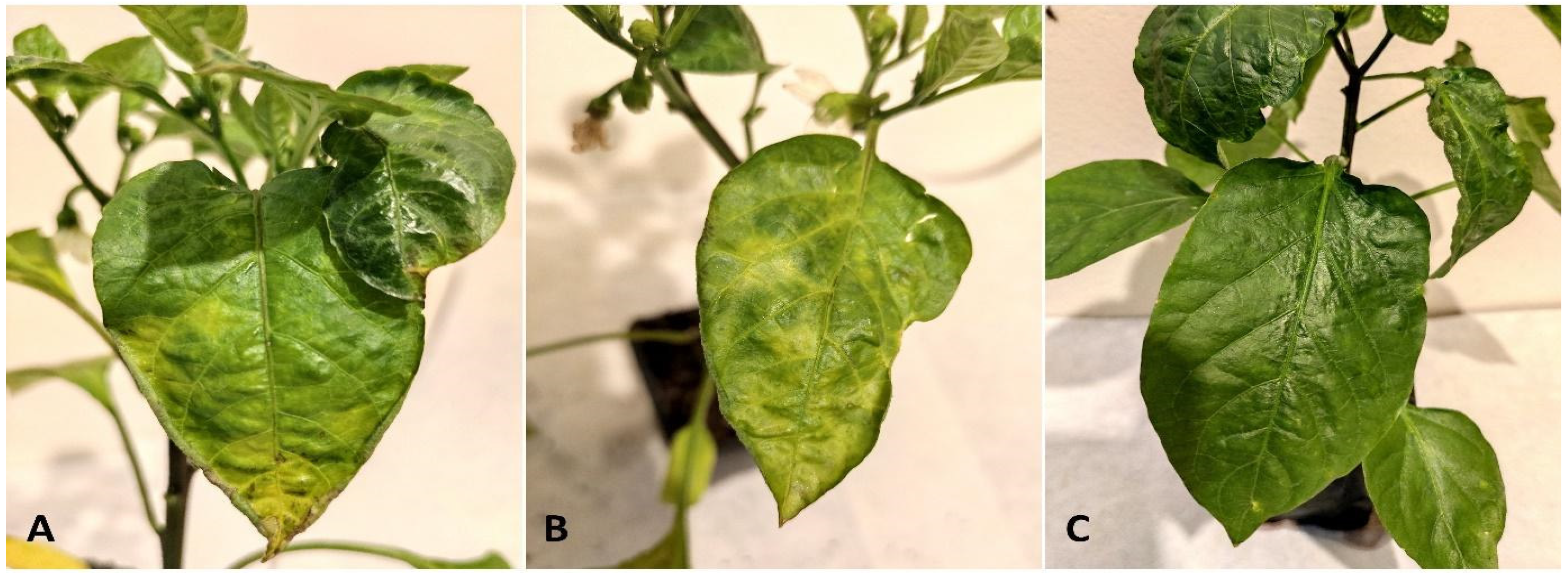
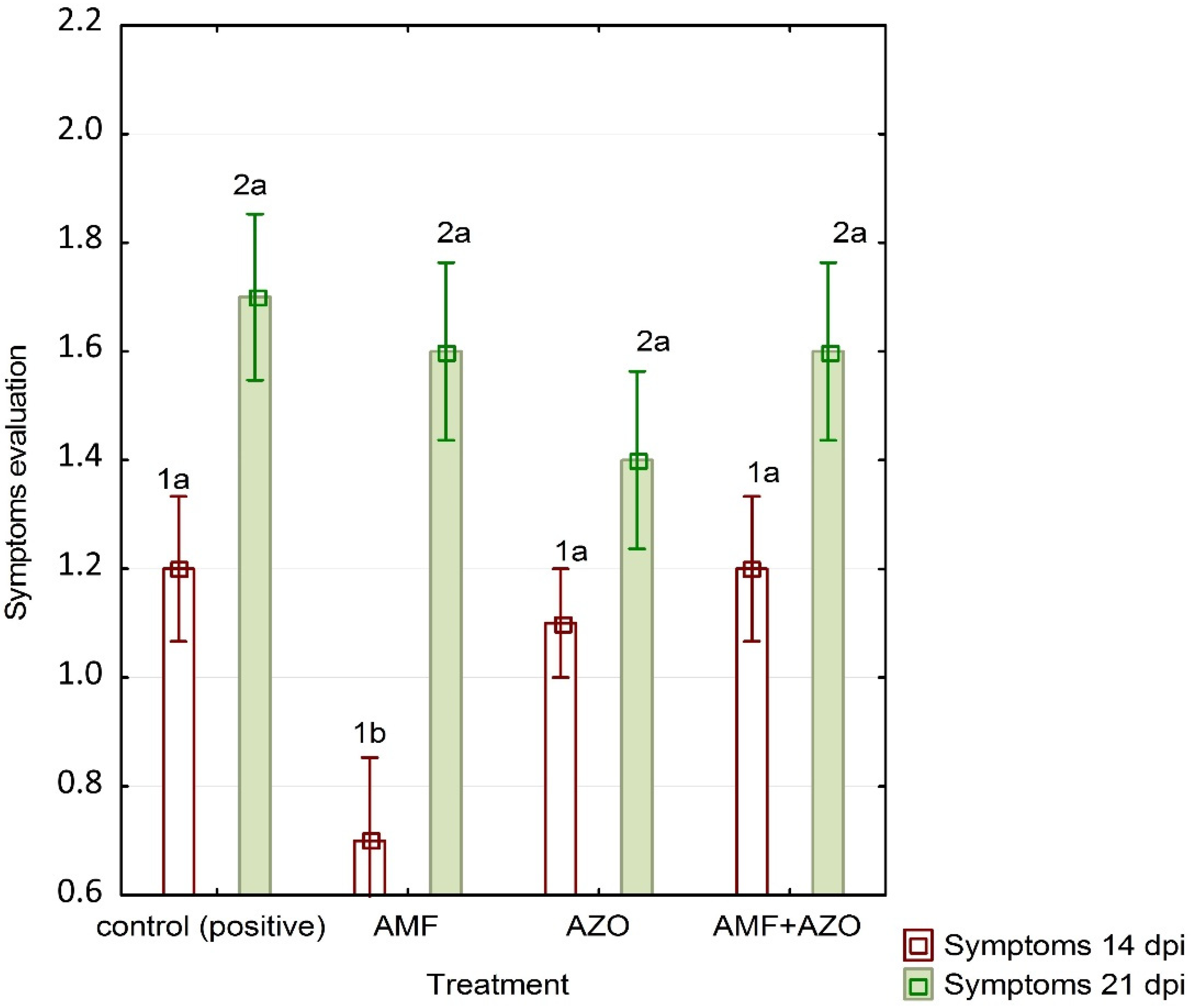
3.1. Symptoms Evaluation

Lowest symptom emergence was observed at pepper plants treated with mycorrhizal fungus (AMF) at 14 days post-inoculation (dpi). No other significant differences were observed in other treatments and at 21 dpi, including positive control plants. No ToBRFV symptoms were observed at non-inoculated control plants (Figure 1 and Figure 2).

3.2. Results of Real-Time qPCR Assay

Results of qPCR assay confirmed a presence of ToBRFV virus in all inoculated variants. The mean Ct (cycle threshold) was around 13 to 14 Ct in positive control plants, plants treated with Funneliformis mosseae (AMF) and plants treated with a combination of both Azospirillum and AMF (Supplementary Table S1). The mean Ct in plants treated with Azospirillum was 17.274. However, according to one-way ANOVA and Fisher´s least significant difference test there was no significant difference in mean Ct between treatments (Table 3). Results of relative quantification of ToBRFV titre in plant tissues confirmed positivity in all ToBRFV inoculated treatments and positive control and no significant differences between them (Supplementary Table S2).

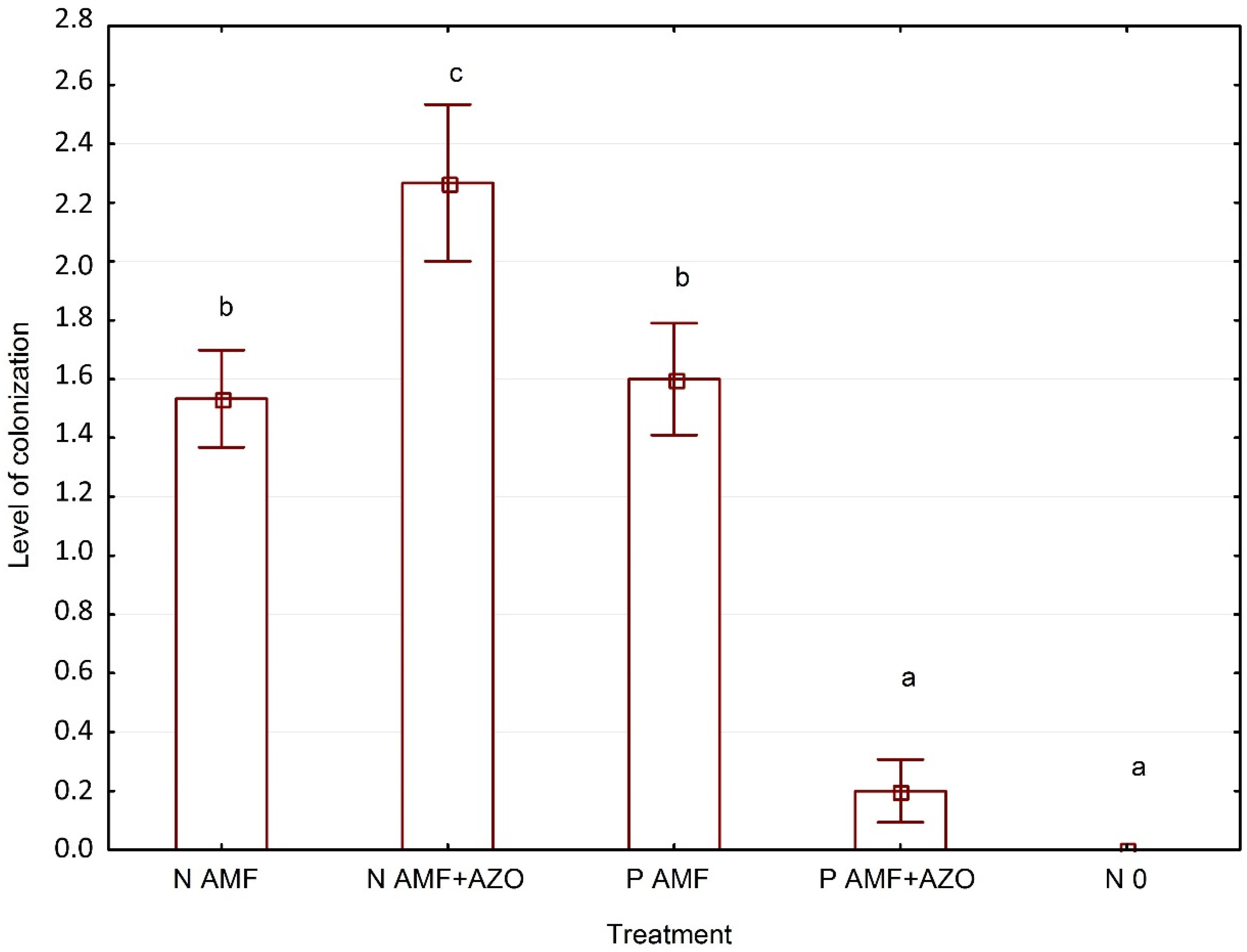
3.3. Evaluation of Funneliformis Mosseae and Azospirillum Brasilense Root System Colonization

Highest intensity (41%) of colonization by Azospirillum bacterium was detected at plants inoculated with ToBRFV virus and treated with both Azospirillum brasilense and arbuscular mycorrhizal fungus. In rest of treatments the colonization went from 20% to 33%. In the roots system of non-inoculated plants, it was detected only colonization trace of 0.4% (Table 4, Figure 3A). According to one-way ANOVA there was no significant difference between treatments, except of negative control plants.
Highest colonization of root system by mycorrhizal fungi (Funneliformis mosseae) was observed at ToBRFV negative plants treated with both AMF and Azospirillum, where the intensity reached 19.3%. Colonization intensity at both, ToBRFV negative and positive plants treated with AMF only, was 4.5% and 6.2%, respectively. Almost no AMF colonization was observed at ToBRFV positive plants treated with combination of AMF and Azospirillum (Table 4, Figure 3B and Figure 4).

4. Discussion

4.1. ToBRFV Symptoms Emergence and Real-Time qPCR Assay Results

Plants treated with AMF (P AMF) had lower symptoms emergence after 14 dpi, however there was no difference in symptoms emergence after 21 dpi within all treatments. Lower symptoms emergence after 14 dpi was not observed on plants treated with both AMF and Azospirillum (P AMF + AZO), what might be explained by very low AMF colonization of roots in this variant (Figure 4). This state might be explained by previous findings where temporary lower viral titre (Tomato aucuba mosaic virus) in tomato plant colonized by Funneliformis macrocarpa detected after 8–12 dpi significantly increased over time [39]. Similar results were observed in tomatoes and strawberries inoculated with Potato virus X (PVX) [39]. Maffei et al. [22] reported a significant reduction of Yellow leaf curl Sardinia virus symptoms of infected tomato plants when colonized with AMF. Increased uptake of nutrients, especially phosphorus may affect the susceptibility of the plant to viral infection [40]. According to study of Borrer et al. higher phosphorous content was associated with increase of barley and cereal yellow dwarf virus [41]. However, better nutrient status of plants treated with mycorhizal fungi, especially under nutrient deficient conditions, leads to vigorous plant growth, which can compensate for viral damage [40] Since there was no significant difference in mean Ct in results of Real-Time qPCR assay, the viral titre was similar in all treatments after 21 dpi. Hao et al. [40] suggested that bioprotection efficiency of AMF might be improved by using AMF with combination with other biological control agents. In this study there was no significant difference in viral titre or symptoms emergence after 21 dpi when using AMF with combination with Azospirillum bacterium compared to use AMF only. According to Lima et al. [42] use of Azospirillum brasilense provided greater tolerance of lettuce to Tospoviruses. Azospirillum genus is also studied for its potential as biocontrol agent against BYDV (Barley Yellow Dwarf Virus). Single inoculation of A. irakense in wheat was not effective, however when Azospirillum was applied before and after infection, the symptoms of BYDV were reduced [43]. In our study only non-significant reduction in symptoms emergence and viral titre in plants treated with Azospirillum (AZO) was detected.

4.2. Funneliformis mosseae and Azospirillum brasilense Root System Colonization

There was no significant difference in root system colonization with Azospirillum bacterium. Colonization intensity in all treated variants went from 20% to 41%. We assume that Azospirillum bacterium was not affected by presence or absence of ToBRFV virus in plant tissues.
By contrast, there were differences in AMF root system colonization. The highest colonization intensity was detected in ToBRFV negative plants treated with both AMF and Azospirillum and the lowest in ToBRFV positive plants with the same treatment (AMF + AZO). Interestingly, there was no significant difference in ToBRFV positive and negative plants treated with AMF only. These results suggest that use of combination of those two beneficial microorganisms in ToBRFV-infected plants negatively affected AMF colonization. Positive interaction between plant growth promoting bacteria (PGPB) and AMF including production of metabolites increasing root cell permeability and hormone synthesis have been reported [44]. On the other hand, a study of Zsögön et al. [45] indicated that ethylene, a plant hormone, can significantly inhibit mycorrhizal colonization, especially under P-deficient conditions. However, the mechanisms by which bacteria stimulate AM colonization are still not fully understood [46]. Aseel et al. [47] reported that roots of the ToMV (Tomato mosaic virus)-infected AMF treated tomato plants showed non-significant reduction of AMF colonization parameters compared to uninfected AMF treated plants. According to a study of Stolyarchuk et al. (2009) the intensity of AMF root colonization was slightly lower on virus infected plats of cucumber and tomato compared to virus-free plants [23]. However, to support possible effects of plant virus on AMF colonization more research must be done.

5. Conclusions

This study examined the potential of use of arbuscular mycorrhizal fungi (Funneliformis mosseae) and bacterium Azospirillum brasilense to enhance the tolerance of pepper plants to ToBRFV virus and reduce symptom severity or viral titre. Results of this study, as well as the results of many other studies focused on AMF effects on plant viruses, are indeterminate. Reduction of symptoms was observed on first evaluation (14 dpi); however, one week later there was no statistical difference in symptom emergence or viral titre in pepper plants. Use of Azospirillum brasilense showed only non-significant reduction in symptoms emergence and viral titre. Results suggest that use or combination of both AMF and Azospirillum bacterium is not more effective.

Supplementary Materials

The following supporting information can be downloaded at: https://www.mdpi.com/article/10.3390/d14080635/s1, Table S1: Complete results from real-time qPCR assay. Table S2: Results of relative quantifiction of ToBRFV titre in plant samples.

Author Contributions

Conceptualization, L.R., E.H., J.Č. and R.P.; methodology, L.R. and E.H.; validation, E.H., L.R., V.F. and G.K.; formal analysis, L.R., E.H. and R.P.; data curation, L.R. and E.H.; writing—original draft preparation, L.R.; writing—review and editing, E.H., R.P. and J.Č.; supervision, J.Č. and R.P.; project administration, J.Č.; funding acquisition, J.Č. All authors have read and agreed to the published version of the manuscript.

Funding

This research was supported by the project IGA-ZF/2021-ST2002: Tomato brown rugose fruit virus—actual threat of tomatoes and peppers production in European Union.

Institutional Review Board Statement

Not applicable.

Informed Consent Statement

Not applicable.

Data Availability Statement

Not applicable.

Conflicts of Interest

The authors declare no conflict of interest.

References

  1. FAO. FAOSTAT. 2021. Available online: https://www.fao.org/faostat/en/#data/QCL (accessed on 1 December 2021).
  2. ÚKZÚZ. Tomato Brown Rugose Fruit Virus (ToBRFV). Rostlinolékařský Portál. 2021. Available online: https://eagri.cz/public/app/srs_pub/fytoportal/public/?key=%2218c5e01b52e298fc5d46cc452f76b389%22#rlp|so|choroby|detail:18c5e01b52e298fc5d46cc452f76b389|popis (accessed on 1 December 2021).
  3. Salem, N.; Mansour, A.; Ciuffo, M.; Falk, B.; Turina, M. A new tobamovirus infecting tomato crops in Jordan. Arch. Virol. 2016, 161, 503–506. [Google Scholar] [CrossRef] [PubMed]
  4. Luria, N.; Smith, E.; Reingold, V.; Bekelman, I.; Lapidot, M.; Levin, I.; Elad, N.; Tam, Y.; Sela, N.; Abu-Ras, A.; et al. A New Israeli Tobamovirus Isolate Infects Tomato Plants Harboring Tm-22 Resistance Genes. PLoS ONE 2017, 12, e0170429. [Google Scholar] [CrossRef] [PubMed] [Green Version]
  5. Panno, S.; Caruso, A.; Davino, S. First Report of Tomato Brown Rugose Fruit Virus on Tomato Crops in Italy. Plant Dis. 2019, 103, 1443. [Google Scholar] [CrossRef]
  6. Menzel, W.; Knierim, D.; Winter, S.; Hamacher, J.; Heupel, M. First report of Tomato brown rugose fruit virus infecting tomato in Germany. New Dis. Rep. 2019, 39, 2044–2588. [Google Scholar] [CrossRef] [Green Version]
  7. Camacho-Beltrán, E.; Pérez-Villarreal, A.; Leyva-López, N.; Rodríguez-Negrete, E.; Ceniceros-Ojeda, E.; Méndez-Lozano, J. Occurrence of Tomato brown rugose fruit virus Infecting Tomato Crops in Mexico. Plant Dis. 2019, 103, 1440. [Google Scholar] [CrossRef]
  8. Alkowni, R.; Alabdallah, O.; Fadda, Z. Molecular identification of tomato brown rugose fruit virus in tomato in Palestine. J. Plant Pathol. 2019, 101, 719–723. [Google Scholar] [CrossRef]
  9. Ling, K.S.; Tian, T.; Gurung, S.; Salati, R.; Gilliard, A. First report of Tomato brown rugose fruit virus infecting greenhouse tomato in the United States. Plant Dis. 2019, 103, 1439. [Google Scholar] [CrossRef]
  10. Yan, Z.; Ma, H.; Han, S.; Geng, C.; Tian, Y.; Li, X. First Report of Tomato brown rugose fruit virus Infecting Tomato in China. Plant Dis. 2019, 103, 2973. [Google Scholar] [CrossRef]
  11. Alfaro-Fernández, A.; Castillo, P.; Sanahuja, E.; Rodríguez-Salido, M.; Font, M. First Report of Tomato Brown Rugose Fruit Virus in Tomato in Spain. Plant Dis. 2021, 105, 515. [Google Scholar] [CrossRef]
  12. Dey, K.; Velez-Climent, M.; Soria, P.; Batuman, O.; Mavrodieva, V.; Wei, G.; Zhou, J.; Adkins, S.; McVay, J. First report of Tomato brown rugose fruit virus infecting tomato in Florida, USA. New Dis. Rep. 2021, 44, e12028. [Google Scholar] [CrossRef]
  13. Sabra, A.; Al-Saleh, M.; Al-Shahwan, I.; Amer, M. First Report of Tomato Brown Rugose Fruit Virus Infecting the Tomato Crop in Saudi Arabia. Plant Dis. 2022, 106, 1310. [Google Scholar] [CrossRef] [PubMed]
  14. Tomato brown rugose fruit virus. EPPO Bull. 2020, 50, 529–534. Available online: https://gd.eppo.int/taxon/TOBRFV/datasheet (accessed on 11 December 2020). [CrossRef]
  15. Hao, L.; Zhang, Z.; Hao, B.; Diao, F.; Zhang, J.; Bao, Z.; Guo, W. Arbuscular mycorrhizal fungi alter microbiome structure of rhizosphere soil to enhance maize tolerance to La. Ecotox. Environ. Saf. 2021, 212, 111996. [Google Scholar] [CrossRef] [PubMed]
  16. Raaijmakers, J.; Paulitz, T.; Steinberg, C.; Alabouvette, C.; Moënne-Loccoz, Y. The rhizosphere: A playground and battlefield for soilborne pathogens and beneficial microorganisms. Plant Soil 2008, 321, 341–361. [Google Scholar] [CrossRef] [Green Version]
  17. Pokluda, R.; Ragasová, L.; Jurica, M.; Kalisz, A.; Komorowska, M.; Niemiec, M.; Sekara, A. Effects of growth promoting microorganisms on tomato seedlings growing in different media conditions. PLoS ONE 2021, 16, e0259380. [Google Scholar] [CrossRef]
  18. Fukami, J.; Nogueira, M.; Araujo, R.; Hungria, M. Accessing inoculation methods of maize and wheat with Azospirillum brasilense. AMB Express 2016, 6, 3. [Google Scholar] [CrossRef]
  19. Jung, S.; Martinez-Medina, A.; Lopez-Raez, J.; Pozo, M. Mycorrhiza-Induced Resistance and Priming of Plant Defenses. J. Chem. Ecol. 2012, 38, 651–664. [Google Scholar] [CrossRef]
  20. Miozzi, L.; Vaira, A.; Brilli, F.; Casarin, V.; Berti, M.; Ferrandino, A.; Nerva, L.; Accotto, G.; Lanfranco, L. Arbuscular Mycorrhizal Symbiosis Primes Tolerance to Cucumber Mosaic Virus in Tomato. Viruses 2020, 12, 675. [Google Scholar] [CrossRef]
  21. Lozano-Durán, R.; Rosas-Díaz, T.; Gusmaroli, G.; Luna, A.; Taconnat, L.; Deng, X.; Bejarano, E. Geminiviruses Subvert Ubiquitination by Altering CSN-Mediated Derubylation of SCF E3 Ligase Complexes and Inhibit Jasmonate Signaling in Arabidopsis thaliana. Plant Cell 2011, 23, 1014–1032. [Google Scholar] [CrossRef] [Green Version]
  22. Maffei, G.; Miozzi, L.; Fiorilli, V.; Novero, M.; Lanfranco, L.; Accotto, G. The arbuscular mycorrhizal symbiosis attenuates symptom severity and reduces virus concentration in tomato infected by Tomato yellow leaf curl Sardinia virus (TYLCSV). Mycorrhiza 2013, 24, 179–186. [Google Scholar] [CrossRef]
  23. Stolyarchuk, I.M.; Shevchenko, T.P.; Polischuk, V.P.; Kripka, A.V. Virus infection course in different plant species under influenceof arbuscular mycorrhiza. Microbiol. Biotechnol. 2009, 6, 70–75. [Google Scholar]
  24. Thiem, D.; Szmidt-Jaworska, A.; Baum, C.; Muders, K.; Niedojadło, K.; Hrynkiewicz, K. Interactive physiological response of potato (Solanum tuberosum L.) plants to fungal colonization and Potato virus Y (PVY) infection. Acta Mycol. 2014, 49, 291–303. [Google Scholar] [CrossRef] [Green Version]
  25. Miozzi, L.; Catoni, M.; Fiorilli, V.; Mullineaux, P.; Accotto, G.; Lanfranco, L. Arbuscular Mycorrhizal Symbiosis Limits Foliar Transcriptional Responses to Viral Infection and Favors Long-Term Virus Accumulation. Mol. Plant-Microbe Interact. 2011, 24, 1562–1572. [Google Scholar] [CrossRef] [Green Version]
  26. Shaul, O.; Galili, S.; Volpin, H.; Ginzberg, I.; Elad, Y.; Chet, I.; Kapulnik, Y. Mycorrhiza-Induced Changes in Disease Severity and PR Protein Expression in Tobacco Leaves. Mol. Plant-Microbe Interact. 1999, 12, 1000–1007. [Google Scholar] [CrossRef] [Green Version]
  27. Pii, Y.; Aldrighetti, A.; Valentinuzzi, F.; Mimmo, T.; Cesco, S. Azospirillum brasilense inoculation counteracts the induction of nitrate uptake in maize plants. J. Exp. Bot. 2019, 70, 1313–1324. [Google Scholar] [CrossRef]
  28. Chanda, B.; Shamimuzzaman, M.; Gilliard, A.; Ling, K. Effectiveness of disinfectants against the spread of tobamoviruses: Tomato brown rugose fruit virus and Cucumber green mottle mosaic virus. Virol. J. 2021, 18, 7. [Google Scholar] [CrossRef] [PubMed]
  29. Jewehan, A.; Salem, N.; Tóth, Z.; Salamon, P.; Szabó, Z. Screening of Solanum (sections Lycopersicon and Juglandifolia) germplasm for reactions to the tomato brown rugose fruit virus (ToBRFV). J. Plant Dis. Prot. 2022, 129, 117–123. [Google Scholar] [CrossRef]
  30. Eichmeier, A.; Baránek, M.; Pidra, M. Analysis of genetic diversity and phylogeny of partial coat protein domain in Czech and Italian GFLV isolates. Plant Prot. Sci. 2010, 46, 145–148. [Google Scholar] [CrossRef] [Green Version]
  31. Livak, K.; Schmittgen, T. Analysis of Relative Gene Expression Data Using Real-Time Quantitative PCR and the 2−ΔΔCT Method. Methods 2001, 25, 402–408. [Google Scholar] [CrossRef]
  32. Menzel, W.; Winter, S. Identification of novel and known tobamoviruses in tomato and other solanaceous crops using a new pair of generic primers and development of a specific RT-qPCR for ToBRFV. Acta Hortic. 2021, 1316, 143–148. [Google Scholar] [CrossRef]
  33. Christensen, N.; Nicolaisen, M.; Hansen, M.; Schulz, A. Distribution of Phytoplasmas in Infected Plants as Revealed by Real-Time PCR and Bioimaging. Mol. Plant-Microbe Interact. 2004, 17, 1175–1184. [Google Scholar] [CrossRef] [Green Version]
  34. Johansen, D. Plant Microtechnique; McGraw Hill: New York, NY, USA, 1940. [Google Scholar]
  35. Vierheilig, H.; Schweiger, P.; Brundrett, M. An overview of methods for the detection and observation of arbuscular mycorrhizal fungi in roots. Physiol. Plant. 2005, 125, 393–404. [Google Scholar] [CrossRef]
  36. Stoffels, M.; Castellanos, T.; Hartmann, A. Design and Application of New 16S rRNA-targeted Oligonucleotide Probes for the Azospirillum-Skermanella-Rhodocista-Cluster. Syst. Appl. Microbiol. 2001, 24, 83–97. [Google Scholar] [CrossRef] [PubMed] [Green Version]
  37. Neef, A.; Amann, R.; Schlesner, H.; Schleifer, K. Monitoring a widespread bacterial group: In situ detection of planctomycetes with 16S rRNA-targeted probes. Microbiology 1998, 144, 3257–3266. [Google Scholar] [CrossRef] [PubMed] [Green Version]
  38. Alarcón, C.; Cuenca, G. Arbuscular mycorrhizas in coastal sand dunes of the Paraguanä Peninsula, Venezuela. Mycorrhiza 2005, 16, 1–9. [Google Scholar] [CrossRef] [PubMed]
  39. Daft, M.; Okusanya, B. Effect of Endogone Mycorrhiza on Plant Growth V. Influence of Infection on the Multiplication of Viruses in Tomato, Petunia and Strawberry. New Phytol. 1973, 72, 975–983. [Google Scholar] [CrossRef]
  40. Hao, Z.; Xie, W.; Chen, B. Arbuscular Mycorrhizal Symbiosis Affects Plant Immunity to Viral Infection and Accumulation. Viruses 2019, 11, 534. [Google Scholar] [CrossRef] [Green Version]
  41. Borer, E.; Seabloom, E.; Mitchell, C.; Power, A. Local context drives infection of grasses by vector-borne generalist viruses. Ecol. Lett. 2010, 13, 810–818. [Google Scholar] [CrossRef]
  42. Lima, A.; Venturoso, L.; Silva, B.; Gomes, A.; Schimidt, O. Eficiência da inoculação de Azospirillum brasilense associado com enraizador no crescimento e na produção de alface. Rev. Verde Agroecol. Desenvol. Sustent. 2017, 12, 233. [Google Scholar] [CrossRef]
  43. Al-Ani, R.A.; Adhab, M.A.; El-Muadhidi, M.A.; Al-Fahad, M.A. Induced systemic resistance and promotion of wheat and barley plants growth by biotic and non-biotic agents against barley yellow dwarf virus. Afr. J. Biotechnol. 2011, 10, 12078–12084. [Google Scholar]
  44. Raklami, A.; Bechtaoui, N.; Tahiri, A.; Anli, M.; Meddich, A.; Oufdou, K. Use of Rhizobacteria and Mycorrhizae Consortium in the Open Field as a Strategy for Improving Crop Nutrition, Productivity and Soil Fertility. Front. Microbiol. 2019, 10, 1106. [Google Scholar] [CrossRef] [PubMed] [Green Version]
  45. Zsögön, A.; Lambais, M.; Benedito, V.; Figueira, A.; Peres, L. Reduced arbuscular mycorrhizal colonization in tomato ethylene mutants. Sci. Agric. 2008, 65, 259–267. [Google Scholar] [CrossRef]
  46. Santoyo, G.; Gamalero, E.; Glick, B. Mycorrhizal-Bacterial Amelioration of Plant Abiotic and Biotic Stress. Front. Sustain. Food Syst. 2021, 5, 672881. [Google Scholar] [CrossRef]
  47. Aseel, D.; Rashad, Y.; Hammad, S. Arbuscular Mycorrhizal Fungi Trigger Transcriptional Expression of Flavonoid and Chlorogenic Acid Biosynthetic Pathways Genes in Tomato against Tomato Mosaic Virus. Sci. Rep. 2019, 9, 9692. [Google Scholar] [CrossRef] [Green Version]
Figure 1. Symptoms development after 14 days post inoculation with ToBRFV on (A) positive control plants (non-treated with AMF or Azospirillum), (B) ToBRFV-infected plants treated with AMF and Azospirillum and (C) negative control plants (ToBRFV negative) treated with AMF and Azospirillum.
Diversity 14 00635 g001
Figure 2. Means (n = 10) (± SE) of symptom evaluation on ToBRFV inoculated plants at 14 and 21 dpi, separated into homogenous groups (1a, 1b and 2a) according to Fisher´s LSD test. Explanatory notes: AMF-arbuscular mycorrhizal fungus (Funneliformis mosseae), AZO-Azospirillum brasilense.
Diversity 14 00635 g002
Figure 3. (A) Colonies of Azospirillum (in red) on root of pepper non-inoculated with ToBRFV, treated with mycorrhizal fungus and Azospirillum (N AMF + AZO). Scale bar 20 µm, lens 40×/1.2 NA; (B) Arbuscular mycorrhizal fungal mycelia (in yellow) growing inside the root of plant non-inoculated with ToBRFV, treated with Funneliformis mosseae fungus (N AMF). Scale bar 20 µm, lens 20×/0.8 NA. (C) Negative control plants for Azospirillum observation (non-treated with Azospirillum or AMF, non-infected with ToBRFV)). Scale bar 20 µm, lens 20×/0.8 NA.; (D) Negative control plants for AMF observation (non treated with AMF or Azospirillum, non-inoculated with ToBRFV). Scale bar 20 µm, lens 20×/0.8 NA.
Diversity 14 00635 g003
Figure 4. Means (±SE) of AMF colonization evaluation, separated into homogenous groups (a, b and c) according to Fisher’s LSD test. Explanatory notes: N AMF-ToBRFV negative plants treated with arbuscular mycorrhizal fungus, N AMF + AZO-ToBRFV negative plants treated with AMF and Azospirillum, P AMF-ToBRFV positive plants treated with arbuscular mycorrhizal fungus, P AMF + AZO-ToBRFV positive plants treated with AMF and Azospirillum, N 0—negative non-treated plants.
Diversity 14 00635 g004
Table 1. Symptom severity scale on inoculated pepper plants.
ScaleSymptoms
0No symptoms
1Mild mosaic or mottling, followed by recovery
2Mild mosaic or mottling with leaf deformation
3Moderate mosaic or mottling and leaf deformation, followed by rolling, necrotic spots and stunting
4Severe mosaic or mottling and leaf deformity, extended necrotic spots
5Severe mosaic or mottling, leaf deformity, shoestring
Modified according to Jewehen et al. [29].
Table 2. Values indicating proportion of root colonization [38].
ValueProportion of Root Colonized by Funneliformis mosseae/Azospirillum brasilense
0Without colonization
1Colonization trace
2Less than 10%
3From 11 to 50%
4From 51% to 90%
5More than 90%
Table 3. Results of Fisher’s LSD test of mean Ct of qPCR assay detecting ToBRFV virus.
LSD Test; Variable Mean Ct (qPCR), Homogenous Groups, Alfa = 0.05000 Error: Mean Sum of sq. = 9.8032, Degrees of Freedom = 16.000
TreatmentMean Ct (Mean)1
P AMF13.33800****
Control (positive)13.39800****
P AMF + AZO14.11600****
P AZO17.27400****
Explanatory notes: P AMF-ToBRFV positive plants treated with arbuscular mycorrhizal fungus, P AMF + AZO-ToBRFV positive plants treated with AMF and Azospirillum, P AZO-ToBRFV positive plants treated with Azospirillum brasilense, ****—Homogenous group
Table 4. Intensity of colonization (M) by Azospirillum brasilense (M) and Funneliformis mosseae within given treatments and non-treated control plants.
Colonization intensity (M) by Azospirillum
TreatmentN AZON AMF + AZOP AZOP AMF + AZOcontrol (negative)
Intensity of colonization (M)20%28%41%33%0.4%
Colonization intensity (M) by funneliformis mosseae
TreatmentN AMFN AMF + AZOP AMFP AMF + AZOcontrol (negative)
Intensity of colonization (M)4.5%19.3%6.2%0.2%0.0%
Publisher’s Note: MDPI stays neutral with regard to jurisdictional claims in published maps and institutional affiliations.

