Article

GmCBP60b Plays Both Positive and Negative Roles in Plant Immunity

1
College of Life Sciences, Zhejiang Normal University, Jinhua 321004, China
2
Zhejiang Provincial Key Laboratory of Biotechnology on Specialty Economic Plants, Zhejiang Normal University, Jinhua 321004, China
*
Author to whom correspondence should be addressed.
Int. J. Mol. Sci. 2024, 25(1), 378; https://doi.org/10.3390/ijms25010378
Submission received: 29 November 2023 / Revised: 20 December 2023 / Accepted: 22 December 2023 / Published: 27 December 2023
(This article belongs to the Special Issue Molecular Genetics and Breeding Mechanisms in Crops 2.0)

Abstract

:
CBP60b (CALMODULIN-BINDING PROTEIN 60b) is a member of the CBP60 transcription factor family. In Arabidopsis, AtCBP60b not only regulates growth and development but also activates the transcriptions in immune responses. So far, CBP60b has only been studied extensively in the model plant Arabidopsis and rarely in crops. In this study, Bean pod mottle virus (BPMV)-mediated gene silencing (BPMV-VIGS) was used to silence GmCBP60b.1/2 in soybean plants. The silencing of GmCBP60b.1/2 resulted in typical autoimmunity, such as dwarfism and enhanced resistance to both Soybean mosaic virus (SMV) and Pseudomonas syringae pv. glycinea (Psg). To further understand the roles of GmCBP60b in immunity and circumvent the recalcitrance of soybean transformation, we generated transgenic tobacco lines that overexpress GmCBP60b.1. The overexpression of GmCBP60b.1 also resulted in autoimmunity, including spontaneous cell death on the leaves, highly induced expression of PATHOGENESIS-RELATED (PR) genes, significantly elevated accumulation of defense hormone salicylic acid (SA), and significantly enhanced resistance to Pst DC3000 (Pseudomonas syrangae pv. tomato DC3000). The transient coexpression of a luciferase reporter gene driven by the promoter of soybean SYSTEMIC ACQUIRED RESISTANCE DEFICIENT 1 (GmSARD1) (ProGmSARD1::LUC), together with GmCBP60b.1 driven by the 35S promoter, led to the activation of the LUC reporter gene, suggesting that GmCBP60b.1 could bind to the core (A/T)AATT motifs within the promoter region of GmSARD1 and, thus, activate the expression of the LUC reporter. Taken together, our results indicate that GmCBP60b.1/2 play both positive and negative regulatory roles in immune responses. These results also suggest that the function of CBP60b is conserved across plant species.

1. Introduction

CALMODULIN-BINDING PROTEIN 60 (CBP60)-like proteins comprise a family of eight members of atypical transcription factors in Arabidopsis: CBP60a–CBP60g and SAR Deficient 1 (SARD1) [1]. The functional roles of CBP60g and SARD1 in immunity have been extensively studied [2,3,4,5,6,7]. They regulate the expression of defense-related genes by directly binding to their promoters [4]. These genes include those involved in SA and N-HYDROXY-PIPECOLIC ACID (NHP) biosynthesis, as well as in PAMP-triggered immunity (PTI), effector-triggered immunity (ETI), and systemic acquired resistance (SAR) pathways [4,7,8,9]. Unlike other CBP60s, SARD1 does not need to bind CALMODULIN (CaM) for its function [4]. SARD1 activates ISOCHORISMATE SYNTHASE 1 (ICS1) expression by binding a GAAATTT motif in its promoter [8]. The simultaneous loss of functions of SARD1 and CBP60g almost completely blocked pathogen-induced SA accumulation, leading to a reduced resistance to various pathogens [2,4]. Contrary to CBP60g or SARD1, CBP60a has been shown to play a negative role in immunity [10]. Recently, two groups provided solid evidence that CBP60b plays both positive and negative roles in Arabidopsis immunity [11,12]. CBP60b is a constitutively expressed gene [13], whereas SARD1 and CBP60g are pathogen-induced genes [2,4]. The loss of function of CBP60b constitutively activated ENHANCED DISEASE SUSCEPTIBILITY 1 (EDS1)- and PHYTOALEXIN DEFICIENT 4 (PAD4)-dependent immunities [11,12], whereas the overexpression of CBP60b activated EDS1- and PAD4-independent immunities [11]. The authors proposed that CBP60b might be indispensable for the expression of a negative regulator of Toll-interleukin-1 receptor–nucleotide-binding leucine-rich repeat (TNL)-mediated immune signaling or that CBP60b might be monitored and guarded by a TNL directly or indirectly, and the loss of its function activates this unidentified TNL and, thus, results in ETI [11,12]. This postulation is supported by the partial requirement for TNL SNC1 (SUPPRESSOR OF NPR1-1, CONSTITUTIVE 1) in the autoimmunity of a cbp60b mutant [11]. The enhanced autoimmunity observed in the cbp60bcbp60g-1 double mutant compared to either single mutant suggests that CBP60b and CBP60g play partially redundant roles in the transcriptional activation of the putative guardee/decoy guarded by unknown TNL [12].
Although extensively studied in model plant Arabidopsis, the functions of CBP60 family members have not been investigated in other plant species, especially in crops. In this report, both GmCBP60b1 and GmCBP60b2 (referred to as GmCBP60b1/2 hereafter) were simultaneously silenced in soybeans via virus-induced gene silencing (VIGS) mediated by BPMV (Bean pod mottle virus), referred to as BPMV-VIGS hereafter. To circumvent the recalcitrance of soybeans in transformation, transgenic Nicotiana tabacum plants overexpressing GmCBP60b1 were generated. Consistent with the observations in Arabidopsis, either silencing GmCBP60b1/2 in soybeans or overexpressing GmCBP60b1 in N. tabacum activated immune responses, indicating that GmCBP60b1/2 play both positive and negative roles in immunity. A transient luciferase reporter assay demonstrated that GmCBP60b.1 could bind to the core motif (A/T)AATT within the promoter region of GmSARD1 and, thus, activate the expression of the LUC reporter, suggesting that the GmCBP60b1 protein binds to a similar core motif as in Arabidopsis. Consistent with its role as a transcription factor, GmCBP60b1 was exclusively localized in nuclei. Taken together, our results indicate that the function of CBP60b is conserved in regulating immunity across plant species.

2. Results

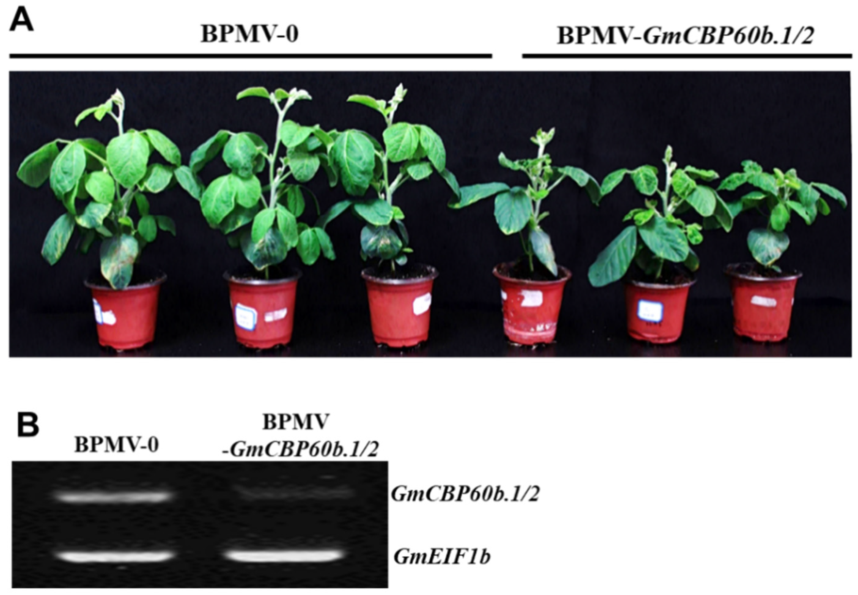
2.1. Silencing GmCBP60b.1 and GmCBP60b.2 Simultaneously Results in Stunted Stature and Enhanced Disease Resistance in Soybean Plants

It has been reported that CBP601b plays a critical role in immunity in the model plant Arabidopsis [11,12]. However, the function of CBP601b homologues in soybean immunity has not been previously investigated. To this end, the BPMV-VIGS system was used to investigate the functions of GmCBP60bs. Soybeans are a paleopolyploid, in which 75% of the genes in their genome have two copies [14]. Consistent with this, two homologous GmCBP60bs sharing 97% identity at the nucleotide level were identified in the soybean genome, and they were referred to as GmCBP60b.1 and GmCBP60b.2. GmCBP60b.1 and GmCBP60b.2 share 67% and 66% similarity with Arabidopsis CBP60b, respectively. To overcome the functional redundancy of these two genes, a 303 bp fragment amplified from GmCBP60b.1 sharing 98% identity at the nucleotide level with the corresponding region of CBP60b.2 was cloned into the BPMV-2 vector [15,16]. To infect the soybean plants, the plasmid DNA of the BPMV1 vector was co-bombarded with plasmid DNA of either the BPMV2 empty vector (referred to as BPMV-0) or BPMV-GmCBP60b.1/2 (referred to as BPMV-GmCBP60b.1/2) into the first two true leaves of 7-day-old soybean seedlings. The viral symptoms were visible on the upper systemic leaves of the bombarded plants at 15 to 20 days post-bombardment (dpb). The systemic leaves with viral symptoms were ground in a phosphate buffer (pH 7.0). The liquid phase (sap), containing numerous viral particles, was collected and stored at −80 °C for subsequent batch inoculations.
The 7-day-old seedlings were rub-inoculated with either the BPMV-0 or BPMV-GmCBP60b.1/2 sap. At 20 days post-inoculation (dpi), the plants inoculated with BPMV-GmCBP60b.1/2 sap displayed a stunted phenotype compared with the those inoculated with BPMV-0 sap (Figure 1A), indicating that simultaneously silencing both GmCBP60b.1 and GmCBP60b.2 dramatically reduced the sizes of the soybean plants. This stunted phenotype is similar to those plants with autoimmunity. Because the cbp60b mutant in Arabidopsis exhibited autoimmunity, it suggests that defense responses are similarly activated in the GmCBP60b.1/2-silenced plants. An RT-PCR analysis indicated that the transcript level of GmCBP60b.1/2 was indeed significantly reduced in the GmCBP60b.1/2-silenced plants compared with the BPMV-0 plants (Figure 1B).

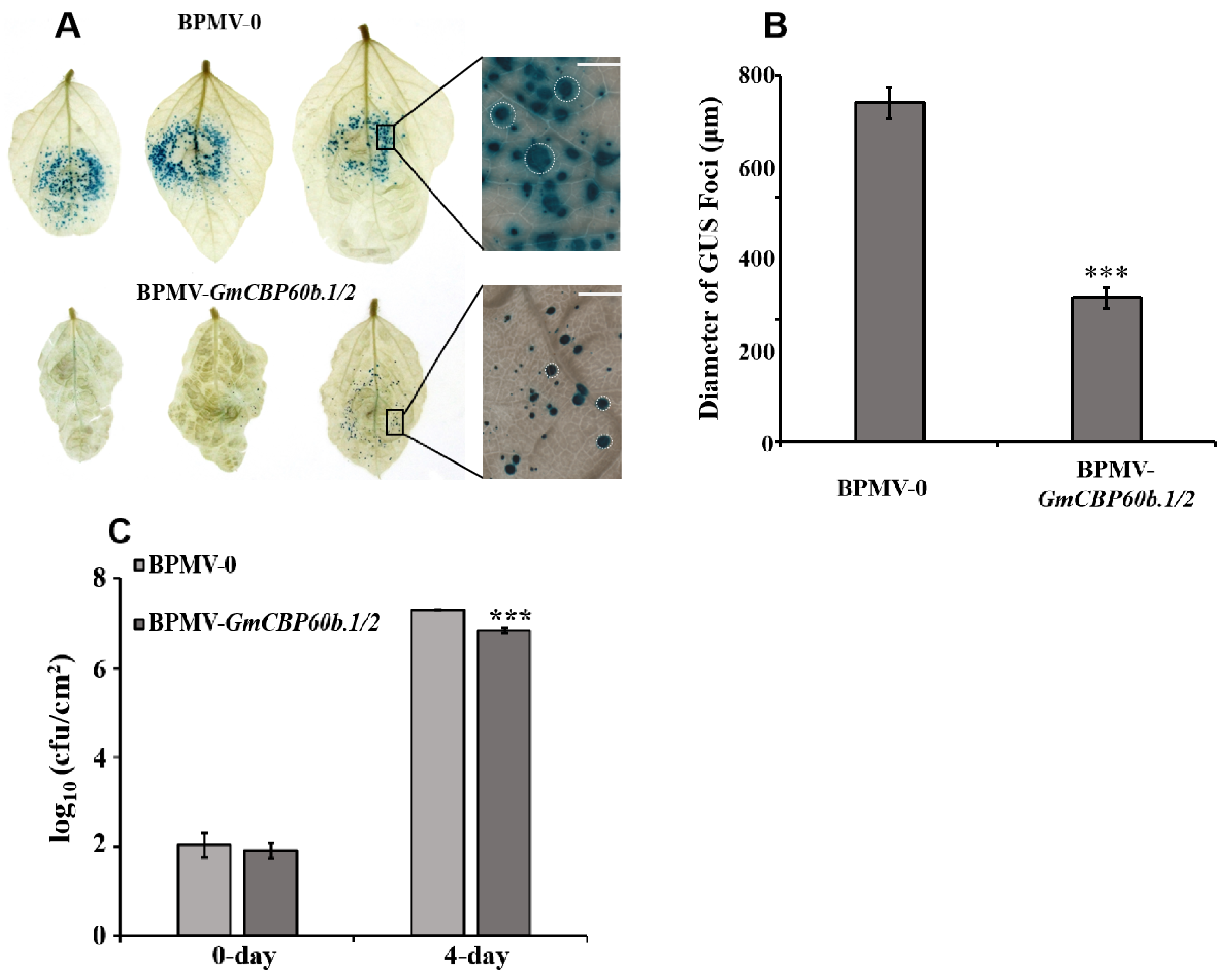
2.2. Silencing GmCBP60b.1/2 Enhances the Resistance of Soybean Plants to Both SMV-N-GUS and Pseudomonas syringae pv. glycinea (Psg)

To test whether the stunted phenotype is associated with activated defense responses, we firstly inoculated BPMV-0 and BPMV-GmCBP60b1/2 plants with the SMV N strain fused with the GUS reporter gene (SMV-N-GUS) [17] via biolistic bombardment. The successful infection of SMV-N-GUS can be visualized by the presence of blue GUS foci. The intensities and diameters of the GUS foci reflect replication efficiency and cell-to-cell movement, respectively. As shown in Figure 2A, much fewer blue GUS foci were observed on the infected leaves of the GmCBP60b1/2-silenced plants than on the infected vector control leaves. In addition, the diameter of the GUS foci was significantly smaller on the infected leaves of the GmCBP60b.1/2-silenced plants than on the infected vector control leaves (Figure 2B), indicating that silencing GmCBP60b.1/2 enhances resistance to SMV-N-GUS.
Next, we examined the effect of GmCBP60b.1/2 silencing on resistance against the bacterial pathogen Pseudomonas syrangae pv. glycinea (Psg). As shown in Figure 2C, the multiplication of Psg on the leaves of the GmCBP60b.1/2-silenced plants was drastically reduced compared with that on the BPMV-0 leaves. These results demonstrate that GmCBP60b.1/2 play a negative role in disease resistance.

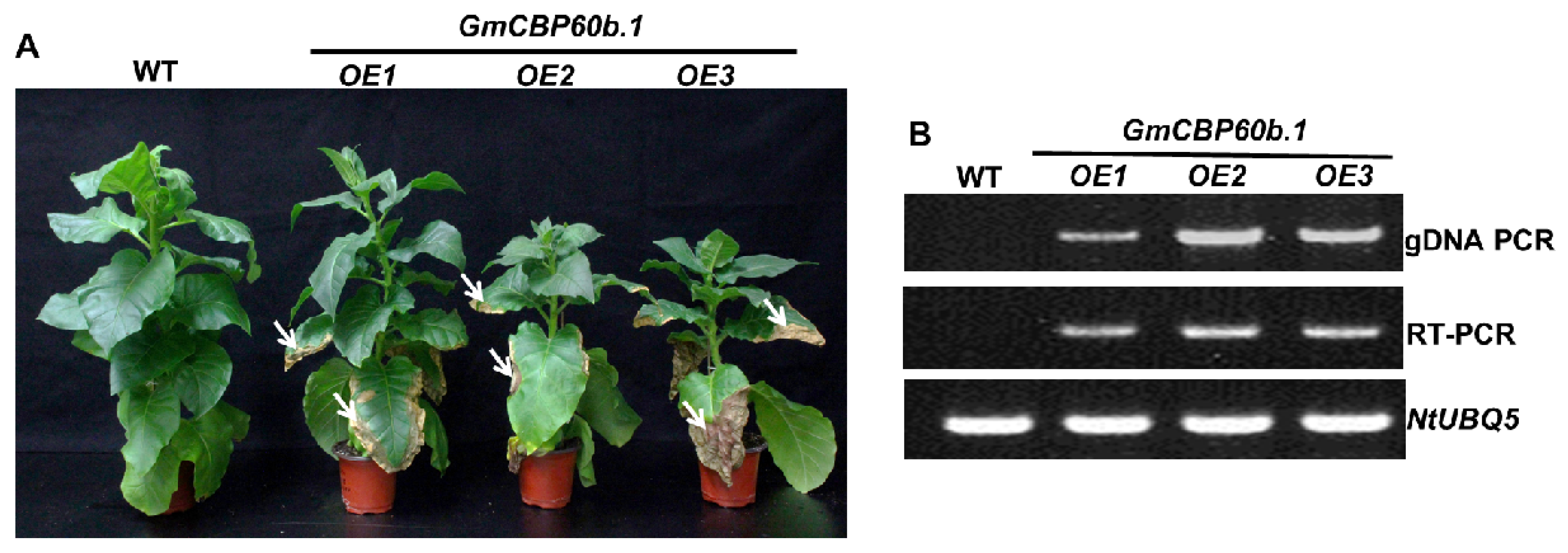
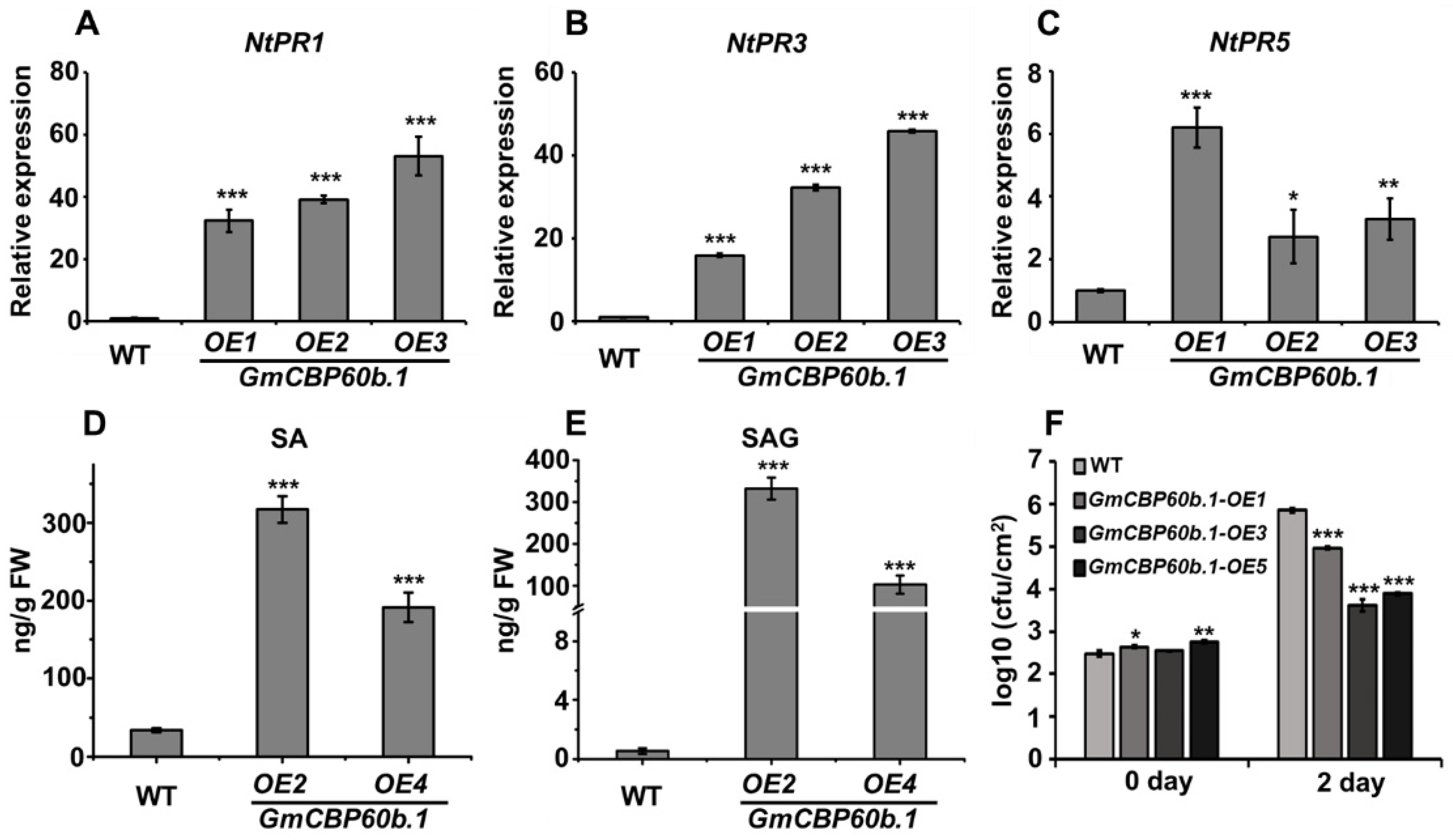
2.3. Overexpression of GmCBP60b.1 in Nicotiana tabacum Activates Immune Responses and Enhances Disease Resistance

To further understand the function of GmCBP60b in immunity and overcome the difficulty of soybean transformation, we generated transgenic tobacco lines that overexpress GmCBP60b.1. The tobacco GmCBP60b.1-overexpressing lines displayed a stunted stature compared to the wide-type (WT) plants (Figure 3A). To our surprise, spontaneous cell death was observed on the leaves of the GmCBP60b.1-overexpressing tobacco lines (Figure 3A, see arrows), suggesting that the immune responses might be activated. The authenticity of the transgenic plants was verified by both genomic PCR and an RT-PCR analysis (Figure 3B).
To confirm that the stunted phenotype and cell death are indeed caused by the activated immunity, we firstly analyzed the expression of multiple PR genes. As shown in Figure 4A–C, the expressions of NtPR1, NtPR3, and NtPR5 were hugely induced in the leaves of the GmCBP60b.1-overexpressing plants compared with the WT leaves. In addition, the accumulation levels of both free SA and conjugated SA were significantly elevated in the GmCBP60b.1-overexpressing plants relative to the WT plants (Figure 4D,E). Taken together, these results demonstrate that the overexpression of GmCBP60b.1 activated the immune responses in tobacco.
Activated immune responses are usually correlated with enhanced disease resistance. To examine whether the activated immunity observed in the GmCBP60b.1-overexpressing plants results in enhanced resistance, we inoculated both the WT and the GmCBP60b.1-overexpressing plants with the bacterial pathogen Pseudomonas syrangae pv. tomato DC3000 (Pst DC3000). As shown in Figure 4F, the multiplication of Pst DC3000 on the leaves of the GmCBP60b.1-overexpressing plants was drastically reduced compared with that on the WT plants, demonstrating that GmCBP60b.1 plays a positive role in defense responses.

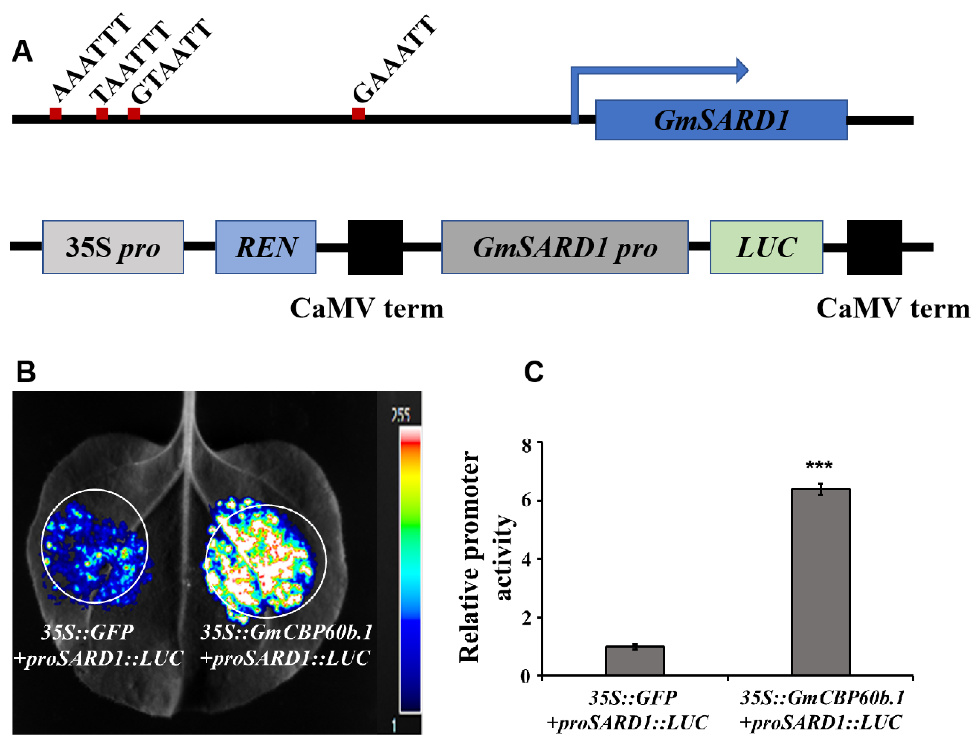
2.4. Transient Overexpression of GmCBP60b.1 Activates the Expression of a Luciferase Gene Driven by GmSARD1 Promoter, Which Contains Multiple CBP60b-Specifc Cis-Elements

In Arabidopsis, CBP60b binds to the core cis-element G(A/T)AATT(T/G) in the promoter regions of defense-related genes, such as SARD1 and ICS1, and it activates their expression [11,12]. We found that there were four core cis-element-like motifs, (G/A/T)AATT, within a 1240 bp promoter region of GmSARD1 (Figure 5A, upper panel). We reasoned that GmCBP60b might be able to bind these motifs and thus activate the expression of GmSARD1. To test this possibility, we first cloned a 1240 bp fragment of the GmSARD1promoter into a pGreenII0800-LUC vector to generate a proGmSARD1::LUC construct (Figure 5A, lower panel). Next, we transiently coexpressed 35S::GmCBP60b.1 with proGmSARD1::LUC in the leaves of N. benthamiana plants via agro-infiltration. If our postulation is correct, the expression of the luciferase gene will be activated in the presence of GmCBP60b in the infiltrated region. As expected, the expression of the luciferase gene was highly activated in the co-infiltrated area (Figure 5B, right), indicating that GmCBP60b could bind to the (G/A/T)AATT motifs and activate the expression of the LUC reporter gene. Because the pGreenII0800-LUC vector also carries a 35S::Rluc cassette, the relative promoter activity of the proGmSARD1 could be normalized by Renilla luciferase, which serves as a control. As shown in Figure 5C, the relative promoter activity of proGmSARD1 was significantly higher in the presence of GmCBP60b.1 than in the presence of GFP, further confirming that GmCBP60b.1 can activate the expression of the LUC reporter gene driven by the GmSARD1 promoter. We found that the LUC reporter gene was also induced to a certain level in the area co-infiltrated with proGmSARD1::LUC and 35S::GFP (Figure 5B, left), which could be activated by one or more endogenous CBP60b-like protein(s).

2.5. GmCBP60b.1-GFP Localizes in the Nucleus

CBP60b is a transcription factor that is supposed to be localized in the nucleus. To experimentally prove this, GmCBP60b.1 was N-terminally fused with GFP, and the resulting 35S::GmCBP60b.1-GFP fusion construct was transiently coexpressed with AtCBL1-mCherry in the leaves of N. benthamiana plants via agro-infiltration, followed by observation under a confocal microscope. Consistent with its role as a transcription factor, GmCBP60b.1-GFP signals were exclusively localized in the nuclei, which were colocalized with the blue signals specifically stained by DAPI (Figure 6, see arrows). As a control, AtCBL1-mCherry was exclusively localized on the plasma membrane (PM).

3. Discussion

The CALMODULIN-BINDING PROTEIN 60 (CBP60) family in Arabidopsis is a small eight-member family of atypical transcription factors. Three members of the CBP60 family have been previously characterized. Two of them function as positive regulators (SARD1 and CBP60g) and the third one functions as a negative regulator (CBP60a) in defense responses [10]. Interestingly, two groups recently reported that the fourth member, CBP60b, functions as both a positive and negative regulator of Arabidopsis immunity [11,12].
CBP60g and SARD1 participate in Arabidopsis immunity by regulating the expression of genes in SA and NHP biosynthesis, as well as in the ETI, PTI, and SAR pathways, by directly binding to the core cis elements on their promoters [2,3,4,5,6,7].
It was unexpectedly found that CBP60b can activate the expression of SARD1 and CBP60g, indicating that CBP60b is a transcription activator. Either the loss of function or overexpression of CBP60b results in similar autoimmunity, including dwarfism, the over-accumulation of ROS and SA, a highly induced expression of PR genes, and an enhanced resistance to the bacterium Pst DC3000 [11,12]. CBP60b probably participates in Arabidopsis immunity by directly regulating the expression of CBP60g and SARD1 [11,12], both of which are required for the biosynthesis of SA/NPH, as well as the activation of defense-related genes [2,3,4,5,6,7]. Whereas the autoimmunity of cbp60b mutant could be fully rescued by EDS1 or PAD4 loss-of-function mutations, the autoimmunity resulting from CBP60b overexpression could not be rescued by the loss function of EDS1 or PAD4, suggesting that the autoimmunity displayed in knock-out and in overexpressing plants results from two different signaling pathways [11].
One may wonder how the loss of function and overexpression of a transcription activator can lead to the same autoimmune phenotype. The authors proposed that CBP60b may serve as a guardee based on the guard/decoy hypothesis [18,19,20]. The level of CBP60b proteins is monitored by the nucleotide-binding leucine-rich repeat receptor (NLR) surveillance system, and the absence of CBP60b can be detected by NLRs, which leads to the activation of ETI [11]. Huang et al. [12] proposed that, in addition to activating the expression of SARD1, CBP60b could also be required for the expression of an unknown gene that encodes a guardee/decoy or a negative regulator of a TNL(s). In the absence of CBP60b, either the absence of the protein encoded by the unknown gene is detected by NLR(s) or the inhibition of NLRs is released, which, in turn, activates ETI.
Consistent with the results in Arabidopsis, we found that either silencing GmCBP60b1/2 in soybean or overexpressing GmCBP60b1 in tobacco resulted in a similar autoimmunity (Figure 1, Figure 2, Figure 3 and Figure 4), indicating that the function of CBP60b homologues is highly conserved in different plant species. CBP60 transcription factors contain a highly conserved DNA-binding domain (DBD). AtCBP60b1 exclusively localizes in the nuclei and activates the expression of defense-related genes, such as ICS1, SARD1, and PRs, by binding to a conserved core cis-element, G(A/T)AATT(T/G) [11,12]. Consistent with its role as a TF, GmCBP60b was found to be exclusively localized in the nuclei (Figure 6). Even though the (A/T)AATT(T/G) core motif was not present within the 1240 bp GmSARD1 promoter fragment (Figure 5A), GmSARD1Pro::LUC could still be activated by GmCBP60b (Figure 5B,C). Accordingly, four similar motifs, (A/T)AATTT or G(A/T)AATT, with only one nucleotide difference with the core cis-element, G(A/T)AATT(T/G), were found in the 1240 bp GmSARD1 promoter fragment (Figure 5A), suggesting that these motifs can still be recognized and bound by GmCBP60b to activate the expression of GmSARD1Pro::LUC (Figure 5B,C). These results raise the possibility that a slight difference may exist between AtCBP60b and GmCBP60b in recognizing and binding to the core cis-elements.
Based on our own results presented in this report and the results reported in Arabidopsis, we proposed a model to explain the positive and negative regulatory roles of GmCBP60b in immunity. In wild-type soybean plants, the expression of GmCBP60b is low, and the expression of immune-related genes is not induced. As a result, no defense responses are activated. In GmCBP60b-overexpressing tobacco plants, the over-accumulated GmCBP60b binds to the cis-elements of immune-related genes (SA/NHP biosynthesis genes, CBP60g/SARD1, and SAR-related genes) and activates the expression of these genes. As a result, the biosynthesis of SA and NHP is significantly enhanced, and immune responses are activated, which leads to a dwarfed stature and spontaneous cell death on the leaves. In GmCBP60b-silenced soybean plants, the significantly reduced GmCBP60b level is either detected by an unidentified NLR(s) [11] or results in a reduced expression of a guardee/decoy or a negative regulator of immunity mediated by TIR (N-terminal Toll-interleukin-1 receptor-like)-domain NLRs [12]. Because the execution of the activated immunity depends on the intact defense signaling pathway, the loss function of any of the downstream proteins, such as EDS1 and PAD4, can abolish the autoimmunity caused by GmCBP60b silencing [11,12].
Increasing evidence suggests that either the loss of function or overexpression of a defense-related gene, such as MEKK1 [21,22], MPK6 [23], Senescence-Associated E3 Ubiquitin Ligase 1 (SAUL1) [24,25,26], and CBP60b [11,12], leads to autoimmunity. These proteins are guarded by NLR proteins [24,27]. Either the loss/disappearance or over-accumulation of these guardees can lead to the activation of NLR-mediated ETI, which depends on EDS1 and PAD4 [24,27].

4. Materials and Methods

4.1. Plant Materials

Soybean (Glycine max, cv. Williams 82) and Nicotiana tabacum cv. Samsun were used in this study. Soybean and tobacco seeds were firstly germinated in soil in a small pot. After 15 days, the soybean and tobacco seedlings were transferred to bigger pots. The soybean and tobacco plants were kept in a growth chamber or growth room at 22 °C (photoperiod: 16 h light/8 h dark).

4.2. BPMV-Mediated VIGS

The BPMV-VIGS system has been described previously [15,16]. The orthologs of GmCBP60b were identified by a BLASTn search of the soybean genome in the Phytozome database (www.phytozome.org, last accessed on 1 June 2019) using the Arabidopsis (Arabidopsis thaliana) CBP60b (AT5G57580) sequence. A 303 bp fragment of GmCBP60b1 (Glyma.17G065500) was amplified via PCR using the following primers: GmCBP60b1-F: 5′-aagGGATCCGTGGTAAAAGAACGTGAAGGA-3′, and GmCBP60b1-R: 5′-ttgGGTACCCACTGGGTAGGAACGGATAG-3′. The bold sequences are BamHI and KpnI restriction sites, respectively. The PCR fragment amplified using the above-mentioned primer pair was cloned into the BPMV-VIGS (IA-D35) vector via double digestion with the same pair of restriction enzymes. The boldface letter indicates the extra nucleotide in reverse primers needed to maintain the reading frame. The following primers were used for silencing verification and internal control:
GmCBP60b.1/2-V-F: aaaTATTCACAGGAGGAAAAGTGG;
GmCBP60b.1/2-V-R: tttGAGAGAATCAGCAGAATAGAATTG;
NtUBQ5-F: CCTAACGGGTAAAACAATCAC;
NtUBQ5-R: AGCCATAAAAGTTCCAGCAC.

4.3. RNA Extraction and RT-qPCR

Total RNA was extracted with Trizol (Invitrogen, Waltham, MA, USA). RT-qPCR was performed as described [22] using the 2× SYBR Green qPCR Mix (Aidlab, Beijing, China) and an ABI550 Real-Time PCR machine (Applied Biosystems, Thermo Fisher Scientific, Austin, TX, USA). The following primers were used:
qRT-NtUBQ5-F: TCTTCATCTCGTGCTCCG;
qRT-NtUBQ5-R: TCCAGAATCATCCACCTTGTA;
qRT-NtPR1-F: GCTGAGGGAAGTGGCGATTTC;
qRT-NtPR1-R: CCTAGCACATCCAACACGAACC;
qRT-NtPR3-F: GACCCATCCAATTGACAAACCAA;
qRT-NtPR3-R: CTGTGGTGTCATCCAGAACC;
qRT-NtPR5-F: GGCGATTGTGGCTCAAACC;
qRT-NtPR5-R: GAAATCTTGCTTCGTACCTGAGA.

4.4. Pseudomonas syringae pv. glycinea (Psg) R4 and P. syringae pv. Tomato DC3000 Growth Assays for the Infected Soybean and Tobacco Leaves

Pst DC3000 (OD600 = 0.00001) was injected into the leaves of different tobacco lines as described [28]. Leaf discs (0.5 cm in diameter) were collected from the infiltrated leaf areas and ground in an extraction buffer (10 mM MgSO4). The extracted saps were diluted by the factors of 10, 100, and 1000 and spread on plates with KB agar medium. Colony-forming units (cfu) was calculated by the number of colonies on a series of plates with different dilution factors.

4.5. SMV-N-GUS Inoculation, GUS Staining, and GUS Foci Measurements

SMV-N-GUS inoculation, GUS staining, and GUS foci measurements were described previously [17]. After SMV-N-GUS infection, the leaves were detached and incubated for 3 days on a piece of moist filter paper in a Petri dish. GUS staining was then performed as described [29]. The leaves were photographed using a stereomicroscope. The diameters of GUS foci were measured using Image-Pro Plus 6.0 (Media Cybernetics, Rockville, MD, USA).

4.6. H2O2 Detection by 3,3′-Diaminobenzidine Staining

H2O2 was detected using the DAB staining method as described previously [30].

4.7. SA Quantification

SA was quantified using an Agilent 1260 HPLC system (Agilent Technologies, Santa Clara, CA, USA) with a diode array detector, a fluorescence detector, and a column, as described previously [31].

4.8. Generation of Transgenic N. tabacum Samsun (NN) Plants That Overexpress GmCBP60b1

The full-length cDNA of GmCBP60b1 was amplified using the following pair of primers with KpnI and XbaI sites (the sequences in bold) attached to the 5′ end and 3′ end, respectively:
GmCBP60b1-pE1776-F: aaaGGTACCATGAAGAAACCAACAA;
GmCBP60b1-pE1776-R: tttTCTAGATTATTCATCTAACTCCTCTATCTG.
The amplified fragment was double-digested with KpnI and XbaI and subsequently cloned into the pE1776 vector [32] predigested with the same pair of restriction enzymes, followed by ligation and transformation. pE1776-GmCBP60b1 was transformed into the Agrobacterium strain GV3101 and then transformed into N. tabacum Samsun (NN) following a protocol described previously [33].

4.9. Subcellular Localization of GmCBP60b1

The open reading frame (ORF) of GmCBP60b1 was amplified from cDNA transcribed from total RNA extracted from tobacco plants using the GmCBP60b.1-KpnI-F: (aaaGGTACCATGAAGAAACCAACAACCGA) and GFP-GmCBP60b.1-R: (GGAGCCACCGCCACCAGAGCCACCACCGCCTTCATCTAACTCCTCTATCTGTGC) primer pair, and GFP was amplified using the GmCBP60b.1-GFP-F (GGCGGTGGTGGCTCTGGTGGCGGTGGCTCCATGGTGAGCAAGGGCGA) and GFP-XbaI-R (tttTCTAGATTACTTGTACAGCTCGTCCATGC) primer pair. GFP-GmCBP60b.1-R and GmCBP60b.1-GFP-F primers are complementary to each other. The GmCBP60b.1-GFP fusion was subsequently amplified by mixing the GmCBP60b1 and EGFP PCR products amplified above by overlapping the PCR approach using the GmCBP60b.1-KpnI-F and EGFP-XbaI-R primer pair. The amplified fusion PCR product was purified and double-digested with KpnI and XbaI and then cloned into the pE1776 binary vector [33]. The bold letters in the primers represent the KpnI and SacI restriction sites.

4.10. Agro-Infiltration and Confocal imaging

This fusion construct was transformed into the Agrobacterium GV3101 strain and subsequently infiltrated into tobacco leaves as described [22]. The infiltrated leaf area was excised at 2 days post-infiltration and was observed under a confocal laser-scanning microscope (LSM 880, Zeiss, Jena, Germany).

4.11. Luciferase Dual-Report Assay

A dual-reporter assay was performed with the reporter constructs as described [11]. A 1240 bp promoter fragment of GmSARD1 was amplified from soybean genomic DNA using the following pair of primers (the bold sequences represent HindIII and BamHI restriction sites, respectively):
ProGmSARD1-HindIII-F: cccAAGCTTGACAACGTTACATTGAATTAACAG;
ProGmSARD1-BamHI-R: cgcGGATCCTGGTGAAGAATCTGGTAA.
The amplified fragment was purified and digested with HindIII and BamHI and subsequently cloned into the pGreen Ⅱ0800-Luc vector [34] pre-digested with the same set of enzymes. A 35S promoter-driven Renilla luciferase reporter carried on the pGreen Ⅱ0800-Luc vector served as an internal control. Agrobacterium carrying pGreen Ⅱ0800-Luc-pGmSARD1::Luc and 35S::Renilla was infiltrated into the leaves of N. benthamiana plants with a final concentration of OD600 = 0.2. To visualize and image the luminescence intensity, N. benthamiana leaves were infiltrated with 100 mM D-luciferin at 2 days post-inoculation with the Agrobacterium and then imaged under a low-light cooled CCD imaging apparatus (Tanon 6600, Biotanon, Shanghai, China). LUC activity was measured with a Dual-LUC Reporter Assay Kit (Promega, Beijing, China). Relative promoter activities were calculated as the ratio of firefly luciferase/Renilla luciferase [11].

Author Contributions

J.-Z.L. designed the experiments and wrote the manuscript; M.-Y.Y. and H.-J.L. performed the experiments. M.-Y.Y. and J.-Z.L. prepared the figures. All authors have read and agreed to the published version of the manuscript.

Funding

This research was funded by the National Natural Science Foundation of China (grant number 32170761 awarded to J.-Z.L.).

Institutional Review Board Statement

Not applicable.

Informed Consent Statement

Not applicable.

Data Availability Statement

Data are contained within the article.

Acknowledgments

We thank Brian Staskawicz, Steven Whitham, and John Hill for kindly providing the pE1776 vector, BPMV-VIGS system, SMV-N-GUS, and the Pseudomonas syringae pv. glycinea (Psg) R4 strain. We also thank Yan Zhang and Sha Li for providing the pGreenII0800-LUC vector.

Conflicts of Interest

The authors declare no conflicts of interest.

Abbreviations

CBP60, CALMODULIN-BINDING PROTEIN 60; SARD1, SAR DEFICIENT 1; VIGS, virus-induced gene silencing; PAMPs, pathogen-associated molecular patterns; PTI, PAMP-triggered immunity; ETI, effector-triggered immunity; SA, salicylic acid; NHP, N-hydroxy-pipecolic acid.

References

  1. Ding, P.; Redkar, A. Pathogens suppress host transcription factors for rampant proliferation. Trends Plant Sci. 2018, 23, 950–953. [Google Scholar] [CrossRef]
  2. Wang, L.; Tsuda, K.; Truman, W.; Sato, M.; Nguyen, L.V.; Katagiri, F.; Glazebrook, J. CBP60g and SARD1 play partially redundant critical roles in salicylic acid signaling. Plant J. 2011, 67, 1029–1041. [Google Scholar] [CrossRef]
  3. Wang, L.; Tsuda, K.; Sato, M.; Cohen, J.D.; Katagiri, F.; Glazebrook, J. Arabidopsis CaM binding protein CBP60g contributes to MAMP-induced SA accumulation and is involved in disease resistance against Pseudomonas syringae. PLoS Pathog. 2009, 5, e1000301. [Google Scholar] [CrossRef]
  4. Zhang, Y.; Xu, S.; Ding, P.; Wang, D.; Cheng, Y.T.; He, J.; Gao, M.; Xu, F.; Li, Y.; Zhu, Z.; et al. Control of salicylic acid synthesis and systemic acquired resistance by two members of a plant-specific family of transcription factors. Proc. Natl. Acad. Sci. USA 2010, 107, 18220–18225. [Google Scholar] [CrossRef]
  5. Truman, W.; Glazebrook, J. Co-expression analysis identifies putative targets for CBP60g and SARD1 regulation. BMC Plant Biol. 2012, 12, 216. [Google Scholar] [CrossRef]
  6. Sun, T.; Zhang, Y.; Li, Y.; Zhang, Q.; Ding, Y.; Zhang, Y. ChIP-seq reveals broad roles of SARD1 and CBP60g in regulating plant immunity. Nat. Commun. 2015, 6, 10159. [Google Scholar] [CrossRef]
  7. Qin, J.; Wang, K.; Sun, L.; Xing, H.; Wang, S.; Li, L.; Chen, S.; Guo, H.-S.; Zhang, J. The plant-specific transcription factors CBP60g and SARD1 are targeted by a Verticillium secretory protein VdSCP41 to modulate immunity. eLife 2018, 7, e34902. [Google Scholar] [CrossRef]
  8. Sun, T.; Busta, L.; Zhang, Q.; Ding, P.; Jetter, R.; Zhang, Y. TGACG-BINDING FACTOR 1 (TGA1) and TGA4 regulate salicylic acid and pipecolic acid biosynthesis by modulating the expression of SYSTEMIC ACQUIRED RESISTANCE DEFICIENT 1 (SARD1) and CALMODULIN-BINDING PROTEIN 60g (CBP60g). New Phytol. 2018, 217, 344–354. [Google Scholar] [CrossRef]
  9. Sun, T.; Huang, J.; Xu, Y.; Verma, V.; Jing, B.; Sun, Y.; Ruiz Orduna, A.; Tian, H.; Huang, X.; Xia, S.; et al. Redundant CAMTA transcription factors negatively regulate the biosynthesis of salicylic acid and N-hydroxypipecolic acid by modulating the expression of SARD1 and CBP60g. Mol. Plant 2020, 13, 144–156. [Google Scholar] [CrossRef]
  10. Truman, W.; Sreekanta, S.; Lu, Y.; Bethke, G.; Tsuda, K.; Katagiri, F.; Glazebrook, J. The CALMODULIN-BINDING PROTEIN60 family includes both negative and positive regulators of plant immunity. Plant Physiol. 2013, 163, 1741–1751. [Google Scholar] [CrossRef]
  11. Li, L.-S.; Ying, J.; Li, E.; Ma, T.; Li, M.; Gong, L.-M.; Wei, G.; Zhang, Y.; Li, S. Calmodulin-Binding Protein 60b is a central transcriptional activator of immunity. Plant Physiol. 2021, 186, 1645–1659. [Google Scholar] [CrossRef]
  12. Huang, W.; Wu, Z.; Tian, H.; Li, X.; Zhang, Y. Arabidopsis CALMODULIN-BINDING PROTEIN 60b plays dual roles in plant immunity. Plant Commun. 2021, 2, 100213. [Google Scholar] [CrossRef]
  13. Reddy, V.S.; Ali, G.S.; Reddy, A.S. Genes encoding calmodulin-binding proteins in the Arabidopsis genome. J. Biol. Chem. 2002, 277, 9840–9852. [Google Scholar] [CrossRef]
  14. Schmutz, J.; Cannon, S.B.; Schlueter, J.; Ma, J.; Mitros, T.; Nelson, W.; Hyten, D.L.; Song, Q.; Thelen, J.J.; Cheng, J. Genome sequence of the palaeopolyploid soybean. Nature 2010, 463, 178–183. [Google Scholar] [CrossRef]
  15. Zhang, C.; Bradshaw, J.D.; Whitham, S.A.; Hill, J.H. The development of an efficient multipurpose bean pod mottle virus viral vector set for foreign gene expression and RNA silencing. Plant Physiol. 2010, 153, 52–65. [Google Scholar] [CrossRef]
  16. Zhang, C.; Yang, C.; Whitham, S.A.; Hill, J.H. Development and use of an efficient DNA-based viral gene silencing vector for soybean. Mol. Plant Microbe Interact. 2009, 22, 123–131. [Google Scholar] [CrossRef]
  17. Wang, L.; Eggenberger, A.; Hill, J.; Bogdanove, A.J. Pseudomonas syringae effector avrB confers soybean cultivar-specific avirulence on Soybean mosaic virus adapted for transgene expression but effector avrPto does not. Mol. Plant Microbe Interact. 2006, 19, 304–312. [Google Scholar] [CrossRef]
  18. Jones, J.D.; Dangl, J.L. The plant immune system. Nature 2006, 444, 323–329. [Google Scholar] [CrossRef]
  19. Dodds, P.N.; Rathjen, J.P. Plant immunity: Towards an integrated view of plant–pathogen interactions. Nat. Rev. Genet. 2010, 11, 539–548. [Google Scholar] [CrossRef]
  20. Kourelis, J.; Van Der Hoorn, R.A. Defended to the nines: 25 Years of resistance gene cloning identifies nine mechanisms for R protein function. Plant Cell 2018, 30, 285–299. [Google Scholar] [CrossRef]
  21. Gao, M.; Liu, J.; Bi, D.; Zhang, Z.; Cheng, F.; Chen, S.; Zhang, Y. MEKK1/MKK1/MKK2 and MPK4 function together in a mitogen-activated protein kinase cascade to regulate innate immunity in plants. Cell Res. 2008, 18, 1190–1198. [Google Scholar] [CrossRef]
  22. Xu, H.Y.; Zhang, C.; Li, Z.C.; Wang, Z.R.; Jiang, X.X.; Shi, Y.F.; Tian, S.N.; Braun, E.; Mei, Y.; Qiu, W.L.; et al. The MAPK kinase GmMEKK1 regulates cell death and defense responses. Plant Physiol. 2018, 178, 907–922. [Google Scholar] [CrossRef]
  23. Liu, J.Z.; Braun, E.; Qiu, W.L.; Shi, Y.F.; Marcelino-Guimarães, F.C.; Navarre, D.; Hill, J.H.; Whitham, S.A. Positive and negative roles for soybean MPK6 in regulating defense responses. Mol. Plant Microbe Interact. 2014, 7, 824–834. [Google Scholar] [CrossRef]
  24. Liang, W.; van Wersch, S.; Tong, M.; Li, X. TIR-NB-LRR immune receptor SOC3 pairs with truncated TIR-NB protein CHS1 or TN2 to monitor the homeostasis of E3 ligase SAUL1. New Phytol. 2019, 221, 2054–2066. [Google Scholar] [CrossRef]
  25. Li, J.-M.; Ye, M.-Y.; Wang, C.; Ma, X.-H.; Wu, N.-N.; Zhong, C.-L.; Zhang, Y.; Cheng, N.; Nakata, P.A.; Zeng, L.; et al. Soybean GmSAUL1, a Bona Fide U-Box E3 Ligase, Negatively Regulates Immunity Likely through Repressing the Activation of GmMPK3. Int. J. Mol. Sci. 2023, 24, 6240. [Google Scholar] [CrossRef]
  26. Li, J.-M.; Ye, M.-Y.; Ma, X.-H.; Wu, N.-N.; Liu, J.-Z. Over-expression of soybean GmSAUL1 enhances disease resistance in Nicotiana tabacum. Agronomy 2023, 13, 1533. [Google Scholar] [CrossRef]
  27. Zhao, T.; Rui, L.; Li, J.; Nishimura, M.T.; Vogel, J.P.; Liu, N.; Liu, S.; Zhao, Y.; Dangl, J.L.; Tang, D. A truncated NLR protein, TIR-NBS2, is required for activated defense responses in the exo70B1 mutant. PLoS Genet. 2015, 11, e1004945. [Google Scholar] [CrossRef]
  28. Tian, S.N.; Liu, D.D.; Zhong, C.L.; Xu, H.Y.; Yang, S.; Fang, Y.; Ran, J.; Liu, J.Z. Silencing GmFLS2 enhances the susceptibility of soybean to bacterial pathogen through attenuating the activation of GmMAPK signaling pathway. Plant Sci. 2020, 292, 110386. [Google Scholar] [CrossRef]
  29. Jefferson, R.A.; Kavanagh, T.A.; Bevan, M.W. GUS fusions: Beta-glucuronidase as a sensitive and versatile gene fusion marker in higher plants. EMBO J. 1987, 6, 3901–3907. [Google Scholar] [CrossRef]
  30. Ren, D.; Yang, H.; Zhang, S. Cell death mediated by MAPK is associated with hydrogen peroxide production in Arabidopsis. J. Biol. Chem. 2002, 277, 559–565. [Google Scholar] [CrossRef]
  31. Zhang, Y.; Zhao, L.; Zhao, J.; Li, Y.; Wang, J.; Guo, R.; Gan, S.; Liu, C.-J.; Zhang, K. S5H/DMR6 Encodes a Salicylic Acid 5-Hydroxylase That Fine-Tunes Salicylic Acid Homeostasis. Plant Physiol. 2017, 175, 1082–1093. [Google Scholar] [CrossRef]
  32. Leister, R.T.; Dahlbeck, D.; Day, B.; Li, Y.; Chesnokova, O.; Staskawicz, B.J. Molecular genetic evidence for the role of SGT1 in the intramolecular complementation of Bs2 protein activity in Nicotiana benthamiana. Plant Cell 2005, 17, 1268–1278. [Google Scholar] [CrossRef]
  33. Horsch, R.B.; Fry, J.E.; Hofmann, N.L.; Eichholtz, D.; Rogers, S.G.; Fraley, R.T. A simple and general method for transferring genes into plants. Science 1985, 227, 1229–1231. [Google Scholar] [CrossRef]
  34. Hellens, R.P.; Allan, A.C.; Friel, E.N.; Bolitho, K.; Grafton, K.; Templeton, M.D.; Karunairetnam, S.; Gleave, A.P.; Laing, W.A. Transient expression vectors for functional genomics, quantification of promoter activity and RNA silencing in plants. Plant Methods 2005, 1, 13. [Google Scholar] [CrossRef]
Figure 1. Silencing GmCBP60b.1/2 simultaneously in soybean results in dwarfed phenotype. (A) The dwarfed phenotype of the GmCBP60b.1/2-silenced soybean plant; (B) RT-PCR analysis showed that GmCBP60b.1/2 were indeed silenced. GmELF1b was used as an internal reference gene.
Ijms 25 00378 g001
Figure 2. Silencing GmCBP60b.1/2 results in enhanced resistance to SMV-N-GUS and Psg. (A) Comparison of the blue foci of SMV-N-GUS on the leaves of the BPMV-0 plants with those on the GmCBP60b.1/2-silenced plants at 3 days post-infection (dpi). The enlarged images were taken under a dissecting microscope (right). Scare bars = 1 mm. (B) Comparison of the diameters of SMV-N-GUS foci on the leaves of the BPMV-0 plants with those on the GmCBP60b.1/2-silenced plants under a dissecting microscope. At least 30 GUS foci were randomly picked for diameter measurement for each infected leaf, and 4 independent infected leaves were measured. Student’s t-test (***, p < 0.001). (C) Comparison of colony-forming units (cfu) between the BPMV-0 plants and the GmCBP60b.1/2-silenced plants at 0 dpi and 4 dpi post-Psg infection. The detailed procedure is described in Section 4. Student’s t-test (***, p < 0.001).
Ijms 25 00378 g002
Figure 3. Overexpression of GmCBP60b.1 leads to dwarfism and spontaneous cell death. (A) Comparison of phenotype between wild-type tobacco and GmCBP60b.1-overexpressing tobacco plants (65 days old). White arrows point to the regions with cell death. (B) The authenticity of the GmCBP60b.1-overexpressing lines was verified by both genomic PCR and RT-PCR. NtUBQ5 was used as the internal reference gene.
Ijms 25 00378 g003
Figure 4. Overexpression of GmCBP60b.1 leads to elevated SA accumulation levels, induced PR gene expression, and enhance resistance against Pst DC3000 in tobacco. (AC) The expression of PR1, PR3, and PR5 was significantly induced in the GmCBP60b.1-overexpressing plants, determined using RT-qPCR analysis. Student’s t-test (*, p < 0.05; **, p < 0.05; ***, p < 0.001). Comparison of free SA content (D) and SAG content (E) in leaves of wild-type tobacco plants and the GmCBP60b.1-overexpressing plants. (F) Comparison of colony-forming units between WT plants and the GmCBP60b.1-overexppressing plants at 0 dpi and 2 dpi of Pst DC3000 infection. Student’s t-test (***, p < 0.001).
Ijms 25 00378 g004
Figure 5. GmCBP60b.1 activates the expression of LUC reporter gene driven by a 1240 bp promoter region of GmSARD1. (A) Identification of GmCBP60b binding site in GmSARD1 promoter region. Four motifs that differ by one nucleotide from G(A/T)AATT(T/G) were found in a 1240 bp promoter region of GmSARD1. The red box in the upper panel indicates the motif site. The schematic diagram of proGmSARD1-LUC fusion vector constructed by cloning the 1240-bp GmSARD1 promoter fragment containing four motifs into pGreenII0800-LUC vector (lower panel). (B) GmCBP60b.1 activates the expression of proGmSARD1::LUC. proGmSARD1::LUC and 35S::GmCBP60b.1 or 35S::GFP and 35S::REN expressing the Renilla luciferase (OD600 = 0.05) were transiently coexpressed in the leaves of N. benthamiana plants mediated by Agrobacterium infiltration. LUC luminescence was detected and captured using a microscope at 40 h post-infiltration (dpi). (C) LUC activity was measured quantitatively using the dual-luciferase reporter assay system. The ratio of firefly LUC/Renilla LUC represents relative promoter activity. Student’s t-test (*** p < 0.001).
Ijms 25 00378 g005
Figure 6. GmCBP60b.1 is localized in the nucleus. (A) The green fluorescence emitted by GmCBP60b.1-GFP is localized in the nuclei (see arrows). (B) The red fluorescence emitted by AtCBL1-mCherry is localized on the plasma membrane (PM). (C) The blue fluorescence of DAPI staining is localized in the nuclei. (D) Superposition of the green, red, and blue channels. Arrows point to the nuclei. Scale bar = 20 μm.
Ijms 25 00378 g006
Disclaimer/Publisher’s Note: The statements, opinions and data contained in all publications are solely those of the individual author(s) and contributor(s) and not of MDPI and/or the editor(s). MDPI and/or the editor(s) disclaim responsibility for any injury to people or property resulting from any ideas, methods, instructions or products referred to in the content.

