Article

Silencing GmATG7 Leads to Accelerated Senescence and Enhanced Disease Resistance in Soybean

1
College of Life Sciences, Zhejiang Normal University, Jinhua 321004, China
2
Institute of Genetics and Developmental Biology, Zhejiang Normal University, Jinhua 321004, China
3
Zhejiang Provincial Key Laboratory of Biotechnology on Specialty Economic Plants, Zhejiang Normal University, Jinhua 321004, China
*
Author to whom correspondence should be addressed.
These authors contributed equally to this work.
Int. J. Mol. Sci. 2023, 24(22), 16508; https://doi.org/10.3390/ijms242216508
Submission received: 7 October 2023 / Revised: 14 November 2023 / Accepted: 16 November 2023 / Published: 20 November 2023
(This article belongs to the Special Issue Molecular Genetics and Breeding Mechanisms in Crops 2.0)

Abstract

:
Autophagy plays a critical role in nutrient recycling/re-utilizing under nutrient deprivation conditions. However, the role of autophagy in soybeans has not been intensively investigated. In this study, the Autophay-related gene 7 (ATG7) gene in soybeans (referred to as GmATG7) was silenced using a virus-induced gene silencing approach mediated by Bean pod mottle virus (BPMV). Our results showed that ATG8 proteins were highly accumulated in the dark-treated leaves of the GmATG7-silenced plants relative to the vector control leaves (BPMV-0), which is indicative of an impaired autophagy pathway. Consistent with the impaired autophagy, the dark-treated GmATG7-silenced leaves displayed an accelerated senescence phenotype, which was not seen on the dark-treated BPMV-0 leaves. In addition, the accumulation levels of both H2O2 and salicylic acid (SA) were significantly induced in the GmATG7-silenced plants compared with the BPMV-0 plants, indicating an activated immunity. Consistently, the GmATG7-silenced plants were more resistant against both Pseudomonas syringae pv. glycinea (Psg) and Soybean mosaic virus (SMV) compared with the BPMV-0 plants. However, the activated immunity in the GmATG7-silenced plant was not dependent upon the activation of MPK3/MPK6. Collectively, our results demonstrated that the function of GmATG7 is indispensable for autophagy in soybeans, and the activated immunity in the GmATG7-silenced plant is a result of impaired autophagy.

1. Introduction

In eukaryotes, macro-autophagy (referred to as autophagy) is a conserved catabolic mechanism that maintains nutrient homeostasis [1]. Under various stress conditions, autophagy mediates the degradation of damaged organelles, protein aggregates that cannot be degraded by 26S proteasome as well as unwanted materials into basic elements in the vacuoles for re-utilization [2,3,4]. In plants, it has been shown that over 40 proteins encoded by autophagy-related genes (ATGs) coordinately participate in the autophagy pathway [2,5]. Autophagy commonly occurs through the formation of double-membrane-bound organelles called autophagosomes that enclose cytoplasmic cargos for autophagic degradation in vacuoles [2]. The formation of autophagosomes starts at a dynamic cup-shaped double membrane structure called the phagophore [6], which is mainly derived from the endoplasmic reticulum (ER) [2,7,8]. After expansion and detachment from the ER, the phagophore surrounds and sequesters damaged or unwanted cytoplasmic materials and organelles followed by fusing and enclosing the double-membraned structure to form an autophagosome [7,8]. The autophagosomes are then delivered to vacuoles, and the outer membranes of the autophagosomes fuse with the tonoplasts to release their contents as autophagic bodies inside vacuoles. The autophagic bodies are degraded to amino acids or other macromolecules and recycled back to the cytosol for re-utilization [2,5,8]. It has been shown that autophagy is involved in numerous biological processes including nutritional starvation responses, growth/development, hormone responses, stress adaptations, senescence, cell death and disease resistance [9,10,11,12,13,14,15,16,17,18].
Autophagy was previously considered a non-selective process. However, it is now apparent that autophagy can be selective to degrade particular cargoes in response to diverse stress conditions [2,15]. Selective autophagy can not only mediate the degradation of ubiquitinated proteins or protein aggregates and damaged organelles but also pathogenic proteins or even entire pathogens [2,18].
Ubiquitin-like Autophagy-related gene 8 (ATG8) mediates selective autophagy. ATG8 protein with lipid phosphatidylethanolamine (PE) attached to its carboxyl terminus (ATG8-PE) is a prerequisite for its anchoring to both the inner and outer membranes of autophagosomes [19]. The C-terminal exposed glycine (Gly) is essential for the formation of ATG8-PE [19]. The exposure of the Gly at the C-terminal of ATG8 is achieved via cleavage by the ATG4 protease from an initially translated longer precursor [2,19].
The formation of ATG8-PE is achieved by a catalytic process similar to ubiquitylation. ATG8 with a Gly exposed at its C terminus is first activated by an ATP-dependent E1-activating enzyme ATG7. The activated ATG7 subsequently binds ATG8 to a conserved cysteine within ATG7 via a thioester linkage. The bound ATG8 is then transferred from ATG7–ATG8 complex to the E2-conjugating enzyme ATG3 by transesterification and ultimately attached to PE catalyzed by an ATG8-specific E3 ligase complex formed by a dimer of the ATG5–ATG12–ATG16 complex [2,19]. Therefore, ATG7, acting as an E1, plays an indispensable role in ATG8–PE formation. The membrane-anchored ATG8s are not only essential for phagophore initiation, elongation, and maturation but also required for selectively recruiting cargoes for autophagic degradation [20,21].
Arabidopsis atg7 mutants display an accelerated senescence phenotype under normal conditions and exhibit an enhanced sensitivity to carbon and nitrogen starvations [22] as well as many other stress conditions including drought, high salt and oxidative stresses [23,24,25,26]. In addition, autophagic bodies are absent in the vacuoles of atg7 mutants under autophagy induction conditions [22]. Furthermore, oxidized peroxisomes are over-accumulated in the atg7 mutant [27,28], suggesting a compromised clearance of damaged organelles. These results demonstrate that the function of ATG7 is indispensable for autophagy.
A loss in the function of ATG7 results in a reduced resistance to the necrotrophic fungal pathogen Alternaria brassicicola [29] but an enhanced resistance to the biotrophic fungal pathogen Golovinomyces cichoracearum [30]. Arabidopsis atg7-2 displays an enhanced sensitivity to iron deficiency [31]. Silencing tomato SlATG7 reduces the thermotolerance of tomatoes [25].
Autophagy has been a hot topic in the past decade and has been extensively studied in the model plant Arabidopsis. However, studies on autophagy in crop plants have just begun. In this study, the function of soybean ATG7 was investigated by the virus-induced silencing approach mediated by Bean pod mottle virus (BPMV). Silencing GmATG7 resulted in an accelerated senescence phenotype under dark treatment, which was accompanied by an over-accumulation of ATG8, indicative of impaired autophagy. In addition, similar to Arabidopsis atg mutants, both hydrogen peroxide (H2O2) and salicylic acid (SA) were significantly accumulated in the GmATG7-silenced plants relative to empty vector control plants, indicating an activated defense response. Consistent with this, the GmATG7-silenced plants exhibited an enhanced resistance against both Soybean mosaic virus (SMV) and Pseudomonas syringae pv. glycine (Psg) compared with the vector control plants. Lastly, the flg22-induced activation of GmMPK3/6 was reduced in GmATG7-silenced plants relative to the control plants, implying that the activated defense responses in the GmATG7-silenced plants are independent of GmMPK3/6 activation.

2. Results

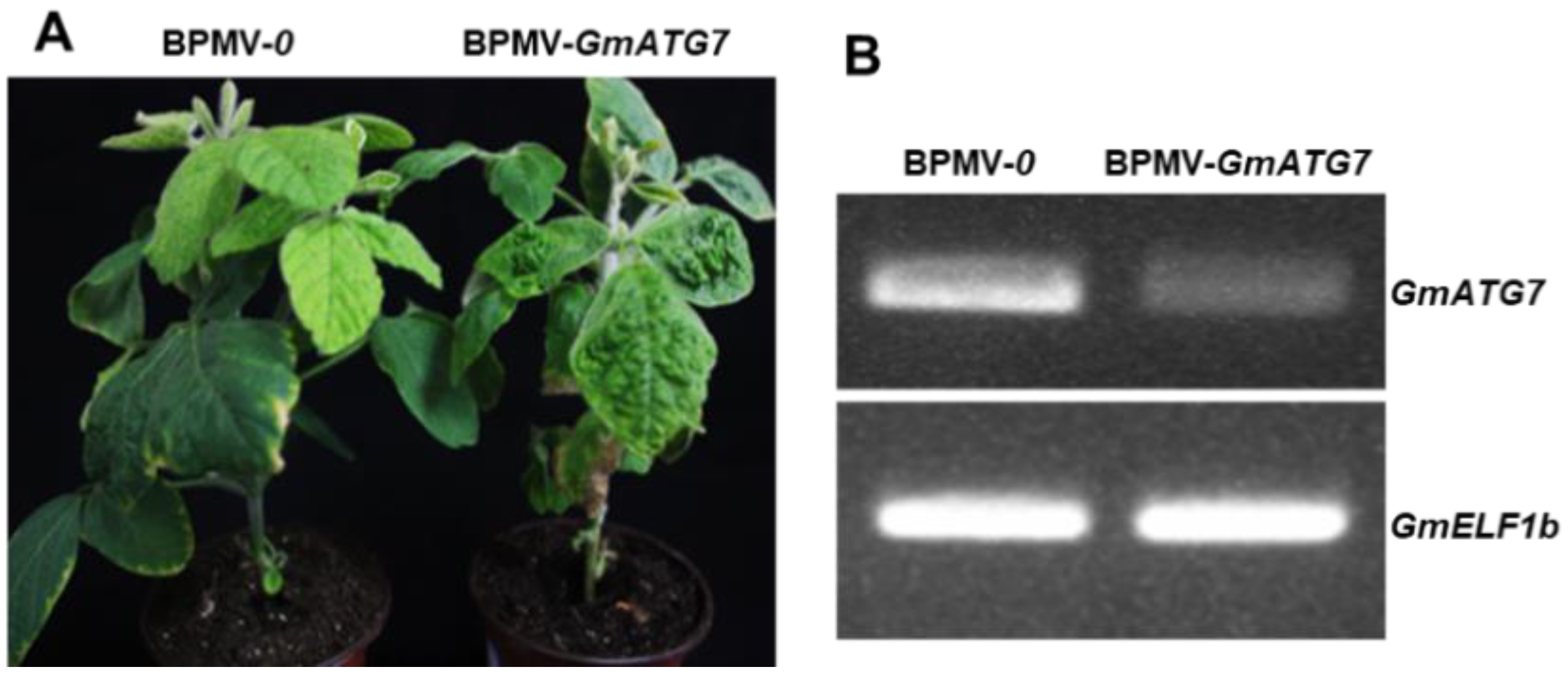
2.1. Silencing GmATG7 Does Not Alter the Morphological Phenotype of Soybean Plants

To understand the roles of autophagy in soybeans, a virus-induced gene silencing approach, mediated by the Bean pod mottle virus (BPMV), was used to silence multiple Autophagy-related genes (ATGs), one of which was the ATG7 homologue. The soybean is a paleotetraploid plant; 75% of the genes in its genome have two copies [32]. However, only one ATG7 homologue was identified in the soybean genome; therefore, it was referred to as GmATG7. A 330-bp fragment of GmATG7 was cloned into BPMV2 [33,34] and formed the BPMV2-GmATG7 silencing construct. GmATG7 silencing was achieved via co-bombardment of BPMV1 and BPMV2-GmATG7 plasmids coated on gold particles onto two expanded true leaves of 7-day-old soybean seedlings. Meanwhile, BPMV1 and BPMV2 were co-bombarded onto different seedlings and used as empty vector controls (referred to as BPMV-0). At 15–20 days post bombardment (dpb), BPMV symptoms were visible on the upper systemic leaves, indicating successful infection. Systemic leaves with BPMV symptoms were collected and ground into powders with a pestle and mortar and then resuspended into a phosphate buffer (pH, 7.0). After centrifugation, the saps were collected and rub inoculated onto two true leaves of a batch of 7-day-old seedlings. At 15 days post inoculation (dpi), RNA samples were extracted from these plants for the verification of silencing by RT-PCR. After verification, these plants could be used for subsequent experiments. No obvious differences were observed between the GmATG7-silenced plants and the empty vector control plants (Figure 1A), indicating that silencing GmATG7 did not affect growth and development in soybeans. A RT-PCR analysis showed that the GmATG7 was indeed silenced, as its transcript level was significantly reduced in the GmATG7-silenced plants relative to the vector control plants (Figure 1B).

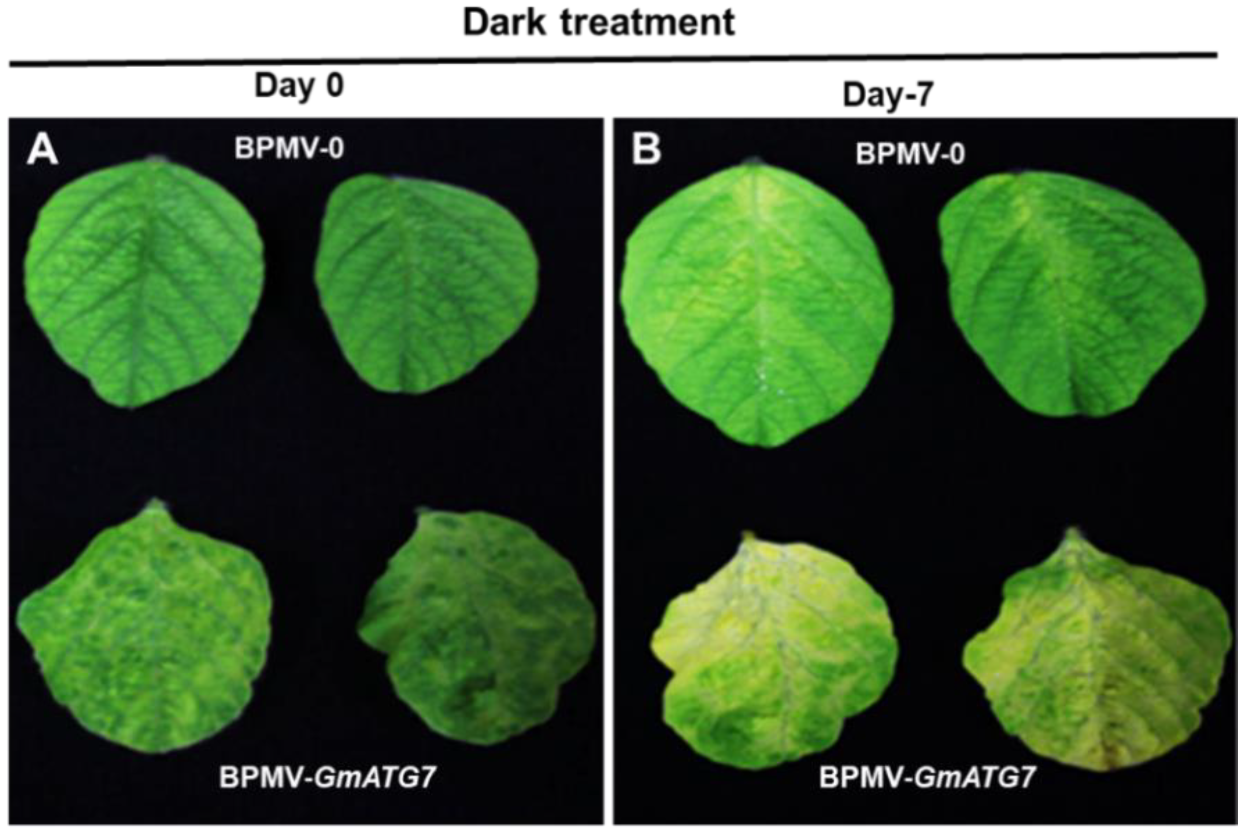
2.2. Silencing GmATG7 Accelerates Leaf Senescence of Soybean Plants under Dark Condition

Under natural conditions, various Arabidopsis atg mutants exhibit an accelerated senescence phenotype [30,35,36,37,38,39]. To examine whether the GmATG7-silenced soybean plants shared a similar phenotype, the leaves from BPMV-0 and GmATG7-silenced plants were collected and dark treated in a Petri dish wrapped with aluminum foil. Long-term dark treatment results in carbon deficiency because of the lack of photosynthesis, which induces autophagy and accelerates senescence. As anticipated, GmATG7-silenced leaves displayed a chlorosis phenotype that was not observed in BPMV-0 leaves after the dark treatment for 7 days (Figure 2). This result demonstrated that silencing GmATG7 leads to autophagy impairment.

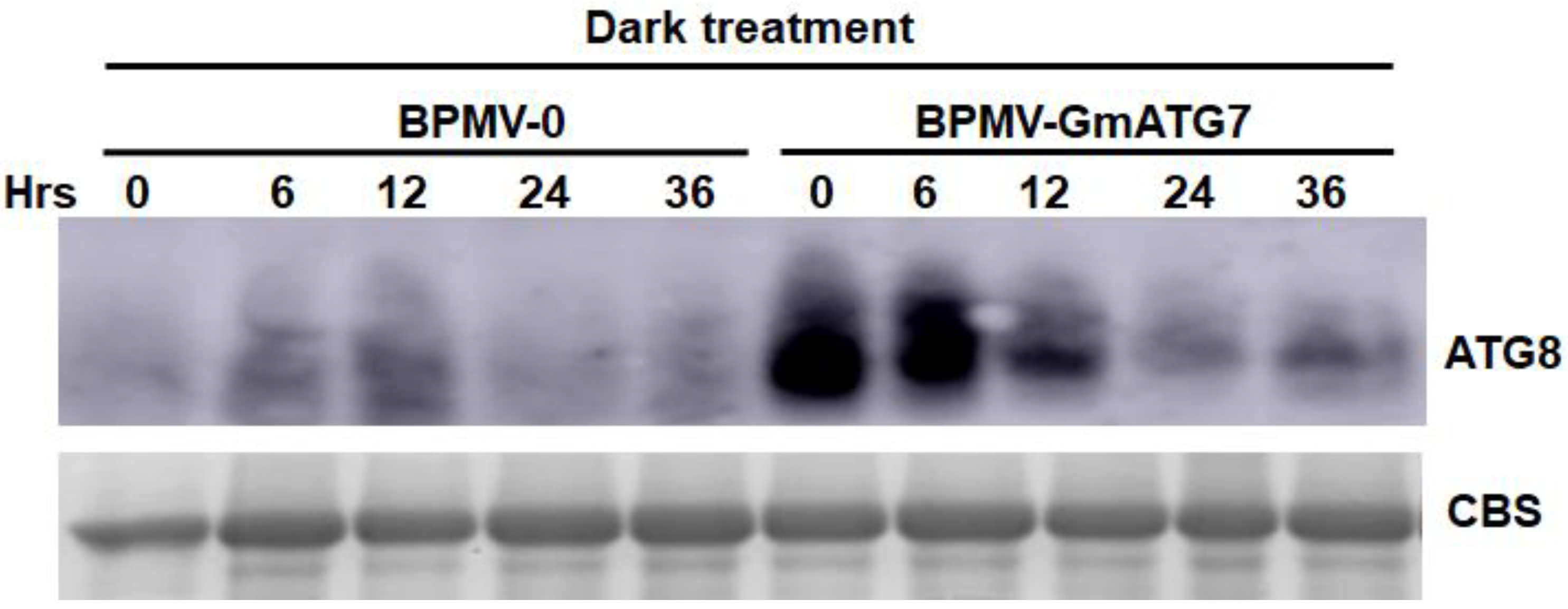
2.3. Silencing GmATG7 Results in Over-Accumulation of GmATG8 in the Soybean Leaves

One of the consequences of compromised autophagy is the over-accumulation of ATG8 and other autophagy-related proteins. To test this, Western blotting was performed for the protein samples prepared from dark-treated empty control leaves and GmATG7-silenced leaves using an antibody specific to Arabidopsis ATG8, which has been shown to cross-react with the soybean ATG8 [40]. As expected, the accumulation level of GmATG8 was much higher in the GmATG7-silenced leaves than in the empty control leaves (Figure 3), indicating again that silencing GmATG7 impairs autophagy in soybeans.

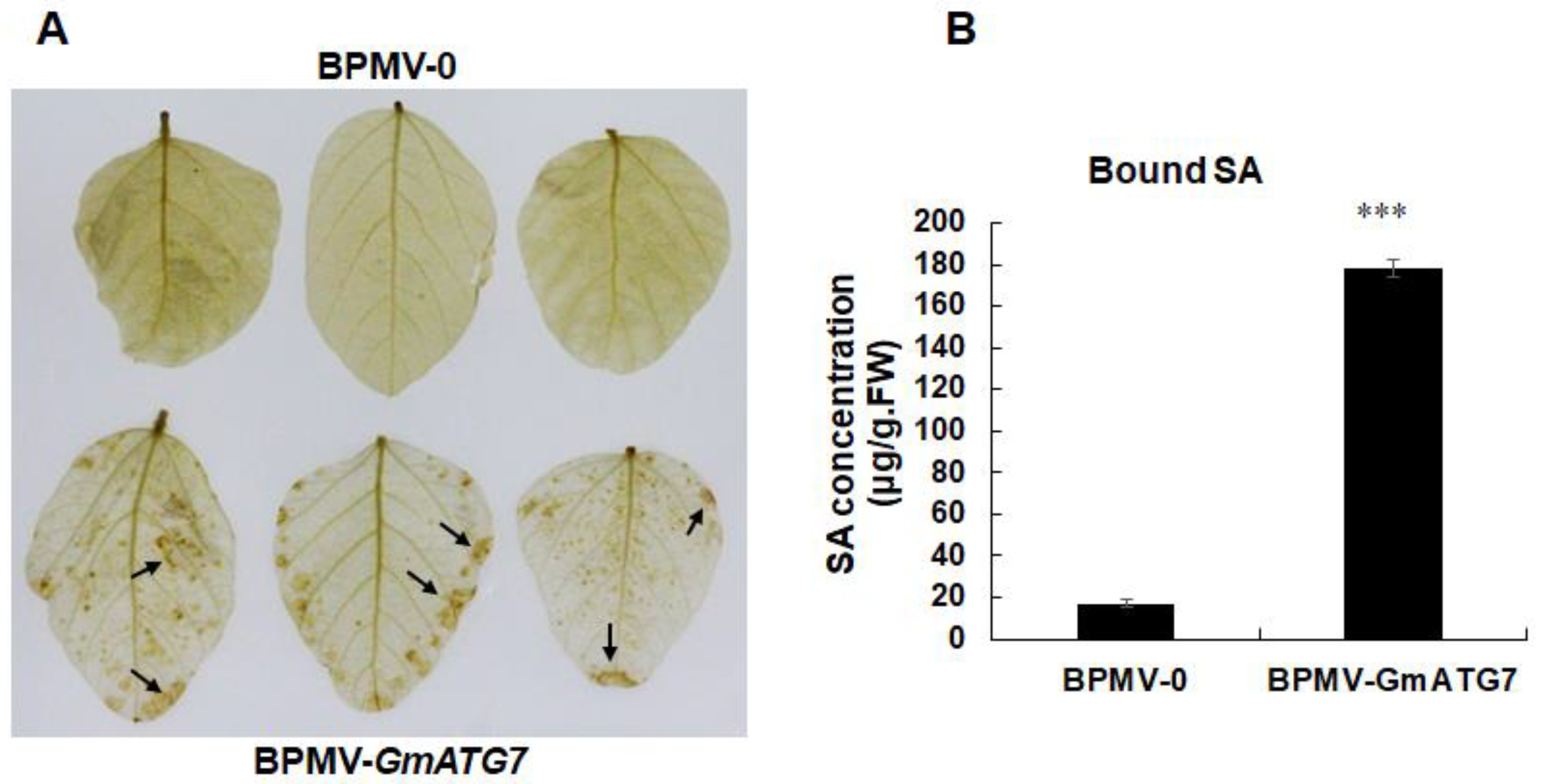
2.4. Silencing GmATG7 Leads to Over-Accumulation of Both H2O2 and Salicylic Acid (SA)

One of the characteristics of Arabidopsis atg mutants is constitutive activated defense responses such as over-accumulation of H2O2 and SA [30,39,41,42]. To test whether the GmATG7-silenced plant shares a similar activated defense response, H2O2 accumulation was firstly visualized for the dark-treated BPMV-0 leaves and GmATG7-silenced leaves using DBA staining. As shown in Figure 4A, the accumulation level of H2O2 was significantly higher in the leaves of the GmATG7-silenced plants than in those of the BPMV-0. In addition, SA accumulation was induced to a much higher level in the leaves of the GmATG7-silenced plants than in those of the BPMV-0 (Figure 4B), indicating that silencing GmATG7 results in constitutive activated defense responses as a result of impaired autophagy.

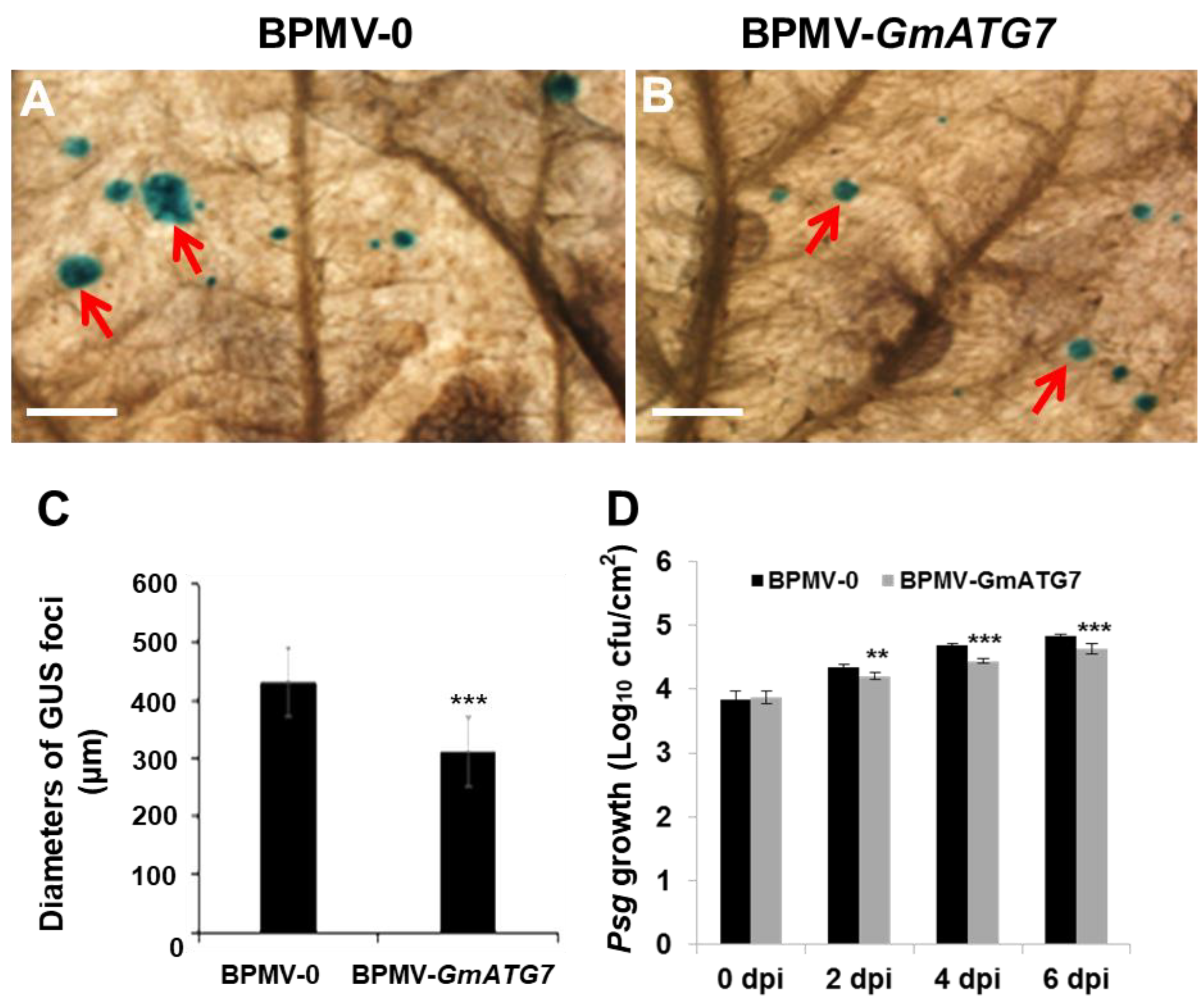
2.5. Silencing GmATG7 Leads to Enhanced Resistance against Soybean mosaic virus (SMV) and Pseudomonas syringae pv. glycinea (Psg)

The results shown above (Figure 4) demonstrated that silencing GmATG7 resulted in the activation of defense responses, which are usually positively correlated with enhanced disease resistance. To test the correlation of the activated defense responses observed in the GmATG7-silenced plants with disease resistance, we first infected the leaves collected from both the vector control plants and the GmATG7-silenced plants with an SMV strain tagged with the GUS reporter gene (SMV-N-GUS) [43] via biolistic particle bombardment. At 3 days post bombardment, the bombarded leaves were incubated with X-Gluc, a substrate of GUS, to visualize the infection foci. A blue GUS foci indicated successful infection, and the diameters of the GUS foci reflect the ability of the replication and cell-to-cell movement of the SMV-N-GUS. As shown in Figure 5A–C, the diameters of the GUS foci on the leaves of the GmATG7-silenced plants were much smaller than those on the BPMV-0 leaves, indicating that silencing GmATG7 leads to an enhanced resistance of soybean plants to SMV.
Next, the BPMV-0 and the GmATG7-silenced plants were individually spray-inoculated with Psg. The upper and lower ends of the leaves were evenly sprayed with a Psg solution. To keep moisture and facilitate infection, all of the plants were then covered with transparent plastic bags after spraying. On different days post inoculation (dpi), leaf discs were collected from the infected leaves for the determination of colony-forming units (cfu). As shown in Figure 5D, the multiplication of Psg was significantly lower in the GmATG7-silenced leaves than in the BPMV-0 leaves, indicating that silencing GmATG7 results in an enhanced resistance of soybean plants to Psg. Collectively, these results demonstrate that GmATG7 plays a negative role in soybean immunity.

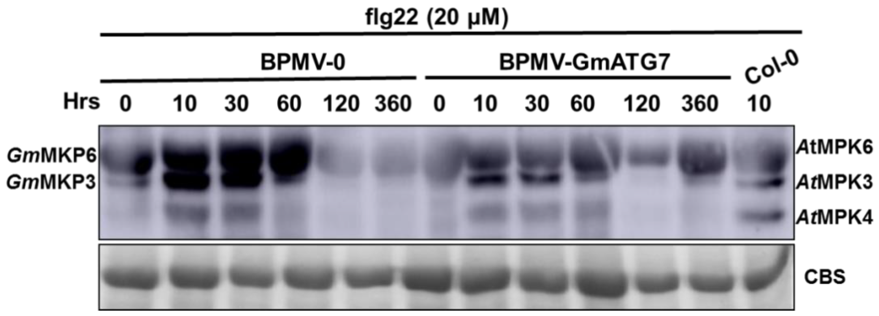
2.6. Silencing GmATG7 Leads to a Reduced Activation of GmMPK3/6 in Response to flg22 Treatment

It has been previously shown that the MAPK signaling pathway is not affected by the loss of ATG function [29]. To examine the effect of GmATG7 silencing on the flg22-induced activation of MAPK, the leaves collected from the GmATG7-silenced plants as well as from the BPMV-0 plants were treated with 20 μM/L flg22 for 0–360 h. Western blotting was performed on the protein samples prepared from these treated leaves using a p44/42 MAP Erk1/2 antibody, which specifically recognizes the phosphorylated forms of MPK3/6. As shown in Figure 6, flg22-induced activation in the GmATG7-silenced leaves was significantly reduced relative to those in the BPMV-0 plants, suggesting that the activated defense responses observed in the GmATG7-silenced plants (Figure 4 and Figure 5) is independent of the activation of GmMPK3/6 and GmATG7 may play a negative role in the flg22-induced activation of GmMPK3/6.

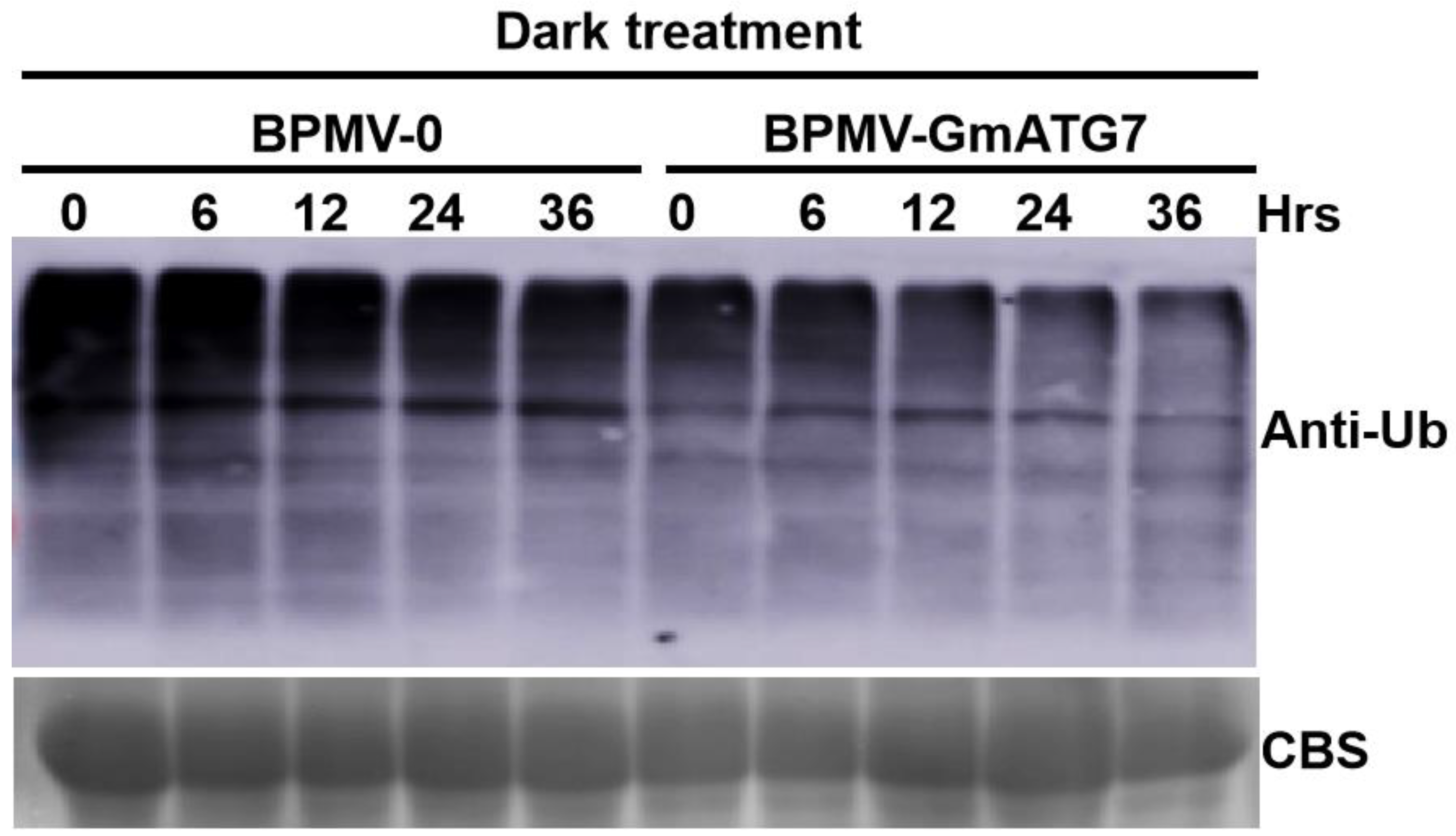
2.7. Silencing GmATG7 Leads to a Reduced Accumulation Level of Ubiquitinated Proteins

Under severe stress conditions, ubiquitinated proteins or protein aggregates, and even the 26S proteasomes, are degraded in vacuoles through the autophagy pathway [44,45]. Ubiquitinated proteins are over-accumulated in various Arabidopsis atg mutants due to impaired autophagy [24,46]. Therefore, the accumulation level of ubiquitinated proteins is used as an important indicator to judge whether the autophagy pathway is functional [2,15]. To examine the effect of GmATG7 silencing on the cellular level of ubiquitinated proteins in soybeans, Western blotting was separately performed for protein samples extracted from the dark-treated leaves of the GmATG7-silenced plants and the BPMV-0 plants using an antibody raised against the Arabidopsis ubiquitin. Contrary to our expectations, a significantly reduced level of ubiquitinated proteins was observed in the GmATG7-silenced plants relative to the BPMV-0 plants (Figure 7), implying that GmATG7 plays a negative role in regulating the level of ubiquitinated proteins. However, the molecular mechanism underpinning this phenomenon is not understood.

3. Discussion

The soybean is a paleotetraploid plant, and 75% of the genes in its genome have two copies due to a duplication event [32]. Consistently, all of the genes we previously studied have two copies [47,48,49,50,51,52,53,54]. However, there is only one ATG7 homolog in soybeans, suggesting that either GmATG7 was not duplicated, or the other copy was lost after duplication during the course of evolution. As an ATP-dependent E1-activating enzyme, ATG7 plays a critical role in the lipidation of ATG8 (ATG8-PE) and insertion of ATG8-PE on both the outer and inner membranes of autophagosomes [2]. The anchoring of ATG8 to autophagosomal membranes is not only important for the autophagosome biogenesis but also for the selective degradation of proteins that interact with ATG8 through an ATG8-interacting motif (AIM). Selective autophagy is involved in various biological processes such as biotic and abiotic stress responses, drought tolerance and hormone signaling [2,18]. In the absence of ATG7, the lipidation of ATG8 and subsequent recruitment of ATG8-PE to the membranes of autophagosomes is disrupted, leading to impaired autophagy.
In plants, it has been reported that autophagy plays important roles in nutrient recycling, growth and development, senescence, biotic and abiotic stress responses [2,4,11,15,16,18]. Autophagy has been intensively studied in model plant Arabidopsis. However, it has not been studied in soybeans until recently. In this study, GmATG7 was successfully silenced in soybeans using a BPMV-VIGS system (Figure 1). Under natural conditions, accelerated senescence and activated defense responses are reported for the Arabidopsis atg mutants [30,35,36,37,38,39]. However, the accelerated senescence and activated defense responses were not observed for the GmATG7-silenced soybean plants (Figure 1A). This could be due to the fact that the silencing efficacy mediated by the BPMV-VIGS system is not 100%. The silencing efficiency of BPMV-VIGS is usually 80–90% [47]. The remaining unsilenced 10–20% of GmATG7 transcript in the GmATG7-silenced plants is sufficient to maintain the basal level autophagy required under normal growth conditions. However, under autophagy induction conditions, the remaining 10–20% of GmATG7 is not sufficient to assemble enough autophagosomes. As a result, an accelerated senescence or early chlorosis was observed for the dark-treated leaves of the GmATG7-silenced plants (Figure 2).
As an essential part of the autophagosome, ATG8 proteins play critical roles not only in autophagosome biogenesis but also in mediating the selective autophagy for a variety of proteins [2,18]. Along with the autophagosomes, ATG8 proteins are destined to vacuoles and ultimately degraded inside vacuoles [2]. Therefore, ATG8 proteins are over-accumulated in the Arabidopsis atg mutants [2]. Consistent with this, GmATG8 proteins were also over-accumulated in the GmATG7-silenced plants (Figure 3), indicating that silencing GmATG7 indeed resulted in the impairment of the autophagy pathway in soybeans.
The over-accumulation of cellular reactive oxygen species (ROS) and SA is one of the primary reasons of plant senescence and defense responses. One of the functions of autophagy is to maintain the homeostasis of cellular ROS through eliminating damaged organelles such as mitochondria, chloroplasts and peroxisomes, from which the vast majority of stress-induced ROS is derived [18,23,55,56,57]. If autophagy is functional, ROS-induced senescence could be prevented or at least delayed. Concurrently, H2O2 was over-accumulated in the GmATG7-silenced plants (Figure 4), which could explain why accelerated senescence was observed in the GmATG7-silenced plants (Figure 2). It has been known for decades that ROS and SA can induce cell death and immune responses and play critical roles in disease resistance [58,59,60]. The activated defense responses observed in the GmATG7-silenced plants (Figure 4B) could be a result of the over-accumulation of both ROS and SA (Figure 4A). Consistent with the activated defense responses, GmATG7-silenced plants separately exhibited enhanced resistance to the viral pathogen SMV-N-GUS and the bacterial pathogen Psg (Figure 5). These results are also consistent with the results from Arabidopsis, which indicated that atg mutants display enhanced resistance to biotrophic, bacterial and fungal pathogens. Together, these results suggest that the function of autophagy is conserved between soybeans and Arabidopsis.
The loss function of ATG2 does not have any effect on the activation of MAK/3/6 [29]. Silencing GmATG2a/2b in soybeans resulted in a reduced activation of GmMPK3/6 induced by flg22 [40]. However, the flg22-induced activation of GmMPK3/6 was elevated in the GmATG10a/10b-silenced plants [54]. These results indicated that Arabidopsis ATG2 and soybean GmATG2 have different effects on the activation MAPK signaling pathway, and different soybean ATG proteins have opposite effects on the activation of GmMPK3/6. Similar to GmATG2a/2b silencing [40], silencing GmATG7 resulted in reduction in flg22-induced GmMPK3/6 activation (Figure 6), suggesting that GmATG7 plays a positive regulatory role in GmMPK3/6 activation. Meanwhile, these results also indicated that the enhanced defense responses and elevated disease resistance observed in the GmATG7-silenced plants (Figure 4 and Figure 5) are independent of GmMPK3/6 activation.
Under stress conditions, ubiquitinated proteins are eliminated by the autophagy pathway [2,15,18]. Impaired autophagy results in an over-accumulation of ubiquitinated proteins. As a result, ubiquitinated proteins are over-accumulated in Arabidopsis atg mutants and GmATG2a/2b-silenced soybean plants [24,27,38,40,46,61]. Contrary to this, silencing GmATG7 resulted in a reduced accumulation of ubiquitinated proteins (Figure 7), suggesting that GmATG7 might have a role in interfering with the ubiquitination process and thus leads to a reduced level of ubiquitinated proteins. The other possibility is that silencing GmATG7 could increase the function of the UPS-26S proteasome pathway, either at a transcription level or at a post-translational level, leading to a reduced accumulation of ubiquitinated proteins. The molecular mechanism underpinning this unexpected result remains to be elucidated.
In conclusion, our results demonstrated that the function of GmATG7 is required for the autophagy pathway in soybeans, and the activated MPK3/MPK6-independent immunity in the GmATG7-silenced plants is a result of impaired autophagy. The direction of future work is to reveal which components and signaling pathways are involved in the activated immunity of the GmATG7-silenced plants.

4. Materials and Methods

4.1. Materials

4.1.1. Soybean Cultivar

The seeds of the soybeans (Glycine max L. ‘Williams 82’) used in this study were harvested from greenhouse-grown plants previously indexed for the absence of BPMV and SMV [47,48]. The soybean plants were grown in a growth chamber at 22 degrees with a photoperiod of 16 h.

4.1.2. Bacterial Stria (Escherichia coli)

DH5α/TOP10 and Pseudomonas syringae pv. glycinea (Psg) R4 strain.

4.1.3. Soybean mosaic virus Strain

SMV-N-GUS [43].

4.2. flg22 Peptides

The flg22 peptide was purchased from HuaBio (Hangzhou, China).

4.3. BPMV-Mediated VIGS

The BPMV-VIGS system and the inoculation of soybean seedlings with a constructed BPMV-GmATG7 vector via biolistic particle bombardments using a Biolistic PDS1000/He system (Bio-Rad Laboratories, Hercules, CA, USA) have been previously described [33,34]. The primers used for the construction of the BPMV-GmATG7 (Glyma.12g010000) are as follows: GmATG7-F: GGATCCATAACAGTTCCACAAGGGTG; ATG7-R: GGTACCCCTGCCTCAATGGGTTAGACAT. The bold sequences are the BamH I and Kpn I restriction sites, which were used for directional cloning.

4.4. RNA Isolation and RT-PCR

RNA isolation and RT-PCR were performed as described elsewhere. These are the primers used for the verification of the silencing effect: GmATG7-V-F: TCACAAGATCCTTTTTGGATTTTA; GmATG7-V-R: TCAATTAAATCACACAAACGTTTAC; These are the primers for GmPR1: GmPR1-F: ATGGGGTTGTGCAAGGTT; GmPR1-R: CTAGTAGGGTCTTTGGCCAA; These are the primers used for the reference gene: GmELF1b-F: GAGCTATGAATTGCCTGATGG; GmELF1b-R: CGTTTCATGAATTCCAGTAGC.

4.5. H2O2 Detection by DAB Staining

H2O2 was detected using the DAB staining method (Sigma-Aldrich, St. Louis, MO, USA), as previously described [62].

4.6. Inoculation of Pseudomonas syringae pv. glycinea (Psg)

A Psg growth assay was performed as described by [52]. Psg was cultivated at 28 degrees to OD = 1.3. The bacterial culture was spun down, and the pellet was re-suspended in an LB medium with an OD = 1. The vector control or GmATG7-silenced plants were sprayed with the re-suspended bacterial solution on both sides. The sprayed plants were immediately covered with transparent plastic bags to maintain moisture and facilitate infection.

4.7. SMV-N-GUS Inoculation, GUS Staining, and GUS Foci Measurements

At 18 dpi infected with only the BPMV vector (BPMV-0) or BPMV-GmATG7 constructs, a second fully expanded soybean trifoliolate leaf, counted from the top, was detached and biolistically inoculated with SMV-N-GUS [51]. Following SMV-N-GUS inoculation, the detached leaves were put into Petri dishes with moist filter papers and kept on a lit growth shelf for 5 days before GUS staining. GUS staining was performed as described by [63]. Photographs of the leaves with GUS foci were taken using a stereo microscope (Olympus SZH10, Center Valley, PA, USA). The numbers of GUS foci were counted, and the diameters of GUS foci were measured using Soft Image System analysis (IA Package; Olympus).

4.8. Western Blotting Analysis

Proteins were extracted from dark-treated BPMV-0 and BPMV-GmATG7 leaves. Western blotting was performed using Anti-Ubq (Agrisera, AS08307, Vännäs, Sweden) or anti-ATG8 (Agrisera, AS142769), as previously described [51].

4.9. GmMPK3/GmMPK6 Kinase Activation Assay

Protein samples were extracted from leaf tissues of indicated soybean plants in the extraction buffer (50 mM Tris-MES pH 8.0, 0.5 M sucrose, 1 mM MgCl2, 10 mM EDTA, 5 mM DTT) in the presence of protease inhibitor cocktail S8830 (Sigma-Aldrich, St. Louis, MO, USA), as described [51]. The protein extracts were spun at 12,000 rpm at 4 °C for 30 min, and the supernatants were collected for Western blotting analysis. Protein separation was conducted using SDS-PAGE; then they were transferred to a PVDF membrane (Millipore, Billerica, MA, USA) via semi-dry electro-transfer (Bio-Rad, Hercules, CA, USA) in accordance with the manufacturer’s manual. The membrane was blocked for 2 h in a 1× TBST buffer containing 5% milk powder. After washing 3 times, the membrane was incubated with human-derived anti–Phospho-p44/p42 MAPK (anti-pTEpY), which specifically recognizes phosphorylated MPK3/MPK4/MPK6 across kingdoms, and then diluted at 1:3000 (Cell Signaling Technology, Danvers, MA, USA), followed by incubation with a secondary antibody. The bands were visualized via incubation with a chemiluminescent HRP substrate (Millipore, Billerica, MA, USA).

4.10. Statistical Analysis

Each experiment was repeated three times, and the representative results are presented. Statistical analyses were performed using SPSS Statistics 27 (Statistical Product and Service Solutions, IBM, Armonk, NY, USA). The test methods are indicated in the figure legends.

Author Contributions

J.-Z.L. designed the experiments, and J.-Z.L. wrote the manuscript; S.M.H., M.-J.H., W.-X.W., T.-Y.L., Y.C., L.-N.L. and H.-J.L. performed the experiments. M.Q.A. revised the manuscript, S.M.H. and J.-Z.L. prepared the figures. All authors have read and agreed to the published version of the manuscript.

Funding

This research was funded by the National Natural Science Foundation of China (grant number 32170761 to J.-Z.L.).

Institutional Review Board Statement

Not applicable.

Informed Consent Statement

Not applicable.

Data Availability Statement

Data are contained within the article.

Acknowledgments

We thank Steven Whitham and John Hill for kindly providing the BPMV-VIGS system, SMV-N-GUS and Pseudomonas syringae pv. glycinea (Psg), R4 strain.

Conflicts of Interest

The authors declare no conflict of interest.

References

  1. Mizushima, N. Autophagy. FEBS Lett. 2010, 584, 1279. [Google Scholar] [CrossRef] [PubMed]
  2. Li, F.; Vierstra, R.D. Autophagy: A multifaceted intracellular system for bulk and selective recycling. Trends Plant Sci. 2012, 17, 526–537. [Google Scholar] [CrossRef] [PubMed]
  3. Liu, Y.; Bassham, D.C. Autophagy: Pathways for self-eating in plant cells. Annu. Rev. Plant Biol. 2012, 63, 215–237. [Google Scholar] [CrossRef]
  4. Avin-Wittenberg, T. Autophagy and its role in plant abiotic stress management. Plant Cell Environ. 2019, 42, 1045–1053. [Google Scholar] [CrossRef]
  5. Qi, H.; Xia, F.N.; Xiao, S. Autophagy in plants: Physiological roles and post-translational regulation. J. Integr. Plant Biol. 2020, 63, 161–179. [Google Scholar] [CrossRef]
  6. Suzuki, K.; Kirisako, T.; Kamada, Y.; Mizushima, N.; Noda, T.; Ohsumi, Y. The pre-autophagosomal structure organized by concerted functions of APG genes is essential for autophagosome formation. EMBO J. 2001, 20, 5971–5981. [Google Scholar] [CrossRef] [PubMed]
  7. Mizushima, N.; Komatsu, M. Autophagy: Renovation of cell1 and tissues. Cell 2011, 147, 728–741. [Google Scholar] [CrossRef]
  8. Lamb, C.A.; Dooley, H.C.; Tooze, S.A. Endocytosis and autophagy: Shared machinery for degradation. Bioessays 2013, 35, 34–45. [Google Scholar]
  9. Liu, Y.; Schiff, M.; Czymmek, K.; Tallóczy, Z.; Levine, B.; Dinesh-Kumar, S.P. Autophagy regulates programmed cell death during the plant innate immune response. Cell 2005, 121, 567–577. [Google Scholar] [CrossRef]
  10. Hofius, D.; Schultz-Larsen, T.; Joensen, J.; Tsitsigiannis, D.I.; Petersen, N.H.T.; Mattsson, O.; Jorgensen, L.B.; Jones, J.D.G.; Mundy, J.; Petersen, M. Autophagic components contribute to hypersensitive cell death in Arabidopsis. Cell 2009, 137, 773–783. [Google Scholar] [CrossRef]
  11. Hofius, D.; Li, L.; Hafren, A.; Coll, N.S. Autophagy as an emerging arena for plant–pathogen interactions. Curr. Opin. Plant Biol. 2017, 8, 117–123. [Google Scholar] [CrossRef]
  12. Üstün, S.; Hafrén, A.; Hofius, D. Autophagy as a mediator of life and death in plants. Curr. Opin. Plant Biol. 2017, 40, 122–130. [Google Scholar] [CrossRef] [PubMed]
  13. Xu, G.; Wang, S.; Han, S.; Xie, K.; Wang, Y.; Li, J.; Liu, Y. Plant Bax inhibitor-1 interacts with ATG6 to regulate autophagy and programmed cell death. Autophagy 2017, 13, 1161–1175. [Google Scholar] [CrossRef]
  14. Ding, X.; Zhang, X.; Otegui, M.S. Plant autophagy: New flavors on the menu. Curr. Opin. Plant Biol. 2018, 46, 113–121. [Google Scholar] [CrossRef] [PubMed]
  15. Marshall, R.S.; Vierstra, R.D. Autophagy: The master of bulk and selective recycling. Annu. Rev. Plant Biol. 2018, 69, 173–208. [Google Scholar] [CrossRef] [PubMed]
  16. Leary, A.Y.; Savage, Z.; Tumtas, Y.; Bozkurt, T.O. Contrasting and emerging roles of autophagy in plant immunity. Curr. Opin. Plant Biol. 2019, 52, 46–53. [Google Scholar] [CrossRef] [PubMed]
  17. Yang, M.; Ismayil, A.; Liu, Y. Autophagy in Plant-Virus Interactions. Annu. Rev. Virol. 2020, 7, 403–419. [Google Scholar] [CrossRef]
  18. Ran, J.; Hashimi, S.M.; Liu, J.Z. Emerging Roles of the Selective Autophagy in Plant Immunity and Stress. Int. J. Mol. Sci. 2020, 21, 6321. [Google Scholar] [CrossRef]
  19. Ohsumi, Y. Molecular dissection of autophagy: Two ubiquitin-like systems. Nat. Rev. Mol. Cell Biol. 2011, 2, 211–216. [Google Scholar] [CrossRef]
  20. Noda, N.N.; Ohsumi, Y.; Inagaki, F. Atg8-family interacting motif crucial for selective autophagy. FEBS Lett. 2010, 584, 1379–1385. [Google Scholar] [CrossRef]
  21. Johansen, T.; Lamark, T. Selective autophagy mediated by autophagic adapter proteins. Autophagy 2011, 7, 279–296. [Google Scholar] [CrossRef] [PubMed]
  22. Thompson, A.R.; Vierstra, R.D. Autophagic recycling: Lessons from yeast help define the process in plants. Curr. Opin. Plant Biol. 2005, 8, 165–173. [Google Scholar] [CrossRef]
  23. Luo, M.; Zhuang, X. Review: Selective degradation of peroxisome by autophagy in plants: Mechanisms, functions, and perspectives. Plant Sci. 2018, 274, 485–491. [Google Scholar] [CrossRef]
  24. Zhou, J.; Zhang, Y.; Qi, J.; Chi, Y.; Fan, B.; Yu, J.Q.; Chen, Z. E3 ubiquitin ligase CHIP and NBR1-mediated selective autophagy protect additively against proteotoxicity in plant stress responses. PLoS Genet. 2014, 10, e1004116. [Google Scholar] [CrossRef] [PubMed]
  25. Zhou, J.; Wang, J.; Yu, J.Q.; Chen, Z. Role and regulation of autophagy in heat stress responses of tomato plants. Front. Plant Sci. 2014, 5, 174. [Google Scholar] [CrossRef]
  26. Zhou, J.; Wang, Z.; Wang, X.; Li, X.; Zhang, Z.; Fan, B.; Zhu, C.; Chen, Z. Dicot-specific ATG8-interacting ATI3 proteins interact with conserved UBAC2 proteins and play critical roles in plant stress responses. Autophagy 2018, 14, 487–504. [Google Scholar] [CrossRef]
  27. Shibata, M.; Oikawa, K.; Yoshimoto, K.; Kondo, M.; Mano, S.; Yamada, K.; Hayashi, M.; Sakamoto, W.; Ohsumi, Y.; Nishimura, M. Highly oxidized peroxisomes are selectively degraded via autophagy in Arabidopsis. Plant Cell 2013, 25, 4967–4983. [Google Scholar] [CrossRef]
  28. Kim, J.; Lee, H.N.; Chung, T. Plant cell remodeling by autophagy: Switching peroxisomes for green life. Autophagy 2014, 10, 702–703. [Google Scholar] [CrossRef]
  29. Lenz, H.D.; Haller, E.; Melzer, E.; Kober, K.; Wurster, K.; Stahl, M.; Bassham, D.C.; Vierstra, R.D.; Parker, J.E.; Bautor, J.; et al. Autophagy differentially controls plant basal immunity to biotrophic and necrotrophic pathogens. Plant J. 2011, 66, 818–830. [Google Scholar] [CrossRef] [PubMed]
  30. Wang, Y.; Nishimura, M.T.; Zhao, T.; Tang, D. ATG2, an autophagy related protein, negatively affects powdery mildew resistance and mildew induced cell death in Arabidopsis. Plant J. 2012, 68, 74–87. [Google Scholar] [CrossRef]
  31. Zhang, T.; Xiao, Z.; Liu, C.; Yang, C.; Li, J.; Li, H.; Gao, C.; Shen, W. Autophagy Mediates the Degradation of Plant ESCRT Component FREE1 in Response to Iron Deficiency. Int. J. Mol. Sci. 2021, 22, 8779. [Google Scholar] [CrossRef]
  32. Schmutz, J.; Cannon, S.B.; Schlueter, J.; Ma, J.; Mitros, T.; Nelson, W.; Hyten, D.L.; Song, Q.; Thelen, J.J.; Cheng, J.; et al. Genome sequence of the palaeopolyploid soybean. Nature 2010, 463, 178–183. [Google Scholar] [CrossRef]
  33. Zhang, C.; Yang, C.; Whitham, S.A.; Hill, J.H. Development and use of an efficient DNA-based viral gene silencing vector for soybean. Mol. Plant Microbe Interact. 2009, 22, 123–131. [Google Scholar] [CrossRef] [PubMed]
  34. Zhang, C.; Bradshaw, J.D.; Whitham, S.A.; Hill, J.H. The development of an efficient multipurpose bean pod mottle virus viral vector set for foreign gene expression and RNA silencing. Plant Physiol. 2010, 153, 52–65. [Google Scholar] [CrossRef]
  35. Doelling, J.H.; Walker, J.M.; Friedman, E.M.; Thompson, A.R.; Vierstra, R.D. The APG8/12-activating enzyme APG7 is required for proper nutrient recycling and senescence in Arabidopsis thaliana. J. Biol. Chem. 2002, 277, 33105–33114. [Google Scholar] [CrossRef] [PubMed]
  36. Hanaoka, H.; Noda, T.; Shirano, Y.; Kato, T.; Hayashi, H.; Shibata, D.; Tabata, S.; Ohsumi, Y. Leaf senescence and starvation-induced chlorosis are accelerated by the disruption of an Arabidopsis autophagy gene. Plant Physiol. 2002, 129, 1181–1193. [Google Scholar] [CrossRef]
  37. Xiong, Y.; Contento, A.L.; Bassham, D.C. AtATG18a is required for the formation of autophagosomes during nutrient stress and senescence in Arabidopsis thaliana. Plant J. 2005, 42, 535–546. [Google Scholar] [CrossRef]
  38. Xiong, Y.; Contento, A.; Nguyen, P.Q.; Bassham, D.C. Degradation of oxidized proteins by autophagy during oxidative stress in Arabidopsis. Plant Physiol. 2007, 143, 291–299. [Google Scholar] [CrossRef]
  39. Yoshimoto, K.; Jikumaru, Y.; Kamiya, Y.; Kusano, M.; Consonni, C.; Panstruga, R.; Ohsumi, Y.; Shirasu, K. Autophagy negatively regulates cell death by controlling NPR1-dependent salicylic acid signaling during senescence and the innate immune response in Arabidopsis. Plant Cell 2009, 21, 2914–2927. [Google Scholar] [CrossRef] [PubMed]
  40. Hashimi, S.; Wu, N.N.; Ran, J.; Liu, J.Z. Silencing autophagy-related gene 2 (ATG2) results in accelerated senescence and enhanced immunity in soybean. Int. J. Mol. Sci. 2021, 22, 11749. [Google Scholar] [CrossRef]
  41. Hayward, A.P.; Tsao, J.; Dinesh-Kumar, S.P. Autophagy and plant innate immunity: Defense through degradation. Semin. Cell Dev. Biol. 2009, 20, 1041–1047. [Google Scholar] [CrossRef] [PubMed]
  42. Yamauchi, S.; Mano, S.; Oikawa, K.; Hikino, K.; Teshima, K.M.; Kimori, Y.; Nishimura, M.; Shimazaki, K.I.; Takemiya, A. Autophagy controls reactive oxygen species homeostasis in guard cells that is essential for stomatal opening. Proc. Natl. Acad. Sci. USA 2019, 116, 19187–19192. [Google Scholar] [CrossRef] [PubMed]
  43. Wang, L.; Eggenberger, A.; Hill, J.; Bogdanove, A.J. Pseudomonas syringae effector avrB confers soybean cultivar-specific avirulence on Soybean mosaic virus adapted for transgene expression but effector avrPto does not. Mol. Plant-Microbe Interact. 2006, 19, 304–312. [Google Scholar] [CrossRef] [PubMed]
  44. Marshall, R.S.; Li, F.; Gemperline, D.C.; Book, A.J.; Vierstra, R.D. Autophagic degradation of the 26S proteasome is mediated by the dual ATG8/Ubiquitin receptor RPN10 in Arabidopsis. Mol. Cell 2015, 58, 1053–1066. [Google Scholar] [CrossRef] [PubMed]
  45. Marshall, R.S.; Hua, Z.; Mali, S.; McLoughlin, F.; Vierstra, R.D. ATG8-Binding UIM Proteins Define a New Class of Autophagy Adaptors and Receptors. Cell 2019, 177, 766–781. [Google Scholar] [CrossRef] [PubMed]
  46. Zhou, J.; Wang, J.; Cheng, Y.; Chi, Y.J.; Fan, B.; Yu, J.Q.; Chen, Z. NBR1-mediated selective autophagy targets insoluble ubiquitinated protein aggregates in plant stress responses. PLoS Genet. 2013, 9, e1003196. [Google Scholar] [CrossRef]
  47. Liu, J.Z.; Graham, M.A.; Pedley, K.F.; Whitham, S.A. Gaining insight into soybean defense responses using functional genomics approaches. Brief. Funct. Genom. 2015, 14, 283–290. [Google Scholar] [CrossRef]
  48. Liu, J.Z.; Fang, Y.; Pang, H. The current status of the soybean-soybean mosaic virus (SMV) pathosystem. Front. Microbiol. 2016, 7, 1906. [Google Scholar] [CrossRef]
  49. Liu, J.Z.; Braun, E.; Qiu, W.L.; Shi, Y.F.; Marcelino-Guimars, F.C.; Navarre, D.; Hill, J.H.; Whitham, S.A. Positive and negative roles for soybean MPK6 in regulating defense responses. Mol. Plant Microbe Interact. 2014, 27, 824–834. [Google Scholar] [CrossRef]
  50. Liu, J.Z.; Horstman, H.D.; Braun, E.; Graham, M.A.; Zhang, C.; Navarre, D.; Qiu, W.L.; Lee, Y.; Nettleton, D.; Hill, J.H.; et al. Soybean homologs of MPK4 negatively regulate defense responses and positively regulate growth and development. Plant Physiol. 2011, 157, 1363–1378. [Google Scholar] [CrossRef]
  51. Xu, H.Y.; Zhang, C.; Li, Z.C.; Wang, Z.R.; Jiang, X.X.; Shi, Y.F.; Tian, S.N.; Braun, E.; Mei, Y.; Qiu, W.L.; et al. The MAPK kinase kinase GmMEKK1 regulates cell death and defense responses. Plant Physiol. 2018, 178, 907–922. [Google Scholar] [CrossRef] [PubMed]
  52. Tian, S.N.; Liu, D.D.; Zhong, C.L.; Xu, H.Y.; Yang, S.; Fang, Y.; Ran, J.; Liu, J.Z. Silencing GmFLS2 enhances the susceptibility of soybeanto bacterial pathogen through attenuating the activation of GmMAPK signaling pathway. Plant Sci. 2020, 292, 110386. [Google Scholar] [CrossRef] [PubMed]
  53. Liu, D.D.; Lan, H.J.; Hashimi, S.M.; Ye, M.Y.; Dai, X.Y.; Zhong, C.L.; Tian, S.N.; Liu, J.Z. Silencing GmBIR1 in Soybean Results in Activated Defense Responses. Int. J. Mol. Sci. 2022, 23, 7450. [Google Scholar] [CrossRef] [PubMed]
  54. Zhou, T.; Ye, M.; Liu, T.; Lan, H.; Hashimi, S.M.; Gou, W.; Liu, J. Silencing GmATG10 results in activation of immune responses in soybean. Chin. J. Biotechnol. 2023, 39, 586–602. [Google Scholar] [CrossRef] [PubMed]
  55. Li, F.; Chung, T.; Vierstra, R.D. AUTOPHAGY-RELATED11 plays a critical role in general autophagy- and senescence-induced mitophagy in Arabidopsis. Plant Cell 2014, 26, 788–807. [Google Scholar] [CrossRef]
  56. Xie, Q.; Michaeli, S.; Peled-Zehavi, H.; Galili, G. Chloroplast degradation: One organelle, multiple degradation pathways. Trends Plant Sci. 2015, 20, 264–265. [Google Scholar] [CrossRef]
  57. Young, P.G.; Bartel, B. Pexophagy and peroxisomal protein turnover in plants. Biochim. Biophys. Acta. 2016, 1863, 999–1005. [Google Scholar] [CrossRef]
  58. Lamb, C.; Dixon, R.A. The oxidative burst in plant disease resistance. Annu. Rev. Plant Physiol. Plant Mol. Biol. 1997, 48, 251–275. [Google Scholar] [CrossRef]
  59. Shirasu, K.; Nakajima, H.; Rajasekhar, V.K.; Dixon, R.A.; Lamb, C. Salicylic acid potentiates an agonist-dependent gain control that amplifies pathogen signals in the activation of defense mechanisms. Plant Cell 1997, 9, 261–270. [Google Scholar]
  60. Torres, M.A.; Dangl, J.L.; Jones, J.D.G. Arabidopsis gp91phox homologues AtrbohD and AtrbohF are required for accumulation of reactive oxygen intermediates in the plant defense response. Proc. Natl. Acad. Sci. USA 2002, 99, 517–522. [Google Scholar] [CrossRef] [PubMed]
  61. Munch, D.; Rodriguez, E.; Bressendorff, S.; Park, O.K.; Hofius, D.; Petersen, M. Autophagy deficiency leads to accumulation of ubiquitinated proteins, ER stress, and cell death in Arabidopsis. Autophagy 2014, 10, 1579–1587. [Google Scholar] [CrossRef] [PubMed]
  62. Ren, D.; Yang, H.; Zhang, S. Cell death mediated by MAPK is associated with hydrogen peroxide production in Arabidopsis. J. Biol. Chem. 2002, 277, 559–565. [Google Scholar] [CrossRef] [PubMed]
  63. Jefferson, R.A.; Kavanagh, T.A.; Bevan, M.W. GUS fusions: Beta-glucuronidase as a sensitive and versatile gene fusion marker in higher plants. EMBO J. 1987, 6, 3901–3907. [Google Scholar] [CrossRef] [PubMed]
Figure 1. Silencing GmATG7 did not alter the morphological phenotypes of soybean plants. (A) Phenotypes of the vector control plants (BPMV-0) and the GmATG7-silenced plants (BPMV-GmATG7) under normal growth conditions; (B) The verification of GmATG7 silencing via RT-PCR analysis.
Ijms 24 16508 g001
Figure 2. Silencing GmATG7 resulted in an accelerated senescence phenotype. (A) A comparison of leaf phenotypes between BPMV-0 plants and GmATG7-silenced plants before dark treatment. (B) A comparison of leaf phenotypes between BPMV-0 plants and the GmATG7-silenced plants after 7 d of dark treatment.
Ijms 24 16508 g002
Figure 3. Silencing GmATG7 resulted in an elevated accumulation level of GmATG8. A comparison of GmATG8 protein accumulation level between BPMV-0 control plants and the BPMV-GmATG7-silenced plants treated in the dark for different periods of time.
Ijms 24 16508 g003
Figure 4. Silencing GmATG7 leads to the activation of immune responses. (A) An elevated accumulation level of H2O2 was observed in the GmATG7-silenced plants via DAB staining. The arrows point to the areas of H2O2 accumulation; (B) Bound SA concentration was significantly higher in the GmATG7-silenced plants than in the vector control plants. *** represents p < 0.001, Student’s t-test. Error bars represent standard deviation (SD).
Ijms 24 16508 g004
Figure 5. Silencing GmATG7 leads to the reduced resistance of soybeans to SMV-N-GUS. (A,B). A comparison of the GUS foci on the leaves of the BPMV-0 (left) and the BPMV-GmATG7 plants (right) under microscopy; arrows point to the GUS foci; bar = 5 mm; (C) A comparison of the diameters of the SMV-N-GUS foci on the leaves of BPMV-0 and BPMV-GmATG7 plants; error bars indicate that standard deviation was calculated by measuring at least 20 lesions (≥60 lesions) in three independent leaves; (D) A growth assay for Psg infection. Cfu = colony forming unit. ** represents p < 0.01 and *** represents p < 0.001, Student’s t-test.
Ijms 24 16508 g005
Figure 6. The activation of flg22-induced GmMPK3 and GmMPK6 is significantly reduced in the GmATG7-silenced plants. The BPMV-0 and GmATG7-silenced plants were treated with 20 μM/L flg22 for the indicated times. Kinase activation was detected via immunoblotting analysis using the phospho-p44/42 MAP Erk1/2 antibody, which specifically recognizes phosphorylated MPK3/4/6. Arabidopsis leaf samples treated with 20 μM/L flg22 for 10 min were used as positive controls. CBS, Coomassie blue staining, was used as a loading control.
Ijms 24 16508 g006
Figure 7. Silencing GmATG7 genes leads to reduced accumulation of the ubiquitinated proteins under dark treatment. Protein samples were extracted from the GmATG7-silenced and the vector plants at 0 min, 6 h, 12 h, 24 h and 36 h post dark treatment. Western blotting was performed by using an antibody raised against the Arabidopsis ubiquitin. CBS staining of the rubisco subunit was used as a loading control (the same as in Figure 3).
Ijms 24 16508 g007
Disclaimer/Publisher’s Note: The statements, opinions and data contained in all publications are solely those of the individual author(s) and contributor(s) and not of MDPI and/or the editor(s). MDPI and/or the editor(s) disclaim responsibility for any injury to people or property resulting from any ideas, methods, instructions or products referred to in the content.

