
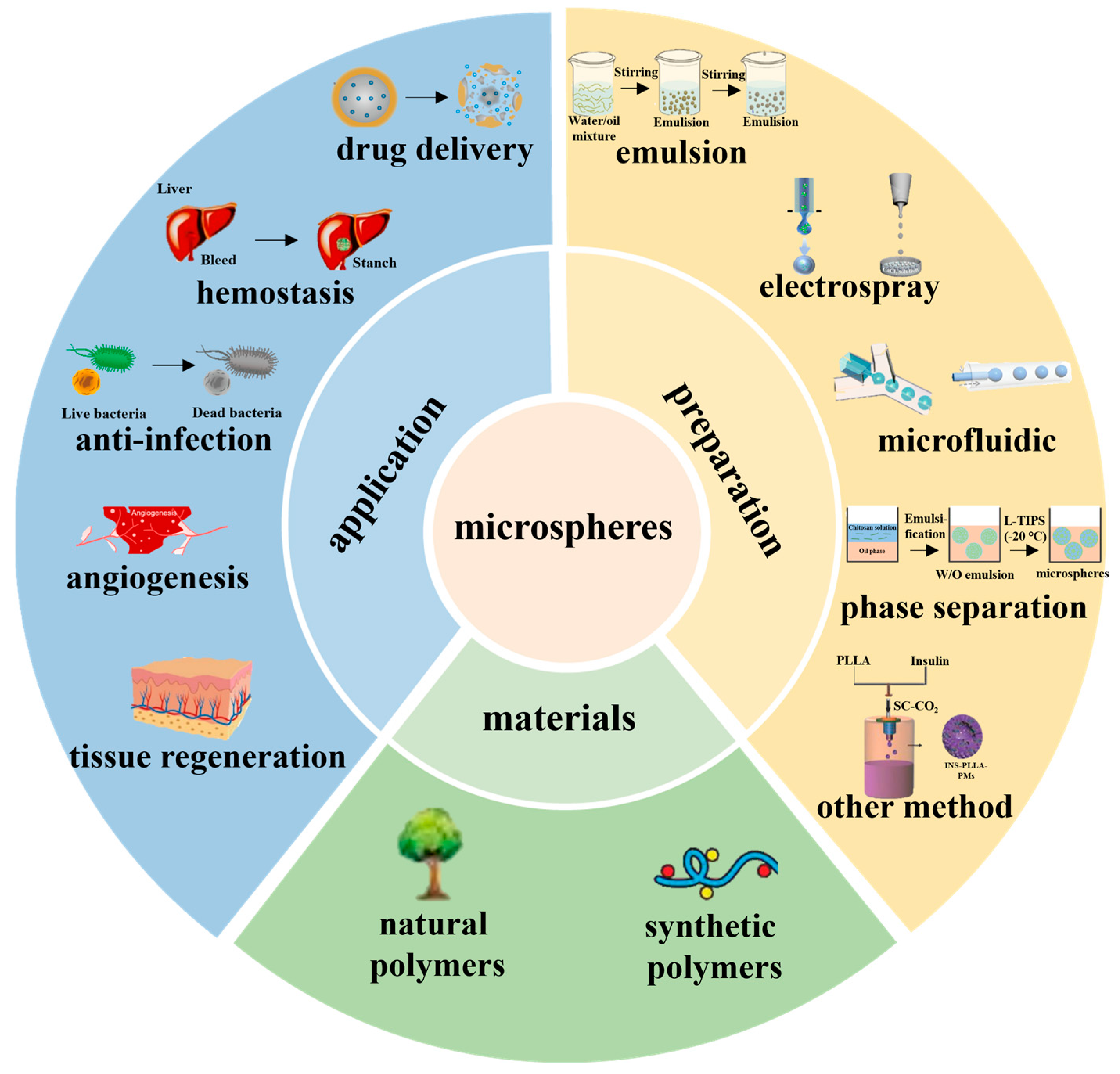
Application of Biomedical Microspheres in Wound Healing

Abstract
:1. Introduction
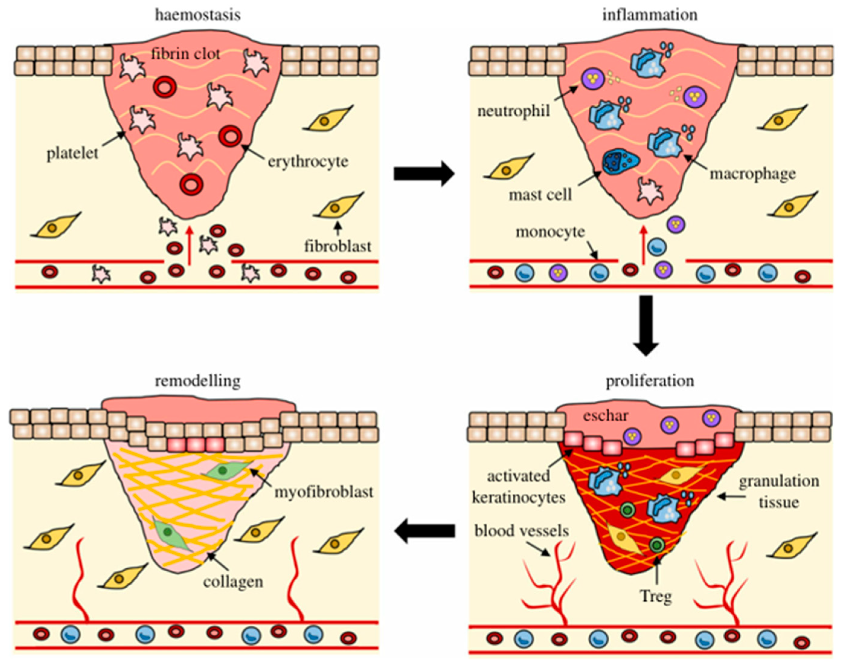
2. The Process of Wound Healing

3. Preparation of Microspheres
3.1. Emulsification
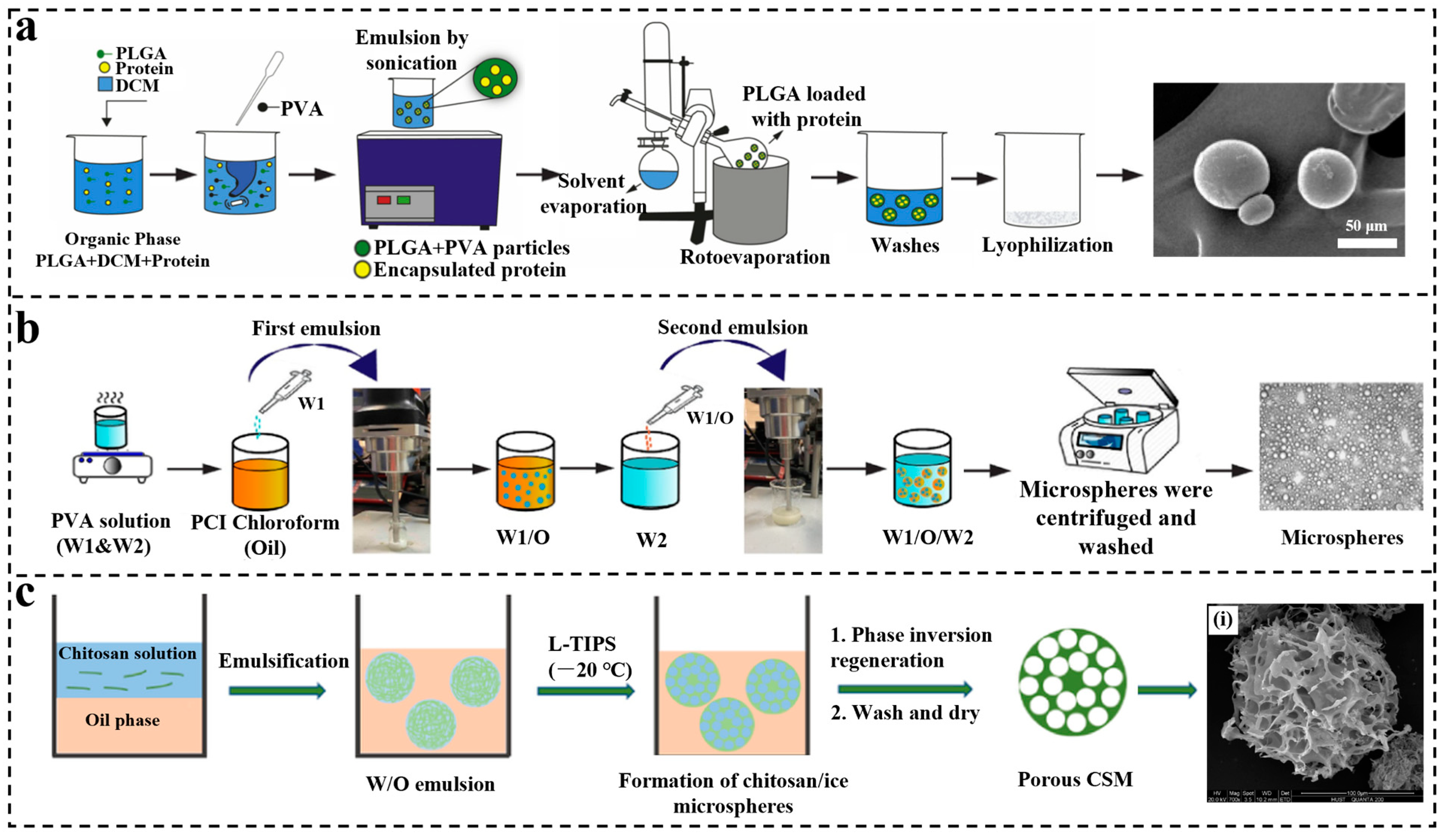
3.1.1. Single Emulsification
3.1.2. Double Emulsification

3.2. Phase Separation
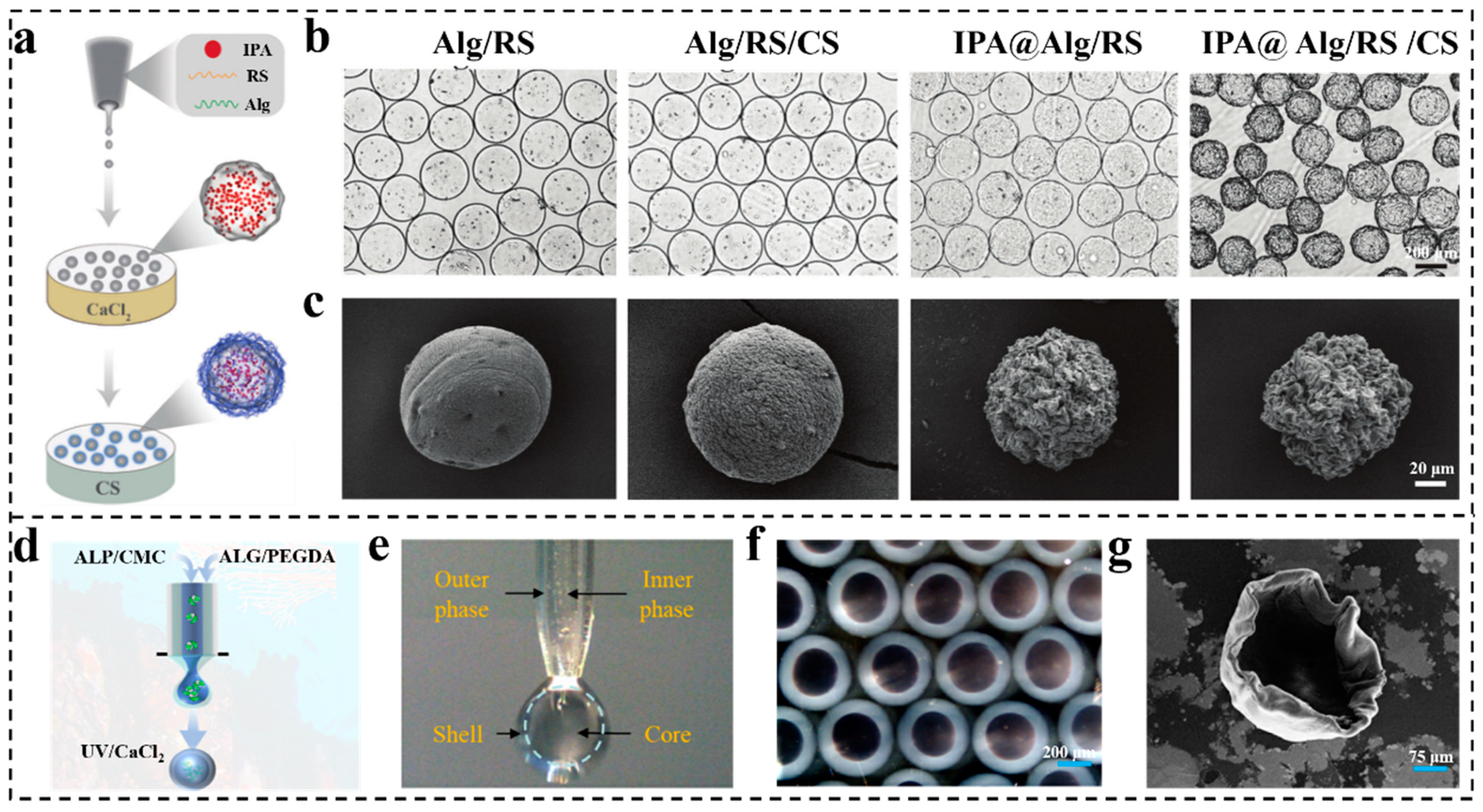
3.3. Electrospray Technology
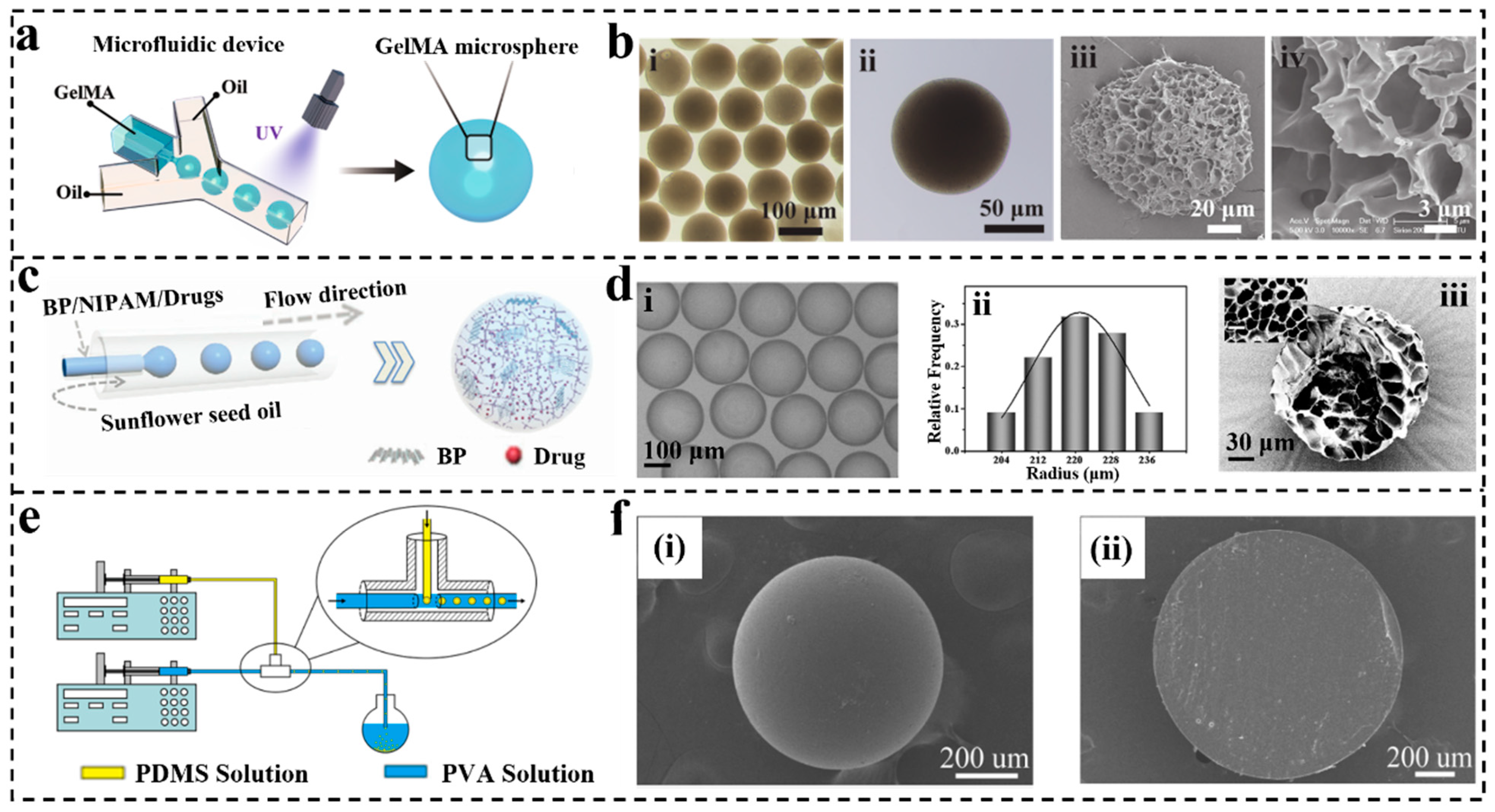
3.4. Microfluidic Technology
3.5. Other Methods
4. The Material for Microspheres
4.1. Natural Polymers
4.1.1. Alginate (Alg)
4.1.2. Chitosan (CS)
4.1.3. Collagen
4.1.4. Gelatin
4.2. Synthetic Polymers
4.2.1. Aliphatic Polyester
4.2.2. Poly(orthoesters)
4.2.3. Polycarbonate
5. Applications in Wound Healing
5.1. Drug Delivery
5.2. Hemostasis

5.3. Anti-Infection
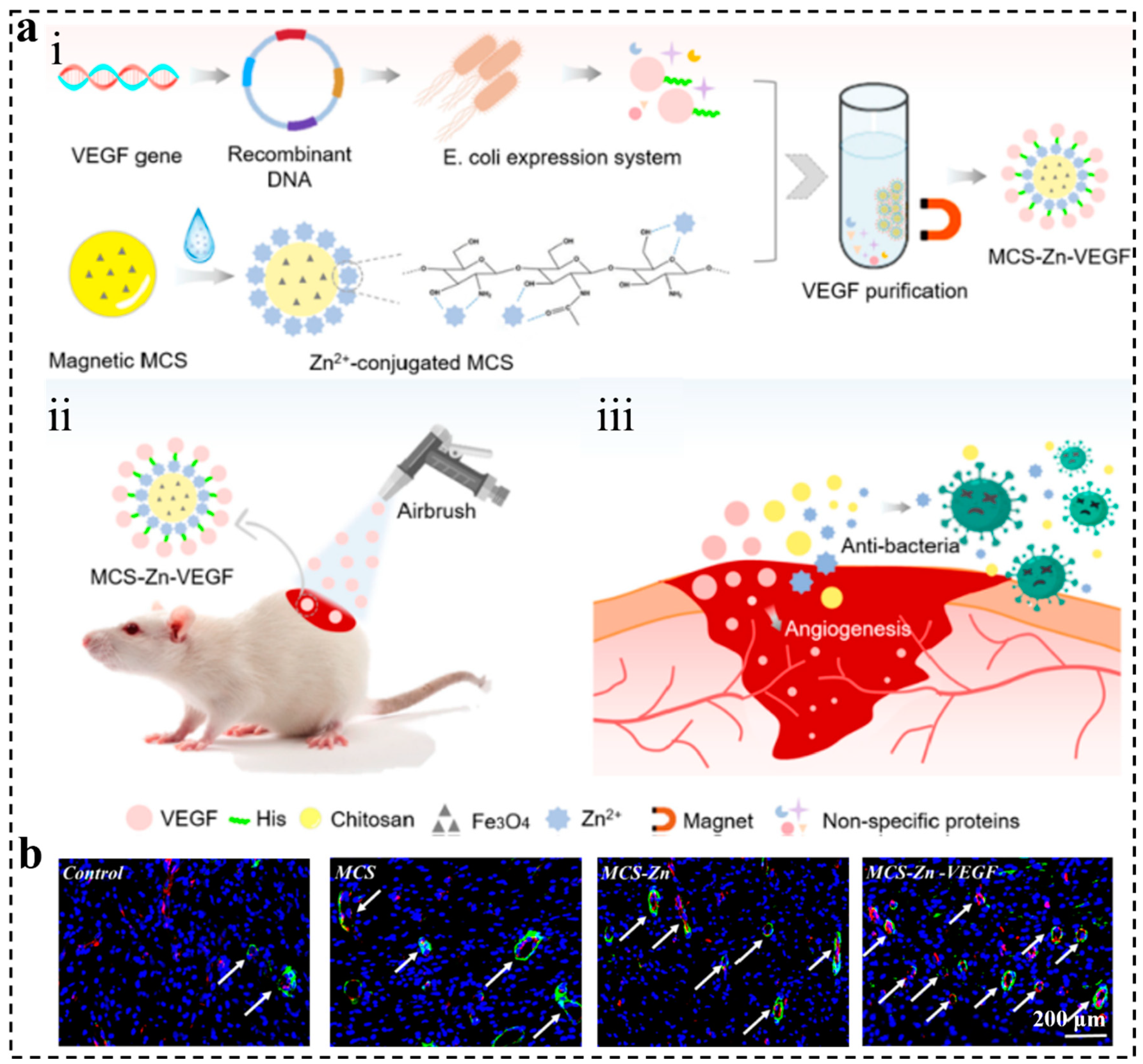
5.4. Angiogenesis
5.5. Tissue Regeneration
5.6. Other Applications
6. Conclusions and Outlook
Author Contributions
Funding
Institutional Review Board Statement
Informed Consent Statement
Data Availability Statement
Conflicts of Interest
References
- Martin, R.F. Wound Healing. Surg. Clin. N. Am. 2020, 100, 9–11. [Google Scholar] [CrossRef] [PubMed]
- Lux, C.N. Wound healing in animals: A review of physiology and clinical evaluation. Vet. Dermatol. 2022, 33, 91-e27. [Google Scholar] [CrossRef]
- Singh, M.; Thakur, V.; Kumar, V.; Raj, M.; Gupta, S.; Devi, N.; Upadhyay, S.K.; Macho, M.; Banerjee, A.; Ewe, D.; et al. Silver Nanoparticles and Its Mechanistic Insight for Chronic Wound Healing: Review on Recent Progress. Molecules 2022, 27, 30. [Google Scholar] [CrossRef] [PubMed]
- Patel, S.; Srivastava, S.; Singh, M.R.; Singh, D. Mechanistic insight into diabetic wounds: Pathogenesis, molecular targets and treatment strategies to pace wound healing. Biomed. Pharmacother. 2019, 112, 108615. [Google Scholar]
- Wilson, S.E. Corneal wound healing. Exp. Eye Res. 2020, 197, 108089. [Google Scholar] [CrossRef] [PubMed]
- Choi, H.J.; Kim, Y.M.; Suh, J.-Y.; Han, J.Y. Beneficial effect on rapid skin wound healing through carboxylic acid-treated chicken eggshell membrane. Mater. Sci. Eng. C. Mater. Biol. Appl. 2021, 128, 112350. [Google Scholar] [CrossRef]
- Neupane, Y.R.; Handral, H.K.; Alkaff, S.A.; Chng, W.H.; Venkatesan, G.; Huang, C.; Lee, C.K.; Wang, J.-W.; Sriram, G.; Dienzo, R.A.; et al. Cell-derived nanovesicles from mesenchymal stem cells as extracellular vesicle-mimetics in wound healing. Acta Pharm. Sin. B, 2022; in press. [Google Scholar]
- Melguizo-Rodríguez, L.; de Luna-Bertos, E.; Ramos-Torrecillas, J.; Illescas-Montesa, R.; Costela-Ruiz, V.J.; García-Martínez, O. Potential Effects of Phenolic Compounds That Can Be Found in Olive Oil on Wound Healing. Foods 2021, 10, 15. [Google Scholar] [CrossRef] [PubMed]
- Naik, S. One Size Does Not Fit All: Diversifying Immune Function in the Skin. J. Immunol. 2022, 2, 227–234. [Google Scholar] [CrossRef]
- Huang, R.; Zhang, X.; Li, W.; Shang, L.; Wang, H.; Zhao, Y. Suction Cups-Inspired Adhesive Patch with Tailorable Patterns for Versatile Wound Healing. Adv. Sci. 2021, 8, e2100201. [Google Scholar] [CrossRef]
- Peng, X.; Xu, X.; Deng, Y.; Xie, X.; Xu, L.; Xu, X.; Yuan, W.; Yang, B.; Yang, X.; Xia, X.; et al. Ultrafast Self-Gelling and Wet Adhesive Powder for Acute Hemostasis and Wound Healing. Adv. Funct. Mater. 2021, 31, 2102583. [Google Scholar] [CrossRef]
- Cerón, A.A.; Nascife, L.; Norte, S.; Costa, S.A.; do Nascimento, J.H.O.; Morisso, F.D.P.; Baruque-Ramos, J.; Oliveira, R.C.; Costa, S.M. Synthesis of chitosan-lysozyme microspheres, physicochemical characterization, enzymatic and antimicrobial activity. Int. J. Biol. Macromol. 2021, 185, 31. [Google Scholar]
- Su, M.; Ruan, L.; Dong, X.; Tian, S.; Lang, W.; Wu, M.; Chen, Y.; Lv, Q.; Lei, L. Current state of knowledge on intelligent-response biological and other macromolecular hydrogels in biomedical engineering: A review. Int. J. Biol. Macromol. 2023, 227, 472–492. [Google Scholar] [PubMed]
- Dhamecha, D.; Movsas, R.; Sano, U.; Menon, J.U. Applications of alginate microspheres in therapeutics delivery and cell culture: Past, present and future. Int. J. Pharm. 2019, 569, 118627. [Google Scholar]
- Chakravarty, P.; Famili, A.; Nagapudi, K.; Al-Sayah, M.A. Using Supercritical Fluid Technology as a Green Alternative During the Preparation of Drug Delivery Systems. Pharmaceutics 2019, 12, 629. [Google Scholar] [CrossRef] [PubMed] [Green Version]
- Sato, Y.; Moritani, T.; Inoue, R.; Takeuchi, H. Preparation and evaluation of sustained release formulation of PLGA using a new injection system based on ink-jet injection technology. Int. J. Pharm. 2023, 635, 122731. [Google Scholar] [CrossRef]
- Andhariya, J.V.; Shen, J.; Wang, Y.; Choi, S.; Burgess, D.J. Effect of minor manufacturing changes on stability of compositionally equivalent PLGA microspheres. Int. J. Pharm. 2019, 566, 532–540. [Google Scholar] [CrossRef]
- Bhattarai, A.; Shah, S.; Bagherieh, S.; Mirmosayyeb, O.; Thapa, S.; Paudel, S.; Gyawali, P.; Khanal, P.; Stankovic, S. Endothelium, Platelets, and Coagulation Factors as the Three Vital Components for Diagnosing Bleeding Disorders: A Simplified Perspective with Clinical Relevance. Int. J. Clin. Pract. 2022, 2022, 5369001. [Google Scholar] [PubMed]
- Komi, D.E.A.; Khomtchouk, K.; Santa Maria, P.L. A Review of the Contribution of Mast Cells in Wound Healing: Involved Molecular and Cellular Mechanisms. Clin. Rev. Allergy Immunol. 2019, 58, 298–312. [Google Scholar] [CrossRef]
- Rifai, L.; Saleh, F.A. A review on acrylamide in food: Occurrence, toxicity, and mitigation strategies. Int. J. Toxicol. 2020, 39, 93–102. [Google Scholar] [CrossRef]
- Overmiller, A.M.; Sawaya, A.P.; Hope, E.D.; Morasso, M.I. Intrinsic Networks Regulating Tissue Repair: Comparative Studies of Oral and Skin Wound Healing. Cold Spring Harb. Perspect. Biol. 2022, 111, a041244. [Google Scholar] [CrossRef]
- Lavrador, P.; Esteves, M.R.; Gaspar, V.M.; Mano, J.F. Stimuli-responsive nanocomposite hydrogels for biomedical applications. Adv. Funct. Mater. 2021, 31, 2005941. [Google Scholar] [CrossRef]
- Ogawa, R. Recent Advances in Scar Biology. Int. J. Mol. Sci. 2018, 19, 1749. [Google Scholar] [CrossRef] [PubMed] [Green Version]
- Shukla, S.K.; Sharma, A.K.; Gupta, V.; Yashavarddhan, M.H. Pharmacological control of inflammation in wound healing. J. Tissue Viability 2019, 28, 218–222. [Google Scholar] [CrossRef] [PubMed]
- Mori, Y.; Nakagami, G.; Kitamura, A.; Minematsu, T.; Kinoshita, M.; Suga, H.; Kurita, M.; Hayashi, C.; Kawasaki, A.; Sanada, H. Effectiveness of biofilm-based wound care system on wound healing in chronic wounds. Wound Repair Regen. 2019, 27, 540–547. [Google Scholar] [CrossRef] [PubMed]
- Takagi, T.; Okayama, T.; Asai, J.; Mizushima, K.; Hirai, Y.; Uchiyama, K.; Ishikawa, T.; Naito, Y.; Itoh, Y. Topical application of sustained released-carbon monoxide promotes cutaneous wound healing in diabetic mice. Biochem. Pharmacol. 2022, 199, 115016. [Google Scholar] [CrossRef]
- Sousa, A.B.; Águas, A.P.; Barbosa, M.A.; Barbosa, J.N. Immunomodulatory biomaterial-based wound dressings advance the healing of chronic wounds via regulating macrophage behavior. Regen. Biomater. 2022, 9, 6. [Google Scholar] [CrossRef]
- Dalisson, B.; Barralet, J. Bioinorganics and Wound Healing. Adv. Healthc. Mater. 2019, 8, e1900764. [Google Scholar] [CrossRef]
- Baczako, A.; Fischer, T.; Konstantinow, A.; Volz, T. Chronische Wunden richtig behandeln. MMW Fortschr. Med. 2019, 161, 48–56. [Google Scholar] [CrossRef]
- Williams, M. Wound infections: An overview. Br. J. Community Nurs. 2021, 26, S22–S25. [Google Scholar] [CrossRef]
- Wilkinson, H.N.; Hardman, M.J. Wound healing: Cellular mechanisms and pathological outcomes. Open Biol. 2020, 10, 200223. [Google Scholar]
- Wiklander, O.P.B.; Brennan, M.Á.; Lötvall, J.; Breakefield, X.O.; El Andaloussi, S. Advances in therapeutic applications of extracellular vesicles. Sci. Transl. Med. 2019, 11, eaav8521. [Google Scholar] [CrossRef]
- Kalluri, R.; LeBleu, V.S. The biology, function, and biomedical applications of exosomes. Science 2020, 367, eaau6977. [Google Scholar] [CrossRef]
- Shen, Y.; Chen, B.; Zuilhof, H.; van Beek, T.A. Microfluidic Chip-Based Induced Phase Separation Extraction as a Fast and Efficient Miniaturized Sample Preparation Method. Molecules 2020, 26, 38. [Google Scholar] [CrossRef] [PubMed]
- Huang, L.; Xiao, L.; Poudel, A.J.; Li, J.; Zhou, P.; Gauthier, M.; Liu, H.; Wu, Z.; Yang, G. Porous chitosan microspheres as microcarriers for 3D cell culture. Carbohydr. Polym. 2018, 202, 611–620. [Google Scholar] [CrossRef] [PubMed] [Green Version]
- He, T.; Jokerst, J.V. Structured micro/nano materials synthesized via electrospray: A review. Biomater. Sci. 2020, 8, 5555–5573. [Google Scholar] [CrossRef] [PubMed]
- Morais, A.Í.S.; Vieira, E.G.; Afewerki, S.; Sousa, R.B.; Honorio, L.M.C.; Cambrussi, A.N.C.O.; Santos, J.A.; Bezerra, R.D.S.; Furtini, J.A.O.; Silva-Filho, E.C.; et al. Fabrication of Polymeric Microparticles by Electrospray: The Impact of Experimental Parameters. J. Funct. Biomater. 2020, 11, 4. [Google Scholar] [CrossRef] [Green Version]
- Niculescu, A.G.; Chircov, C.; Bîrcă, A.C.; Grumezescu, A.M. Fabrication and Applications of Microfluidic Devices: A Review. Int. J. Mol. Sci. 2021, 4, 2011. [Google Scholar] [CrossRef]
- Debski, P.R.; Sklodowska, K.; Michalski, J.A.; Korczyk, P.M.; Dolata, M.; Jakiela, S. Continuous Recirculation of Microdroplets in a Closed Loop Tailored for Screening of Bacteria Cultures. Micromachines 2018, 9, 469. [Google Scholar] [CrossRef] [Green Version]
- Mo, F.; Chen, Q.; Zhang, X. Synthesis of Hollow Calcium Carbonate Microspheres by a Template Method for DOX Loading and Release with Carbon Dots Photosensitivity. Materials 2022, 15, 8768. [Google Scholar] [CrossRef]
- Escobar, E.L.N.; da Silva, T.A.; Pirich, C.L.; Corazza, M.L.; Ramos, L.P. Supercritical Fluids: A Promising Technique for Biomass Pretreatment and Fractionation. Front. Bioeng. Biotechnol. 2020, 8, 252. [Google Scholar] [CrossRef] [Green Version]
- Nanaki, S.; Viziridou, A.; Zamboulis, A.; Kostoglou, M.; Papageorgiou, G.Z.; Bikiaris, D.N. New Biodegradable Poly(l-lactide)-Block-Poly(propylene adipate) Copolymer Microparticles for Long-Acting Injectables of Naltrexone Drug. Polymers 2020, 12, 852. [Google Scholar] [CrossRef] [PubMed] [Green Version]
- Ding, S.; Serra, C.A.; Vandamme, T.F.; Yu, W.; Anton, N. Double emulsions prepared by two-step emulsification: History, state-of-the-art and perspective. J. Control Release 2019, 295, 31–49. [Google Scholar] [CrossRef] [PubMed]
- Aragón-Navas, A.; Rodrigo, M.J.; Garcia-Herranz, D.; Martinez, T.; Subias, M.; Mendez, S.; Ruberte, J.; Pampalona, J.; Bravo-Osuna, I.; Garcia-Feijoo, J.; et al. Mimicking chronic glaucoma over 6 months with a single intracameral injection of dexamethasone/fibronectin-loaded PLGA microspheres. Drug Deliv. 2022, 29, 2357–2374. [Google Scholar] [CrossRef] [PubMed]
- Hong, S.J.; Garcia, C.V.; Shin, G.H.; Kim, J.T. Enhanced bioaccessibility and stability of iron through W/O/W double emulsion-based solid lipid nanoparticles and coating with water-soluble chitosan. Int. J. Biol. Macromol. 2022, 209, 895–903. [Google Scholar] [CrossRef]
- Hong, S.J.; Garcia, C.V.; Shin, G.H.; Kim, J.T. Microspheres made by w/o/o emulsion method with reduced initial burst for long-term delivery of endostar, a novel recombinant human endostatin. Int. J. Biol. Macromol. 2022, 209, 895–903. [Google Scholar] [CrossRef]
- Ospina-Villa, J.D.; Gómez-Hoyos, C.; Zuluaga-Gallego, R.; Triana-Chávez, O. Encapsulation of proteins from Leishmania panamensis into PLGA particles by a single emulsion-solvent evaporation method. J. Microbiol. Methods 2019, 162, 1–7. [Google Scholar] [CrossRef]
- Mashhadian, A.; Afjoul, H.; Shamloo, A. An integrative method to increase the reliability of conventional double emulsion method. Anal. Chim. Acta. 2022, 1197, 339523. [Google Scholar] [CrossRef]
- Aldossary, A.M.; Ekweremadu, C.S.M.; Offe, I.M.; Alfassam, H.A.; Han, S.; Onyali, V.C.; Ozoude, C.H.; Ayeni, E.A.; Nwagwu, C.S.; Halwani, A.A.; et al. A guide to oral vaccination: Highlighting electrospraying as a promising manufacturing technique toward a successful oral vaccine development. Saudi. Pharm. J. 2022, 6, 655–668. [Google Scholar] [CrossRef]
- Zhang, D.; Zhang, Y.; Yin, F.; Qin, Q.; Bi, H.; Liu, B.; Qiao, L. Microfluidic filter device coupled mass spectrometry for rapid bacterial antimicrobial resistance analysis. Analyst 2021, 146, 515–520. [Google Scholar] [CrossRef]
- Reyes, C.G.; Lagerwall, J.P.F. Disruption of Electrospinning due to Water Condensation into the Taylor Cone. ACS Appl. Mater. Interfaces 2020, 12, 26566–26576. [Google Scholar] [CrossRef]
- Batens, M.; Dewaele, L.; Massant, J.; Teodorescu, B.; Clasen, C.; Van den Mooter, G. Feasibility of electrospraying fully aqueous bovine serum albumin solutions. Eur. J. Pharm. Biopharm. 2020, 147, 102–110. [Google Scholar] [CrossRef] [PubMed]
- Uko, L.; Noby, H.; Zkria, A.; ElKady, M. Electrospraying of Bio-Based Chitosan Microcapsules Using Novel Mixed Cross-Linker: Experimental and Response Surface Methodology Optimization. Materials 2022, 23, 8447. [Google Scholar] [CrossRef] [PubMed]
- Yang, K.; Wang, X.; Huang, R.; Wang, H.; Lan, P.; Zhao, Y. Prebiotics and Postbiotics Synergistic Delivery Microcapsules from Microfluidics for Treating Colitis. Adv. Sci. 2022, 9, e2104089. [Google Scholar] [CrossRef]
- Zhao, C.; Chen, G.; Wang, H.; Zhao, Y.; Chai, R. Bio-inspired intestinal scavenger from microfluidic electrospray for detoxifying lipopolysaccharide. Bioact. Mater. 2020, 6, 1653–1662. [Google Scholar] [CrossRef]
- Groper, E.R.; Barnes, J.A.; McEwen, R.; Messaddeq, Y.; Oleschuk, R.D.; Loock, H.P. Fabrication and characterization of laser-heated, multiplexed electrospray emitter. Analyst 2021, 146, 2834–2841. [Google Scholar] [CrossRef] [PubMed]
- André, E.; Pannacci, N.; Dalmazzone, C.; Colin, A. A new way to measure viscosity in droplet-based microfluidics for high throughput analysis. Soft Matter. 2019, 15, 504–514. [Google Scholar] [CrossRef]
- Giuliani, C. The Flavonoid Quercetin Induces AP-1 Activation in FRTL-5 Thyroid Cells. Antioxidants 2019, 8, 112. [Google Scholar] [CrossRef] [PubMed] [Green Version]
- Zhuo, Z.; Wang, J.; Luo, Y.; Zeng, R.; Zhang, C.; Zhou, W.; Guo, K.; Wu, H.; Sha, W.; Chen, H. Targeted extracellular vesicle delivery systems employing superparamagnetic iron oxide nanoparticles. Acta Biomater. 2021, 134, 13–31. [Google Scholar] [CrossRef]
- Chen, K.; Wang, F.; Ding, R.; Cai, Z.; Zou, T.; Zhang, A.; Guo, D.; Ye, B.; Cui, W.; Xiang, M. Adhesive and Injectable Hydrogel Microspheres for Inner Ear Treatment. Small 2022, 18, e2106591. [Google Scholar] [CrossRef]
- Fan, L.; Zhang, X.; Liu, X.; Sun, B.; Li, L.; Zhao, Y. Responsive Hydrogel Microcarrier-Integrated Microneedles for Versatile and Controllable Drug Delivery. Adv. Healthc. Mater. 2021, 10, e2002249. [Google Scholar] [CrossRef]
- Zhang, Z.; Chu, F.; Wang, X.; Zhou, X.; Xiong, G. Microfluidic Fabrication of a PDMS Microlens for Imaging Tunability. Langmuir 2022, 38, 4059–4064. [Google Scholar] [CrossRef] [PubMed]
- Han, J.H.; Kim, C.M.; Kim, T.H.; Jin, S.; Kim, G.M. Development of In Situ Microfluidic System for Preparation of Controlled Porous Microsphere for Tissue Engineering. Pharmaceutics 2022, 11, 2345. [Google Scholar] [CrossRef]
- Thaweeskulchai, T.; Schulte, A. Basic Guide to Multilayer Microfluidic Fabrication with Polyimide Tape and Diode Laser. Micromachines 2023, 2, 324. [Google Scholar] [CrossRef] [PubMed]
- Ibadat, N.F.; Ongkudon, C.M.; Saallah, S.; Mission, M. Synthesis and Characterization of Polymeric Microspheres Template for a Homogeneous and Porous Monolith. Polymers 2021, 21, 3639. [Google Scholar] [CrossRef] [PubMed]
- Franco, P.; De Marco, I. Formation of Rutin-β-Cyclodextrin Inclusion Complexes by Supercritical Antisolvent Precipitation. Polymers 2021, 2, 246. [Google Scholar] [CrossRef]
- Zhao, K.; Cheng, L.; Ye, F.; Cheng, S.; Cui, X. Preparation and Performance of Si3N4 Hollow Microspheres by the Template Method and Carbothermal Reduction Nitridation. ACS Appl. Mater. Interfaces 2019, 11, 39054–39061. [Google Scholar] [CrossRef]
- Penoy, N.; Grignard, B.; Evrard, B.; Piel, G. A supercritical fluid technology for liposome production and comparison with the film hydration method. Int. J. Pharm. 2021, 592, 120093. [Google Scholar] [CrossRef]
- Elmowafy, M.; Shalaby, K.; Salama, A.; Soliman, G.M.; Alruwaili, N.K.; Mostafa, E.M.; Mohammed, E.F.; Moustafa, A.E.G.A.; Zafar, A. Soy isoflavone-loaded alginate microspheres in thermosensitive gel base: Attempts to improve wound-healing efficacy. J. Pharm. Pharmacol. 2019, 71, 774–787. [Google Scholar] [CrossRef]
- Budhiraja, M.; Zafar, S.; Akhter, S.; Alrobaian, M.; Rashid, M.A.; Barkat, M.A.; Beg, S.; Ahmad, F.J. Mupirocin-Loaded Chitosan Microspheres Embedded in Piper betle Extract Containing Collagen Scaffold Accelerate Wound Healing Activity. AAPS PharmSciTech. 2022, 23, 77. [Google Scholar] [CrossRef]
- Zhang, Y.; Liang, H.; Luo, Q.; Chen, J.; Zhao, N.; Gao, W.; Pu, Y.; He, B.; Xie, J. In vivo inducing collagen regeneration of biodegradable polymer microspheres. Regen. Biomater. 2021, 8, rbab042. [Google Scholar] [CrossRef]
- Gelse, K. Collagens—Structure, function, and biosynthesis. Regen. Biomater. 2021, 8, rbab042. [Google Scholar] [CrossRef] [PubMed] [Green Version]
- Al-Nimry, S.; Dayah, A.A.; Hasan, I.; Daghmash, R. Cosmetic, Biomedical and Pharmaceutical Applications of Fish Gelatin/Hydrolysates. Mar. Drugs 2021, 19, 145. [Google Scholar] [CrossRef] [PubMed]
- Tavares, M.T.; Gaspar, V.M.; Monteiro, M.V.; Farinha, J.P.S.; Baleizão, C.; Mano, J.F. GelMA/bioactive silica nanocomposite bioinks for stem cell osteogenic differentiation. Biofabrication 2021, 13, 035012. [Google Scholar] [CrossRef] [PubMed]
- Stefaniak, K.; Masek, A. Green Copolymers Based on Poly(Lactic Acid)—Short Review. Materials 2021, 14, 5254. [Google Scholar] [CrossRef] [PubMed]
- Wang, Y.; Wang, P.; Ji, H.; Ji, G.; Wang, M.; Wang, X. Analysis of Safety and Effectiveness of Sodium Alginate/Poly(γ-glutamic acid) Microspheres for Rapid Hemostasis. ACS Appl. Bio. Mater. 2021, 4, 6539–6548. [Google Scholar] [CrossRef] [PubMed]
- Yoon, S.K.; Chung, D.J. In Vivo Degradation Studies of PGA-PLA Block Copolymer and Their Histochemical Analysis for Spinal-Fixing Application. Polymers 2022, 14, 3322. [Google Scholar] [CrossRef]
- Hu, J.; Chen, K.; Yao, Z.; Li, C. Unlocking solid-state conversion batteries reinforced by hierarchical microsphere stacked polymer electrolyte. Sci. Bull. 2021, 66, 694–707. [Google Scholar] [CrossRef]
- Guo, X.H.; Ding, F.; Lian, X.; Cui, W.; Li, Z.; Xing, Y. The efficiency and mechanism of a new absorption enhancer, malic acid, for enhancing the oral bioavailability of docetaxel. Pharm. Dev. Technol. 2021, 26, 592–598. [Google Scholar] [CrossRef]
- Ebhodaghe, S.O. Natural Polymeric Scaffolds for Tissue Engineering Applications. J. Biomater. Sci. Polym. Ed. 2021, 16, 2144–2194. [Google Scholar] [CrossRef]
- Filippi, M.; Born, G.; Chaaban, M.; Scherberich, A. Natural Polymeric Scaffolds in Bone Regeneration. Front. Bioeng. Biotechnol. 2020, 8, 474. [Google Scholar] [CrossRef]
- Soares, R.M.D.; Siqueira, N.M.; Prabhakaram, M.P.; Ramakrishna, S. Electrospinning and electrospray of bio-based and natural polymers for biomaterials development. Mater. Sci. Eng. C. Mater. Biol. Appl. 2018, 92, 969–982. [Google Scholar] [CrossRef]
- Stanisci, A.; Tøndervik, A.; Gaardløs, M.; Lervik, A.; Skjåk-Bræk, G.; Sletta, H.; Aachmann, F.L. Identification of a Pivotal Residue for Determining the Block Structure-Forming Properties of Alginate C-5 Epimerases. ACS Omega 2020, 5, 4352–4361. [Google Scholar] [CrossRef]
- Riseh, R.S.; Skorik, Y.A.; Thakur, V.K.; Pour, M.M.; Tamanadar, E.; Noghabi, S.S. Encapsulation of Plant Biocontrol Bacteria with Alginate as a Main Polymer Material. Int. J. Mol. Sci. 2021, 22, 11165. [Google Scholar] [CrossRef] [PubMed]
- Khlibsuwan, R.; Khunkitti, W.; Pongjanyakul, T. Alginate-poloxamer beads for clotrimazole delivery: Molecular interactions, mechanical properties, and anticandidal activity. Int. J. Biol. Macromol. 2020, 148, 1061–1071. [Google Scholar] [CrossRef] [PubMed]
- Nützl, M.; Schrottenbaum, M.; Müller, T.; Müller, R. Mechanical properties and chemical stability of alginate-based anisotropic capillary hydrogels. J. Mech. Behav. Biomed. Mater. 2022, 134, 105397. [Google Scholar] [CrossRef] [PubMed]
- Yuan, Y.; Xu, X.; Gong, J.; Mu, R.; Li, Y.; Wu, C.; Pang, J. Fabrication of chitosan-coated konjac glucomannan/sodium alginate/graphene oxide microspheres with enhanced colon-targeted delivery. Int. J. Biol. Macromol. 2019, 131, 209–217. [Google Scholar] [CrossRef]
- Noureen, S.; Noreen, S.; Ghumman, S.A.; Batool, F.; Hameed, H.; Hasan, S.; Noreen, F.; Elsherif, M.A.; Bukhari, S.N.A. Prunus armeniaca Gum-Alginate Polymeric Microspheres to Enhance the Bioavailability of Tramadol Hydrochloride: Formulation and Evaluation. Pharmaceutics 2022, 5, 916. [Google Scholar] [CrossRef]
- Telange, D.R.; Pandharinath, R.R.; Pethe, A.M.; Jain, S.P.; Pingale, P.L. Calcium Ion-Sodium Alginate-Piperine-Based Microspheres: Evidence of Enhanced Encapsulation Efficiency, Bio-Adhesion, Controlled Delivery, and Oral Bioavailability of Isoniazid. AAPS PharmSciTech. 2022, 4, 99. [Google Scholar] [CrossRef]
- Rankothgedera, S.; Atukorala, I.; Fernando, C.; Munidasa, D.; Wijayaratne, L.; Udagama, P. A potential diagnostic serum immunological marker panel to differentiate between primary and secondary knee osteoarthritis. PLoS ONE 2021, 16, e0257507. [Google Scholar] [CrossRef]
- Nah, J.W.; Jeong, G.W. Preparation and encapsulation techniques of chitosan microsphere for enhanced bioavailability of natural antioxidants. Carbohydr. Res. 2021, 500, 108218. [Google Scholar] [CrossRef]
- El Kadib, A. Green and Functional Aerogels by Macromolecular and Textural Engineering of Chitosan Microspheres. Chem. Rec. 2020, 20, 753–772. [Google Scholar] [CrossRef]
- Muñoz-Nuñez, C.; Cuervo-Rodríguez, R.; Echeverría, C.; Fernández-García, M.; Muñoz-Bonilla, A. Synthesis and characterization of thiazolium chitosan derivative with enhanced antimicrobial properties and its use as component of chitosan-based films. Carbohydr. Polym. 2023, 302, 120438. [Google Scholar] [PubMed]
- Carrera, C.; Bengoechea, C.; Carrillo, F.; Calero, N. Effect of deacetylation degree and molecular weight on surface properties of chitosan obtained from biowastes. Food Hydrocoll. 2022, 137, 108383. [Google Scholar] [CrossRef]
- Li, J.; Wu, Y.; Zhao, L. Antibacterial activity and mechanism of chitosan with ultra-high molecular weight. Carbohydr. Polym. 2016, 148, 200–205. [Google Scholar] [CrossRef]
- Abd-Allah, H.; Abdel-Aziz, R.T.A.; Nasr, M. Chitosan nanoparticles making their way to clinical practice: A feasibility study on their topical use for acne treatment. Int. J. Biol. Macromol. 2020, 156, 262–270. [Google Scholar] [PubMed]
- Uke, N.; Singh, S.; Sorensen, G.E.; Frost, J.; Venable, A.; Burge, B.; Terziyski, I.; Payberah, E.; Griswold, J. The Ideal Donor Site Dressing: A Comparison of a Chitosan-Based Gelling Dressing to Traditional Dressings. J. Burn. Care Res. 2022, 3, 652–656. [Google Scholar] [CrossRef]
- Massarelli, E.; Silva, D.; Pimenta, A.F.R.; Fernandes, A.I.; Mata, J.L.G.; Armês, H.; Salema-Oom, M.; Saramago, B.; Serro, A.P. Polyvinyl alcohol/chitosan wound dressings loaded with antiseptics. Int. J. Pharm. 2021, 593, 120110. [Google Scholar] [CrossRef]
- Sarbacher, C.A.; Halper, J.T. Connective Tissue and Age-Related Diseases. Subcell. Biochem. 2019, 91, 281–310. [Google Scholar]
- Lee, J.; Sabatini, C. Glutaraldehyde collagen cross-linking stabilizes resin-dentin interfaces and reduces bond degradation. Eur. J. Oral. Sci. 2017, 125, 63–71. [Google Scholar] [CrossRef]
- Michopoulou, A.; Koliakou, E.; Terzopoulou, Z.; Rousselle, P.; Palamidi, A.; Anestakis, D.; Konstantinidou, P.; Roig-Rosello, E.; Demiri, E.; Bikiaris, D. Benefit of coupling heparin to crosslinked collagen I/III scaffolds for human dermal fibroblast subpopulations′ tissue growth. J. Biomed. Mater. Res. A 2022, 110, 797–811. [Google Scholar] [CrossRef] [PubMed]
- Mony, M.P.; Anilkumar, T.V. Controlled cross-linking of porcine cholecyst extracellular matrix for preparing tissue engineering scaffold. J. Biomed. Mater. Res. B Appl. Biomater. 2020, 108, 1057–1067. [Google Scholar] [CrossRef]
- Lin, Q.; Huo, Q.; Qin, Y.; Zhao, Z.; Tao, F. Development of ligustrazine hydrochloride carboxymethyl chitosan and collagen microspheres: Formulation optimization, characterization, and vitro release. Bioengineered 2017, 1, 55–60. [Google Scholar] [CrossRef] [Green Version]
- Rezaei, N.; Lyons, A.; Forde, N.R. Nano-Mechanical Studies of Collagen: The Influence of Ionic Strength, pH and Collagen Sources on Molecular Flexibility. Biophys. J. 2017, 113, 488a. [Google Scholar] [CrossRef]
- Yang, C.; Wu, H.; Wang, J. Formulation and evaluation of controlled-release of steroidal saponins-loaded collagen microspheres. Mater. Technol. 2019, 34, 534–539. [Google Scholar] [CrossRef]
- Amin, U.; Khan, M.U.; Majeed, Y.; Rebezov, M.; Khayrullin, M.; Bobkova, E.; Shariati, M.A.; Chung, I.M.; Thiruvengadam, M. Potentials of polysaccharides, lipids and proteins in biodegradable food packaging applications. Int. J. Biol. Macromol. 2021, 183, 2184–2198. [Google Scholar] [PubMed]
- Ionescu, O.M.; Mignon, A.; Minsart, M.; Van Hoorick, J.; Gardikiotis, I.; Caruntu, I.D.; Giusca, S.E.; Van Vlierberghe, S.; Profire, L. Gelatin-Based Versus Alginate-Based Hydrogels: Providing Insight in Wound Healing Potential. Macromol. Biosci. 2021, 21, e2100230. [Google Scholar] [CrossRef]
- Escutia-Guadarrama, L.; Morales, D.; Pérez-Calixto, D.; Burillo, G. Development of Polyphenol-Functionalized Gelatin-Poly(vinylpyrrolidone) IPN for Potential Biomedical Applications. Polymer 2022, 21, 4705. [Google Scholar] [CrossRef]
- Fan, H.Y.; Duquette, D.; Dumont, M.-J.; Simpson, B.K. Salmon skin gelatin-corn zein composite films produced via crosslinking with glutaraldehyde: Optimization using response surface methodology and characterization. Int. J. Biol. Macromol. 2018, 120, 263–273. [Google Scholar] [CrossRef] [PubMed]
- Samirah; Budiatin, A.S.; Mahyudin, F.; Khotib, J. Fabrication and characterization of bovine hydroxyapatite-gelatin-alendronate scaffold cross-linked by glutaraldehyde for bone regeneration. J. Basic. Clin. Physiol. Pharmacol. 2021, 32, 555–560. [Google Scholar] [CrossRef]
- Pei, Y.; Zheng, Y.; Li, Z.; Liu, J.; Zheng, X.; Tang, K.; Kaplan, D.L. Ethanol-induced coacervation in aqueous gelatin solution for constructing nanospheres and networks: Morphology, dynamics and thermal sensitivity. J. Colloid Interface Sci. 2021, 582, 610–618. [Google Scholar] [CrossRef]
- Ferreira, S.; Nicoletti, V.R. Microencapsulation of ginger oil by complex coacervation using atomization: Effects of polymer ratio and wall material concentration. J. Food Eng. 2021, 291, 110214. [Google Scholar] [CrossRef]
- Wang, W.; Zhang, X.; Teng, A.; Liu, A. Mechanical reinforcement of gelatin hydrogel with nanofiber cellulose as a function of percolation concentration. Int. J. Biol. Macromol. 2017, 103, 226–233. [Google Scholar] [CrossRef]
- Tutar, R.; Yüce-Erarslan, E.; İzbudak, B.; Bal-Öztürk, A. Photocurable silk fibroin-based tissue sealants with enhanced adhesive properties for the treatment of corneal perforations. J. Mater. Chem. B. 2022, 15, 2912–2925. [Google Scholar] [CrossRef] [PubMed]
- Yue, K.; Li, X.; Schrobback, K.; Sheikhi, A.; Annabi, N.; Leijten, J.; Zhang, W.; Zhang, Y.S.; Hutmacher, D.W.; Klein, T.J.; et al. Structural analysis of photocrosslinkable methacryloyl-modified protein derivatives. Biomaterials 2017, 139, 163–171. [Google Scholar] [CrossRef] [PubMed] [Green Version]
- Li, Z.; Zhang, S.; Chen, Y.; Ling, H.; Zhao, L.; Luo, G.; Wang, X.; Hartel, M.C.; Liu, H.; Xue, Y.; et al. Gelatin methacryloyl-based tactile sensors for medical wearables. Adv. Funct. Mater. 2020, 49, 2003601. [Google Scholar] [CrossRef] [PubMed]
- Im, G.B.; Lin, R.Z. Bioengineering for vascularization: Trends and directions of photocrosslinkable gelatin methacrylate hydrogels. Front. Bioeng. Biotechnol. 2022, 10, 1053491. [Google Scholar] [CrossRef]
- Terzopoulou, Z.; Zamboulis, A.; Koumentakou, I.; Michailidou, G.; Noordam, M.J.; Bikiaris, D.N. Biocompatible Synthetic Polymers for Tissue Engineering Purposes. Biomacromolecules 2022, 5, 1841–1863. [Google Scholar] [CrossRef]
- Chandakavathe, B.N.; Kulkarni, R.G.; Dhadde, S.B. Grafting of Natural Polymers and Gums for Drug Delivery Applications: A Perspective Review. Crit. Rev. Ther. Drug Carr. Syst. 2022, 6, 45–83. [Google Scholar] [CrossRef] [PubMed]
- Schmidt, S.J.; Holt, B.D.; Arnold, A.M.; Sydlik, S.A. Polyester functional graphenic materials as a mechanically enhanced scaffold for tissue regeneration. RSC Adv. 2020, 10, 8548–8557. [Google Scholar] [CrossRef]
- Yang, P.; Ju, Y.; Hu, Y.; Xie, X.; Fang, B.; Lei, L. Emerging 3D bioprinting applications in plastic surgery. Biomater. Res. 2023, 1, 1–27. [Google Scholar] [CrossRef]
- Da Silva, D.; Kaduri, M.; Poley, M.; Adir, O.; Krinsky, N.; Shainsky-Roitman, J.; Schroeder, A. Biocompatibility, biodegradation and excretion of polylactic acid (PLA) in medical implants and theranostic systems. Chem. Eng. J. 2018, 340, 9–14. [Google Scholar] [CrossRef]
- Butreddy, A.; Gaddam, R.P.; Kommineni, N.; Dudhipala, N.; Voshavar, C. PLGA/PLA-Based Long-Acting Injectable Depot Microspheres in Clinical Use: Production and Characterization Overview for Protein/Peptide Delivery. Int. J. Mol. Sci. 2021, 22, 8884. [Google Scholar] [CrossRef]
- Liu, S.; Yu, J.; Li, H.; Wang, K.; Wu, G.; Wang, B.; Liu, M.; Zhang, Y.; Wang, P.; Zhang, J.; et al. Controllable Drug Release Behavior of Polylactic Acid (PLA) Surgical Suture Coating with Ciprofloxacin (CPFX)—Polycaprolactone (PCL)/Polyglycolide (PGA). Polymers 2020, 12, 288. [Google Scholar] [CrossRef] [Green Version]
- Liu, S.; Wu, G.; Zhang, X.; Yu, J.; Liu, M.; Zhang, Y.; Wang, P.; Yin, X.; Zhang, J.; Li, F.; et al. Preparation and properties of poly (lactic acid) (PLA) suture loaded with PLA microspheres enclosed drugs (PM-Ds). J. Text. Inst. 2019, 110, 1596–1605. [Google Scholar] [CrossRef]
- Chaiyasat, P.; Pholsrimuang, P.; Boontung, W.; Chaiyasat, A. Influence of Poly (L-lactic acid) Molecular Weight on the Encapsulation Efficiency of Urea in Microcapsule Using a Simple Solvent Evaporation Technique. Polym. Plast. Technol. Mater. 2016, 55, 1131–1136. [Google Scholar] [CrossRef]
- Ochi, M.; Wan, B.; Bao, Q.; Burgess, D.J. Influence of PLGA molecular weight distribution on leuprolide release from microspheres. Int J Pharm. 2021, 599, 120450. [Google Scholar] [CrossRef]
- Evrova, O.; Hosseini, V.; Milleret, V.; Palazzolo, G.; Zenobi-Wong, M.; Sulser, T.; Buschmann, J.; Eberli, D. Hybrid Randomly Electrospun Poly(lactic-co-glycolic acid):Poly(ethylene oxide) (PLGA:PEO) Fibrous Scaffolds Enhancing Myoblast Differentiation and Alignment. ACS Appl. Mater. Interfaces 2016, 8, 31574–31586. [Google Scholar] [CrossRef] [PubMed]
- Worm, M.; Kang, B.; Dingels, C.; Wurm, F.R.; Frey, H. Acid-Labile Amphiphilic PEO-b-PPO-b-PEO Copolymers: Degradable Poloxamer Analogs. Macromol. Rapid Commun. 2016, 37, 775–780. [Google Scholar] [CrossRef]
- Casanova, L.; Ceriani, F.; Pedeferri, M.; Ormellese, M. Addition of Organic Acids during PEO of Titanium in Alkaline Solution. Coatings 2022, 12, 143. [Google Scholar] [CrossRef]
- Wang, J.; Zhang, L.; Wang, X.; Fu, S.; Yan, G. Acid-labile poly (amino alcohol ortho ester) based on low molecular weight polyethyleneimine for gene delivery. J. Biomater. Appl. 2017, 3, 349–361. [Google Scholar] [CrossRef] [PubMed]
- Sun, S.; Chen, C.; Zhang, J.; Hu, J. Biodegradable smart materials with self-healing and shape memory function for wound healing. RSC Adv. 2023, 13, 3155–3163. [Google Scholar] [CrossRef]
- Sun, J.; Birnbaum, W.; Anderski, J.; Picker, M.T.; Mulac, D.; Langer, K.; Kuckling, D. Use of Light-Degradable Aliphatic Polycarbonate Nanoparticles As Drug Carrier for Photosensitizer. Biomacromolecules 2018, 19, 4677–4690. [Google Scholar] [CrossRef] [PubMed]
- Weems, A.C.; Arno, M.C.; Yu, W.; Huckstepp, R.T.R.; Dove, A.P. 4D polycarbonates via stereolithography as scaffolds for soft tissue repair. Nat. Commun. 2021, 12, 3771. [Google Scholar] [CrossRef] [PubMed]
- Kumaraswamy, S.; Thangasundaralingam, S.R.; Sekar, R.; Jayakrishnan, A. A floating-type dosage form of repaglinide in polycarbonate microspheres. J. Drug Deliv. Sci. Technol. 2017, 41, 99–105. [Google Scholar] [CrossRef]
- Hu, B.; Du, H.J.; Yan, G.P.; Zhuo, R.X.; Wu, Y.; Fan, C.L. Magnetic polycarbonate microspheres for tumor-targeted delivery of tumor necrosis factor. Drug Deliv. 2014, 21, 204–212. [Google Scholar] [CrossRef] [PubMed] [Green Version]
- Talbott, H.E.; Mascharak, S.; Griffin, M.; Wan, D.C.; Longaker, M.T. Wound healing, fibroblast heterogeneity, and fibrosis. Cell Stem Cell 2022, 29, 1161–1180. [Google Scholar]
- Mateus, A.P.; Anjos, L.; Cardoso, J.R.; Power, D.M. Chronic stress impairs the local immune response during cutaneous repair in gilthead sea bream (Sparus aurata, L.). Mol. Immunol. 2017, 87, 267–283. [Google Scholar] [CrossRef]
- Wilkinson, H.N.; Hardman, M.J. Senescence in Wound Repair: Emerging Strategies to Target Chronic Healing Wounds. Front. Cell Dev. Biol. 2020, 8, 773. [Google Scholar]
- Thaarup, I.C.; Gummesson, C.; Bjarnsholt, T. Measuring enzymatic degradation of degradable starch microspheres using confocal laser scanning microscopy. Acta Biomater. 2021, 131, 464–471. [Google Scholar] [CrossRef]
- Bian, J.; Cai, F.; Chen, H.; Tang, Z.; Xi, K.; Tang, J.; Wu, L.; Xu, Y.; Deng, L.; Gu, Y.; et al. Modulation of Local Overactive Inflammation via Injectable Hydrogel Microspheres. Nano Lett. 2021, 21, 2690–2698. [Google Scholar] [CrossRef]
- Lee, Y.H.; Park, H.I.; Chang, W.S.; Choi, J.S. Triphenylphosphonium-conjugated glycol chitosan microspheres for mitochondria-targeted drug delivery. Int. J. Biol. Macromol. 2021, 167, 35–45. [Google Scholar] [CrossRef] [PubMed]
- Mayorova, O.A.; Sindeeva, O.A.; Lomova, M.V.; Gusliakova, O.I.; Tarakanchikova, Y.V.; Tyutyaev, E.V.; Pinyaev, S.I.; Kulikov, O.A.; German, S.V.; Pyataev, N.A.; et al. Endovascular addressing improves the effectiveness of magnetic targeting of drug carrier. Comparison with the conventional administration method. Nanomedicine 2020, 28, 102184. [Google Scholar] [CrossRef] [PubMed]
- Han, C.; Zhang, X.; Pang, G.; Zhang, Y.; Pan, H.; Li, L.; Cui, M.; Liu, B.; Kang, R.; Xue, X.; et al. Hydrogel microcapsules containing engineered bacteria for sustained production and release of protein drugs. Biomaterials 2022, 287, 121619. [Google Scholar] [CrossRef]
- Paolini, M.S.; Fenton, O.S.; Bhattacharya, C.; Andresen, J.L.; Langer, R. Polymers for extended-release administration. Biomed. Microdevices 2019, 21, 45. [Google Scholar] [PubMed]
- Spyker, D.A.; Dart, R.C.; Yip, L.; Reynolds, K.; Brittain, S.; Yarema, M. Population pharmacokinetic analysis of acetaminophen overdose with immediate release, extended release and modified release formulations. Clin. Toxicol 2022, 60, 1113–1121. [Google Scholar] [CrossRef]
- Xu, K.; Chai, B.; Zhang, K.; Xiong, J.; Zhu, Y.; Xu, J.; An, N.; Xia, W.; Ji, H.; Wu, Y.; et al. Topical Application of Fibroblast Growth Factor 10-PLGA Microsphere Accelerates Wound Healing via Inhibition of ER Stress. Oxidative Med. Cell Longev. 2020, 2020, 8586314. [Google Scholar] [CrossRef] [PubMed]
- Annamalai, R.T.; Turner, P.A.; Carson, W.F., 4th; Levi, B.; Kunkel, S.; Stegemann, J.P. Harnessing macrophage-mediated degradation of gelatin microspheres for spatiotemporal control of BMP2 release. Biomaterials 2018, 161, 216–227. [Google Scholar] [CrossRef]
- Tsiailanis, A.D.; Tzakos, A.G.; Mavromoustakos, T. Advancing the Therapeutic Efficacy of Bioactive Molecules by Delivery Vehicle Platforms. Curr. Med. Chem. 2021, 28, 2697–2706. [Google Scholar] [CrossRef]
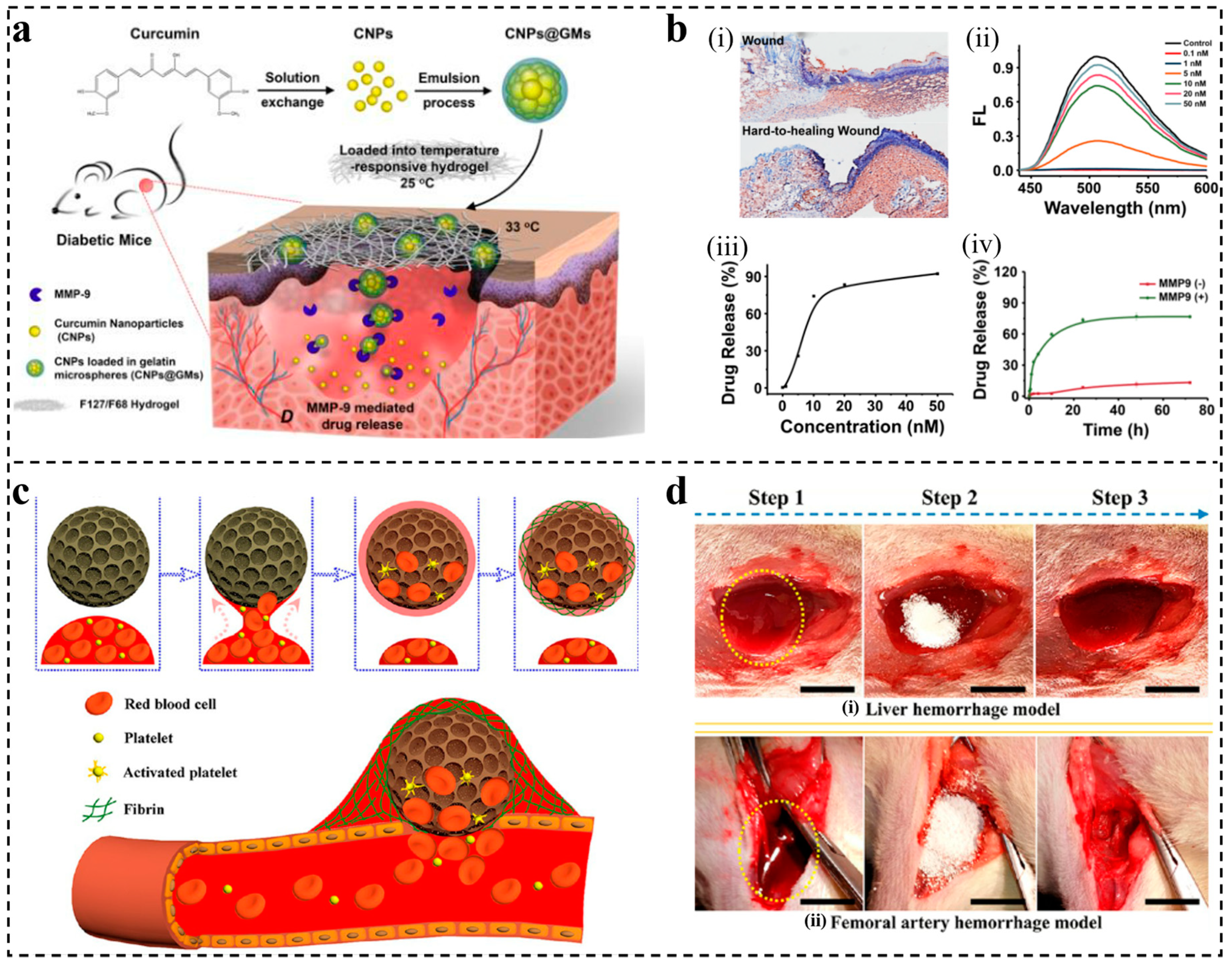
- Liu, J.; Chen, Z.; Wang, J.; Li, R.; Li, T.; Chang, M.; Yan, F.; Wang, Y. Encapsulation of Curcumin Nanoparticles with MMP9-Responsive and Thermos-Sensitive Hydrogel Improves Diabetic Wound Healing. ACS Appl. Mater. Interfaces 2018, 10, 16315–16326. [Google Scholar] [CrossRef]
- Hajiahmadi, F.; Alikhani, M.Y.; Shariatifar, H.; Arabestani, M.R.; Ahmadvand, D. The bactericidal effect of lysostaphin coupled with liposomal vancomycin as a dual combating system applied directly on methicillin-resistant Staphylococcus aureus infected skin wounds in mice. Int. J. Nanomed. 2019, 14, 5943–5955. [Google Scholar] [CrossRef] [Green Version]
- Birk, S.E.; Boisen, A.; Nielsen, L.H. Polymeric nano- and microparticulate drug delivery systems for treatment of biofilms. Adv. Drug Deliv. Rev. 2021, 174, 30–52. [Google Scholar] [CrossRef] [PubMed]
- Ameh, T.; Zarzosa, K.; Dickinson, J.; Braswell, W.E.; Sayes, C.M. Nanoparticle surface stabilizing agents influence antibacterial action. Adv. Drug Deliv. Rev. 2021, 174, 30–52. [Google Scholar] [CrossRef] [PubMed]
- Godoy-Gallardo, M.; Eckhard, U.; Delgado, L.M.; de Roo Puente, Y.J.D.; Hoyos-Nogués, M.; Gil, F.J.; Perez, R.A. Antibacterial approaches in tissue engineering using metal ions and nanoparticles: From mechanisms to applications. Bioact. Mater. 2021, 6, 4470–4490. [Google Scholar]
- Scheiner, K.C.; Maas-Bakker, R.F.; van Steenbergen, M.J.; Schwendeman, S.P.; Hennink, W.E.; Kok, R.J. Post-loading of proangiogenic growth factors in PLGA microspheres. Eur. J. Pharm. Biopharm. 2021, 158, 1–10. [Google Scholar] [CrossRef] [PubMed]
- Huang, J.; Ren, J.; Chen, G.; Li, Z.; Liu, Y.; Wang, G.; Wu, X. Tunable sequential drug delivery system based on chitosan/hyaluronic acid hydrogels and PLGA microspheres for management of non-healing infected wounds. Mater. Sci. Eng. C. Mater. Biol. Appl. 2018, 89, 213–222. [Google Scholar] [CrossRef]
- Malik, A.; Rehman, F.U.; Shah, K.U.; Naz, S.S.; Qaisar, S. Hemostatic strategies for uncontrolled bleeding: A comprehensive update. J. Biomed. Mater. Res. B. Appl. Biomater. 2021, 109, 1465–1477. [Google Scholar] [CrossRef]
- Hu, B.; Bao, G.; Xu, X.; Yang, K. Topical hemostatic materials for coagulopathy. J. Mater. Chem. B. 2022, 10, 1946–1959. [Google Scholar] [CrossRef]
- Tao, K.; Bai, X.; Ji, P.; Zhang, Y.; Cao, T.; Han, F.; Zhang, Z.; Guan, H.; Hu, D. A Composite of Hepatocyte Growth Factor- and 5α-Dihydrotestosterone-Gelatin Microspheres with Adipose-Derived Stem Cells Enhances Wound Healing. Skin Pharmacol. Physiol. 2022, 35, 206–214. [Google Scholar] [CrossRef]
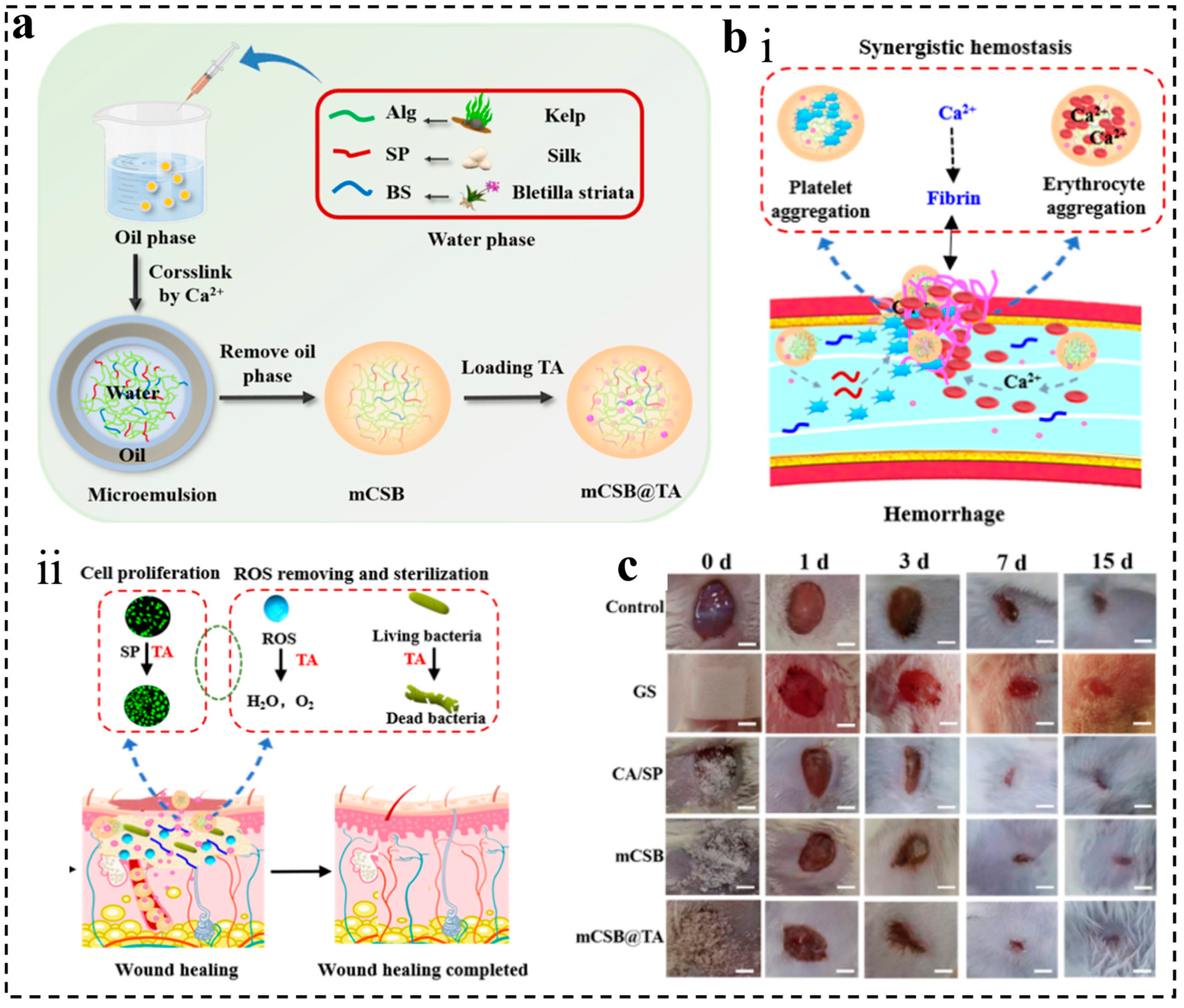
- Wang, N.; Tian, X.; Cheng, B.; Guang, S.; Xu, H. Calcium alginate/silk fibroin peptide/Bletilla striata polysaccharide blended microspheres loaded with tannic acid for rapid wound healing. Int. J. Biol. Macromol. 2022, 220, 1329–1344. [Google Scholar] [CrossRef]
- Wu, X.; Tang, Z.; Liao, X.; Wang, Z.; Liu, H. Fabrication of chitosan@calcium alginate microspheres with porous core and compact shell, and application as a quick traumatic hemostat. Carbohydr. Polym. 2020, 247, 116669. [Google Scholar] [CrossRef]
- Kuznetsova, T.A.; Andryukov, B.G.; Makarenkova, I.D.; Zaporozhets, T.S.; Besednova, N.N.; Fedyanina, L.N.; Kryzhanovsky, S.P.; Shchelkanov, M.Y. The Potency of Seaweed Sulfated Polysaccharides for the Correction of Hemostasis Disorders in COVID-19. Molecules 2021, 9, 2618. [Google Scholar] [CrossRef]
- Xi, G.; Liu, W.; Chen, M.; Li, Q.; Hao, X.; Wang, M.; Yang, X.; Feng, Y.; He, H.; Shi, C.; et al. Polysaccharide-Based Lotus Seedpod Surface-Like Porous Microsphere with Precise and Controllable Micromorphology for Ultrarapid Hemostasis. ACS Appl. Mater. Interfaces 2019, 11, 46558–46571. [Google Scholar] [CrossRef]
- Lu, W.; Bao, D.; Ta, F.; Liu, D.; Zhang, D.; Zhang, Z.; Fan, Z. Multifunctional Alginate Hydrogel Protects and Heals Skin Defects in Complex Clinical Situations. ACS Omega 2020, 5, 17152–17159. [Google Scholar] [CrossRef]
- Grolman, J.M.; Singh, M.; Mooney, D.J.; Eriksson, E.; Nuutila, K. Antibiotic-Containing Agarose Hydrogel for Wound and Burn Care. J. Burn Care Res. 2019, 40, 900–906. [Google Scholar] [CrossRef]
- Rashki, S.; Asgarpour, K.; Tarrahimofrad, H.; Hashemipour, M.; Ebrahimi, M.S.; Fathizadeh, H.; Khorshidi, A.; Khan, H.; Marzhoseyni, Z.; Salavati-Niasari, M.; et al. Chitosan-based nanoparticles against bacterial infections. Carbohydr. Polym. 2021, 251, 117108. [Google Scholar] [CrossRef]
- Li, J.; Tang, R.; Zhang, P.; Yuan, M.; Li, H.; Yuan, M. The Preparation and Characterization of Chitooligosaccharide-Polylactide Polymers, and In Vitro Release of Microspheres Loaded with Vancomycin. J. Funct. Biomater. 2022, 13, 113. [Google Scholar] [CrossRef]
- Yu, X.; Pan, Q.; Zheng, Z.; Chen, Y.; Chen, Y.; Weng, S.; Huang, L. pH-responsive and porous vancomycin-loaded PLGA microspheres: Evidence of controlled and sustained release for localized inflammation inhibition in vitro. RSC Adv. 2018, 8, 37424–37432. [Google Scholar] [CrossRef] [Green Version]
- León-Buitimea, A.; Garza-Cárdenas, C.R.; Garza-Cervantes, J.A.; Lerma-Escalera, J.A.; Morones-Ramírez, J.R. The Demand for New Antibiotics: Antimicrobial Peptides, Nanoparticles, and Combinatorial Therapies as Future Strategies in Antibacterial Agent Design. Front. Microbiol. 2020, 11, 1669. [Google Scholar] [CrossRef] [PubMed]
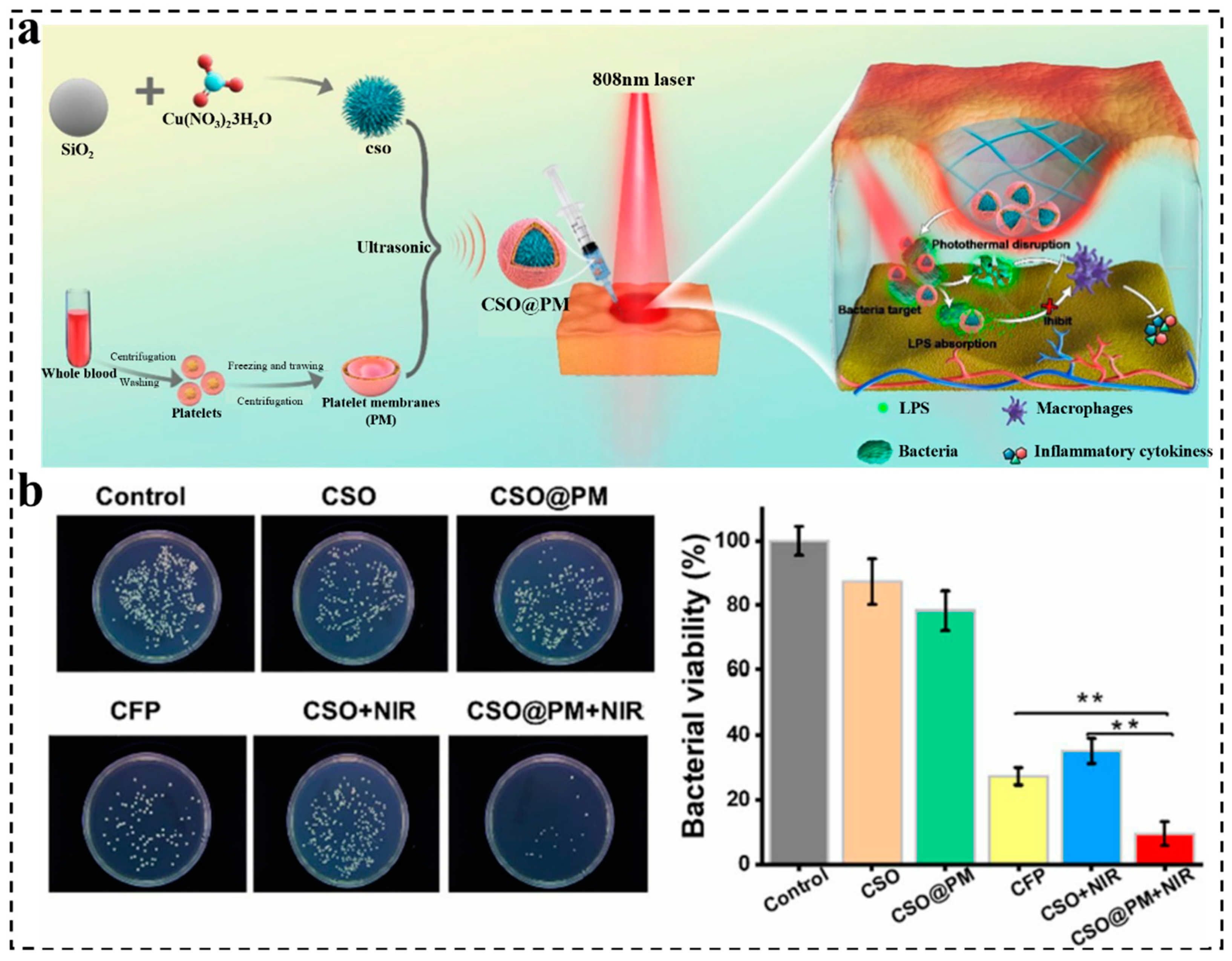
- Peng, Z.; Zhang, X.; Yuan, L.; Li, T.; Chen, Y.; Tian, H.; Ma, D.; Deng, J.; Qi, X.; Yin, X. Integrated endotoxin-adsorption and antibacterial properties of platelet-membrane-coated copper silicate hollow microspheres for wound healing. J. Nanobiotechnol. 2021, 19, 383. [Google Scholar] [CrossRef] [PubMed]
- Nitzsche, B.; Rong, W.W.; Goede, A.; Hoffmann, B.; Scarpa, F.; Kuebler, W.M.; Secomb, T.W.; Pries, A.R. Coalescent angiogenesis—Evidence for a novel concept of vascular network maturation. Angiogenesis 2022, 25, 35–45. [Google Scholar] [CrossRef] [PubMed]
- Liu, Y.; Yang, Y.; Wang, Z.; Fu, X.; Chu, X.M.; Li, Y.; Wang, Q.; He, X.; Li, M.; Wang, K.; et al. Insights into the regulatory role of circRNA in angiogenesis and clinical implications. Atherosclerosis 2020, 298, 14–26. [Google Scholar] [CrossRef] [PubMed] [Green Version]
- Lei, L.; Lv, Q.; Jin, Y.; An, H.; Shi, Z.; Hu, G.; Yang, Y.; Wang, X.; Yang, L. Angiogenic Microspheres for the Treatment of a Thin Endometrium. ACS Biomater. Sci. Eng. 2021, 7, 4914–4920. [Google Scholar] [CrossRef] [PubMed]
- Chen, Y.; Ding, B.S. Comprehensive Review of the Vascular Niche in Regulating Organ Regeneration and Fibrosis. Stem Cells Transl. Med. 2022, 11, 1135–1142. [Google Scholar] [CrossRef] [PubMed]
- Lu, W.; Li, X. PDGFs and their receptors in vascular stem/progenitor cells: Functions and therapeutic potential in retinal vasculopathy. Mol. Asp. Med. 2018, 62, 22–32. [Google Scholar] [CrossRef]
- Lee, J.S.; Guo, P.; Klett, K.; Hall, M.; Sinha, K.; Ravuri, S.; Huard, J.; Murphy, W.L. VEGF-attenuated platelet-rich plasma improves therapeutic effect on cartilage repair. Biomater. Sci. 2022, 10, 2172–2181. [Google Scholar] [CrossRef]
- Lei, L.; Wang, X.; Zhu, Y.; Su, W.; Lv, Q.Z.; Li, D. Antimicrobial hydrogel microspheres for protein capture and wound healing. Mater. Des. 2022, 215, 110478. [Google Scholar] [CrossRef]
- Ren, B.; Lu, J.; Li, M.; Zou, X.; Liu, Y.; Wang, C.; Wang, L. Anti-inflammatory effect of IL-1ra-loaded dextran/PLGA microspheres on Porphyromonas gingivalis lipopolysaccharide-stimulated macrophages in vitro and in vivo in a rat model of periodontitis. Biomed. Pharmacother. 2021, 134, 111171. [Google Scholar] [CrossRef]
- Zhong, R.; Zhong, Q.; Huo, M.; Yang, B.; Li, H. Preparation of biocompatible nano-ZnO/chitosan microspheres with multi-functions of antibacterial, UV-shielding and dye photodegradation. Int. J. Biol. Macromol. 2020, 146, 939–945. [Google Scholar] [CrossRef]
- Yang, L.; Liu, Y.; Sun, L.; Zhao, C.; Chen, G.; Zhao, Y. Biomass Microcapsules with Stem Cell Encapsulation for Bone Repair. Nanomicro. Lett. 2021, 14, 4. [Google Scholar] [CrossRef]
- Zhou, X.; Zhu, L.; Li, W.; Liu, Q. An integrated microfluidic chip for alginate microsphere generation and 3D cell culture. Anal Methods 2022, 14, 1181–1186. [Google Scholar] [CrossRef]
- Jayachandran, V.; Murugan, S.S.; Dalavi, P.A.; Vishalakshi, Y.D.G.; Seong, G.H. Alginate-based Composite Microspheres: Preparations and Applications for Bone Tissue Engineering. Curr. Pharm. Des. 2022, 28, 1067–1081. [Google Scholar] [CrossRef]
- Zhang, A.; Xiao, Z.; Liu, Q.; Li, P.; Xu, F.; Liu, J.; Tao, H.; Feng, L.; Song, S.; Liu, Z.; et al. CaCO3 -Encapuslated Microspheres for Enhanced Transhepatic Arterial Embolization Treatment of Hepatocellular Carcinoma. Adv. Healthc. Mater. 2021, 10, e2100748. [Google Scholar] [CrossRef] [PubMed]
- Peticone, C.; Thompson, D.S.; Dimov, N.; Jevans, B.; Glass, N.; Micheletti, M.; Knowles, J.C.; Kim, H.W.; Cooper-White, J.J.; Wall, I.B. Characterisation of osteogenic and vascular responses of hMSCs to Ti-Co doped phosphate glass microspheres using a microfluidic perfusion platform. J. Tissue Eng. 2020, 11, 2041731420954712. [Google Scholar] [CrossRef] [PubMed]
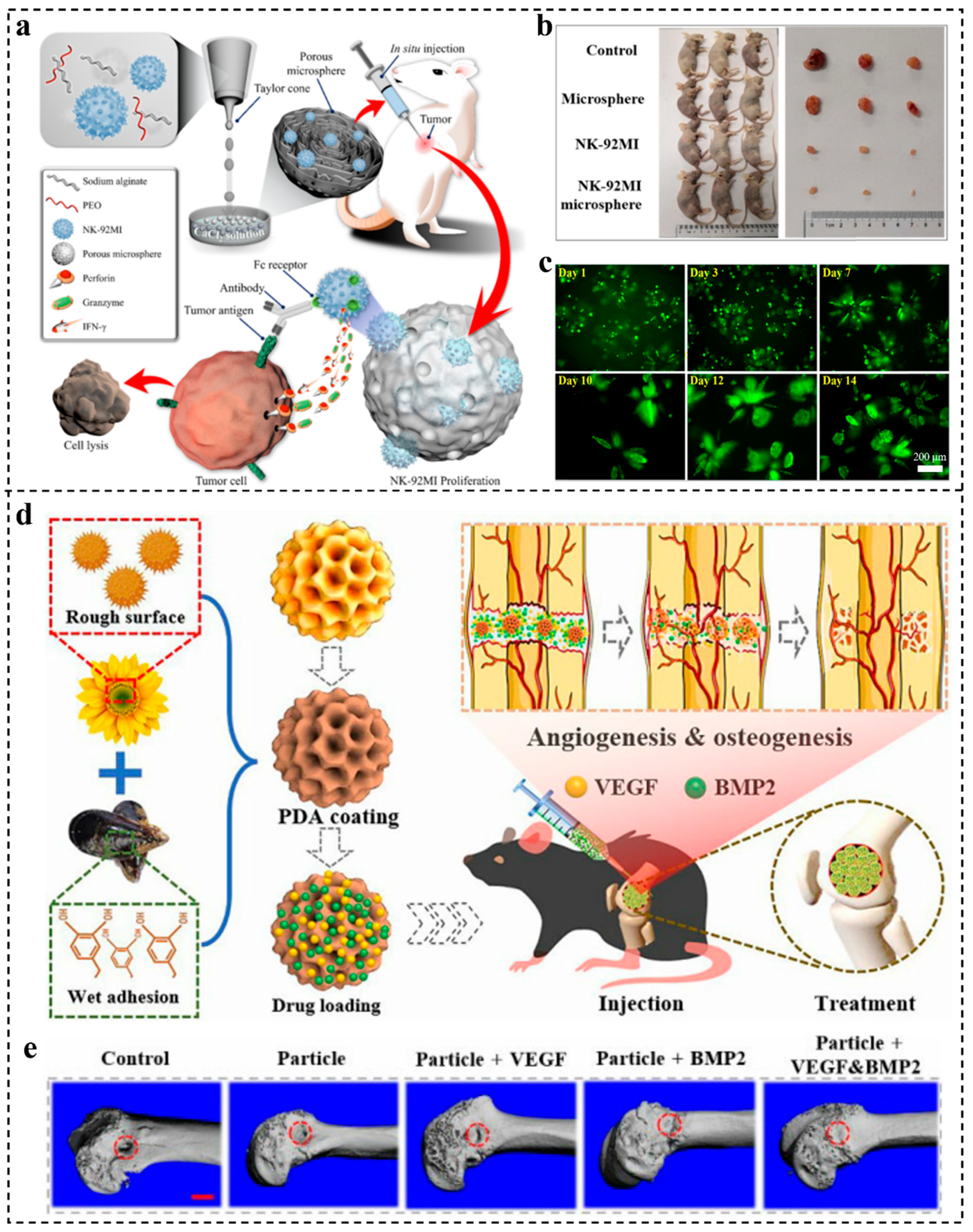
- Wu, D.; Yu, Y.; Zhao, C.; Shou, X.; Piao, Y.; Zhao, X.; Zhao, Y.; Wang, S. NK-Cell-Encapsulated Porous Microspheres via Microfluidic Electrospray for Tumor Immunotherapy. ACS Appl. Mater. Interfaces 2019, 11, 33716–33724. [Google Scholar] [CrossRef] [PubMed]
- Yang, L.; Wang, X.; Yu, Y.; Shang, L.; Xu, W.; Zhao, Y. Bio-inspired dual-adhesive particles from microfluidic electrospray for bone regeneration. Nano Res. 2022. [Google Scholar] [CrossRef]
- Wu, C.; Luo, X.; Baldursdottir, S.G.; Yang, M.; Sun, X.; Mu, H. In vivo evaluation of solid lipid microparticles and hybrid polymer-lipid microparticles for sustained delivery of leuprolide. Eur. J. Pharm. Biopharm. 2019, 142, 315–321. [Google Scholar] [CrossRef]


| Method | Advantages | Disadvantages | References |
|---|---|---|---|
| Emulsification | 1. W1/O/W2 does not require adjustment of the pH and significant change in the temperature 2. Single emulsification is relatively simple | 1. Waste generation 2. Use of one or more surfactants 3. Requires multiple steps 4. Low yield and high purification cost | [32,33] |
| Phase separation | 1. Simple equipment 2. Wide range of polymer materials 3. Encapsulation of variety of drugs | 1. The problems of the adhesion and aggregation of microspheres 2. The conditions are difficult to control during the process of the microspheres | [34,35] |
| Electrospray | 1. Preparation of high-purity microspheres 2. Suitable for many types of polymers 3. The operation is relatively simple | 1. In some cases, a crosslinking agent is used 2. There are many factors affecting particle size | [36,37] |
| Microfluidics | 1. Low cost 2. High size controllability 3. Small droplet volume 4. The particle size is highly homogeneous 5. The device is relatively simple | 1. Use of various solvents to remove the oil phase 2. The precision requirements of fluidic devices are high | [38,39] |
| Template synthesis | 1. The condition is relatively mild 2. The biological template material is non-toxic to the human body | 1. Templates need to be embellished 2. No template is used narrowly | [40] |
| Supercritical fluids | 1. Good process reproducibility 2. Little effect on the stability of the drug | 1. Microspheres may adhere to the inside of the spray dryer, causing material loss | [41] |
| Property | Composition | Molecular Structure | Method | Application | Reference |
|---|---|---|---|---|---|
| Natural polymers | Sodium Alginate |  | Electrospray | Wound dressing | [54,69] |
| Chitosan |  | Supercritical fluids | Wound dressing | [69,70] | |
| Collagen |  | Emulsification | Skin repair | [71,72] | |
| Gelatin |  | Electrospray | Packaging for food | [73] | |
| GelMA |  | Microfluidic | Skin closure | [74] | |
| Synthetic polymers | Polylactic acid (PLA) |  | Co-solvent evaporation | Sutures | [75] |
| Polyglycolic acid (PGA) |  | Emulsification | Surgical Suture | [76] | |
| PGA-PLA copolymer |  | Emulsification | Implants | [77] | |
| Poly- (orthoesters) (PEO) |  | Emulsification | Drug delivery | [78] | |
| Polycarbonate |  | Emulsification | Drug delivery | [79] |
Disclaimer/Publisher’s Note: The statements, opinions and data contained in all publications are solely those of the individual author(s) and contributor(s) and not of MDPI and/or the editor(s). MDPI and/or the editor(s) disclaim responsibility for any injury to people or property resulting from any ideas, methods, instructions or products referred to in the content. |
© 2023 by the authors. Licensee MDPI, Basel, Switzerland. This article is an open access article distributed under the terms and conditions of the Creative Commons Attribution (CC BY) license (https://creativecommons.org/licenses/by/4.0/).
Share and Cite
Yang, C.; Zhang, Z.; Gan, L.; Zhang, L.; Yang, L.; Wu, P. Application of Biomedical Microspheres in Wound Healing. Int. J. Mol. Sci. 2023, 24, 7319. https://doi.org/10.3390/ijms24087319
Yang C, Zhang Z, Gan L, Zhang L, Yang L, Wu P. Application of Biomedical Microspheres in Wound Healing. International Journal of Molecular Sciences. 2023; 24(8):7319. https://doi.org/10.3390/ijms24087319
Chicago/Turabian StyleYang, Caihong, Zhikun Zhang, Lu Gan, Lexiang Zhang, Lei Yang, and Pan Wu. 2023. "Application of Biomedical Microspheres in Wound Healing" International Journal of Molecular Sciences 24, no. 8: 7319. https://doi.org/10.3390/ijms24087319