
Development of Small-Molecule Fluorescent Probes Targeting Enzymes


Abstract
:1. Introduction
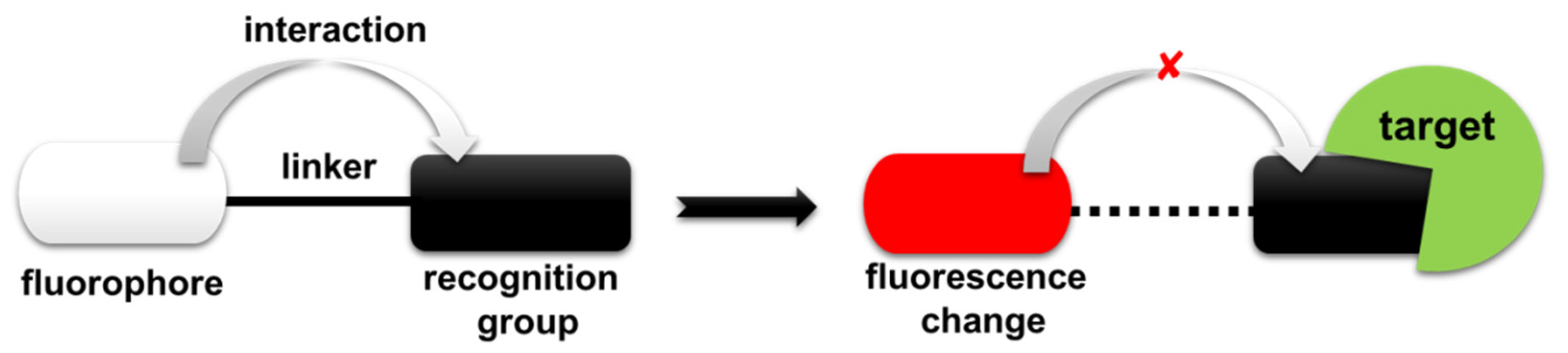
2. Design Principles of Fluorescent Probes
3. Categorization of Fluorescent Probes
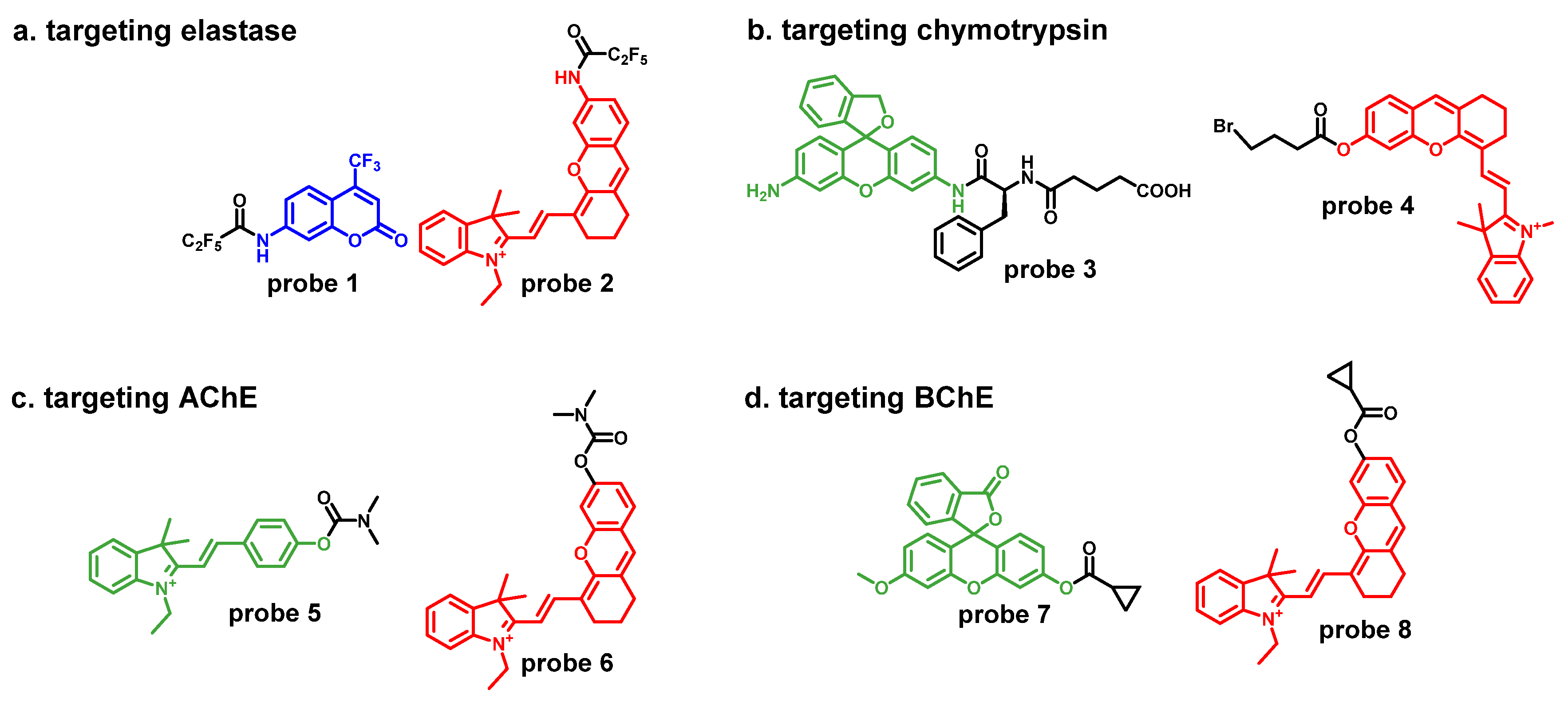
3.1. Fluorescent Probes Targeting Serine Hydrolases
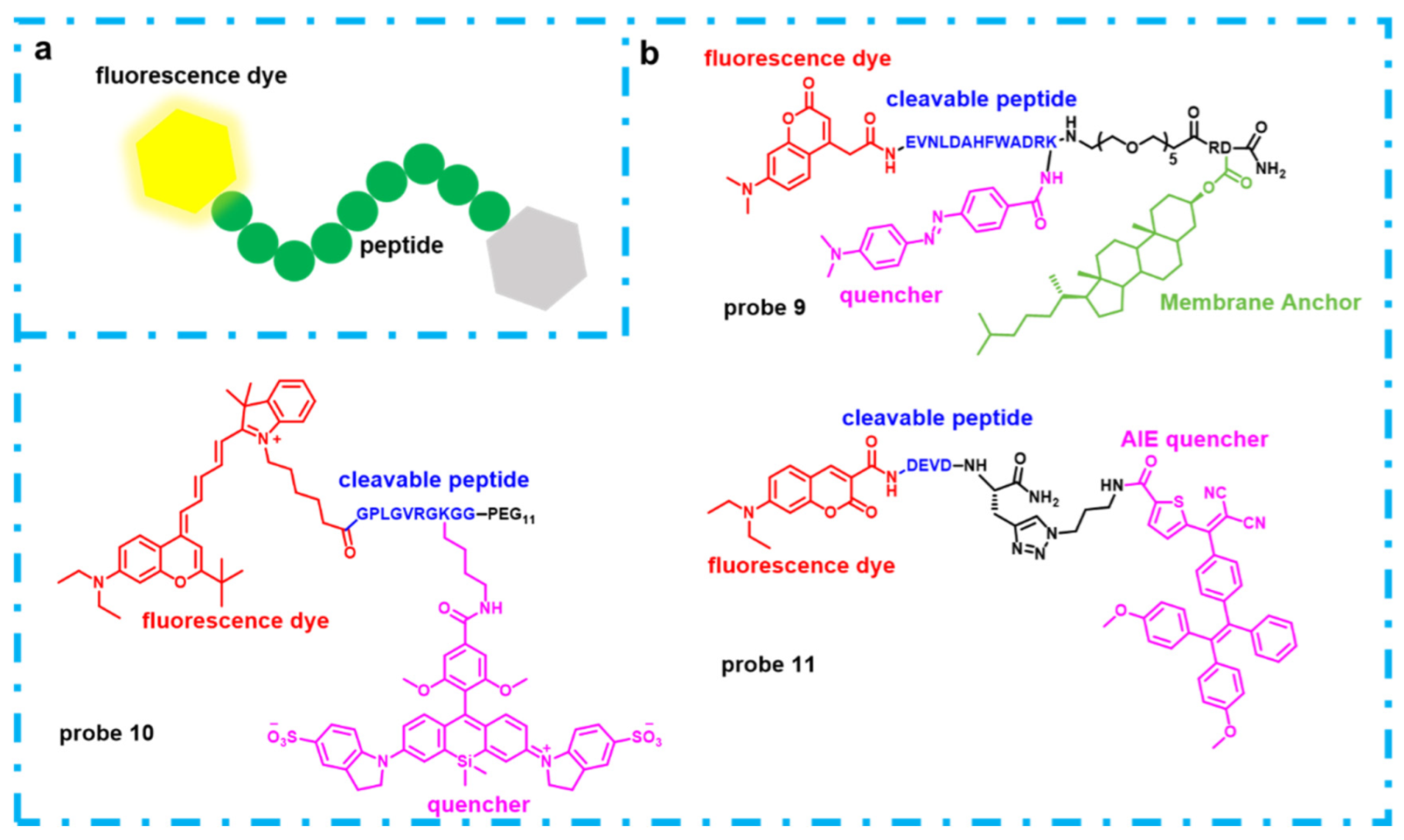
3.2. Fluorescent Probes Targeting Proteases
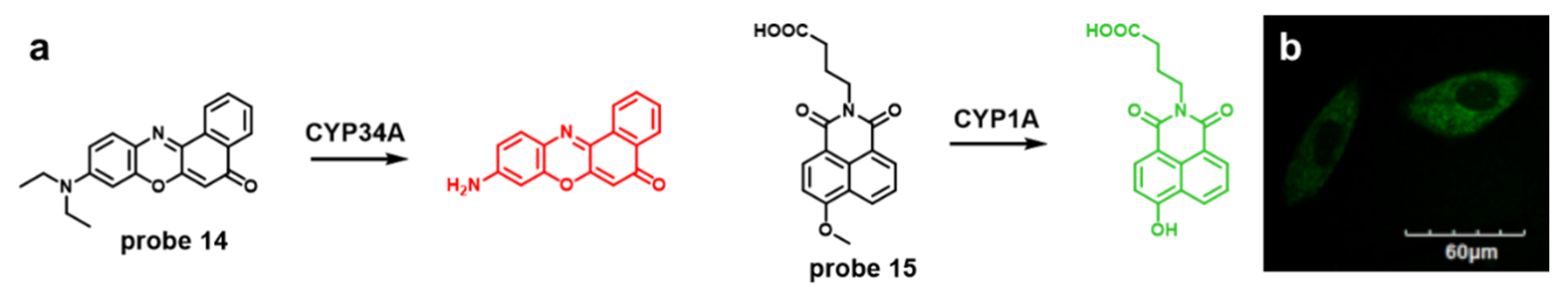
3.3. Fluorescent Probes Targeting Oxidoreductases
4. Application of Fluorescent Probes Targeting Enzymes in Fluorescence-Guided Surgery
5. Future Development of Fluorescent Probes
Author Contributions
Funding
Conflicts of Interest
References
- Hoshino, Y.; Hanaoka, K.; Sakamoto, K.; Yasunaga, M.; Kojima, T.; Kotani, D.; Nomoto, A.; Sasaki, E.; Komatsu, T.; Ueno, T.; et al. Molecular Design of Near-Infrared (NIR) Fluorescent Probes Targeting Exopeptidase and Application for Detection of Dipeptidyl Peptidase 4 (DPP-4) Activity. RSC Chem. Biol. 2022. [Google Scholar] [CrossRef]
- Yagishita, A.; Ueno, T.; Tsuchihara, K.; Urano, Y. Amino BODIPY-Based Blue Fluorescent Probes for Aldehyde Dehydrogenase 1-Expressing Cells. Bioconjug. Chem. 2021, 32, 234–238. [Google Scholar] [CrossRef] [PubMed]
- Chin, J.; Kim, H.J. Near-Infrared Fluorescent Probes for Peptidases. Coord. Chem. Rev. 2018, 354, 169–181. [Google Scholar] [CrossRef]
- Yanagi, K.; Komatsu, T.; Ogihara, S.; Okabe, T.; Kojima, H.; Nagano, T.; Ueno, T.; Hanaoka, K.; Urano, Y. Establishment of Live-Cell-Based Coupled Assay System for Identification of Compounds to Modulate Metabolic Activities of Cells. Cell Reports 2021, 36, 109311. [Google Scholar] [CrossRef]
- Sakamoto, S.; Komatsu, T.; Watanabe, R.; Zhang, Y.; Inoue, T.; Kawaguchi, M.; Nakagawa, H.; Ueno, T.; Okusaka, T.; Honda, K.; et al. Multiplexed Single-Molecule Enzyme Activity Analysis for Counting Disease-Related Proteins in Biological Samples. Sci. Adv. 2020, 6, eaay0888. [Google Scholar] [CrossRef] [Green Version]
- Chen, L.; Li, J.; Du, L.; Li, M. Strategies in the Design of Small-Molecule Fluorescent Probes for Peptidases. Med. Res. Rev. 2014, 34, 1217–1241. [Google Scholar] [CrossRef]
- Fei, X.; Gu, Y. Progress in Modifications and Applications of Fluorescent Dye Probe. Prog. Nat. Sci. 2009, 19, 1–7. [Google Scholar] [CrossRef]
- Chen, X.; Bi, Y.; Wang, T.; Li, P.; Yan, X.; Hou, S.; Bammert, C.E.; Ju, J.; Gibson, K.M.; Pavan, W.J.; et al. Lysosomal Targeting with Stable and Sensitive Fluorescent Probes (Superior LysoProbes): Applications for Lysosome Labeling and Tracking during Apoptosis. Sci. Rep. 2015, 5, 1–10. [Google Scholar] [CrossRef] [Green Version]
- Rajapaksha, A.A.; Fu, Y.X.; Guo, W.Y.; Liu, S.Y.; Li, Z.W.; Xiong, C.Q.; Yang, W.C.; Yang, G.F. Review on the Recent Progress in the Development of Fluorescent Probes Targeting Enzymes. Methods Appl. Fluoresc. 2021, 9, 032001. [Google Scholar] [CrossRef]
- Liu, R.; Lu, P.; Chu, J.W.K.; Sharom, F.J. Characterization of Fluorescent Sterol Binding to Purified Human NPC1. J. Biol. Chem. 2009, 284, 1840–1852. [Google Scholar] [CrossRef] [Green Version]
- Demchenko, A.P. Visualization and Sensing of Intermolecular Interactions with Two-Color Fluorescent Probes. FEBS Lett. 2006, 580, 2951–2957. [Google Scholar] [CrossRef] [PubMed] [Green Version]
- Arrowsmith, C.H.; Audia, J.E.; Austin, C.; Baell, J.; Bennett, J.; Blagg, J.; Bountra, C.; Brennan, P.E.; Brown, P.J.; Bunnage, M.E.; et al. The Promise and Peril of Chemical Probes. Nat. Chem. Biol. 2015, 11, 536–541. [Google Scholar] [CrossRef] [PubMed] [Green Version]
- Dai, N.; Kool, E.T. Fluorescent DNA-based enzyme sensors. Chem. Soc. Rev. 2011, 40, 5756–5770. [Google Scholar] [CrossRef] [PubMed] [Green Version]
- Terai, T.; Nagano, T. Fluorescent Probes for Bioimaging Applications. Curr. Opin. Chem. Biol. 2008, 12, 515–521. [Google Scholar] [CrossRef]
- Sabanayagam, C.R.; Eid, J.S.; Meller, A. Long Time Scale Blinking Kinetics of Cyanine Fluorophores Conjugated to DNA and Its Effect on Förster Resonance Energy Transfer. J. Chem. Phys. 2005, 123, 1–7. [Google Scholar] [CrossRef] [Green Version]
- Doose, S.; Neuweiler, H.; Sauer, M. Fluorescence Quenching by Photoinduced Electron Transfer: A Reporter for Conformational Dynamics of Macromolecules. ChemPhysChem 2009, 10, 1389–1398. [Google Scholar] [CrossRef]
- Santucci, A.; Bernardini, G.; Braconi, D.; Petricci, E.; Manetti, F. 4-Hydroxyphenylpyruvate Dioxygenase and Its Inhibition in Plants and Animals: Small Molecules as Herbicides and Agents for the Treatment of Human Inherited Diseases. J. Med. Chem. 2017, 60, 4101–4125. [Google Scholar] [CrossRef]
- Hao, M.; Chi, W.; Wang, C.; Xu, Z.; Li, Z.; Liu, X. Molecular Origins of Photoinduced Backward Intramolecular Charge Transfer. J. Phys. Chem. C 2020, 124, 16820–16826. [Google Scholar] [CrossRef]
- Kwon, J.E.; Park, S.Y. Advanced Organic Optoelectronic Materials: Harnessing Excited-State Intramolecular Proton Transfer (ESIPT) Process. Adv. Mater. 2011, 23, 3615–3642. [Google Scholar] [CrossRef]
- Hong, Y.; Lam, J.W.Y.; Tang, B.Z. Aggregation-Induced Emission. Chem. Soc. Rev. 2011, 40, 5361–5388. [Google Scholar] [CrossRef] [Green Version]
- Liu, H.W.; Chen, L.; Xu, C.; Li, Z.; Zhang, H.; Zhang, X.B.; Tan, W. Recent Progresses in Small-Molecule Enzymatic Fluorescent Probes for Cancer Imaging. Chem. Soc. Rev. 2018, 47, 7140–7180. [Google Scholar] [CrossRef]
- Liu, S.Y.; Qu, R.Y.; Li, R.R.; Yan, Y.C.; Sun, Y.; Yang, W.C.; Yang, G.F. An Activity-Based Fluorogenic Probe Enables Cellular and in Vivo Profiling of Carboxylesterase Isozymes. Anal. Chem. 2020, 92, 9205–9213. [Google Scholar] [CrossRef] [PubMed]
- Sun, Q.; Li, J.; Liu, W.N.; Dong, Q.J.; Yang, W.C.; Yang, G.F. Non-Peptide-Based Fluorogenic Small-Molecule Probe for Elastase. Anal. Chem. 2013, 85, 11304–11311. [Google Scholar] [CrossRef] [PubMed]
- Yamashita, S.; Sakabe, M.; Ishizawa, T.; Hasegawa, K.; Urano, Y.; Kokudo, N. Visualization of the Leakage of Pancreatic Juice Using a Chymotrypsin-Activated Fluorescent Probe. Br. J. Surg. 2013, 100, 1220–1228. [Google Scholar] [CrossRef] [PubMed]
- Mu, S.; Xu, Y.; Zhang, Y.; Guo, X.; Li, J.; Wang, Y.; Liu, X.; Zhang, H. A Non-Peptide NIR Fluorescent Probe for Detection of Chymotrypsin and Its Imaging Application. J. Mater. Chem. B 2019, 7, 2974–2980. [Google Scholar] [CrossRef]
- Ma, J.; Si, T.; Yan, C.; Li, Y.; Li, Q.; Lu, X.; Guo, Y. Near-Infrared Fluorescence Probe for Evaluating Acetylcholinesterase Activity in PC12 Cells and in Situ Tracing AChE Distribution in Zebrafish. ACS Sens. 2020, 5, 83–92. [Google Scholar] [CrossRef]
- Wang, X.; Li, P.; Ding, Q.; Wu, C.; Zhang, W.; Tang, B. Observation of Acetylcholinesterase in Stress-Induced Depression Phenotypes by Two-Photon Fluorescence Imaging in the Mouse Brain. J. Am. Chem. Soc. 2019, 141, 2061–2068. [Google Scholar] [CrossRef]
- Yang, S.H.; Sun, Q.; Xiong, H.; Liu, S.Y.; Moosavi, B.; Yang, W.C.; Yang, G.F. Discovery of a Butyrylcholinesterase-Specific Probe via a Structure-Based Design Strategy. Chem. Commun. 2017, 53, 3952–3955. [Google Scholar] [CrossRef]
- Liu, S.Y.; Xiong, H.; Yang, J.Q.; Yang, S.H.; Li, Y.; Yang, W.C.; Yang, G.F. Discovery of Butyrylcholinesterase-Activated Near-Infrared Fluorogenic Probe for Live-Cell and in Vivo Imaging. ACS Sensors 2018, 3, 2118–2128. [Google Scholar] [CrossRef]
- Lian, I.; Ong, H.; Lian, I.; Ong, H. Recent Developments in Protease Activity Assays and Sensors. Analyst 2017, 142, 1867–1881. [Google Scholar] [CrossRef] [Green Version]
- Agbowuro, A.A.; Huston, W.M.; Gamble, A.B.; Tyndall, J.D. Proteases and protease inhibitors in infectious diseases. Med. Res. Rev. 2018, 38, 1295–1331. [Google Scholar] [CrossRef] [PubMed]
- Ghosh, A.K.; Brindisi, M.; Tang, J. Developing β-Secretase Inhibitors for Treatment of Alzheimer’s Disease. J. Neurochem. 2012, 120, 71–83. [Google Scholar] [CrossRef] [PubMed] [Green Version]
- Folk, D.S.; Torosian, J.C.; Hwang, S.; McCafferty, D.G.; Franz, K.J. Monitoring β-Secretase Activity in Living Cells with a Membrane-Anchored FRET Probe. Angew. Chemie Int. Ed. 2012, 51, 10795–10799. [Google Scholar] [CrossRef] [PubMed] [Green Version]
- Isaacson, K.J.; Martin Jensen, M.; Subrahmanyam, N.B.; Ghandehari, H. Matrix-Metalloproteinases as Targets for Controlled Delivery in Cancer: An Analysis of Upregulation and Expression. J. Control. Release 2017, 259, 62–75. [Google Scholar] [CrossRef] [PubMed]
- Myochin, T.; Hanaoka, K.; Iwaki, S.; Ueno, T.; Komatsu, T.; Terai, T.; Nagano, T.; Urano, Y. Development of a Series of Near-Infrared Dark Quenchers Based on Si-Rhodamines and Their Application to Fluorescent Probes. J. Am. Chem. Soc. 2015, 137, 4759–4765. [Google Scholar] [CrossRef]
- Yamaguchi, Y.; Kuranaga, E.; Nakajima, Y.I.; Koto, A.; Takemoto, K.; Miura, M. In vivo monitoring of caspase activation using a fluorescence resonance energy transfer-based fluorescent probe. In Methods in Enzymology. Acad. Press 2014, 544, 299–325. [Google Scholar] [CrossRef]
- Yuan, Y.; Zhang, R.; Cheng, X.; Xu, S.; Liu, B. A FRET Probe with AIEgen as the Energy Quencher: Dual Signal Turn-on for Self-Validated Caspase Detection. Chem. Sci. 2016, 7, 4245–4250. [Google Scholar] [CrossRef] [Green Version]
- Sellés Vidal, L.; Kelly, C.L.; Mordaka, P.M.; Heap, J.T. Review of NAD(P)H-Dependent Oxidoreductases: Properties, Engineering and Application. Biochim. Et Biophys. Acta (BBA)-Proteins Proteom. 2018, 1866, 327–347. [Google Scholar] [CrossRef]
- Qiao, J.; Wang, M.; Cui, M.; Fang, Y.; Li, H.; Zheng, C.; Li, Z.; Xu, Y.; Hua, H.; Li, D. Small-Molecule Probes for Fluorescent Detection of Cellular Hypoxia-Related Nitroreductase. J. Pharm. Biomed. Anal. 2021, 203, 114199. [Google Scholar] [CrossRef]
- Gao, J.; Yin, X.; Li, M.; Chen, J.A.; Tan, J.; Zhao, Z.; Gu, X. Rational Design of Fluorescent Probes for Targeted: In Vivo Nitroreductase Visualization. Org. Biomol. Chem. 2020, 18, 4744–4747. [Google Scholar] [CrossRef]
- Xu, A.; Tang, Y.; Lin, W. Endoplasmic Reticulum-Targeted Two-Photon Turn-on Fluorescent Probe for Nitroreductase in Tumor Cells and Tissues. Spectrochim. Acta-Part A Mol. Biomol. Spectrosc. 2018, 204, 770–776. [Google Scholar] [CrossRef] [PubMed]
- Coon, M.J. Omega Oxygenases: Nonheme-Iron Enzymes and P450 Cytochromes. Biochem. Biophys. Res. Commun. 2005, 338, 378–385. [Google Scholar] [CrossRef] [PubMed]
- Wang, D.; Guo, Y.; Wrighton, S.A.; Cooke, G.E.; Sadee, W. Intronic Polymorphism in CYP3A4 Affects Hepatic Expression and Response to Statin Drugs. Pharm. J. 2011, 11, 274–286. [Google Scholar] [CrossRef] [Green Version]
- Lampe, J.N.; Fernandez, C.; Nath, A.; Atkins, W.M. Nile Red Is a Fluorescent Allosteric Substrate of Cytochrome P450 3A4. Biochemistry 2008, 47, 509–516. [Google Scholar] [CrossRef] [PubMed]
- Lu, J.; Shang, X.; Zhong, W.; Xu, Y.; Shi, R.; Wang, X. New Insights of CYP1A in Endogenous Metabolism: A Focus on Single Nucleotide Polymorphisms and Diseases. Acta Pharm. Sin. B 2020, 10, 91–104. [Google Scholar] [CrossRef] [PubMed]
- Dai, Z.R.; Ge, G.B.; Feng, L.; Ning, J.; Hu, L.H.; Jin, Q.; Wang, D.D.; Lv, X.; Dou, T.Y.; Cui, J.N.; et al. A Highly Selective Ratiometric Two-Photon Fluorescent Probe for Human Cytochrome P450 1A. J. Am. Chem. Soc. 2015, 137, 14488–14495. [Google Scholar] [CrossRef]
- Faletrov, Y.V.; Frolova, N.S.; Hlushko, H.V.; Rudaya, E.V.; Edimecheva, I.P.; Mauersberger, S.; Shkumatov, V.M. Evaluation of the Fluorescent Probes Nile Red and 25-NBD-Cholesterol as Substrates for Steroid-Converting Oxidoreductases Using Pure Enzymes and Microorganisms. FEBS J. 2013, 280, 3109–3119. [Google Scholar] [CrossRef]
- Lee, W.S.; Ham, W.; Kim, J. Roles of NAD(P)H:Quinone Oxidoreductase 1 in Diverse Diseases. Life 2021, 11, 1301. [Google Scholar] [CrossRef]
- Carpino, L.A.; Triolo, S.A.; Berglund, R.A. Reductive Lactonization of Strategically Methylated Quinone Propionic Acid Esters and Amides. J. Org. Chem. 1989, 54, 3303–3310. [Google Scholar] [CrossRef]
- Dai, M.; Song, C.W.; Yang, Y.J.; Kim, H.R.; Reo, Y.J.; Ahn, K.H. Toward Ratiometric Detection of NAD(P)H Quinone Oxidoreductase-1: Benzocoumarin-Based Fluorescent Probes. Sens. Actuators B Chem. 2021, 330, 129277. [Google Scholar] [CrossRef]
- Yang, Y.P.; Qi, F.J.; Qian, Y.P.; Bao, X.Z.; Zhang, H.C.; Ma, B.; Dai, F.; Zhang, S.X.; Zhou, B. Developing Push-Pull Hydroxylphenylpolyenylpyridinium Chromophores as Ratiometric Two-Photon Fluorescent Probes for Cellular and Intravital Imaging of Mitochondrial NQO1. Anal. Chem. 2021, 93, 2385–2393. [Google Scholar] [CrossRef] [PubMed]
- Zhang, Y.; Zhang, G.; Zeng, Z.; Pu, K. Activatable Molecular Probes for Fluorescence-Guided Surgery, Endoscopy and Tissue Biopsy. Chem. Soc. Rev. 2022, 51, 566–593. [Google Scholar] [CrossRef] [PubMed]
- Mochida, A.; Ogata, F.; Nagaya, T.; Choyke, P.L.; Kobayashi, H. Activatable Fluorescent Probes in Fluorescence-Guided Surgery: Practical Considerations. Bioorganic Med. Chem. 2018, 26, 925–930. [Google Scholar] [CrossRef] [PubMed]
- Li, H.; Yao, Q.; Sun, W.; Shao, K.; Lu, Y.; Chung, J.; Kim, D.; Fan, J.; Long, S.; Du, J.; et al. Aminopeptidase N Activatable Fluorescent Probe for Tracking Metastatic Cancer and Image-Guided Surgery via in Situ Spraying. J. Am. Chem. Soc. 2020, 142, 6381–6389. [Google Scholar] [CrossRef]
- Tang, C.; Du, Y.; Liang, Q.; Cheng, Z.; Tian, J. Development of a Novel Histone Deacetylase-Targeted Near-Infrared Probe for Hepatocellular Carcinoma Imaging and Fluorescence Image-Guided Surgery. Mol. Imaging Biol. 2020, 22, 476–485. [Google Scholar] [CrossRef]
- Widen, J.C.; Tholen, M.; Yim, J.J.; Antaris, A.; Casey, K.M.; Rogalla, S.; Klaassen, A.; Sorger, J.; Bogyo, M. AND-Gate Contrast Agents for Enhanced Fluorescence-Guided Surgery. Nat. Biomed. Eng. 2021, 5, 264–277. [Google Scholar] [CrossRef]


| NO | λex (nm) | λem (nm) | LOD | Target | Application | Ref. |
|---|---|---|---|---|---|---|
| probe 1 | 340 | 505 | 68 ng/mL | elastase | inhibitor screening | [23] |
| probe 2 | 670 | 700 | 29.6 ng/mL | elastase | cell and mice imaging | [22] |
| probe 3 | 490 | 520 | / | chymotrypsin | clinical detection | [24] |
| probe 4 | 670 | 695 | 13 mU/mL | chymotrypsin | cell and mice imaging | [25] |
| probe 5 | 520 | 560 | 0.36 U/mL | AChE | cell and mice imaging | [27] |
| probe 6 | 670 | 700 | 117.3 mU/mL | AChE | cell and zebrafish imaging | [26] |
| probe 7 | 455 | 515 | / | BChE | inhibitor screening and cell imaging | [28] |
| probe 8 | 665 | 705 | / | BChE | cell, zebrafish and mice imaging | [29] |
| probe 9 | 390 | 470 | / | BACE1 | inhibitor screening and cell imaging | [33] |
| probe 10 | 720 | 750 | / | MMPs | cell and mice imaging | [35] |
| probe 11 | 405 | 465 and 665 | / | Caspases | cell imaging | [37] |
| probe 12 | 450 | 505 | 22 ng/mL | NTR | cell and mice imaging | [40] |
| probe 13 | 440 | 543 | 36 ng/mL | NTR | cell and tissue slices imaging | [41] |
| probe 14 | 470 | 570 | / | CYP34A | / | [44] |
| probe 15 | 452 | 564 | 0.02 nmol/mL | CYP1A | cell and tissue slices imaging | [46] |
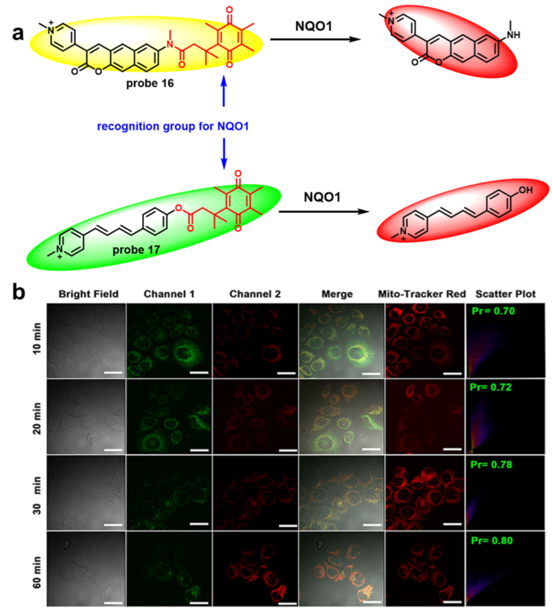
| probe 16 | 405 | 650/555 | 4.99 μg/mL | NOQ1 | cell imaging | [50] |
| probe 17 | 407 | 564/480 | 0.9 nM | NQO1 | cell and mice imaging | [49] |
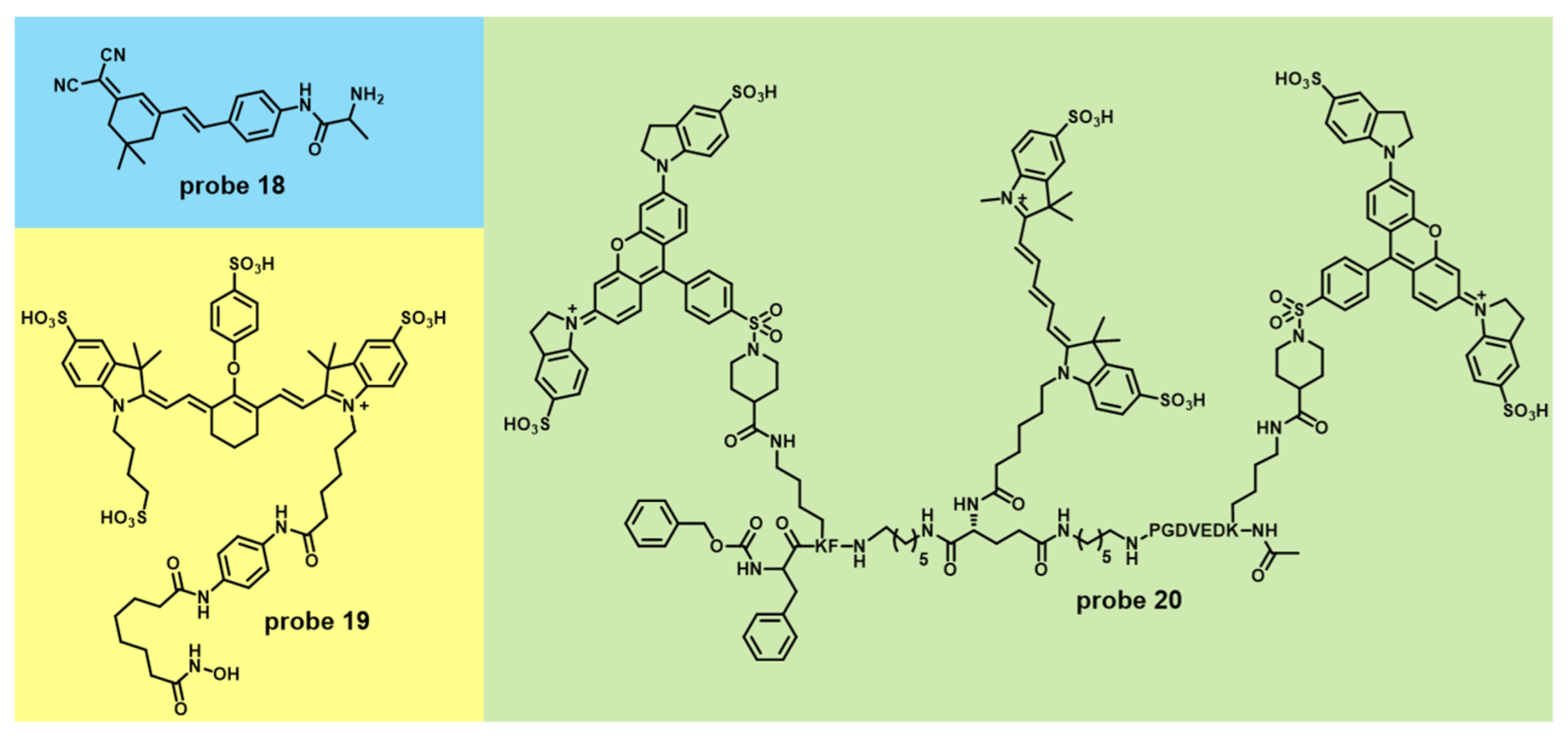
| probe 18 | 445 | 650 | 0.13 ng/ml | APN | cell imaging and fluorescence-guided surgery | [53] |
| probe 19 | 775 | 801 | / | HDACs | fluorescence-guided surgery | [54] |
| probe 20 | 640 | 670 | / | cathepsin and caspase 3 | cell, mice imaging and fluorescence-guided surgery | [55] |
Publisher’s Note: MDPI stays neutral with regard to jurisdictional claims in published maps and institutional affiliations. |
© 2022 by the authors. Licensee MDPI, Basel, Switzerland. This article is an open access article distributed under the terms and conditions of the Creative Commons Attribution (CC BY) license (https://creativecommons.org/licenses/by/4.0/).
Share and Cite
Li, Y.-X.; Xie, D.-T.; Yang, Y.-X.; Chen, Z.; Guo, W.-Y.; Yang, W.-C. Development of Small-Molecule Fluorescent Probes Targeting Enzymes. Molecules 2022, 27, 4501. https://doi.org/10.3390/molecules27144501
Li Y-X, Xie D-T, Yang Y-X, Chen Z, Guo W-Y, Yang W-C. Development of Small-Molecule Fluorescent Probes Targeting Enzymes. Molecules. 2022; 27(14):4501. https://doi.org/10.3390/molecules27144501
Chicago/Turabian StyleLi, Yuan-Xiang, Dong-Tai Xie, Ya-Xi Yang, Zhao Chen, Wu-Yingzheng Guo, and Wen-Chao Yang. 2022. "Development of Small-Molecule Fluorescent Probes Targeting Enzymes" Molecules 27, no. 14: 4501. https://doi.org/10.3390/molecules27144501