

A Hydroxytricyanopyrrole-Based Fluorescent Probe for Sensitive and Selective Detection of Hypochlorous Acid
 , and
, and 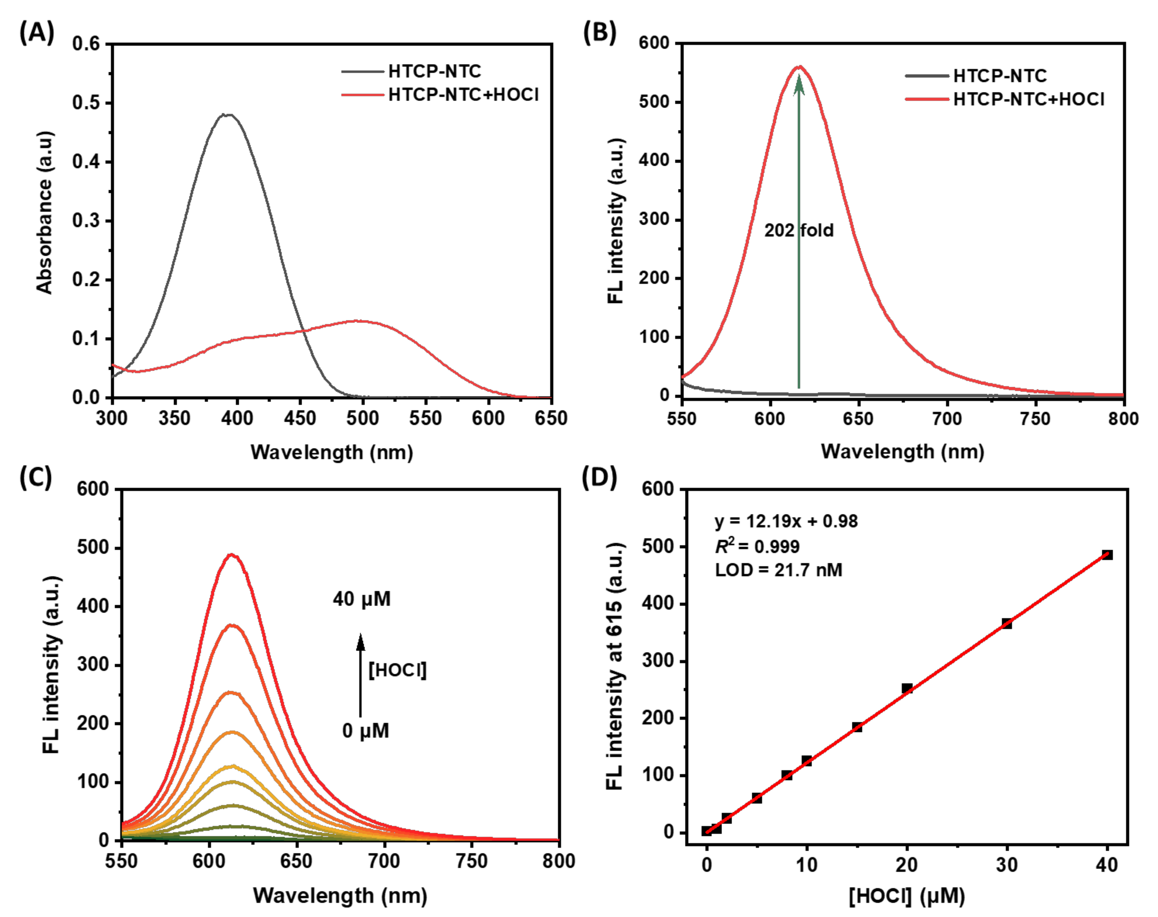
Abstract
:1. Introduction
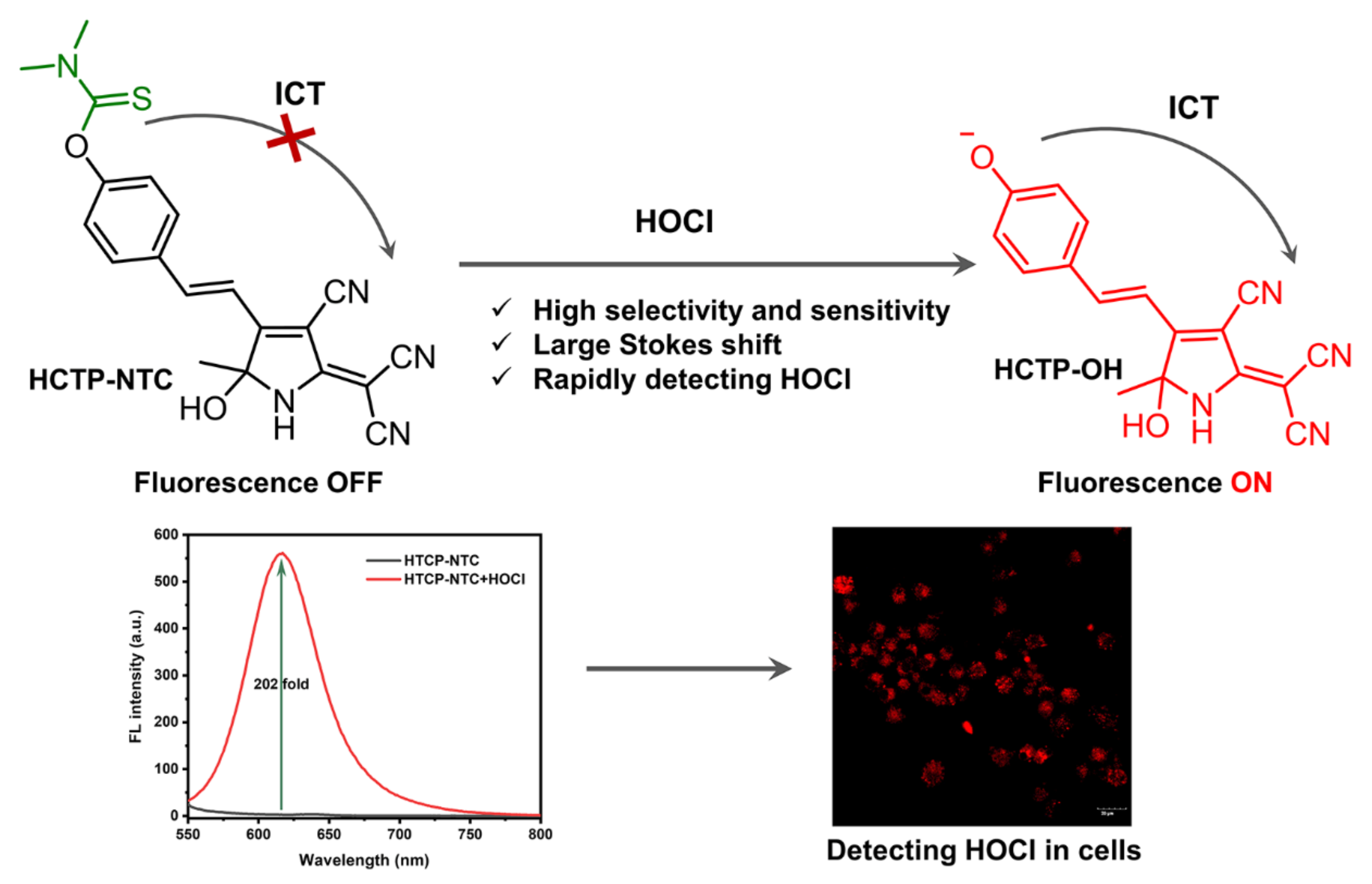
2. Results and Discussion
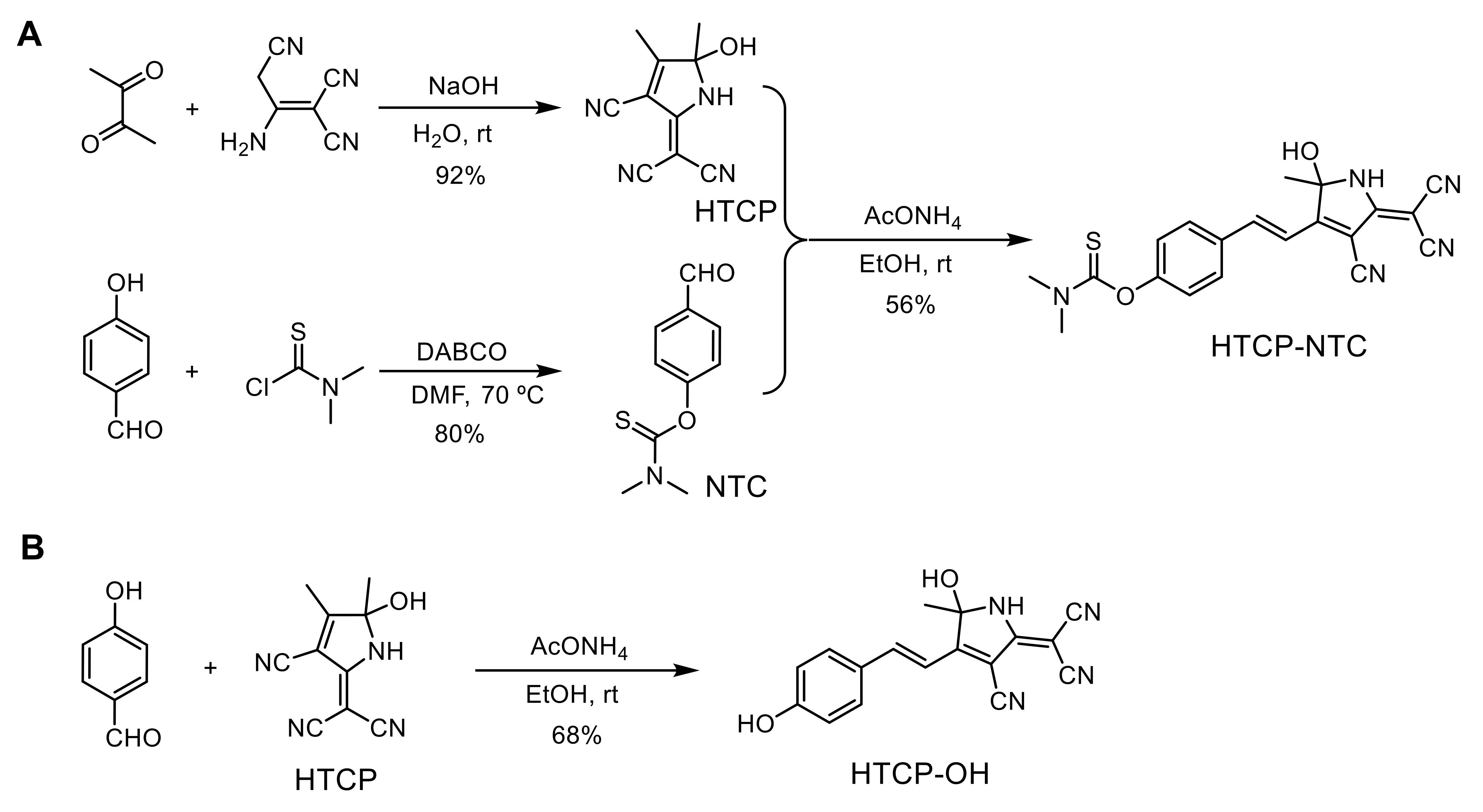
2.1. Design and Synthesis of HTCP-NTC
2.2. The Spectroscopic Properties of HTCP-NTC
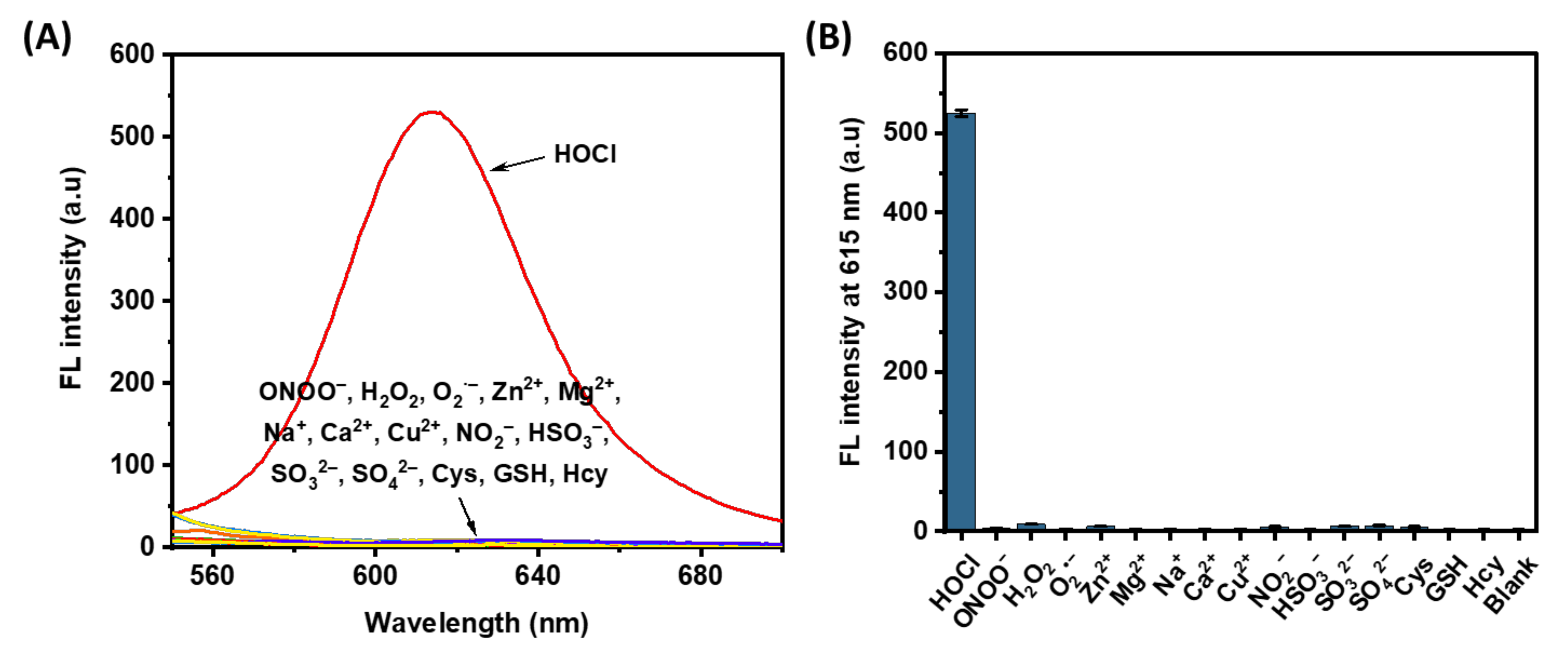
2.3. Selectivity of the HTCP-NTC
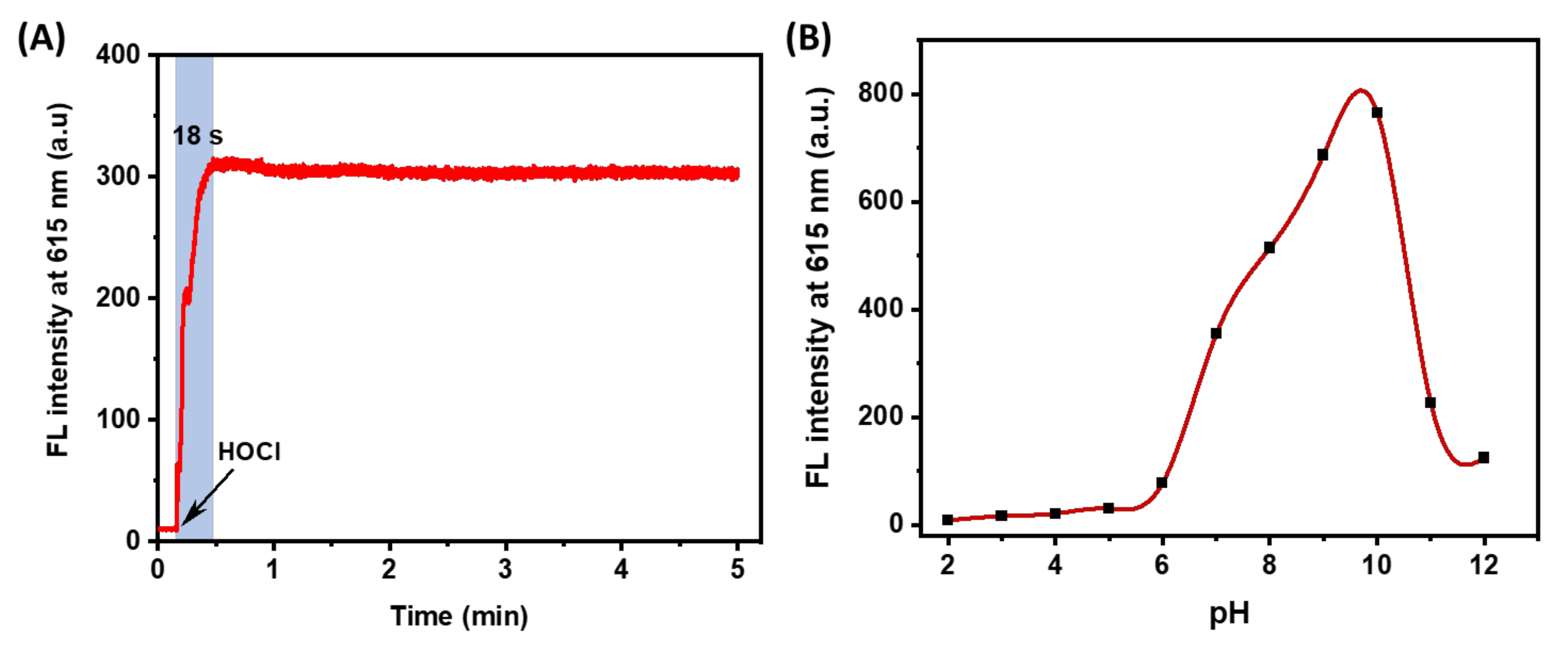
2.4. Reaction Kinetics and Environment Effects
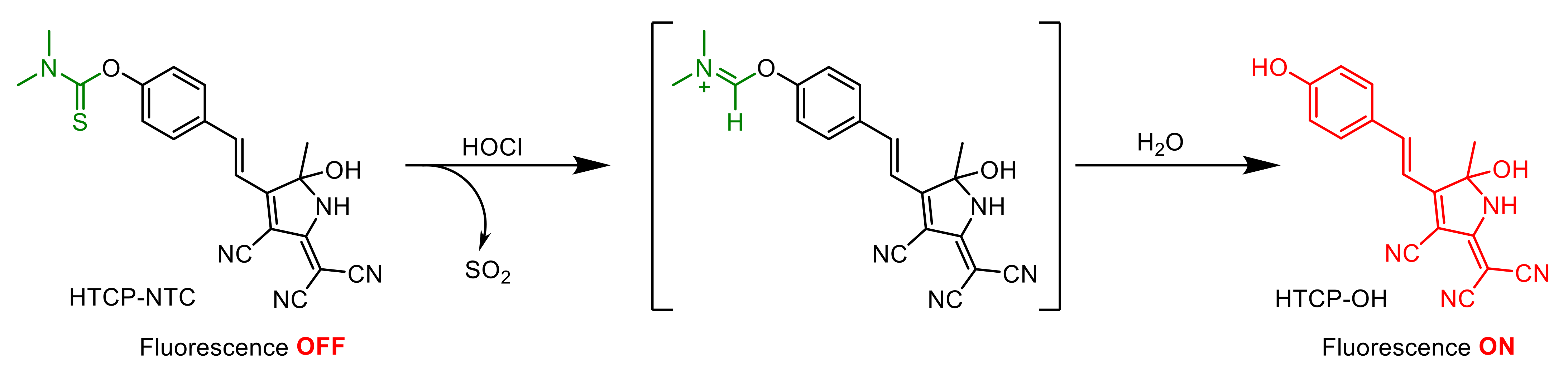
2.5. Mechanism of HTCP-NTC Detection of HOCl
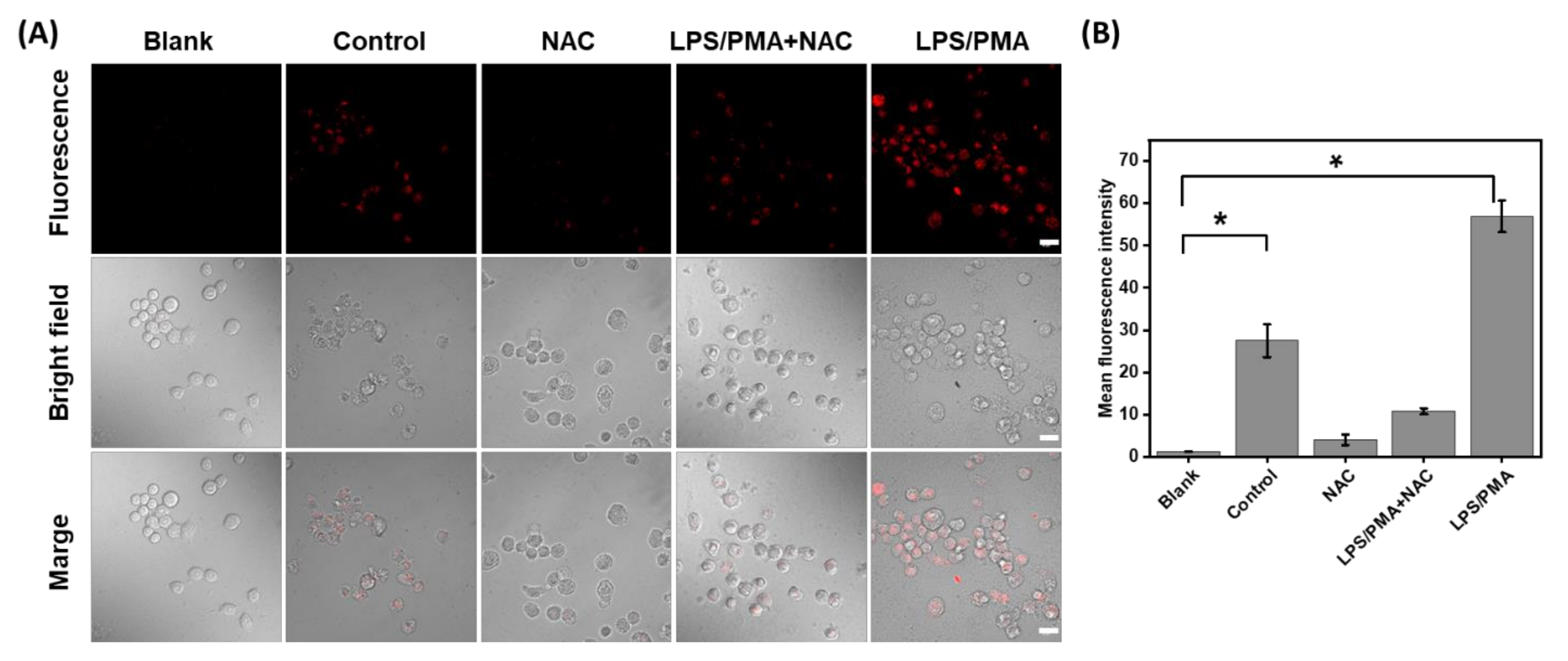
2.6. Fluorescence Imaging of HOCl in Living Cells
3. Materials and Methods
3.1. Reagents and Apparatus
3.2. Synthesis of the HTCP
3.3. Synthesis of the NTC
3.4. Synthesis of HTCP-NTC
3.5. Synthesis of HTCP-OH
3.6. Spectrum of HTCP-NTC with HOCl
3.7. Determination of the Fluorescence Quantum Yield
3.8. Determination of LOD
3.9. Reaction Kinetics of HTCP-NTC with HOCl
3.10. Cell Culture and Cytotoxicity Assays
3.11. Confocal Fluorescence Imaging
4. Conclusions
Supplementary Materials
Author Contributions
Funding
Institutional Review Board Statement
Informed Consent Statement
Data Availability Statement
Acknowledgments
Conflicts of Interest
Sample Availability
References
- Chen, X.; Wang, F.; Hyun, J.Y.; Wei, T.; Qiang, J.; Ren, X.; Shin, I.; Yoon, J. Recent Progress in the Development of Fluorescent, Luminescent and Colorimetric Probes for Detection of Reactive Oxygen and Nitrogen Species. Chem. Soc. Rev. 2016, 45, 2976–3016. [Google Scholar] [CrossRef] [PubMed]
- Butterfield, D.A.; Halliwell, B. Oxidative Stress, Dysfunctional Glucose Metabolism and Alzheimer Disease. Nat. Rev. Neurosci. 2019, 20, 148–160. [Google Scholar] [CrossRef] [PubMed]
- Zhang, W.; Li, P.; Yang, F.; Hu, X.; Sun, C.; Zhang, W.; Chen, D.; Tang, B. Dynamic and Reversible Fluorescence Imaging of Superoxide Anion Fluctuations in Live Cells and in Vivo. J. Am. Chem. Soc. 2013, 135, 14956–14959. [Google Scholar] [CrossRef] [PubMed]
- Finkel, T.; Serrano, M.; Blasco, M.A. The Common Biology of Cancer and Ageing. Nature 2007, 448, 767–774. [Google Scholar] [CrossRef] [Green Version]
- Panth, N.; Paudel, K.R.; Parajuli, K. Reactive Oxygen Species: A Key Hallmark of Cardiovascular Disease. Adv. Med. 2016, 2016, 9152732. [Google Scholar] [CrossRef] [Green Version]
- Moris, D.; Spartalis, M.; Spartalis, E.; Karachaliou, G.S.; Karaolanis, G.I.; Tsourouflis, G.; Tsilimigras, D.I.; Tzatzaki, E.; Theocharis, S. The Role of Reactive Oxygen Species in the Pathophysiology of Cardiovascular Diseases and the Clinical Significance of Myocardial Redox. Ann. Transtl. Med. 2017, 5, 326. [Google Scholar] [CrossRef] [Green Version]
- Barnham, K.J.; Masters, C.L.; Bush, A.I. Neurodegenerative Diseases and Oxidative Stress. Nat. Rev. Drug Discov. 2004, 3, 205–214. [Google Scholar] [CrossRef]
- Hammer, A.; Desoye, G.; Dohr, G.; Sattler, W.; Malle, E. Myeloperoxidase-Dependent Generation of Hypochlorite-Modified Proteins in Human Placental Tissues During Normal Pregnancy. Lab. Investig. 2001, 81, 543–554. [Google Scholar] [CrossRef] [Green Version]
- da Cruz Nizer, W.S.; Inkovskiy, V.; Overhage, J. Surviving Reactive Chlorine Stress: Responses of Gram-Negative Bacteria to Hypochlorous Acid. Microorganisms 2020, 8, 1220. [Google Scholar] [CrossRef]
- Sultana, S.; Foti, A.; Dahl, J.U. Bacterial Defense Systems against the Neutrophilic Oxidant Hypochlorous Acid. Infect. Immun. 2020, 88, e00964-19. [Google Scholar] [CrossRef]
- Wigginton, K.R.; Pecson, B.M.; Sigstam, T.; Bosshard, F.; Kohn, T. Virus Inactivation Mechanisms: Impact of Disinfectants on Virus Function and Structural Integrity. Environ. Sci. Technol. 2012, 46, 12069–12078. [Google Scholar] [CrossRef] [PubMed]
- Hawkins, C.L.; Pattison, D.I.; Davies, M.J. Hypochlorite-Induced Oxidation of Amino Acids, Peptides and Proteins. Amino Acids. 2003, 25, 259–274. [Google Scholar] [CrossRef] [PubMed]
- Hawkins, C.L.; Davies, M.J. Inactivation of Protease Inhibitors and Lysozyme by Hypochlorous Acid: Role of Side-Chain Oxidation and Protein Unfolding in Loss of Biological Function. Chem. Res. Toxicol. 2005, 18, 1600–1610. [Google Scholar] [CrossRef] [PubMed]
- Summers, F.A.; Forsman Quigley, A.; Hawkins, C.L. Identification of Proteins Susceptible to Thiol Oxidation in Endothelial Cells Exposed to Hypochlorous Acid and N-Chloramines. Biochem. Biophys. Res. Commun. 2012, 425, 157–161. [Google Scholar] [CrossRef] [PubMed]
- Wang, B.; Yuan, F.; Wang, S.; Duan, R.; Ren, W.X.; Hou, J.-T. Detection of Atherosclerosis-Associated HOCl Using a Mitochondria-Targeted Fluorescent Probe. Sens. Actuators B 2021, 348, 130695. [Google Scholar] [CrossRef]
- Wei, P.; Yuan, W.; Xue, F.; Zhou, W.; Li, R.; Zhang, D.; Yi, T. Deformylation Reaction-Based Probe for in Vivo Imaging of HOCl. Chem. Sci. 2018, 9, 495–501. [Google Scholar] [CrossRef] [Green Version]
- Feng, H.; Zhang, Z.; Meng, Q.; Jia, H.; Wang, Y.; Zhang, R. Rapid Response Fluorescence Probe Enabled in Vivo Diagnosis and Assessing Treatment Response of Hypochlorous Acid-Mediated Rheumatoid Arthritis. Adv. Sci. 2018, 5, 1800397. [Google Scholar] [CrossRef]
- He, X.; Chen, H.; Xu, C.; Fan, J.; Xu, W.; Li, Y.; Deng, H.; Shen, J. Ratiometric and Colorimetric Fluorescent Probe for Hypochlorite Monitor and Application for Bioimaging in Living Cells, Bacteria and Zebrafish. J. Hazard. Mater. 2020, 388, 122029. [Google Scholar] [CrossRef]
- Jin, L.; Tan, X.; Dai, L.; Zhao, C.; Wang, W.; Wang, Q. A Novel Coumarin-Based Fluorescent Probe with Fine Selectivity and Sensitivity for Hypochlorite and Its Application in Cell Imaging. Talanta 2019, 202, 190–197. [Google Scholar] [CrossRef]
- Modrzejewska, J.; Szala, M.; Grzelakowska, A.; Zakłos-Szyda, M.; Zielonka, J.; Podsiadły, R. Novel Boronate Probe Based on 3-Benzothiazol-2-Yl-7-Hydroxy-Chromen-2-One for the Detection of Peroxynitrite and Hypochlorite. Molecules 2021, 26, 5940. [Google Scholar] [CrossRef]
- Wang, S.; Zhu, B.; Wang, B.; Cao, X.; Zhu, L.; Hou, J.-T.; Zeng, L. Revealing HOCl Burst from Endoplasmic Reticulum in Cisplatin-Treated Cells Via a Ratiometric Fluorescent Probe. Chin. Chem. Lett. 2021, 32, 1795–1798. [Google Scholar] [CrossRef]
- Wu, L.; Shi, Y.; Yu, H.; Zhang, J.; Li, Z.; Yang, X.-F. Bromination-Induced Spirocyclization of Rhodamine Dyes Affording a Fret-Based Ratiometric Fluorescent Probe for Visualization of Hypobromous Acid (HOBr) in Live Cells and Zebrafish. Sens. Actuators B 2021, 337, 129790. [Google Scholar] [CrossRef]
- Yuan, Q.; Zhao, Z.-M.; Zhang, Y.-R.; Su, L.; Miao, J.-Y.; Zhao, B.-X. A Lysosome-Targeted Ratiometric Fluorescent Probe for Detection of Hypochlorous Acid in Living Cells. Sens. Actuators B 2017, 247, 736–741. [Google Scholar] [CrossRef]
- Ali, F.; Aute, S.; Sreedharan, S.; Anila, H.A.; Saeed, H.K.; Smythe, C.G.; Thomas, J.A.; Das, A. Tracking HOCl Concentrations across Cellular Organelles in Real Time Using a Super Resolution Microscopy Probe. Chem. Commun. 2018, 54, 1849–1852. [Google Scholar] [CrossRef] [Green Version]
- Emrullahoglu, M.; Ucuncu, M.; Karakus, E. A BODIPY Aldoxime-Based Chemodosimeter for Highly Selective and Rapid Detection of Hypochlorous Acid. Chem. Commun. 2013, 49, 7836–7838. [Google Scholar] [CrossRef] [Green Version]
- Gai, L.; Mack, J.; Liu, H.; Xu, Z.; Lu, H.; Li, Z. A BODIPY Fluorescent Probe with Selective Response for Hypochlorous Acid and Its Application in Cell Imaging. Sens. Actuators B 2013, 182, 1–6. [Google Scholar] [CrossRef]
- Wang, X.; Tao, Y.; Zhang, J.; Chen, M.; Wang, N.; Ji, X.; Zhao, W. Selective Detection and Visualization of Exogenous/Endogenous Hypochlorous Acid in Living Cells Using a BODIPY-Based Red-Emitting Fluorescent Probe. Chem. Asian J. 2020, 15, 770–774. [Google Scholar] [CrossRef]
- Zhang, Z.; Fan, J.; Cheng, G.; Ghazali, S.; Du, J.; Peng, X. Fluorescence Completely Separated Ratiometric Probe for HClO in Lysosomes. Sens. Actuators B 2017, 246, 293–299. [Google Scholar] [CrossRef]
- Zhu, H.; Zhang, Z.; Long, S.; Du, J.; Fan, J.; Peng, X. Synthesis of an Ultrasensitive BODIPY-Derived Fluorescent Probe for Detecting HOCl in Live Cells. Nat. Protoc. 2018, 13, 2348–2361. [Google Scholar] [CrossRef]
- Shen, S.L.; Huang, X.Q.; Jiang, H.L.; Lin, X.H.; Cao, X.Q. A Rhodamine B-Based Probe for the Detection of HOCl in Lysosomes. Anal. Chim. Acta 2019, 1046, 185–191. [Google Scholar] [CrossRef]
- Shen, S.-L.; Ning, J.-Y.; Zhang, X.-F.; Miao, J.-Y.; Zhao, B.-X. Through-Bond Energy Transfer-Based Ratiometric Fluorescent Probe for the Imaging of HOCl in Living Cells. Sens. Actuators B 2017, 244, 907–913. [Google Scholar] [CrossRef]
- Shen, S.-L.; Zhang, X.-F.; Ge, Y.-Q.; Zhu, Y.; Cao, X.-Q. A Novel Ratiometric Fluorescent Probe for the Detection of HOCl Based on Fret Strategy. Sens. Actuators B 2018, 254, 736–741. [Google Scholar] [CrossRef]
- Tian, L.; Ma, H.; Song, B.; Dai, Z.; Zheng, X.; Zhang, R.; Chen, K.; Yuan, J. Time-Gated Luminescence Probe for Ratiometric and Luminescence Lifetime Detection of Hypochorous Acid in Lysosomes of Live Cells. Talanta 2020, 212, 120760. [Google Scholar] [CrossRef] [PubMed]
- Zhang, M.; Wang, T.; Lin, X.; Fan, M.; Zho, Y.; Li, N.; Cui, X. Boron-Substituted Rhodamine for Ratiometric Monitoring Dynamic of H2O2 and HOCl in Vivo. Sens. Actuators B 2021, 331, 129411. [Google Scholar] [CrossRef]
- Zhang, Y.R.; Chen, X.P.; Jing, S.; Zhang, J.Y.; Yuan, Q.; Miao, J.Y.; Zhao, B.X. A Ratiometric Fluorescent Probe for Sensing HOCl Based on a Coumarin-Rhodamine Dyad. Chem. Commun. 2014, 50, 14241–14244. [Google Scholar] [CrossRef]
- Duan, Q.; Jia, P.; Zhuang, Z.; Liu, C.; Zhang, X.; Wang, Z.; Sheng, W.; Li, Z.; Zhu, H.; Zhu, B.; et al. Rational Design of a Hepatoma-Specific Fluorescent Probe for HOCl and Its Bioimaging Applications in Living HepG2 Cells. Anal. Chem. 2019, 91, 2163–2168. [Google Scholar] [CrossRef]
- Leslie, K.G.; Jacquemin, D.; New, E.J.; Jolliffe, K.A. Expanding the Breadth of 4-Amino-1,8-Naphthalimide Photophysical Properties through Substitution of the Naphthalimide Core. Chem. Eur. J. 2018, 24, 5569–5573. [Google Scholar] [CrossRef]
- Zhang, H.; Huo, F.; Zhang, Y.; Yin, C. Mono- or Di- Naphthalimides as Fluorophore to Detect Hypochlorous Acid (HOCl) by Ratiometric Fluorescent Signal and Their Biological Application. Sens. Actuators B 2018, 269, 180–188. [Google Scholar] [CrossRef]
- Zhang, Y.; Zuo, Y.; Yang, T.; Gou, Z.; Wang, X.; Lin, W. Novel Fluorescent Probe with a Bridged Si-O-Si Bond for the Reversible Detection of Hypochlorous Acid and Biothiol Amino Acids in Live Cells and Zebrafish. Analyst 2019, 144, 5075–5080. [Google Scholar] [CrossRef]
- Wang, X.; Zuo, Y.; Gou, Z.; Lin, W. Thiethylated Naphthalimide Functional Silica Nanomaterials: A Fluorescent Nanosensor for Detection of HClO in Living Cells. Dyes Pigm. 2021, 185, 108936. [Google Scholar] [CrossRef]
- Li, L.-K.; Hou, Y.-M.; Liu, X.-C.; Tian, M.-J.; Ma, Q.-J.; Zhu, N.-N.; Liu, S.-Z. An TCT-FRET-Based Fluorescent Probe for the Ratiometric Sensing of Hypochlorous Acid Based on a Coumarin–Naphthalimide Derivative. New J. Chem. 2022, 46, 6596–6602. [Google Scholar] [CrossRef]
- Adegoke, O.; Forbes, P.B.C. Challenges and Advances in Quantum Dot Fluorescent Probes to Detect Reactive Oxygen and Nitrogen Species: A Review. Anal. Chim. Acta 2015, 862, 1–13. [Google Scholar] [CrossRef] [PubMed] [Green Version]
- Pak, Y.L.; Swamy, K.M.K.; Yoon, J. Recent Progress in Fluorescent Imaging Probes. Sensors 2015, 15, 24374–24396. [Google Scholar] [CrossRef] [PubMed] [Green Version]
- Wu, D.; Chen, L.Y.; Xu, Q.L.; Chen, X.Q.; Yoon, J.Y. Design Principles, Sensing Mechanisms, and Applications of Highly Specific Fluorescent Probes for HOCl/OCl. Acc. Chem. Res. 2019, 52, 2158–2168. [Google Scholar] [CrossRef] [PubMed]
- Yan, Y.H.; He, X.Y.; Su, L.; Miao, J.Y.; Zhao, B.X. A New Fret-Based Ratiometric Fluorescence Probe for Hypochlorous Acid and Its Imaging in Living Cells. Talanta 2019, 201, 330–334. [Google Scholar] [CrossRef]
- Yudhistira, T.; Mulay, S.V.; Kim, Y.; Halle, M.B.; Churchill, D.G. Imaging of Hypochlorous Acid by Fluorescence and Applications in Biological Systems. Chem.-Asian J. 2019, 14, 3048–3084. [Google Scholar] [CrossRef]
- Zhang, D.; Guo, S.B.; Li, L.; Shang, K. H2O2/HOCl-Based Fluorescent Probes for Dynamically Monitoring Pathophysiological Processes. Analyst 2020, 145, 7477–7487. [Google Scholar] [CrossRef]
- Fang, Y.Y.; Dehaen, W. Fluorescent Probes for Selective Recognition of Hypobromous Acid: Achievements and Future Perspectives. Molecules 2021, 26, 363. [Google Scholar] [CrossRef]
- Ikeno, T.; Hanaoka, K.; Urano, Y. Development of a Small-Molecule-Based Activatable Photoacoustic Probe. In Methods in Enzymology; Academic Press: Cambridge, MA, USA, 2021; Volume 657, pp. 1–19. [Google Scholar] [CrossRef]
- Kwon, N.; Kim, D.; Swamy, K.M.K.; Yoon, J. Metal-Coordinated Fluorescent and Luminescent Probes for Reactive Oxygen Species (ROS) and Reactive Nitrogen Species (RNS). Coord. Chem. Rev. 2021, 427, 213581. [Google Scholar] [CrossRef]
- Reut, V.E.; Gorudko, I.V.; Grigorieva, D.V.; Sokolov, A.V.; Panasenko, O.M. Fluorescent Probes for HOCl Detection in Living Cells. Russ. J. Bioorg. Chem. 2022, 48, 467–490. [Google Scholar] [CrossRef]
- Yao, L.L.; Yin, C.X.; Huo, F.J. Small-Molecule Fluorescent Probes for Detecting Several Abnormally Expressed Substances in Tumors. Micromachines 2022, 13, 1328. [Google Scholar] [CrossRef] [PubMed]
- Hu, J.J.; Ye, S.; Yang, D. Fluorescent Probes for HOCl Imaging. Isr. J. Chem. 2017, 57, 251–258. [Google Scholar] [CrossRef]
- Hou, J.-T.; Kwon, N.; Wang, S.; Wang, B.; He, X.; Yoon, J.; Shen, J. Sulfur-Based Fluorescent Probes for HOCl: Mechanisms, Design, and Applications. Coord. Chem. Rev. 2022, 450, 214232. [Google Scholar] [CrossRef]
- Fedoseev, S.V.; Belikov, M.Y.; Ievlev, M.Y. Synthesis and Optical Properties of the First Representatives of N,N-Disubstituted Aminostyryl D–π–A Chromophores with Tunable Hydroxytricyanopyrrole (HTCP) Acceptor. Dyes Pigm. 2022, 204, 110455. [Google Scholar] [CrossRef]
- Fedoseev, S.V.; Belikov, M.Y.; Ievlev, M.Y.; Ershov, O.V.; Tafeenko, V.A. Tuning Solid-State Fluorescence of a Novel Group D–π–A Chromophores with a Reactive Hydroxytricyanopyrrole (HTCP) Acceptor. Dyes Pigm. 2019, 165, 451–457. [Google Scholar] [CrossRef]
- Belikov, M.Y.; Fedoseev, S.V.; Ievlev, M.Y.; Ershov, O.V.; Lipin, K.V.; Tafeenko, V.A. Direct Synthesis of Variously Substituted Negative Photochromes of Hydroxytricyanopyrrole (HTCP) Series. Synth. Commun. 2020, 50, 2413–2421. [Google Scholar] [CrossRef]
- Chib, R.; Raut, S.; Sabnis, S.; Singhal, P.; Gryczynski, Z.; Gryczynski, I. Associated Anisotropy Decays of Ethidium Bromide Interacting with DNA. Methods Appl. Fluoresc. 2014, 2, 015003. [Google Scholar] [CrossRef]
- Qian, X.; Yu, H.; Zhu, W.; Yao, X.; Liu, W.; Yang, S.; Zhou, F.; Liu, Y. Near Infrared Fluorescent Probe for in Vivo Bioimaging of Endogenous Hypochlorous Acid. Dyes Pigm. 2021, 188, 109218. [Google Scholar] [CrossRef]
- Yuan, L.; Lin, W.; Zhao, S.; Gao, W.; Chen, B.; He, L.; Zhu, S. A Unique Approach to Development of Near-Infrared Fluorescent Sensors for in Vivo Imaging. J. Am. Chem. Soc. 2012, 134, 13510–13523. [Google Scholar] [CrossRef]
- Dai, M.; Reo, Y.J.; Song, C.W.; Yang, Y.J.; Ahn, K.H. Development of Photo- and Chemo-Stable Near-Infrared-Emitting Dyes: Linear-Shape Benzo-Rosol and Its Derivatives as Unique Ratiometric Bioimaging Platforms. Chem. Sci. 2020, 11, 8901–8911. [Google Scholar] [CrossRef]
- Fedoseev, S.V.; Belikov, M.Y.; Ievlev, M.Y.; Ershov, O.V.; Tafeenko, V.A. Three-Component Synthesis of Alkylammonium 4-Cyano-5-(Dicyanomethylene)-2-Hydroxy-2,5-Dihydropyrrol-1-Ides. Res. Chem. Intermed. 2018, 44, 3565–3579. [Google Scholar] [CrossRef]
- He, M.; Ye, M.; Wang, Z.; Liu, P.; Li, H.; Lu, C.; Wang, Y.; Liang, T.; Li, H.; Li, C. A Ratiometric Near-Infrared Fluorescent Probe with a Large Emission Peak Shift for Sensing and Imaging Hypochlorous Acid. Sens. Actuators B 2021, 343, 130063. [Google Scholar] [CrossRef]
- Shi, D.; Chen, S.; Dong, B.; Zhang, Y.; Sheng, C.; James, T.D.; Guo, Y. Evaluation of HOCl-Generating Anticancer Agents by an Ultrasensitive Dual-Mode Fluorescent Probe. Chem. Sci. 2019, 10, 3715–3722. [Google Scholar] [CrossRef] [PubMed] [Green Version]
- Zhu, B.; Li, P.; Shu, W.; Wang, X.; Liu, C.; Wang, Y.; Wang, Z.; Wang, Y.; Tang, B. Highly Specific and Ultrasensitive Two-Photon Fluorescence Imaging of Native HOCl in Lysosomes and Tissues Based on Thiocarbamate Derivatives. Anal. Chem. 2016, 88, 12532–12538. [Google Scholar] [CrossRef]
- Zhang, Y.; Guan, L.; Yu, H.; Yan, Y.; Du, L.; Liu, Y.; Sun, M.; Huang, D.; Wang, S. Reversible Fluorescent Probe for Selective Detection and Cell Imaging of Oxidative Stress Indicator Bisulfite. Anal. Chem. 2016, 88, 4426–4431. [Google Scholar] [CrossRef] [PubMed]
- Pak, Y.L.; Park, S.J.; Wu, D.; Cheon, B.; Kim, H.M.; Bouffard, J.; Yoon, J. N-Heterocyclic Carbene Boranes as Reactive Oxygen Species-Responsive Materials: Application to the Two-Photon Imaging of Hypochlorous Acid in Living Cells and Tissues. Angew Chem. Int. Ed. Engl. 2018, 57, 1567–1571. [Google Scholar] [CrossRef] [PubMed]
- Wang, B.; Li, P.; Yu, F.; Song, P.; Sun, X.; Yang, S.; Lou, Z.; Han, K. A Reversible Fluorescence Probe Based on Se-BODIPY for the Redox Cycle between HClO Oxidative Stress and H2S Repair in Living Cells. Chem. Commun. 2013, 49, 1014–1016. [Google Scholar] [CrossRef]
- MacDougall, D.; Crummett, W.B. Guidelines for Data Acquisition and Data Quality Evaluation in Environmental Chemistry. Anal. Chem. 2002, 52, 2242–2249. [Google Scholar] [CrossRef]
- Wang, L.; Li, B.; Jiang, C.; Sun, R.; Hu, P.; Chen, S.; Wu, W. A BODIPY based fluorescent probe for the rapid detection of hypochlorite. J. Fluoresc. 2018, 28, 933–941. [Google Scholar] [CrossRef]
- Tang, X.; Zhu, Z.; Liu, R.; Tang, Y. A novel ratiometric and colorimetric fluorescent probe for hypochlorite based on cyanobiphenyl and its applications. Spectrochim. Acta Part A Mol. Biomol. Spectrosc. 2019, 219, 576–581. [Google Scholar] [CrossRef]
- Mao, G.J.; Liang, Z.Z.; Bi, J.; Zhang, H.; Meng, H.M.; Su, L.; Gong, Y.-J.; Feng, S.; Zhang, G. A near-infrared fluorescent probe based on photostable Si-rhodamine for imaging hypochlorous acid during lysosome-involved inflammatory response. Anal. Chim. Acta 2019, 1048, 143–153. [Google Scholar] [CrossRef] [PubMed]
- Ning, Y.; Cui, J.; Lu, Y.; Wang, X.; Xiao, C.; Wu, S.; Li, J.; Zhang, Y. De novo design and synthesis of a novel colorimetric fluorescent probe based on naphthalenone scaffold for selective detection of hypochlorite and its application in living cells. Sens. Actuators B Chem. 2018, 269, 322–330. [Google Scholar] [CrossRef]
- Wang, N.; Xu, W.; Song, D.; Ma, P. A fluorescein-carbazole-based fluorescent probe for imaging of endogenous hypochlorite in living cells and zebrafish. Spectrochim. Acta Part A Mol. Biomol. Spectrosc. 2020, 227, 117692. [Google Scholar] [CrossRef]
- Xu, C.; Wu, T.; Duan, L.; Zhou, Y. A naphthalimide-derived hypochlorite fluorescent probe from ACQ to AIE effect transformation. Chem. Commun. 2021, 57, 11366–11369. [Google Scholar] [CrossRef]
- Cheng, W.; Ren, C.; Liu, S.; Jiang, W.; Zhu, X.; Jia, W.; Cheng, J.; Liu, Z. A highly selective A–π–A “turn-on” fluorescent probe for hypochlorite in tap water. New J. Chem. 2022, 46, 18010–18017. [Google Scholar] [CrossRef]
- Shelar, D.S.; Malankar, G.S.; Manikandan, M.; Patra, M.; Butcher, R.J.; Manjare, S.T. Selective detection of hypochlorous acid in living cervical cancer cells with an organoselenium-based BOPPY probe. New J. Chem. 2022, 46, 17610–17618. [Google Scholar] [CrossRef]


Publisher’s Note: MDPI stays neutral with regard to jurisdictional claims in published maps and institutional affiliations. |
© 2022 by the authors. Licensee MDPI, Basel, Switzerland. This article is an open access article distributed under the terms and conditions of the Creative Commons Attribution (CC BY) license (https://creativecommons.org/licenses/by/4.0/).
Share and Cite
Zeng, C.; Chen, Z.; Yang, M.; Lv, J.; Li, H.; Gao, J.; Yuan, Z. A Hydroxytricyanopyrrole-Based Fluorescent Probe for Sensitive and Selective Detection of Hypochlorous Acid. Molecules 2022, 27, 7237. https://doi.org/10.3390/molecules27217237
Zeng C, Chen Z, Yang M, Lv J, Li H, Gao J, Yuan Z. A Hydroxytricyanopyrrole-Based Fluorescent Probe for Sensitive and Selective Detection of Hypochlorous Acid. Molecules. 2022; 27(21):7237. https://doi.org/10.3390/molecules27217237
Chicago/Turabian StyleZeng, Chunhua, Zhengjun Chen, Mingyan Yang, Jiajia Lv, Hongyu Li, Jie Gao, and Zeli Yuan. 2022. "A Hydroxytricyanopyrrole-Based Fluorescent Probe for Sensitive and Selective Detection of Hypochlorous Acid" Molecules 27, no. 21: 7237. https://doi.org/10.3390/molecules27217237