
A Review of the Health Protective Effects of Phenolic Acids against a Range of Severe Pathologic Conditions (Including Coronavirus-Based Infections)


Abstract
:1. Introduction
2. Structure, Herbal Sources and Extraction of the Most Common Naturally Occurring Phenolic Acids
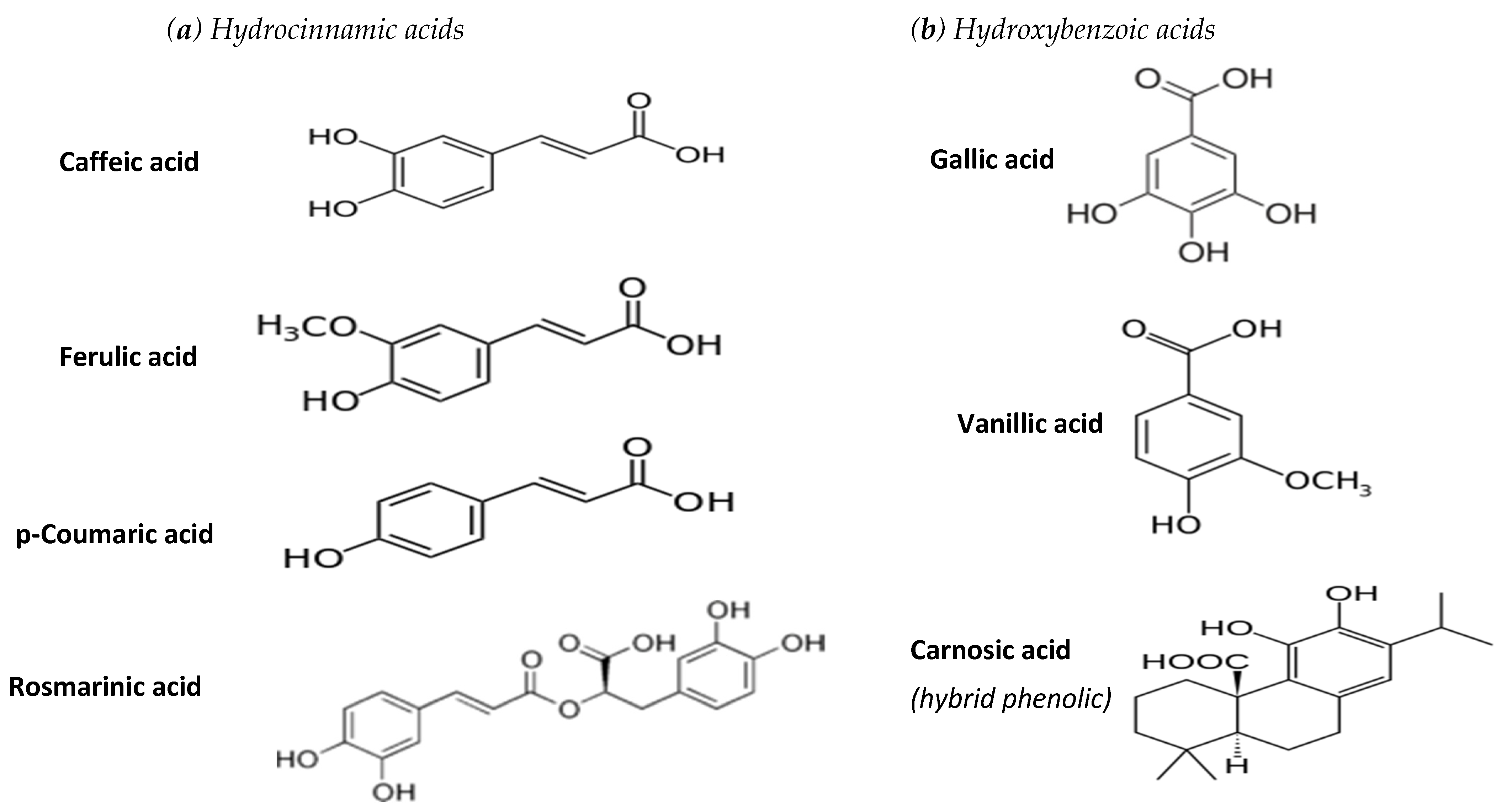
2.1. Structural Classidication of Natural Phenolic Acids
2.2. Herbal Sources and Extraction of Phenolic Acids
2.3. Extraction of Phenolic Acids from Their Natural Sources
3. Biochemical and Health Properties of the Examined Phenolic Acids
3.1. Effects against Cancer
3.1.1. Individual Phenolic Acids
3.1.2. Natural Extracts Rich in Phenolic Acids
3.2. Effects against Cardiovascular Diseases
3.2.1. Individual Phenolic Acids
3.2.2. Natural Extracts Rich in Phenolic Acids
3.3. Effects against Hepatotoxicity and Liver Disorders
3.3.1. Individual Phenolic Acids
3.3.2. Natural Extracts Rich in Phenolic Acids
3.4. Effects against Neurological Disorders
3.4.1. Individual Phenolic Acids
3.4.2. Natural Extracts Rich in Phenolic Acids
3.5. Protective Effects against Microbial and Viral Infections (Incl. COVID-19)
3.5.1. Antimicrobial Activity of Phenolic Acids-Mechanism of Action
3.5.2. Potential Protective Effects of Phenolic Acids against Coronavirus-Based Infections
4. Conclusions and Prospects
Author Contributions
Funding
Institutional Review Board Statement
Informed Consent Statement
Data Availability Statement
Conflicts of Interest
Abbreviations
| AChE | acetylcholine |
| AD | Alzheimer’s disease |
| CA | caffeic acid |
| CAPE | caffeic acid phenethyl ester |
| CarA | carnocic acid |
| CC14 | carbon tetrachloride |
| COVID-19 | coronavirus disease 2019 |
| CVDs | cardiovascular diseases |
| DMBA | dimethylbenz[a]anthracene |
| DNA | deoxyribonucleic acid |
| EGCG | epigallocatechin-3-gallate |
| FA | ferulic acid |
| GA | gallic acid |
| HDL | high density lipoproteins |
| HPLC | high performance liquid chromatography |
| HSPA5 | heat shock protein A5 |
| LDL | low density lipoproteins |
| p-CA-p | coumaric acid |
| RA | rosmarinic acid |
| ROS | reactive active oxygen species |
| SARS | severe acute respiratory syndrome |
| TAA | thioacetamide |
| VA | vanillic Acid |
References
- Lobo, V.; Patil, A.; Phatak, A.; Chandra, N. Free radicals, antioxidants and functional foods: Impact on human health. Pharmacogen. Rev. 2010, 4, 118–126. [Google Scholar] [CrossRef] [Green Version]
- Yan, M.; Lo, C.-J.; Edwards, T.-J.; Baran, S.-P. Radicals: Reactive intermediates with translational potential. J. Am. Chem. Soc. 2016, 138, 12692–12714. [Google Scholar] [CrossRef]
- Dizdaroglu, M.; Jaruga, P. Mechanisms of free radical-induced damage to DNA. Free Radic. Res. 2012, 46, 382–419. [Google Scholar] [CrossRef] [PubMed]
- Mamede, A.C.; Tavares, S.D.; Abrantes, A.M.; Trindade, J.; Maia, J.M.; Botelho, M.F. The role of vitamins in cancer: A review. Nutr. Cancer 2011, 63, 479–494. [Google Scholar] [CrossRef] [PubMed]
- Kiokias, S.; Proestos, C.; Oreopoulou, V. Effect of natural Food Antioxidants against LDL and DNA Oxidative damages. Antioxidants 2018, 7, 133. [Google Scholar] [CrossRef] [PubMed] [Green Version]
- Kiokias, S.; Proestos, C.; Oreopoulou, V. Beneficial Health Properties of Common Natural Phenolic Acids. Encyclopedia. 2020. Available online: https://encyclopedia.pub/1205 (accessed on 13 January 2021).
- Yemiş, G.P.; Pagotto, F.; Bach, S.; Delaquis, P. Effect of Vanillin, Ethyl Vanillin, and Vanillic Acid on the Growth and Heat Resistance of Cronobacter Species. J. Food Protect. 2011, 74, 2062–2069. [Google Scholar] [CrossRef] [PubMed]
- Jungbauer, A.; Medjakovic, S. Anti-inflammatory properties of culinary herbs and spices that ameliorate the effects of metabolic syndrome. Maturitas 2012, 71, 227–239. [Google Scholar] [CrossRef]
- Kiokias, S. Antioxidant effects of vitamins C, E and provitamin A compounds as monitored by use of biochemical oxidative indicators linked to atherosclerosis and carcinogenesis. Intern. J. Nutr. Res. 2019, 1, 1–13. [Google Scholar]
- Manuja, R.; Sachdeva, S.; Jain, A.; Chaudhary, J. A comprehensive review on biological activities of p-hydroxy benzoic acid and its derivatives. Int. J. Pharm. Sci. Rev. Res. 2013, 22, 109–115. [Google Scholar]
- Kiokias, S.; Proestos, C.; Oreopoulou, V. Phenolic acids of plant origin—A review on their antioxidant activity in vitro (o/w emulsion systems) along with their in vivo health biochemical properties. Foods 2020, 9, 534. [Google Scholar] [CrossRef]
- Liu, Y.; Carver, J.A.; Calabrese, A.N.; Pukala, T.L. Gallic acid interacts with α-synuclein to prevent the structural collapse necessary for its aggregation. Biochim. Biophys. Acta (BBA)-Prot. Proteom. 2014, 1844, 1481–1485. [Google Scholar] [CrossRef]
- Tsimogiannis, D.; Oreopoulou, V. Classification of phenolic compounds in Plants. In Polyphenols in Plants Isolation Purification and Extract Preparation, 2nd ed.; Watson, R.R., Ed.; Elsevier Inc.: London, UK, 2019; pp. 263–284. [Google Scholar]
- Dalbem, L.; Costa Monteiro, C.M.; Anderson, J.T. Anticancer properties of hydroxycinnamic acids—A Review. Canc. Clin. Oncol. 2012, 1, 109–121. [Google Scholar]
- Cabras, P.; Angioni, A.; Tuberoso, C.; Floris, I.; Reniero, F.; Guillou, C.; Ghelli, S. Homogentisic acid: A phenolic acid as a marker of strawberry-tree (Arbutus unedo) honey. J. Agric. Food Chem. 1999, 47, 4064–4067. [Google Scholar] [CrossRef]
- Berté, K.A.; Beux, M.R.; Spada, P.K.; Salvador, M.; Hoffmann-Ribani, R. Chemical composition and antioxidant activity of yerba-mate (Ilex paraguariensis A.St.-Hil., Aquifoliaceae) extract as obtained by spray drying. J. Agric. Food Chem 2011, 25, 5523–5527. [Google Scholar] [CrossRef]
- Sugahara, S.; Chiyo, A.; Fukuoka, Κ.; Ueda, Y.; Tokunaga, Y.; Nishida, Y.; Kinoshita, H.; Matsuda, Y.; Igoshi, K.; Ono, M.; et al. Unique antioxidant effects of herbal leaf tea and stem tea from Moringa oleifera L. especially on superoxide anion radical generation systems. Biosci. Biotechnol. Biochem. 2018, 82, 1973–1984. [Google Scholar] [CrossRef] [Green Version]
- Žugić, A.; Đorđević, S.; Arsić, I.; Marković, G.; Živković, J.; Jovanovic, S.; Tadić, V. Antioxidant activity and phenolic compounds in 10 selected herbs from Vrujci Spa, Serbia. Indust. Crops Prod. 2014, 52, 519–527. [Google Scholar] [CrossRef]
- Loussouarn, M.; Krieger-Liszkay, A.; Svilar, L.; Bily, A.; Birtić, S.; Havaux, M. Carnosic Acid and Carnosol, Two Major Antioxidants of Rosemary, Act through Different Mechanisms. Plant Physiol. 2017, 175, 1381–1394. [Google Scholar] [CrossRef] [PubMed] [Green Version]
- Raes, K.; Doolaege, E.H.A.; Deman, S.; Vossen, E.; De Smet, S. Effect of carnosic acid, quercetin and α-tocopherol on lipid and protein oxidation in anin vitrosimulated gastric digestion model. Intern. J. Food Sci. Nutr. 2015, 66, 216–221. [Google Scholar] [CrossRef]
- Flanagan, J.; Bily, A.; Rolland, Y.; Roller, M. Lipolytic Activity of Svetol®, a Decaffeinated Green Coffee Bean Extract. Phytoth. Res. 2013, 28, 946–948. [Google Scholar] [CrossRef] [PubMed]
- Mojica, L.; Meyer, A.; Berhow, M.; González, E. Bean cultivars (Phaseolus vulgaris L.) have similar high antioxidant capacity, in vitro inhibition of α-amylase and α-glucosidase while diverse phenolic composition and concentration. Food Res. Intern. 2015, 69, 38–48. [Google Scholar] [CrossRef]
- Wei, W.L.; Zeng, R.; Gu, C.M.; Qu, Y.; Huang, L.F. Angelica sinensis in China-A review of botanical profile, ethnopharmacology, phytochemistry and chemical analysis. J. Ethnopharmacol. 2016, 190, 116–141. [Google Scholar] [CrossRef]
- Pandurangan, A.K.; Mohebali, N.; Norhaizan, M.E.; Looi, C.Y. Gallic acid attenuates dextran sulfate sodium-induced experimental colitis in BALB/c mice. Drug Des. Dev. Ther. 2015, 9, 3923–3934. [Google Scholar] [CrossRef] [Green Version]
- Zucca, P.; Rosa, A.; Tuberoso, C.; Piras, A.; Rinaldi, A.C.; Sanjust, E.; Dessi, A.; Rescigno, A. Evaluation of Antioxidant Potential of “Maltese Mushroom” (Cynomorium coccineum) by Means of Multiple Chemical and Biological Assays. Nutrients 2013, 5, 149–161. [Google Scholar] [CrossRef] [Green Version]
- Rangani, I.; Kumari, A.; Patel, M.; Brahmbhatt, H.; Parida, A.K. Phytochemical profiling, polyphenol composition, and antioxidant activity of the leaf extract from the medicinal halophyte Thespesia populnea reveal a potential source of bioactive compounds and nutraceuticals. J. Food Biochem. 2019, 43, e12731. [Google Scholar] [CrossRef] [PubMed]
- Trisha, S. Role of hesperdin, luteolin and coumaric acid in arthritis management: A Review. Int. J. Phys. Nutr. Phys. Educ. 2018, 3, 1183–1186. [Google Scholar]
- Kavita, P.; Gandhi, P. Rediscovering the therapeutic potential of Amaranthus species: A review. Egypt J. Basic Appl. Sci. 2017, 4, 196–205. [Google Scholar] [CrossRef] [Green Version]
- Woogyeong, K.; Dahae, L.; Jinju, K. p-Coumaric Acid, a Major Active Compound of Bambusae Caulis in Taeniam, Suppresses Cigarette Smoke-Induced Pulmonary Inflammation. Am. J. Chin. Med. 2018, 46, 407–421. [Google Scholar] [CrossRef]
- Multari, S.; Pihlava, J.M.; Ollennu-Chuasam, P.; Hietaniemi, V.; Yang, B.; Suomela, J.P. Identification and Quantification of Avenanthramides and Free and Bound Phenolic Acids in Eight Cultivars of Husked Oat (Avena sativa L) from Finland. J. Agric. Food Chem. 2018, 68, 2900–2908. [Google Scholar] [CrossRef] [Green Version]
- Yashin, A.; Yashin, Y.; Xia, X.; Nemzer, V. Antioxidant Activity of Spices and Their Impact on Human Health: A Review. Antioxidants 2017, 6, 70. [Google Scholar] [CrossRef] [Green Version]
- Tsimogiannis, D.; Choulitoudi, E.; Bimpilas, A.; Mitropoulou, G.; Kourkoutas, Y.; Oreopoulou, V. Exploitation of the biological potential of Satureja thymbra essential oil and distillation by-products. J. Appl. Res. Med. Aromat. Plants 2016, 4, 12–20. [Google Scholar] [CrossRef]
- Pacheco-Palencia, L.A.; Mertens, T.S.; Talcott, S.-T. Chemical composition, antioxidant properties, and thermal stability of a phytochemical enriched oil from Acai (Euterpe oleracea Mart.). J. Agric. Food Chem. 2008, 56, 4631–4636. [Google Scholar] [CrossRef]
- Chengke, Z.; Yuan, J.; Fachuang, L. Angelica Stem: A Potential Low-Cost Source of Bioactive Phthalides and Phytosterols. Molecules 2018, 23, 3065. [Google Scholar] [CrossRef] [Green Version]
- Oreopoulou, A.; Tsimogiannis, D.; Oreopoulou, V. Extraction of Polyphenols From Aromatic and Medicinal Plants: An Overview of the Methods and the Effect of Extraction Parameters. In Polyphenols in Plants Isolation Purification and Extract Preparation, 2nd ed.; Watson, R.R., Ed.; Elsevier Inc.: London, UK, 2019; pp. 243–260. [Google Scholar]
- Psarrou, I.; Oreopoulou, A.; Tsimogiannis, D.; Oreopoulou, V. Extraction Kinetics of Phenolic Antioxidants from the Hydro Distillation Residues of Rosemary and Effect of Pretreatment and Extraction Parameters. Molecules 2020, 25, 4520. [Google Scholar] [CrossRef]
- Oreopoulou, A.; Goussias, G.; Tsimogiannis, D.; Oreopoulou, V. Hydro-alcoholic Extraction Kinetics of Phenolics from Oregano: Optimization of the Extraction Parameters. Food Bioprod. Proc. 2020, 123, 378–389. [Google Scholar] [CrossRef]
- Corbin, C.; Fidel, T.; Leclerc, E.A.; Barakzoy, E.; Sagot, N.; Falguiéres, A.; Lainé, E. Development and validation of an efficient ultrasound assisted extraction of phenolic compounds from flax (Linum usitatissimum L.) seeds. Ultras. Sonochem. 2015, 26, 176–185. [Google Scholar] [CrossRef]
- De AR Oliveira, G.; De Oliveira, A.E.; Da Conceição, E.C.; Leles, M.I. Multiresponse optimization of an extraction procedure of carnosol and rosmarinic and carnosic acids from rosemary. Food Chem. 2016, 211, 465–473. [Google Scholar] [CrossRef] [PubMed]
- Max, B.; Salgado, J.M.; Cortés, S.; Domínguez, J.M. Extraction of phenolic acids by alkaline hydrolysis from the solid residue obtained after prehydrolysis of trimming vine shoots. J. Agric. Food Chem. 2009, 58, 1909–1917. [Google Scholar] [CrossRef]
- Gonzales, G.B.; Raes, K.; Vanhoutte, H.; Coelus, S.; Smagghe, G.; Van Camp, J. Liquid chromatography–mass spectrometry coupled with multivariate analysis for the characterization and discrimination of extractable and nonextractable polyphenols and glucosinolates from red cabbage and Brussels sprout waste streams. J. Chrom. A 2015, 1402, 60–70. [Google Scholar] [CrossRef]
- Kumar, N.; Goel, N. Phenolic acids: Natural versatile molecules with promising therapeutic applications. Biotechnol. Rep. 2019, 24, e00370. [Google Scholar] [CrossRef]
- Kehan, P.; Ou, J.; Huanga, J.; Oua, S. Coumaric acid and itsconjugates: Dietary sources, pharmacokinetic properties and biological activities. J. Sci. Food Agric. 2016, 96, 2952–2962. [Google Scholar] [CrossRef]
- Proestos, C.; Komaitis, M. Application of microwave-assisted extraction to the fast extraction of plant phenolic compounds. LWT-Food Sci. Technol. 2008, 41, 652–659. [Google Scholar] [CrossRef]
- Bernatoniene, J.; Cizauskaite, U.; Ivanauskas, L.; Jakstas, V.; Kalveniene, Z.; Kopustinskiene, D.M. Novel approaches to optimize extraction processes of ursolic, oleanolic and rosmarinic acids from Rosmarinus officinalis leaves. Ind. Crops Prod. 2016, 84, 72–79. [Google Scholar] [CrossRef]
- Ciulu, M.; de la Luz Cádiz-Gurrea, M.; Segura-Carretero, A. Extraction and Analysis of Phenolic Compounds in Rice: A Review. Molecules 2018, 23, 2890. [Google Scholar] [CrossRef] [Green Version]
- Pyrzynska, K.; Sentkowska, A. Chromatographic Analysis of Polyphenols. In Polyphenols in Plants Isolation Purification and Extract Preparation, 2nd ed.; Watson, R.R., Ed.; Elsevier Inc.: London, UK, 2019; pp. 353–364. [Google Scholar]
- Vinas, P.; Campillo, N. Gas Chromatography: Mass Spectrometry Analysis of Polyphenols in Foods. In Polyphenols in Plants Isolation Purification and Extract Preparation, 2nd ed.; Watson, R.R., Ed.; Elsevier Inc.: London, UK, 2019; pp. 285–316. [Google Scholar]
- Κiokias, S.; Varzakas, T. Activity of flavonoids and beta-carotene during the auto-oxidative deterioration of model food oil-in water emulsions. Food Chem. 2014, 150, 280–286. [Google Scholar] [CrossRef]
- Nourah, A.; Al Zahrani, N.A.; El-Shishtawy, R.M.; Asiri, A.M. Recent developments of gallic acid derivatives and their hybrids in medicinal chemistry: A review. Eur. J. Med. Chem. 2020, 204, 112609. [Google Scholar] [CrossRef]
- Abotaleb, Μ.; Liskova, A.; Kubatka, P.; Büsselberg, D. Therapeutic Potential of Plant Phenolic Acids in the Treatment of Cancer. Biomolecules 2020, 10, 221. [Google Scholar] [CrossRef] [Green Version]
- Gaglione, Μ.; Malgieri, G.; Pacifico, S.; Severino, V.; D’Abrosca, P.; Russo, L.; Fiorentino, A.; Messere, A. Synthesis and Biological Properties of Caffeic Acid-PNA Dimers Containing Guanine. Molecules 2013, 18, 9147–9162. [Google Scholar] [CrossRef] [Green Version]
- Wang, J.; Bhargava, P.; Yu, Y.; Sari, A.N.; Zhang, H.; Ishii, N. Novel Caffeic Acid Phenethyl Ester-Mortalin Antibody Nanoparticles Offer Enhanced Selective Cytotoxicity to Cancer Cells. Cancers 2020, 12, 2370. [Google Scholar] [CrossRef]
- Zhang, A.; Lin, A.; Jiang, R.; Li, H.; Wan, H. Ferulic acid exerts antitumour activity and inhibits metastasis in breast cancer cells by regulating epithelial to mesenchymal transition. Oncol. Rep. 2016, 36, 271–278. [Google Scholar] [CrossRef] [Green Version]
- Al-Mutairi, A.; Rahman, A.; Rao, M.S. Low Doses of Thymoquinone and Ferulic Acid in Combination Effectively Inhibit Proliferation of Cultured MDA-MB 231 Breast Adenocarcinoma Cells. Nutr. Cancer 2020, 72, 8. [Google Scholar] [CrossRef] [PubMed]
- Solomonov, Y.; Hadad, N.; Levy, R. The combined anti-inflammatory effect of astaxanthin, lyc-O-mato and carnosic acid in vitro and in vivo in a mouse model of peritonitis. J. Nutr. Food Sci. 2018, 8, 653. [Google Scholar] [CrossRef]
- Sun, G.L.; Wang, D. Gallic acid from Terminalia chebula inhibited the growth of esophageal carcinoma cells by suppressing the Hippo signal pathway. Iran. J. Basic Med. Sci. 2020, 23, 1401–1408. [Google Scholar] [CrossRef] [PubMed]
- Sales, M.S.; Roy, A.; Antony, L.; Banu, S.K.; Jeyaraman, S.; Manikkam, R. Octyl gallate and gallic acid isolated from Terminalia bellarica regulates normal cell cycle in human breast cancer cell lines. Biomed. Pharmacother. 2018, 103, 1577–1584. [Google Scholar] [CrossRef]
- Afonso, F.A.; Pereira, O.R.; Cardoso, S.M. Health-Promoting Effects of Thymus Phenolic-Rich Extracts: Antioxidant, Anti-Inflammatory and Antitumoural Properties. Antioxidants 2020, 9, 814. [Google Scholar] [CrossRef]
- Luo, Y.; Ma, Z.; Xu, X.; Qi, H.; Cheng, Z.; Chen, L. Anticancer effects of Rosmarinic acid in human oral cancee cells is mediated via endoplasmic reticulum stress, apoptosis, G2/M cell cycle arrest and inhibition of cell migration. J. BUON 2020, 25, 1245–1250. [Google Scholar] [PubMed]
- Messeha, S.S.; Zarmouh, N.O.; Asiri, A.; Soliman, K.F.A. Rosmarinic acid-induced apoptosis and cell cycle arrest in triple-negative breast cancer cells. Eur. J. Pharmacol. 2020, 885, 173419. [Google Scholar] [CrossRef]
- Anbalagan, V.; Raju, K.; Shanmugam, K. Assessment of Lipid Peroxidation and Antioxidant Status in Vanillic Acid Treated 7,12-Dimethylbenz[a]anthracene Induced Hamster Buccal Pouch Carcinogenesis. J. Clin. Diagn. Res. 2017, 11, BF01. [Google Scholar] [CrossRef]
- Gong, J.; Zhou, S.; Yang, S. Vanillic Acid Suppresses HIF-1α Expression via Inhibition of mTOR/p70S6K/4E-BP1 and Raf/MEK/ERK Pathways in Human Colon Cancer HCT116 Cells. Int. J. Mol. Sci. 2019, 22, 465. [Google Scholar] [CrossRef] [Green Version]
- Taner, G.; Ozkan Vardar, D.; Aydin, S.; Aytac, Z.; Basaran, A.; Basaran, N. Use of in vitro assays to assess the potential cytotoxic, genotoxic and antigenotoxic effects of vanillic and cinnamic acid. Drug Chem. Toxicol. 2017, 40, 183–190. [Google Scholar] [CrossRef]
- Jeong, N.; Phan, H.; Jong-Whan, C. Anti-cancer effects of polyphenolic compounds in epidermal growth factor receptor tyrosine kinase inhibitor-resistant non-small cell lung cancer. Pharmacogn. Mag. 2017, 13, 595–599. [Google Scholar] [CrossRef]
- Trivellini, A.; Lucchesini, M.; Maggini, R.; Mosadegh, H.; Villamarin, T.-S.; Vernieri, P.; Mensuali-Sodi, A.; Pardossi, A. Lamiaceae phenols as multifaceted compounds: Bioactivity, industrial prospects and role of “positive-stress”. Industr. Crops Product. 2016, 83, 241–254. [Google Scholar] [CrossRef]
- Oreopoulou, A.; Papavassilopoulou, E.; Bardouki, H.; Vamvakias, M.; Bimpilas, A.; Oreopoulou, V. Antioxidant recovery from hydrodistillation residues of selected Lamiaceae species by alkaline extraction. J. Appl. Res. Medic. Aromat. Plants 2018, 8, 83–89. [Google Scholar] [CrossRef]
- Fernandes, A.; Mazzei, H.J.; Evangelista, H.; Da Costa Marques Calderari, M.J.; Ferraz, E.R.A.; Felzenszwalb, I. Protection against UV-induced oxidative stress and DNA damage by Amazon moss extracts. J. Photoch. Photob. B Biol. 2018, 183, 331–341. [Google Scholar] [CrossRef] [PubMed]
- Koyuncu, I.; Gönel, A.; Akdağ, A.; Yilmaz, M.A. Identification of phenolic compounds, antioxidant activity and anti-cancer effects of the extract obtained from the shoots of Ornithogalum narbonense L. Cell Mol. Biol. 2018, 64, 75–83. [Google Scholar] [CrossRef] [PubMed]
- Waheed, M.; Hussain, M.B.; Javed, A.; Mushtaq, Z.; Hassan, S.; Sariati, M.-A.; Khan, M.-U.; Majeed, M.; Nigam, M.; Mishra, A.-P.; et al. Honey and cancer: A mechanistic review. Clin. Nutr. 2019, 38, 2499–2503. [Google Scholar] [CrossRef]
- Ali, S.S.; Ahmad, W.A.; Budin, S.B.; Zainalabidin, S. Implication of dietary phenolic acids on inflammation in cardiovascular disease. Rev. Cardiovasc. Med. 2020, 21, 225–240. [Google Scholar] [CrossRef] [PubMed]
- Silva, H.; Lopes., N.M. Cardiovascular Effects of Caffeic Acid and Its Derivatives: A Comprehensive Review. Front. Physiol. 2020, 27, 595516. [Google Scholar] [CrossRef]
- Olas, B. Honey and its phenolic compounds as an effective natural medicine for cardiovascular diseases in humans? Nutrients 2020, 12, 283. [Google Scholar] [CrossRef] [Green Version]
- Salazar-López, N.Z.; Astiazarán-García, H.; González-Aguilar, A.; Loarca-Piña, G.; Ezquerra-Brauer, J.M.; Domínguez Avila, J.A.; Robles-Sánchez, M. Ferulic Acid on Glucose Dysregulation, Dyslipidemia, and Inflammation in Diet-Induced Obese Rats: An Integrated Study. Nutrients 2017, 29, 675. [Google Scholar] [CrossRef] [PubMed] [Green Version]
- Bumrungpert, A.; Lilitchan, S.; Tuntipopipat, S.; Tirawanchai, N.; Komindr, S. Ferulic Acid Supplementation Improves Lipid Profiles, Oxidative Stress, and Inflammatory Status in Hyperlipidemic Subjects: A Randomized, Double-Blind, Placebo-Controlled Clinical Trial. Nutrients 2018, 10, 713. [Google Scholar] [CrossRef] [PubMed] [Green Version]
- Akbari, G. Molecular mechanisms underlying gallic acid effects against cardiovascular diseases: An update review. J. Phytomed. 2020, 10, 11–23. [Google Scholar]
- Ibitoye, O.B.; Ajiboy, O.T. Dietary phenolic acids reverse insulin resistance, hyperglycaemia, dyslipidaemia, inflammation and oxidative stress in high-fructose diet-induced metabolic syndrome rats. Arch. Physiol. Biochem. 2018, 124, 410–417. [Google Scholar] [CrossRef] [PubMed]
- Sherratt, S.C.R.; Villeneuve, P.; Durand, E.; Mason, R.P. Rosmarinic acid and its esters inhibit membrane cholesterol domain formation through an antioxidant mechanism based, in nonlinear fashion, on alkyl chain length. Biochim. Biophys. Acta Biomembr. 2019, 1861, 550–555. [Google Scholar] [CrossRef] [PubMed]
- Murino Rafacho, B.P.; Dos Santos, P.P.; Gonçalves, A.; Henrique Fernandes, A.A.; Okoshi, K.; Chiuso-Minicucci, F.; Azevedo, P.S.; Zornoff, L.A.M.; Minicucci, M.F.; Wang, X.-D.; et al. Rosemary supplementation (Rosmarinus ofici nallis L.) attenuates cardiac remodeling after myocardial infarction in rats. PLoS ONE 2017, 12, e0177521. [Google Scholar] [CrossRef]
- Faponle, A.S.; Atunnise, A.K.; Adegbesan, B.O. Separate and co-administration of Amaranthus spinosus and vitamin C modulates cardiovascular disease risk in high fat diet-fed experimental rats. J. Pharmac. Phytoth. 2015, 7, 27–34. [Google Scholar] [CrossRef]
- Fatma, G.; Khantouche, L.; Amel, M.; Bellamine, H.; Landoulsi, A. Histopathological and Biochemical Effects of Thyme Essential Oil on H2O2 Stress in Heart Tissues. Heart Lung Circ. 2020, 29, 308–314. [Google Scholar] [CrossRef]
- Panda, V.; Laddha, A.; Nandave, M.; Srinath, S. Dietary Phenolic Acids of Macrotyloma uniflorum (Horse Gram) Protect the Rat Heart against Isoproterenol-Induced Myocardial Infarction. Phytother. Res. 2016, 30, 1146–1155. [Google Scholar] [CrossRef]
- Cianciosi, D.; Forbes-Hernández, T.W.; Afrin, S.; Gasparrini, M.; Reboredo-Rodriguez, P.; Reboredo-Rodriguez, P.; Pia Manna, P.; Zhang, J.; Bravo Lamas, L.; Martínez Flórez, S.; et al. Phenolic Compounds in Honey and Their Associated Health Benefits: A Review. Molecules 2018, 23, 2322. [Google Scholar] [CrossRef] [Green Version]
- Aćimović, M.G.; Milić, N.B. Perspectives of the Apiaceae Hepatoprotective Effects—A Review. Nat. Prod. Commun. 2017, 12, 309–317. [Google Scholar] [CrossRef] [Green Version]
- Ajiboye, T.O.; Ajala-Lawal, R.A.; Adeyiga, A.B. Caffeic acid abrogates 1,3-dichloro-2-propanol-induced hepatotoxicity by upregulating nuclear erythroid-related factor 2 and downregulating nuclear factor-kappa B. Hum. Exp. Toxicol. 2019, 38, 1092–1101. [Google Scholar] [CrossRef]
- Mu, H.Ν.; Li, Q.; Fan, J.Y.; Pan, C.S.; Liu, Y.Y.; Yan, L. Caffeic acid attenuates rat liver injury after transplantation involving PDIA3-dependent regulation of NADPH oxidase. Free Radic. Biol. Med. 2018, 129, 202–214. [Google Scholar] [CrossRef]
- Hao, R.; Ge, J.; Ren, Y.; Song, X.; Jiang, J.; Waterhouse, D.S.; Li, F. Caffeic acid phenethyl ester mitigates cadmium-induced hepatotoxicity in mice: Role of miR-182-5p/TLR4 axis. Ecotox. Environ. Saf. 2021, 207, 111578. [Google Scholar] [CrossRef] [PubMed]
- Chen, S.; Lin, Y.; Miao, L.; Pan, W.; Jiang, W.; Qian, L.; Hao, J.; Xi, B.; Liu, B.; Ge, X. Ferulic acid alleviates lipopolysaccharide-induced acute liver injury in Megalobrama amblycephala. Aquaculture 2021, 532, 735972. [Google Scholar] [CrossRef]
- Owumi, S.; Najophe, E.S.; Farombi, E.O.; Oyelere, A.K. Gallic acid protects against Aflatoxin B1-induced oxidative and inflammatory stress damage in rats kidneys and liver. J. Food Biochem. 2020, 44, e13316. [Google Scholar] [CrossRef] [PubMed]
- Hussein, R.M.; Anwar, M.M.; Farghaly, H.S.; Kandeil, A.M. Gallic acid and ferulic acid protect the liver from thioacetamide-induced fibrosis in rats via differential expression of miR-21, miR-30 and miR-200 and impact on TGF-β1/Smad3 signaling. Chem. Biol. Interact. 2020, 324, 109098. [Google Scholar] [CrossRef]
- Lee, Y.H.; Lim, W.; Sung, M.K. Carnosic Acid Modulates Increased Hepatic Lipogenesis and Adipocytes Differentiation in Ovariectomized Mice Fed Normal or High-Fat Diets. Nutrients 2018, 15, 1984. [Google Scholar] [CrossRef] [PubMed] [Green Version]
- Oguz, A.; Boyuk, A.; Ekinsi, A.; Alabalik, U.; Turkoglou, A.; Tuncer, M.-C.; Ekingen, A.; Deveci, E.; Gültürk, B.; Aday, U. Investigation on the antioxidant effects of rosmarinic acid on liver, lung and kidney in rats; A biochemical and histopathological study. Folia Morpholog. 2020, 2, 288–295. [Google Scholar] [CrossRef] [PubMed]
- Meng, X.; Tang, G.Y.; Liu, P.H.; Liu, Q.; Li, H.B.; Zhao, C.J. Antioxidant activity and hepatoprotective effect of 10 medicinal herbs on CCl4-induced liverinjury in mice. World J. Gastroent. 2020, 26, 5629–5645. [Google Scholar] [CrossRef]
- Meharie, B.G.; Amare, G.G.; Belayneh, Y.M. Evaluation of Hepatoprotective Activity of the Crude Extract and Solvent Fractions of against CCl4-Induced Hepatotoxicity in Mice. J. Exp. Pharmacol. 2020, 12, 137–150. [Google Scholar] [CrossRef]
- Hewage, K.; Nadeeka, I.; Herath, M.; Cho, J.; Kim, A.; Eom, T.K.; Kim, J.S.; Kim, J.B.; Doh, Y.H.; Jee, Y. Phenolic acid and flavonoid-rich fraction of Sasa quelpaertensis Nakai leaves prevent alcohol induced fatty liver through AMPK activation. J. Ethnopharmacol. 2018, 224, 335–348. [Google Scholar] [CrossRef]
- Mbarki, S.; Alimi, H.; Bouzenna, H.; Elfeki, A.; Hfaiedh, N. Phytochemical study and protective effect of Trigonella foenum graecum (Fenugreek seeds) against carbon tetrachloride-induced toxicity in liver and kidney of male rat. Biomed. Pharmacother. 2017, 88, 19–26. [Google Scholar] [CrossRef]
- Grewal, S.; Sharma, N.; Singh, S.; Kanojia, S.; Thapa, K.; Swami, R.; Rupanshi, G. Molecular docking guided screening of phenolic compounds from Ginko Bilobaas multipotent anti Alzheimer’s agents. Plant Arch. 2020, 20, 3297–3308. [Google Scholar]
- Habtemariam, S. Protective Effects of Caffeic Acid and the Alzheimer’s Brain: An Update. Mini-Rev. Med. Chem. 2017, 17, 667–674. [Google Scholar] [CrossRef]
- Zhang, Y.; Wu, O.; Zhang, L.; Wang, O.; Yang, Z.; Liu, J.; Feng, L. Caffeic acid reduces A53T α-synuclein by activating JNK/Bcl-2-mediated autophagy in vitro and improves behaviour and protects dopaminergic neurons in a mouse model of Parkinson’s disease. Pharmacol. Res. 2019, 150, 104538. [Google Scholar] [CrossRef]
- Ferreira, R.M.; Dos Santos, A.G.; Martins, N.M.; Fernandes, L.S.; Dos Santos, A.S. Caffeic Acid Phenethyl Ester (CAPE) Protects PC12 Cells from Cisplatin-Induced Neurotoxicity by Activating the NGF-Signaling Pathway. Neurotox. Res. 2018, 34, 32–46. [Google Scholar] [CrossRef] [PubMed]
- Gießel, J.M.; Loesche, A.; Csuk, R. Caffeic acid phenethyl ester (CAPE)-derivatives act as selective inhibitors of acetylcholinesterase. Eur. J. Med. Chem. 2019, 177, 259–268. [Google Scholar] [CrossRef] [PubMed]
- Bahri, S.; Jameleddine, S.; Shlyonsky, V. Relevance of carnosic acid to the treatment of several health disorders: Molecular targets and mechanisms. Biomed. Pharmacother. 2016, 84, 569–582. [Google Scholar] [CrossRef] [PubMed]
- Rehman, S.U.; Ali, T.; Alam, S.I.; Ullah, R.; Zeb, A.; Lee, K.W.; Rutten, B.P.F.; Kim, O.M. Ferulic Acid Rescues LPS-Induced Neurotoxicity via Modulation of the TLR4 Receptor in the Mouse Hippocampus. Mol. Neurobiol. 2019, 56, 2774–2790. [Google Scholar] [CrossRef]
- Mori, T.; Koyama, N.; Tan, J.; Segawa, T.; Maeda, M.; Town, T. Combined treatment with the phenolics (-)-epigallocatechin-3-gallate and ferulic acid improves cognition and reduces Alzheimer-like pathology in mice. J. Biol. Chem. 2019, 294, 2714–2731. [Google Scholar] [CrossRef] [Green Version]
- Shabani, S.; Rabiei, Z.; Amini-Khoei, H. Exploring the multifaceted neuroprotective actions of gallic acid, a review. Int. J. Food Prop. 2020, 23, 736–752. [Google Scholar] [CrossRef]
- Liu, Y.L.; Hsu, C.C.; Huang, Y.L.; Chang, C.J.; Sun, S.H.; Yuh Lin, A.M. Gallic Acid Attenuated LPS-Induced Neuroinflammation: Protein Aggregation and Necroptosis. Mol. Neurobiol. 2020, 57, 96–104. [Google Scholar] [CrossRef] [PubMed]
- Khoshnam, S.E.; Sarkaki, A.; Rashno, M.; Farbood, Y. Memory deficits and hippocampal inflammationin cerebral hypoperfusion and reperfusion in male rats: Neuroprotective role of vanillic acid. Life Sci. 2018, 211, 126–132. [Google Scholar] [CrossRef]
- Rizk, Z.A.; Masoud, W.; Maher, O.W. Prophylactic effects of ellagic acid and rosmarinic acid on doxorubicin-induced neurotoxicity in rats. J. Biochem. Mol. Toxicol. 2017, 31, 12. [Google Scholar] [CrossRef] [PubMed]
- Salau, V.F.; Erukainure, O.L.; Ibeji, C.U.; Olasehinde, T.A.; Koorbanally, N.A. Vanillin and vanillic acid modulate antioxidant defense system via amelioration of metabolic complications linked to Fe2+-induced brain tissues damage. Metab. Brain Dis. 2020, 35, 727–738. [Google Scholar] [CrossRef] [PubMed]
- Anand, A.; Khurana, R.; Wahal, N.; Mahajan, S.; Mehta, M.; Satija, S.; Sharma, N.; Vyas, M.; Khurana, N. Vanillin: A comprehensive review of pharmacological activities. Plant Arch. 2019, 1000–1004. [Google Scholar]
- El-Sawi, S.A.; Ezzat, S.A.; Aly, H.F.; Merghany, R.M.; Meselhy, M.R. Neuroprotective effect of Salvia splendens extract and its constituents against AlCl3-induced Alzheimer’s disease in rats. Adv. Tradit. Med. 2020, 20, 381–393. [Google Scholar] [CrossRef]
- Khoshnam, S.E.; Farbood, Y.; Fathi Moghaddam, H.; Sarkaki, A.; Badavi, M.; Khorsandi, L. Vanillic acid attenuates cerebral hyperemia, blood-brain barrier disruption and anxiety-like behaviors in rats following transient bilateral common carotid occlusion and reperfusion. Metab. Brain Dis. 2018, 33, 785–793. [Google Scholar] [CrossRef]
- Panzella, L.; Eidenberger, T.; Napolitano, A. Anti-Amyloid aggregation activity of black sesame pigment: Toward a Novel Alzheimer’s Disease Preventive Agent. Molecules 2018, 23, 676. [Google Scholar] [CrossRef] [Green Version]
- Liang, Z.; Yuan, Z.P.; Ding, T.; Yan, L.; Ling, L.; Zhou, X.F.; Zeng, Y.Q. Neuroprotective Effect of Fagopyrum dibotrys Extract against Alzheimer’s Disease. Evid.-Based Complement. Alternat. Med. 2017, 2017, 3294586. [Google Scholar] [CrossRef]
- Yilmaz, Y.; Toledo, R.T. Health aspects of functional grape seed constituents. Trends Food Sci. Technol. 2004, 9, 422–433. [Google Scholar] [CrossRef]
- Kaur, N.; Bains, A.; Kaushik, R.; Dhull, S.B.; Chawla, P. Review on Antifungal Efficiency of Plant Extracts Entrenched Polysaccharide-Based Nanohydrogels. Nutrients 2021, 13, 2055. [Google Scholar] [CrossRef]
- Semeniuc, C.A.; Socaciu, M.A.; Socaci, S.A.; Muresan, V.; Fogarasi, M.; Rotar, A.M. Chemometric Comparison and Classification of Some Essential Oils Extracted from Plants Belonging to Apiaceae and Lamiaceae Families Based on Their Chemical Composition and Biological Activities. Molecules 2018, 23, 2261. [Google Scholar] [CrossRef] [Green Version]
- Liu, J.; Du, C.; Beaman, H.T.; Monroe, M.B. Characterization of Phenolic Acid Antimicrobial and Antioxidant Structure—Property Relationships. Pharmaceutics 2020, 12, 419. [Google Scholar] [CrossRef]
- Elegir, G.; Kindl, A.; Yilmaz, Y.; Orland, S.M. Development of antimicrobial cellulose packaging through laccase-mediated grafting of phenolic compounds. Enzym. Microb. Technol. 2008, 43, 84–92. [Google Scholar] [CrossRef]
- Merkl, R.; Hrádková, I.; Filip, V.; Šmidrkal, J. Antimicrobial and antioxidant properties of phenolic acids alkyl esters. Czech. J. Food Sci. 2010, 28, 275–279. [Google Scholar] [CrossRef] [Green Version]
- Wu, Y.H.; Zhang, B.Y.; Qiu, L.P.; Guan, R.F.; Ye, Z.H.; Yu, X.P. Structure Properties and Mechanisms of Action of Naturally Originated Phenolic Acids and Their Derivatives against Human Viral Infections. Curr. Med. Chem. 2017, 24, 4279–4302. [Google Scholar] [CrossRef] [PubMed]
- Paulo, F.; Santos, L. Microencapsulation of caffeic acid and its release using a w/o/w double emulsion method: Assessment of formulation parameters. Drying Techn. 2019, 37, 950–961. [Google Scholar] [CrossRef]
- Langland, J.; Jacobs, B.; Wagner, C.E.; Ruiz, G.; Cahill, T.M. Antiviral Activity of Metal Chelates of Caffeic Acid and Similar Compounds towards Herpes Simplex, VSV-Ebola Pseudotyped and Vaccinia Viruses. Antiviral Res. 2018, 160, 143–150. [Google Scholar] [CrossRef]
- Zhang, E.; Zhai, T.; Haide, S.; Li, Y.; Huang, H.J. Synergistic Effect of Chlorogenic Acid and Caffeic Acid with Fosfomycin on Growth Inhibition of a Resistant Listeria monocytogenes Strain. ACS Omega 2020, 5, 7537–7544. [Google Scholar] [CrossRef] [Green Version]
- De Camargo, A.C.; Regitano-d’Arce, M.A.B.; Rasera, G.B.; Canniatti-Brazaca, S.G.; Do Prado-Silva, L.; Alvarenga, L.O.; Sant’Ana, A.S.; Shahidi, F. Phenolic acids and flavonoids of peanut by-products: Antioxidant capacity and antimicrobial effects. Food Chem. 2017, 237, 538–544. [Google Scholar] [CrossRef] [PubMed]
- Baidoo, M.F.; Mensah, A.Y.; Ossei, P.P.S.; Kwatia, E.A.; Amponsah, I.K. Wound healing, antimicrobial and antioxidant properties of the leaf and stem bark of Entada africana Guill. & Perr. S. Afr. J. Bot. 2020, 137, 52–59. [Google Scholar] [CrossRef]
- Attia, Y.A.; Alagawany, M.M.; Farag, M.R.; Alkhatib, F.M.; Khafaga, A.F.; Abdel-Moneim, A.M.; Asiry, A.; Mesalam, N.M.; Shafi, M.E.; Al-Harthi, A.; et al. Phytogenic Products and Phytochemicals as a Candidate Strategy to Improve Tolerance to Coronavirus. Front. Vet. Sci. 2020, 7, 573159. [Google Scholar] [CrossRef] [PubMed]
- Mani, J.S.; Johnson, J.B.; Steel, J.C.; Broszczak, D.A.; Neilsen, P.M.; Walsh, K.B.; Naiker, M. Natural product-derived phytochemicals as potential agents against coronaviruses: A review. Virus Res. 2020, 284, 197989. [Google Scholar] [CrossRef] [PubMed]
- Elfiky, A.A. Natural products may interfere with SARS-CoV-2 attachment to the host cell. J. Biomol. Struct. Dyn. 2020, 1–10. [Google Scholar] [CrossRef]
- Kumar, V.; Dhanjal, J.K.; Kaur, J.; Bhargava, P.; Kaul, A.; Wang, J.; Zhang, H.; Kaul, S.C.; Wadhwa, R.; Sundar, D. Withanone and Withaferin-A are predicted to interact with transmembrane protease serine 2 (TMPRSS2) and block entry of SARS-CoV-2 into cells. J. Biomol. Struct. Dyn. 2020, 39, 3194–3203. [Google Scholar] [CrossRef]
- Khalil, A.; Tazeddinova, D. The upshot of Polyphenolic compounds on immunity amid COVID-19 pandemic and other emerging communicable diseases: An apprais. Nat. Prod. Bioprosp. 2020, 10, 411–429. [Google Scholar] [CrossRef]
- Weng, J.R.; Lin, C.S.; Lai, H.C.; Lin, Y.P.; Wang, C.Y.; Wang, C.Y.; Tsai, Y.C.; Wu, K.C.; Huang, S.H.; Lin, C.W. Antiviral activity of Sambucus FormosanaNakai ethanol extract and related phenolic acid constituents against human coronavirus NL63. Virus Res. 2019, 273, 197767. [Google Scholar] [CrossRef]
- Adem, S.; Eyupoglu, V.; Sarfraz, I.; Rasul, A.; Zahoor, A.F.; Ali, M.; Abdalla, M.; Ibrahim, I.M.; Elfiky, A.A. Caffeic acid derivatives (CAFDs) as inhibitors of SARS-CoV-2: CAFDs-based functional foods as a potential alternative approach to combat COVID-19. Phytomedicine 2020, 85, 153310. [Google Scholar] [CrossRef] [PubMed]
- Gasparotto Junior, A.; Lima Tolouei, S.A.; Dos Reis Lívero, F.A.; Gasparotto, F.; Boeing, T.; de Souza, P. Natural Agents Modulating ACE-2: A Review of Compounds with Potential against SARS-CoV-2 Infections. Curr. Farm. Des. 2021, 27, 1588–1596. [Google Scholar] [CrossRef]


| Health Disease | Phenolic Treatment & Conditions | Conclusion of Study/Health Effect | References |
|---|---|---|---|
| ANTICANCER PROTECTION | Effect of Thymoquinone (TQ-25 µM) and FA (250 µM) on proliferation and apoptosis of a breast cancer cell line MDA-MB 231. | FA in combination with TQ significantly reduced cell proliferation/anticancer effect | [55] |
| Human EC cells (EC9706 and KYSE450) were treated with different concentrations (10–40 μg/mL) of GA | GA reduced the growth of xenograft tumour and promoted apoptosis in a concentration dependent manner. | [57] | |
| Rats subject to DMBA induced oral carcinogenesis were supplemented with VA (200 mg/kg bw p.o) for 14 weeks | VA significantly restored the disturbances in antioxidants status {superoxide dismutase, catalase) to near normal range in DMBA treated hamsters/anti-cancer effects | [62] | |
| CARDIO-PROTECTION | Male Wistar rats supplemented with either lard at 310 g/kg (HFD) or lard and FA at 2 g/kg (HFD + FA) for 8 weeks. | The rats fed with HFD + FA had significantly lower plasma lipids and glucose levels compared with the HFD group. | [74] |
| Daily dietary supplementation of male Wistar rats with Rosemary leaves (11–110 mg) rich in RA | Rosemary attenuated cardiac function improving metabolism & decreasing oxidative stress. | [79] | |
| LIVER PROTECTION | Activity of CA on 1,3-dichloro-2-propanol-induced hepatotoxicity in rats that received CA (10 or 20 mg/kg bw) for 7 days. | CA protected against hepatotoxicity by enhancing the cytoprotective enzymes and lowering inflammation. | [85] |
| Dietary supplementation of fish (Megalobrama amblycephala) with FA at 50–100 mg/kg bw | FA decreased pro-inflammatory cytokines alleviating acute liver injury. | [88] | |
| Rats exposed to aflatoxin B1AFB1 (75 µg/kg bw) were treated with GA (20 or 40 mg/kg bw) for 28 days. | GA ameliorated AFB1-induced hepatorenal dysfunction by decreasing oxidative stress and inflammation in rats hepatotoxicity. | [89] | |
| 32 rats exposed to hepatic ischaemia/reperfusion injury were subsequently treated with RA dose of 50 mg/kg via oral gavage. | RA significantly reduced oxidative stress and abnormal histopathological findings in liver. | [92] | |
| NEURO-PROTECTION | Systemic administration of neuroinflammatory rat with GA (100 mg/kg) | Clear neuroprotective effect of GA in treated rats compared to placebo | [106] |
| Transgenic mice supplemented orally with epigallocatechin-3-gallate (EGCG) and/or FA (30 mg/kg each) daily for 3 months data | The combined EGCG-FA treatment reversed cognitive impairment, presenting AD therapeutic effect. | [104] | |
| Dietary supplementation of rats with 500 mg/kg body weight) of methanolic extracts of Salvia splendens (rich in RA and CA) for 4 weeks | The treatment significantly attenuated AlCl3-induced behavioral impairment (AD like). | [111] | |
| VA was tested against Fe2+- induced oxidative toxicity in brain tissues (neuronal cell lines—HT22). | VA exerted a clear neuroprotective activity. | [109] |
| Phenolic Acid Treatment | Activity against Microorganism/Infections | References |
|---|---|---|
| CA enhanced with chelated inorganic ions (or a metal such as iron) | Antiviral activity towards herpes simplex (HSV), VSV-Ebola pseudotypes and vaccinia viruses occurred early in the virus replication cycle. | [123] |
| CA (1.5 mg/mL) and chlorogenic acid (3 mg/mL) | Phenolic cocktail significantly inhibit the growth of the food born pathogen Listeria monocytogenes. | [99] |
| Methanol extracts of the leaf and stem of E. africana (rich in GA) | Antioxidant and antibacterial activity against Staphylococcus (S. aureus and S. pyogenes) with a minimum inhibitory concentration of 1.56 mg/mL—great potential for treatment of open wounds | [126] |
| CA and GA | CA and GA were found to exert good anti-viral activity against human coronavirus NL63 (HCoV-NL63) | [132] |
| CA and p-CA | Phenolic acids were found to bind to cell-surface HSPA5 competing for recognition by SARS-CoV-2 spike protein | [129] |
| Screening of 27 CA derivatives against 5 proteins of SARS-CoV-2 | 5 CA derivatives exerted anti-viral efficacy against COVID-19 via molecular docking and molecular dynamics simulation. | [133] |
Publisher’s Note: MDPI stays neutral with regard to jurisdictional claims in published maps and institutional affiliations. |
© 2021 by the authors. Licensee MDPI, Basel, Switzerland. This article is an open access article distributed under the terms and conditions of the Creative Commons Attribution (CC BY) license (https://creativecommons.org/licenses/by/4.0/).
Share and Cite
Kiokias, S.; Oreopoulou, V. A Review of the Health Protective Effects of Phenolic Acids against a Range of Severe Pathologic Conditions (Including Coronavirus-Based Infections). Molecules 2021, 26, 5405. https://doi.org/10.3390/molecules26175405
Kiokias S, Oreopoulou V. A Review of the Health Protective Effects of Phenolic Acids against a Range of Severe Pathologic Conditions (Including Coronavirus-Based Infections). Molecules. 2021; 26(17):5405. https://doi.org/10.3390/molecules26175405
Chicago/Turabian StyleKiokias, Sotirios, and Vassiliki Oreopoulou. 2021. "A Review of the Health Protective Effects of Phenolic Acids against a Range of Severe Pathologic Conditions (Including Coronavirus-Based Infections)" Molecules 26, no. 17: 5405. https://doi.org/10.3390/molecules26175405