


Cancers 2024, 16(8), 1543; https://doi.org/10.3390/cancers16081543 - 18 Apr 2024
Viewed by 283
Abstract
►
Show Figures
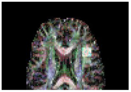
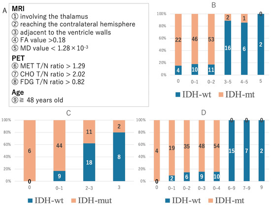
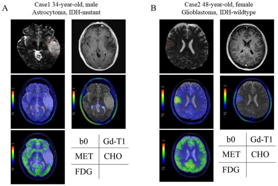
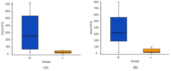
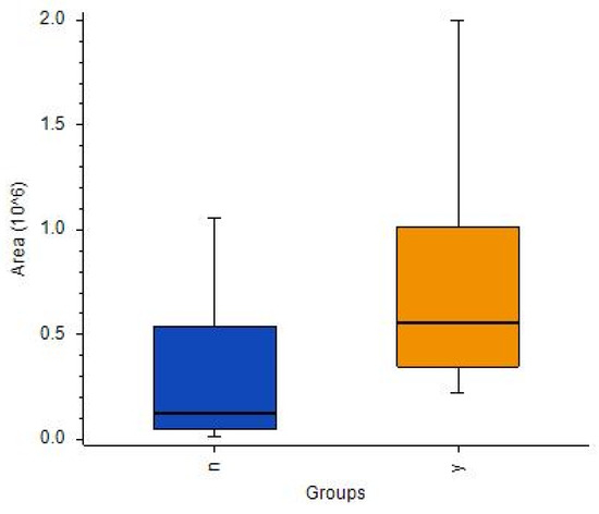
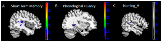
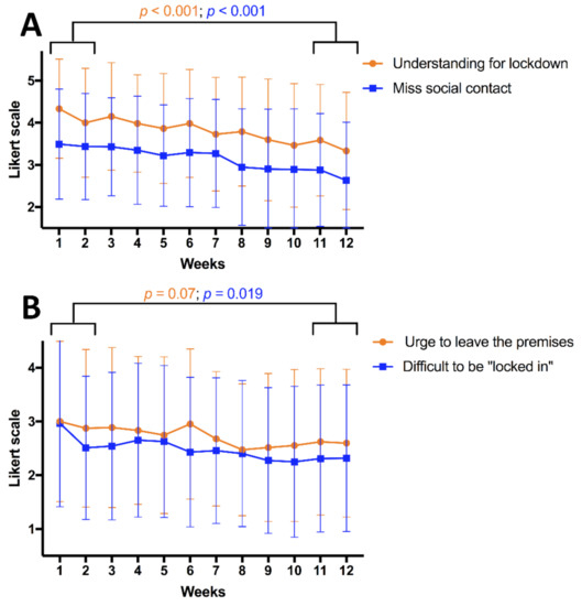
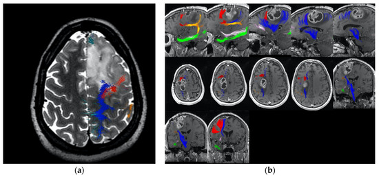
We aimed to differentiate the isocitrate dehydrogenase (IDH) status among non-enhanced astrocytic tumors using preoperative MRI and PET. We analyzed 82 patients with non-contrast-enhanced, diffuse, supratentorial astrocytic tumors (IDH mutant [IDH-mut], 55 patients; IDH-wildtype [IDH-wt], 27 patients) who underwent MRI and PET between
[...] Read more.
We aimed to differentiate the isocitrate dehydrogenase (IDH) status among non-enhanced astrocytic tumors using preoperative MRI and PET. We analyzed 82 patients with non-contrast-enhanced, diffuse, supratentorial astrocytic tumors (IDH mutant [IDH-mut], 55 patients; IDH-wildtype [IDH-wt], 27 patients) who underwent MRI and PET between May 2012 and December 2022. We calculated the fractional anisotropy (FA) and mean diffusivity (MD) values using diffusion tensor imaging. We evaluated the tumor/normal brain uptake (T/N) ratios using 11C-methionine, 11C-choline, and 18F-fluorodeoxyglucose PET; extracted the parameters with significant differences in distinguishing the IDH status; and verified their diagnostic accuracy. Patients with astrocytomas were significantly younger than those with glioblastomas. The following MRI findings were significant predictors of IDH-wt instead of IDH-mut: thalamus invasion, contralateral cerebral hemisphere invasion, location adjacent to the ventricular walls, higher FA value, and lower MD value. The T/N ratio for all tracers was significantly higher for IDH-wt than for IDH-mut. In a composite diagnosis based on nine parameters, including age, 84.4% of cases with 0–4 points were of IDH-mut; conversely, 100% of cases with 6–9 points were of IDH-wt. Composite diagnosis using all parameters, including MRI and PET findings with significant differences, may help guide treatment decisions for early-stage gliomas.
Full article
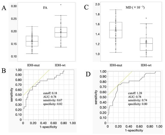
Figure 1

