Optical Genome Mapping in Human Neurogenetic Diagnostics: New Advances and Future Trends

A special issue of Biomolecules (ISSN 2218-273X). This special issue belongs to the section "Molecular Genetics".

Deadline for manuscript submissions: closed (30 June 2023) | Viewed by 7952

Special Issue Editor


E-Mail Website
Guest Editor
Department of Molecular Neuroscience, Queen Square Institute of Neurology, University College London (UCL), London, UK
Interests: sequencing; technologies; neurodevelopmental disorders; genetic epilepsies; next-generation sequencing; optical genome mapping; rare diseases

Special Issue Information

Dear Colleagues,

In recent years, optical genome mapping (OGM) has developed into a highly promising method of detecting large-scale structural variants in human genomes. It affords an effective technology for the detection of SVs, especially those that are mosaic, since these remain difficult to detect with current NGS technologies and with conventional chromosomal microarrays. It promises to be feasible as a first-line diagnostic tool, permitting insight into a new realm of previously unknown variants. However, due to its novelty, little experience with OGM is available to infer best practices for its application or to clarify which features cannot be detected.

The aim of this Special Issue would be to contribute to and expand the knowledge of applying OGM in genetically undiagnosed neurological patients, providing scientists with a useful collection of recent updates and advances in the field.

The aim of this Special Issue is to stimulate discussion around the use and adoption of OGM for clinical diagnostics in genetic neurological disorders, and more specifically to:

  • Carry out an evaluation of OGM in different types of neurological disorders compared to conventional approaches or other novel technologies for structural and molecular classification;
  • Introduce interpretative systems, software tools, or other bioinformatics approaches to improve data analysis and reporting in neurological disorders;
  • Provide technical improvements or processes in neurological disease diagnosis using OGM to improve analysis, diagnostic sensitivity, etc.;
  • Produce novel research findings that may lead to novel diagnostic insights in the future.

In this Special Issue, original research articles and reviews are welcome. Research areas may include (but are not limited to) the following:

  • Repeat disorders;
  • Genetic epilepsies;
  • Neurodevelopmental disorders;
  • Movement disorders.

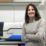
Dr. Stephanie Efthymiou
Guest Editor

Manuscript Submission Information

Manuscripts should be submitted online at www.mdpi.com by registering and logging in to this website. Once you are registered, click here to go to the submission form. Manuscripts can be submitted until the deadline. All submissions that pass pre-check are peer-reviewed. Accepted papers will be published continuously in the journal (as soon as accepted) and will be listed together on the special issue website. Research articles, review articles as well as short communications are invited. For planned papers, a title and short abstract (about 100 words) can be sent to the Editorial Office for announcement on this website.

Submitted manuscripts should not have been published previously, nor be under consideration for publication elsewhere (except conference proceedings papers). All manuscripts are thoroughly refereed through a single-blind peer-review process. A guide for authors and other relevant information for submission of manuscripts is available on the Instructions for Authors page. Biomolecules is an international peer-reviewed open access monthly journal published by MDPI.

Please visit the Instructions for Authors page before submitting a manuscript. The Article Processing Charge (APC) for publication in this open access journal is 2700 CHF (Swiss Francs). Submitted papers should be well formatted and use good English. Authors may use MDPI's English editing service prior to publication or during author revisions.

Keywords

  • structural variation
  • optical genome mapping
  • neurodevelopmental disorders
  • repeat disorders

Published Papers (4 papers)

Order results
Result details
Select all
Export citation of selected articles as:

Research

Jump to: Other

14 pages, 2706 KiB  
Article
Optical Genome Mapping for the Molecular Diagnosis of Facioscapulohumeral Muscular Dystrophy: Advancement and Challenges
by Stephanie Efthymiou, Richard J. L. F. Lemmers, Venugopalan Y. Vishnu, Natalia Dominik, Benedetta Perrone, Stefano Facchini, Elisa Vegezzi, Sabrina Ravaglia, Lindsay Wilson, Patrick J. van der Vliet, Rinkle Mishra, Alisha Reyaz, Tanveer Ahmad, Rohit Bhatia, James M. Polke, Mv Padma Srivastava, Andrea Cortese, Henry Houlden, Silvère M. van der Maarel, Michael G. Hanna and Enrico Bugiardiniadd Show full author list remove Hide full author list
Biomolecules 2023, 13(11), 1567; https://doi.org/10.3390/biom13111567 - 24 Oct 2023
Cited by 2 | Viewed by 1683
Abstract
Facioscapulohumeral muscular dystrophy (FSHD) is the second most common muscular dystrophy in adults, and it is associated with local D4Z4 chromatin relaxation, mostly via the contraction of the D4Z4 macrosatellite repeat array on chromosome 4q35. In this study, we aimed to investigate the [...] Read more.
Facioscapulohumeral muscular dystrophy (FSHD) is the second most common muscular dystrophy in adults, and it is associated with local D4Z4 chromatin relaxation, mostly via the contraction of the D4Z4 macrosatellite repeat array on chromosome 4q35. In this study, we aimed to investigate the use of Optical Genome Mapping (OGM) as a diagnostic tool for testing FSHD cases from the UK and India and to compare OGM performance with that of traditional techniques such as linear gel (LGE) and Pulsed-field gel electrophoresis (PFGE) Southern blotting (SB). A total of 6 confirmed and 19 suspected FSHD samples were processed with LGE and PFGE, respectively. The same samples were run using a Saphyr Genome-Imaging Instrument (1-color), and the data were analysed using custom EnFocus FSHD analysis. OGM was able to confirm the diagnosis of FSHD1 in all FSHD1 cases positive for SB (n = 17), and D4Z4 sizing highly correlated with PFGE-SB (p < 0.001). OGM correctly identified cases with mosaicism for the repeat array contraction (n = 2) and with a duplication of the D4Z4 repeat array. OGM is a promising new technology able to unravel structural variants in the genome and seems to be a valid tool for diagnosing FSHD1. Full article
Show Figures

Figure 1

11 pages, 2105 KiB  
Article
Optical Genome Mapping Enables Detection and Accurate Sizing of RFC1 Repeat Expansions
by Stefano Facchini, Natalia Dominik, Arianna Manini, Stephanie Efthymiou, Riccardo Currò, Bianca Rugginini, Elisa Vegezzi, Ilaria Quartesan, Benedetta Perrone, Shahedah Koya Kutty, Valentina Galassi Deforie, Ricardo P. Schnekenberg, Elena Abati, Anna Pichiecchio, Enza Maria Valente, Cristina Tassorelli, Mary M. Reilly, Henry Houlden, Enrico Bugiardini and Andrea Cortese
Biomolecules 2023, 13(10), 1546; https://doi.org/10.3390/biom13101546 - 19 Oct 2023
Cited by 2 | Viewed by 1814
Abstract
A recessive Short Tandem Repeat expansion in RFC1 has been found to be associated with cerebellar ataxia, neuropathy and vestibular areflexia syndrome (CANVAS), and to be a frequent cause of late onset ataxia and sensory neuropathy. The usual procedure for sizing these expansions [...] Read more.
A recessive Short Tandem Repeat expansion in RFC1 has been found to be associated with cerebellar ataxia, neuropathy and vestibular areflexia syndrome (CANVAS), and to be a frequent cause of late onset ataxia and sensory neuropathy. The usual procedure for sizing these expansions is based on Southern Blotting (SB), a time-consuming and a relatively imprecise technique. In this paper, we compare SB with Optical Genome Mapping (OGM), a method for detecting Structural Variants (SVs) based on the measurement of distances between fluorescently labelled probes, for the diagnosis of RFC1 CANVAS and disease spectrum. The two methods are applied to 17 CANVAS patients’ blood samples and resulting sizes compared, showing a good agreement. Further, long-read sequencing is used for two patients to investigate the agreement of sizes with either SB or OGM. Our study concludes that OGM represents a viable alternative to SB, allowing for a simpler technique, a more precise sizing of the expansion and ability to expand analysis of SV in the entire genome as opposed to SB which is a locus specific method. Full article
Show Figures

Figure 1

Other

Jump to: Research

8 pages, 2880 KiB  
Brief Report
Deep Intronic LINE-1 Insertions in NF1: Expanding the Spectrum of Neurofibromatosis Type 1-Associated Rearrangements
by Viola Alesi, Silvia Genovese, Francesca Romana Lepri, Giorgia Catino, Sara Loddo, Valeria Orlando, Silvia Di Tommaso, Alessandra Morgia, Licia Martucci, Maddalena Di Donato, Maria Cristina Digilio, Bruno Dallapiccola, Antonio Novelli and Rossella Capolino
Biomolecules 2023, 13(5), 725; https://doi.org/10.3390/biom13050725 - 23 Apr 2023
Cited by 2 | Viewed by 1768
Abstract
Neurofibromatosis type 1 is an autosomal-dominant condition caused by NF1 gene inactivation. Clinical diagnosis is corroborated by genetic tests on gDNA and cDNA, which are inconclusive in approximately 3–5% of cases. Genomic DNA approaches may overlook splicing-affecting intronic variants and structural rearrangements, especially [...] Read more.
Neurofibromatosis type 1 is an autosomal-dominant condition caused by NF1 gene inactivation. Clinical diagnosis is corroborated by genetic tests on gDNA and cDNA, which are inconclusive in approximately 3–5% of cases. Genomic DNA approaches may overlook splicing-affecting intronic variants and structural rearrangements, especially in regions enriched in repetitive sequences. On the other hand, while cDNA-based methods provide direct information about the effect of a variant on gene transcription, they are hampered by non-sense-mediated mRNA decay and skewed or monoallelic expression. Moreover, analyses on gene transcripts in some patients do not allow tracing back to the causative event, which is crucial for addressing genetic counselling, prenatal monitoring, and developing targeted therapies. We report on a familial NF1, caused by an insertion of a partial LINE-1 element inside intron 15, leading to exon 15 skipping. Only a few cases of LINE-1 insertion have been reported so far, hampering gDNA studies because of their size. Often, they result in exon skipping, and their recognition of cDNA may be difficult. A combined approach, based on Optical Genome Mapping, WGS, and cDNA studies, enabled us to detect the LINE-1 insertion and test its effects. Our results improve knowledge of the NF1 mutational spectrum and highlight the importance of custom-built approaches in undiagnosed patients. Full article
Show Figures

Figure 1

11 pages, 2351 KiB  
Brief Report
Genetic Predisposition to Neurological Complications in Patients with COVID-19
by Nikhil Shri Sahajpal, Alex R. Hastie, Maximilian Schieck, Ashis K. Mondal, Marc Felde, Caspar I. van der Made, Janet S. Chou, Adrienne G. Randolph, Thomas Illig, Michael C. Zody, Catherine A. Brownstein, Alan H. Beggs, Alexander Hoischen, Alka Chaubey and Ravindra Kolhe
Biomolecules 2023, 13(1), 133; https://doi.org/10.3390/biom13010133 - 9 Jan 2023
Viewed by 2013
Abstract
Several studies have identified rare and common genetic variants associated with severe COVID-19, but no study has reported genetic determinants as predisposition factors for neurological complications. In this report, we identified rare/unique structural variants (SVs) implicated in neurological functions in two individuals with [...] Read more.
Several studies have identified rare and common genetic variants associated with severe COVID-19, but no study has reported genetic determinants as predisposition factors for neurological complications. In this report, we identified rare/unique structural variants (SVs) implicated in neurological functions in two individuals with neurological manifestations of COVID-19. This report highlights the possible genetic link to the neurological symptoms with COVID-19 and calls for a collective effort to study these cohorts for a possible genetic linkage. Full article
Show Figures

Figure 1

Back to TopTop