Opinion

New Paradigms for Thoracic Outlet Compression and Thoracic Outlet Syndrome, with or without Complications or Sequelae: A Trans-Continental and Trans-Disciplinary Opinion Paper

1
Department of Sports Medicine, University Hospital, F-49100 Angers, France
2
Université Angers, INSERM, CNRS, MITOVASC, Equipe CarMe, SFR ICAT, F-49000 Angers, France
3
Gonda Vascular Center, Mayo Clinic, Rochester, MN 55901, USA
4
Department of Cardiology and Angiology, Justus-Liebig-University, 35390 Giessen, Germany
5
Vascular Surgery Department of XuanWu Hospital, Capital Medical University, No.45, Chuangchun Street, Xi Cheng District, Beijing 100053, China
6
Higher Institute of Health Sciences, Nazi Boni University, Bobo-Dioulasso 01 BP 1091, Burkina Faso
7
Institute for Health and Care Improvement, York St John University, York YO31 7EX, UK
8
Angiology Department, Lausanne University Hospital (CHUV), Lausanne University (UNIL), 1011 Lausanne, Switzerland
9
Unit of Angiology, AULSS 1 Dolomiti, 32100 Belluno, Italy
10
Department of Vascular Medicine, University Hospital, F-49000 Angers, France
*
Author to whom correspondence should be addressed.
J. Vasc. Dis. 2023, 2(4), 413-418; https://doi.org/10.3390/jvd2040032
Submission received: 10 May 2023 / Revised: 14 August 2023 / Accepted: 4 September 2023 / Published: 2 November 2023

Abstract

:
The anatomy of the human shoulder predisposes the neurovascular bundle to compression at different levels of the thoracic outlet during abduction of the arm. There are four possible levels of compression at the thoracic outlet pathway: at the costo-clavicular angle, the inter-scalenic angle, under the pectoralis minor muscle or at the level of the humeral head. The positional thoracic outlet compression (TOC) often remains completely asymptomatic. When symptomatic, compressions are collectively referred to as thoracic outlet syndrome (TOS) and may require surgery if physical therapy fails to improve symptoms. The “thoracic outlet compression with complications or sequelae” (the acronym of which is “TOC-CS”, which can be simplified as “TOX”) will almost invariably lead to surgery to release the compression, and other possible treatment targeting the complications as required. There is a continuum between TOC, TOS and TOX, which are simply different clinical stages of the same mechanical issue, just like the Rutherford grades represent different stages of lower extremity arterial disease. We believe that discriminating between TOC, TOS and TOX clarifies clinical definitions and their respective treatment options. TOC is to be considered as a physiological positional phenomenon, TOS requires medical or surgical treatment and surgery should be considered as a primary option in TOX.

1. Introduction

The anatomy of the human shoulder, with the evolutionary changes in its morphological characteristics from our simian ancestors [1,2], predisposes the neurovascular bundle to compression at different levels of the thoracic outlet. This is mostly evident during abduction of the arm [3,4]. There are four different levels of potential compression: the costo-clavicular angle, behind the pectoralis minor muscle, the inter-scalenic angle, and in front of the humeral head. The possibility of conflict between the neurovascular bundle and the adjacent structures was reported in the 19th century and often described as “scalenius anticus syndrome” [5,6] or “costo-clavicular syndrome” [7,8], while the expression “thoracic outlet syndrome” (TOS) has prevailed since the fifties to describe all four positional compressions. The recent suggestion to individualize symptoms resulting from pectoralis minor compressions with the PMS acronym [3] does not account for the other two possible sites of compression and, although it may influence treatment options, is scarcely used. Lastly, there is some confusion in the literature, with TOS being used to describe compressions with or without complications or sequelae (CS) and a lack of an accepted abbreviation to describe positional compressions that can be found in asymptomatic subjects.

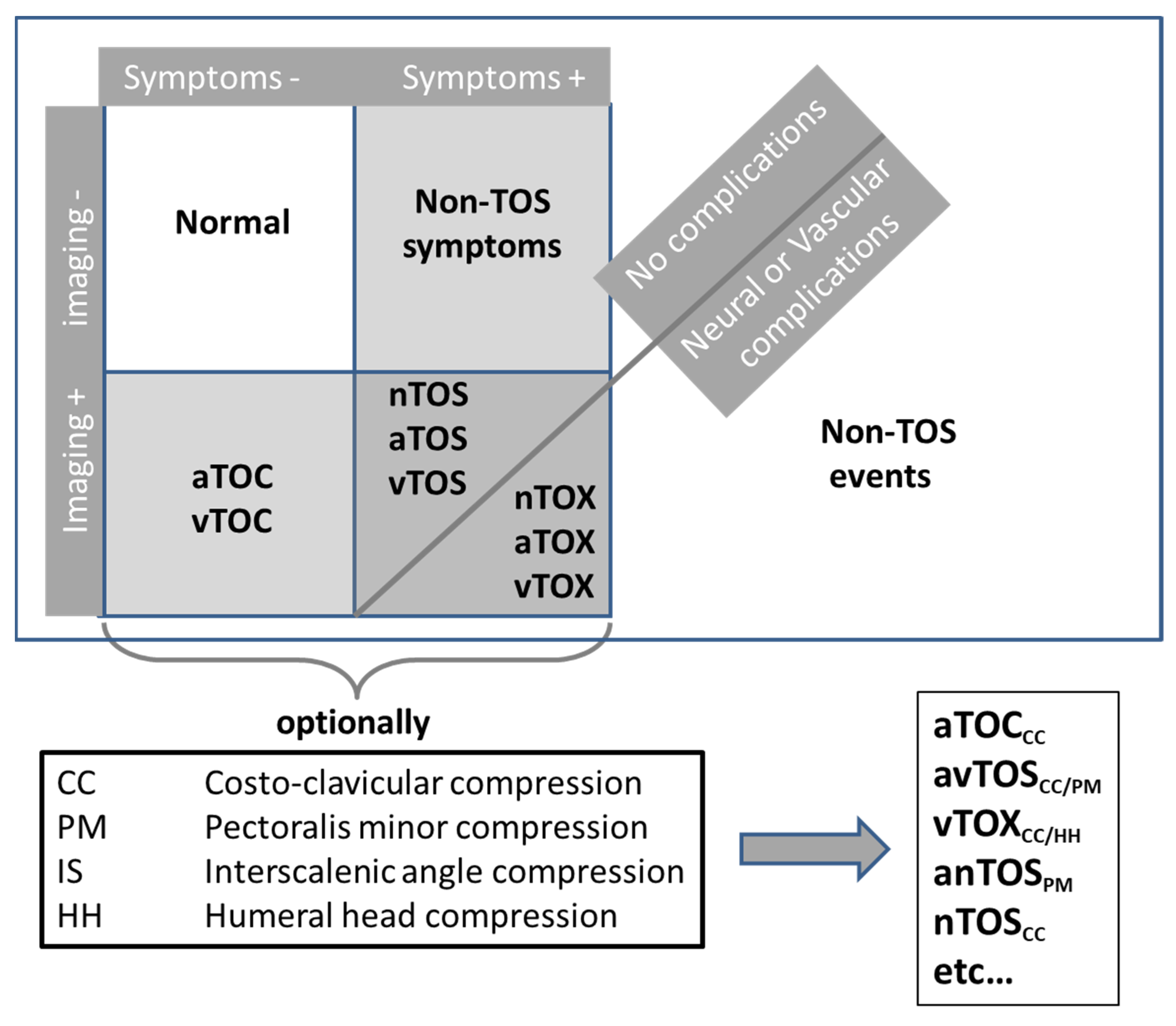
2. New Paradigm for Clinical Classification

The positional thoracic outlet compression (TOC) may remain completely asymptomatic. Indeed, it has been estimated that TOC may be present in about 30% of the completely asymptomatic adult population [9,10]. When symptomatic, the compression of nerves or vessels by adjacent structures during upper limb movements can result in a large variety of signs and symptoms, which are collectively referred to as thoracic outlet syndrome (TOS) [3,4,11]. It is clear that patients with TOC but without any signs or symptoms cannot be classified as having TOS, as by definition, a ‘syndrome’ is a group of signs and symptoms that characterize a particular abnormality or condition. It is proposed that the term TOC should be used specifically to refer to the asymptomatic form of positional neurovascular compression at the thoracic outlet level.
Most symptomatic patients with TOS are known to benefit from physical therapy [12,13]. Physical therapy leads to the resolution of symptoms in the majority of these patients despite the persistence of TOC. Indeed, most of these patients will never require surgery and this is reserved only when physical therapy fails to improve the symptoms [14]. Ultimately, a significant number of patients with TOC will develop complications or sequelae (CS) as a result of the chronic conflict between the neuro-vascular bundle and the adjacent structures. Remarkably, in accordance with the reporting standards of the Society of Vascular Surgery [3], only vascular CS are considered when it comes to classifying patients into different TOS sub-groups. This is likely inadequate as CS can affect veins (Paget Schroetter syndrome) or arteries (sub-clavicular aneurysm or ischemia from distal emboli) but also nerves (amyotrophy and abnormal nerve conduction at rest, specifically of C8 or T1 roots) [15,16,17]. Further, it is currently difficult to provide clinical proof of ischemia or arm swelling. As a result, to date, more than 90% of TOS cases are considered to be of neural origin [11].
Vascular complications may present as primary events in previously asymptomatic patients with undiagnosed TOC [18,19]. The “thoracic outlet compression with complications or sequelae” (the acronym of which should be spelled TOC-CS but can be simplified as “TOX”) will almost invariably lead to surgery to release the compression, and other possible treatment targeting the complications as required. There is no obvious reason why a neurological cause would not also be sought, only in cases of neurological complications. Electromyographic signs of denervation affecting the lower plexus branches at rest, with or without amyotrophy, provide clear evidence of neural complications, as much as ultrasound or angiography provide evidence for vascular sequelae [18,19]. Consequently, thoracic outlet compression with complications or sequelae could be classified as being of neural (nTOX), arterial (aTOX) and/or venous (vTOX) origin, depending on the nature of the complication, which should be objectively recorded. Simultaneous neural, arterial and/or venous complications could lead to letter associations such as anTOX, nvTOX, avTOX and anvTOX.
Patients with TOS should be classified as neural (nTOS), arterial (aTOS), and/or venous (vTOS) origin depending on the clinical positional symptoms together with any objective evidence of neuro-vascular bundle compression during provocative tests. The combination of symptoms of arterial, venous, and/or neural origin would then result in anTOS, avTOS, nvTOS, or anvTOS encoding. Optionally, and for treatment possibilities, indices may indicate whether the compression is found at the costo-clavicular (CC), pectoralis minor (PM), inter-scalenic angle (IS) or humeral head (HH) levels (Figure 1).
The remaining asymptomatic thoracic outlet positional compression (TOC) shall become a specific clinical entity that should be called neither TOS nor TOX. Note that, to the best of our knowledge, asymptomatic positional compression has been reported for veins and/or arteries during vascular investigations, but has never been reported for nerves, and then conceptually, one could discriminate aTOC and vTOC, but not nTOC. There is a continuum between TOC, TOS and TOX, which are simply different clinical stages of the same compression of the neurovascular bundle at the thoracic outlet, just like the Rutherford grades represent different stages of the same pathology in lower extremity arterial disease. This TOC/TOS/TOX paradigm might help to put an end to some old controversies of the literature.

3. Discussion

Multiple reports suggest that clinical, ultrasound and radio-vascular investigations show a high rate of positive positional results in control subjects or in the asymptomatic arm of patients with unilateral symptoms [9,20]. Consequently, several authors suggest that many of these tests are inadequate in the diagnosis of TOS, especially in extreme arm positions [20,21,22]. This suggestion attributes the inconsistency between the results of the investigation and the symptoms to sub-optimal investigations. However, in our opinion, the problem is inadequate use of the expression TOS. TOS should be considered only in the presence of symptoms and TOX only when CS arise. The other situations where there are no symptoms or CS should be classified as TOC and not TOS. Clearly, the presence of a compression (TOC) does not imply that symptoms will occur, and not all arm and shoulder symptoms result from TOC. Furthermore, TOX (e.g., sub-clavicular aneurysms) may remain completely asymptomatic and be discovered accidentally [23]. In the absence of complications or sequelae, symptoms that are present at rest in a limb with TOC are very unlikely to be secondary to the positional compression of the neurovascular bundle as this occurs mainly in the non-resting state. TOC with resting symptoms only shall remain TOC, whereas compressions with symptoms occurring following upper limb provocative movement might indicate TOS. It should be noted that the movement itself rarely causes immediate pain. As seen in claudication, from our experience, there is generally a pain-free interval during the first seconds of the provocative maneuver. Pain occurring during initial mobilization of the arm is rather indicative of an osteo-articular or tendinous problem.
Finally, the major issue in symptomatic patients remains attributing the symptoms to the presence of TOC, thus establishing a diagnosis of TOS. The first question here is why does TOC remain asymptomatic? There are many possible explanations that should be investigated in the future. Inflammation could be proposed as a factor that results in the conversion of TOC (asymptomatic) to TOS (symptomatic). Another possibility is that TOS occurs if the frequency or duration of compression and, hence, of its consequences on neurovascular function surpasses the level of post-compression recovery. This might explain the high prevalence of TOS in certain professions that are characterized by repetitive movements [24,25,26]. It is likely that many symptomatic individuals develop avoidance behavior when facing difficulties in their daily routine (such as using a headset or earplugs to use their phone rather than holding it in their hand) [27]. As a consequence, they no longer report complaints once their avoidance techniques have become totally integrated in their behavior.
The second question to ask is is the presence of vascular compression diagnostic of the vascular origin of pain per se? Obviously not! Firstly, vascular compression is only an indicator of the presence of a conflict between the neurovascular bundle and adjacent structures and can be associated with neurological symptoms only and thus classified as nTOS. Secondly, a compression of the subclavian vein is very unlikely to result in symptoms (and thus is classified as vTOS) if collateral or pre-clavicular veins normalize venous outflow. Similarly, a non-occlusive compression of the subclavian artery is very unlikely to result in symptoms (and thus is classified as aTOS) if the persisting arterial inflow is sufficient to cover oxygen requirements. Lastly, with the current classification, an arterial etiology is considered only in the presence of complications, leading to the suggestion that a white hand (a relatively frequent observation in TOS during provocative movement) results from an adrenergic vasoconstriction of neural origin. This is quite odd as the presence of a white foot is exceptional in sciatica (another example of neural compression). Once again, the underlying etiology is mislabeled in view of the patients’ symptomatology with the current classification. It should be noted that hand ischemia resulting from arterial positional occlusion may result in paresthesia [28,29]. Therefore, paresthesia is not specific to nTOS. Ischemia-induced paresthesia will rather affect the whole hand, while neural compression is more likely to result in paresthesia in the C5-T1 dermatome.
The TOC/TOS/TOX classification proposed here is quite simple. We believe that discriminating between TOC, TOS and TOX clarifies clinical definitions and their respective treatment options. TOC is to be considered as a physiological positional phenomenon, TOS requires medical or surgical treatment and surgery should be considered as a primary option in TOX.
Ultimately, attributing symptoms to the presence of compression of the neurovascular bundle to ascertain a diagnosis of neural, arterial and/or venous TOS remains based on a holistic diagnostic approach. Currently, this relies heavily on a careful description of symptoms (either self-reported or induced by provocative maneuvers) which clearly requires some clinical experience and expertise. In an ideal world, objective measurements of positional neural dysfunction and objective proof of positional measurable ischemia or positional measurable swelling would strengthen the association between symptoms and neurovascular compression to diagnose TOS [3]. Indeed, many possible causes of upper limb pain may be erroneously labeled as TOS. Recent approaches such as STIR-MRI [30,31] or positional ENMG for diagnosing objective neural functional impairment [32], transcutaneous oximetry for arterial ischemia [33,34], and venous photo-plethysmography for venous positional edema [35] are future options for attributing the symptoms to the TOC, thus providing more robust evidence for neural, arterial or venous TOS, respectively.

Author Contributions

Draft writing: P.A. and S.H., critical revision and review: P.W.W., P.B., Y.G., N.O., L.G., G.T., L.M. and R.M. Each of the authors read and approved the submitted version and agrees to be personally accountable for the author’s own contributions and for ensuring that questions related to the accuracy or integrity of any part of the work, even ones in which the author was not personally involved, are appropriately investigated, resolved and documented in the literature. All authors have read and agreed to the published version of the manuscript.

Funding

This work received no external funding.

Acknowledgments

The authors thank Ruth Sicluna for support reviewing the grammar and style.

Conflicts of Interest

The authors declare no conflict of interest.

References

  1. Larson, S.G.; Jungers, W.L.; Morwood, M.J.; Sutikna, T.; Jatmiko; Saptomo, E.W.; Due, R.A.; Djubiantono, T. Homo floresiensis and the evolution of the hominin shoulder. J. Hum. Evol. 2007, 53, 718–731. [Google Scholar] [CrossRef]
  2. Voisin, J.L. Clavicle, a neglected bone: Morphology and relation to arm movements and shoulder architecture in primates. Anat. Rec. A Discov. Mol. Cell Evol. Biol. 2006, 288, 944–953. [Google Scholar] [CrossRef] [PubMed]
  3. Illig, K.A.; Donahue, D.; Duncan, A.; Freischlag, J.; Gelabert, H.; Johansen, K.; Jordan, S.; Sanders, R.; Thompson, R. Reporting standards of the Society for Vascular Surgery for thoracic outlet syndrome: Executive summary. J. Vasc. Surg. 2016, 64, 797–802. [Google Scholar] [CrossRef] [PubMed]
  4. Illig, K.A.; Rodriguez-Zoppi, E. How Common Is Thoracic Outlet Syndrome? Thorac. Surg. Clin. 2021, 31, 11–17. [Google Scholar] [CrossRef] [PubMed]
  5. Wyllie, W.G. Scalenus Anticus Syndrome in a Girl aged 11 Years. Proc. R. Soc. Med. 1939, 32, 218. [Google Scholar] [CrossRef] [PubMed]
  6. Craig, W.M.; Knepper, P.A. Cervical Rib and the Scalenus Anticus Syndrome. Ann. Surg. 1937, 105, 556–563. [Google Scholar] [CrossRef] [PubMed]
  7. Falconer, M.A. The costoclavicular syndrome. Br. Med. J. 1947, 2, 69–70. [Google Scholar] [CrossRef] [PubMed]
  8. Fletcher, E.; Fletcher, A. The costoclavicular syndrome. Br. Med. J. 1947, 1, 465–466. [Google Scholar] [CrossRef]
  9. Bishop, L.; Bartlett, M. Doppler waveform analysis during provocative manoeuvres in the assessment for arterial thoracic outlet syndrome results in high false-positive rates; a cross-sectional study. JRSM Cardiovasc. Dis. 2021, 10, 20480040211006571. [Google Scholar] [CrossRef]
  10. Nord, K.M.; Kapoor, P.; Fisher, J.; Thomas, G.; Sundaram, A.; Scott, K.; Kothari, M.J. False positive rate of thoracic outlet syndrome diagnostic maneuvers. Electromyogr. Clin. Neurophysiol. 2008, 48, 67–74. [Google Scholar]
  11. Li, N.; Dierks, G.; Vervaeke, H.E.; Jumonville, A.; Kaye, A.D.; Myrcik, D.; Paladini, A.; Varrassi, G.; Viswanath, O.; Urits, I. Thoracic Outlet Syndrome: A Narrative Review. J. Clin. Med. 2021, 10, 962. [Google Scholar] [CrossRef] [PubMed]
  12. Collins, E.; Orpin, M. Physical Therapy Management of Neurogenic Thoracic Outlet Syndrome. Thorac. Surg. Clin. 2021, 31, 61–69. [Google Scholar] [CrossRef] [PubMed]
  13. Balderman, J.; Abuirqeba, A.A.; Eichaker, L.; Pate, C.; Earley, J.A.; Bottros, M.M.; Jayarajan, S.N.; Thompson, R.W. Physical therapy management, surgical treatment, and patient-reported outcomes measures in a prospective observational cohort of patients with neurogenic thoracic outlet syndrome. J. Vasc. Surg. 2019, 70, 832–841. [Google Scholar] [CrossRef] [PubMed]
  14. Pesser, N.; Goeteyn, J.; van der Sanden, L.; Houterman, S.; van Alfen, N.; van Sambeek, M.; van Nuenen, B.F.L.; Teijink, J.A.W. Feasibility and Outcomes of a Multidisciplinary Care Pathway for Neurogenic Thoracic Outlet Syndrome: A Prospective Observational Cohort Study. Eur. J. Vasc. Endovasc. Surg. 2021, 61, 1017–1024. [Google Scholar] [CrossRef] [PubMed]
  15. Matsubara, T.; Kurokawa, K.; Sakurai, K.; Yasutomi, H.; Yamawaki, T. The Gilliatt-Sumner hand: A diagnostic clue of neurogenic thoracic outlet syndrome. QJM 2018, 111, 831–832. [Google Scholar] [CrossRef] [PubMed]
  16. McGillicuddy, J.E. Cervical radiculopathy, entrapment neuropathy, and thoracic outlet syndrome: How to differentiate? Invited submission from the Joint Section Meeting on Disorders of the Spine and Peripheral Nerves, March 2004. J. Neurosurg. Spine 2004, 1, 179–187. [Google Scholar] [CrossRef] [PubMed]
  17. Tilki, H.E.; Stalberg, E.; Incesu, L.; Basoglu, A. Bilateral neurogenic thoracic outlet syndrome. Muscle Nerve 2004, 29, 147–150. [Google Scholar] [CrossRef] [PubMed]
  18. Cruz-Martinez, A.; Arpa, J. Electrophysiological assessment in neurogenic thoracic outlet syndrome. Electromyogr. Clin. Neurophysiol. 2001, 41, 253–256. [Google Scholar]
  19. Smith, T.; Trojaborg, W. Diagnosis of thoracic outlet syndrome. Value of sensory and motor conduction studies and quantitative electromyography. Arch. Neurol. 1987, 44, 1161–1163. [Google Scholar] [CrossRef]
  20. Dessureault-Dober, I.; Bronchti, G.; Bussieres, A. Diagnostic Accuracy of Clinical Tests for Neurogenic and Vascular Thoracic Outlet Syndrome: A Systematic Review. J. Manip. Physiol. Ther. 2018, 41, 789–799. [Google Scholar] [CrossRef]
  21. Hixson, K.M.; Horris, H.B.; McLeod, T.C.V.; Bacon, C.E.W. The Diagnostic Accuracy of Clinical Diagnostic Tests for Thoracic Outlet Syndrome. J. Sport Rehabil. 2017, 26, 459–465. [Google Scholar] [CrossRef] [PubMed]
  22. Stapleton, C.; Herrington, L.; George, K. Sonographic evaluation of the subclavian artery during thoracic outlet syndrome shoulder manoeuvres. Man. Ther. 2009, 14, 19–27. [Google Scholar] [CrossRef] [PubMed]
  23. DiFiore, J.W.; Reid, J.R.; Drummond-Webb, J. Thoracic outlet syndrome in a child—Transaxillary resection of anomalous first rib. J. Pediatr. Surg. 2002, 37, 1220–1222. [Google Scholar] [CrossRef] [PubMed]
  24. Franklin, G.M. Work-Related Neurogenic Thoracic Outlet Syndrome: Diagnosis and Treatment. Phys. Med. Rehabil. Clin. N. Am. 2015, 26, 551–561. [Google Scholar] [CrossRef] [PubMed]
  25. Sanders, R.J.; Hammond, S.L. Outcome of surgery for thoracic outlet syndrome in Washington state workers’ compensation. Neurology 2000, 55, 1594–1595. [Google Scholar] [CrossRef] [PubMed]
  26. Ritter, A.; Sensat, M.L.; Harn, S.D. Thoracic outlet syndrome: A review of the literature. J. Dent. Hyg. 1999, 73, 205–207. [Google Scholar] [PubMed]
  27. Abraham, P.; Henni, S. Symptoms and absence of symptoms while using a telephone: The paradox of thoracic outlet syndrome. Musculoskelet. Sci. Pract. 2021, 51, 102266. [Google Scholar] [CrossRef]
  28. Hammacher, E.R.; Eikelboom, B.C.; van Lier, H.J.; Skotnicki, S.H.; Wijn, P.F. Brachial artery lesions after cardiac catheterisation. Eur. J. Vasc. Surg. 1988, 2, 145–149. [Google Scholar] [CrossRef]
  29. Huber, T.S.; Brown, M.P.; Seeger, J.M.; Lee, W.A. Midterm outcome after the distal revascularization and interval ligation (DRIL) procedure. J. Vasc. Surg. 2008, 48, 926–932; discussion 932–923. [Google Scholar] [CrossRef]
  30. Zhang, T.; Xu, Z.; Chen, J.; Liu, Z.; Wang, T.; Hu, Y.; Shen, L.; Xue, F. A Novel Approach for Imaging of Thoracic Outlet Syndrome Using Contrast-Enhanced Magnetic Resonance Angiography (CE-MRA), Short Inversion Time Inversion Recovery Sampling Perfection with Application-Optimized Contrasts Using Different Flip Angle Evolutions (T2-STIR-SPACE), and Volumetric Interpolated Breath-Hold Examination (VIBE). Med. Sci. Monit. 2019, 25, 7617–7623. [Google Scholar] [CrossRef]
  31. Magill, S.T.; Brus-Ramer, M.; Weinstein, P.R.; Chin, C.T.; Jacques, L. Neurogenic thoracic outlet syndrome: Current diagnostic criteria and advances in MRI diagnostics. Neurosurg. Focus 2015, 39, E7. [Google Scholar] [CrossRef] [PubMed]
  32. Haghighi, S.S.; Baradarian, S.; Bagheri, R. Sensory and motor evoked potentials findings in patients with thoracic outlet syndrome. Electromyogr. Clin. Neurophysiol. 2005, 45, 149–154. [Google Scholar] [PubMed]
  33. Abraham, P.; Hersant, J.; Ramondou, P.; Thouveny, F.; Feuilloy, M.; Picquet, J.; Henni, S. Comparison of transcutaneous oximetry with symptoms and arteriography in thoracic outlet syndrome. Clin. Hemorheol. Microcirc. 2020, 75, 107–119. [Google Scholar] [CrossRef] [PubMed]
  34. Henni, S.; Hersant, J.; Ammi, M.; Mortaki, F.E.; Picquet, J.; Feuilloy, M.; Abraham, P. Microvascular Response to the Roos Test Has Excellent Feasibility and Good Reliability in Patients With Suspected Thoracic Outlet Syndrome. Front. Physiol. 2019, 10, 136. [Google Scholar] [CrossRef] [PubMed]
  35. Hersant, J.; Ramondou, P.; Chavignier, V.; Chavanon, A.; Feuilloy, M.; Picquet, J.; Henni, S.; Abraham, P. Forearm Volume Changes Estimated by Photo-Plethysmography During an Original Candlestick/Prayer Maneuver in Patients with Suspected Thoracic Outlet Syndrome. Front. Physiol. 2021, 12, 652456. [Google Scholar] [CrossRef]
Figure 1. Schematic representation of the proposed new paradigm. TOC, TOS and TOX are for “thoracic outlet compression”, “Thoracic outlet syndrome” and “thoracic outlet compression with complications or sequelae”, respectively.
Jvd 02 00032 g001
Disclaimer/Publisher’s Note: The statements, opinions and data contained in all publications are solely those of the individual author(s) and contributor(s) and not of MDPI and/or the editor(s). MDPI and/or the editor(s) disclaim responsibility for any injury to people or property resulting from any ideas, methods, instructions or products referred to in the content.

