Opinion

The Prostate Is Not a Pill and Therefore Has No Capsule

Department of Pathology, Medical University of Vienna, 1090 Wien, Austria
*
Author to whom correspondence should be addressed.
Anatomia 2023, 2(3), 253-260; https://doi.org/10.3390/anatomia2030023
Submission received: 5 July 2023 / Revised: 2 August 2023 / Accepted: 5 August 2023 / Published: 14 August 2023
(This article belongs to the Special Issue Anatomy for Modern Urology)

Abstract

:
Tumor staging of prostate cancer is a fundamental principle in management and therapy, with a hallmark being tumor growth beyond the organ boundary. Often, this is referred to as “capsule penetration”, suggesting the existence of a true prostatic capsule that would facilitate the determination of tumor penetration. In fact, the prostate does not have a true capsule and, depending on the anatomic area, it blends with the surrounding fibrous, adipose and muscular tissue. This makes it sometimes difficult or impossible to unequivocally identify extraprostatic tumor extension. It is necessary to appreciate this difficulty in order to better understand the significance of extraprostatic tumor extension.

1. Introduction

Prostate carcinoma (PCa) is the most common cancer in men, and the determination of the tumor extent in radical prostatectomy specimens is important for staging and patient management [1]. One significant difference in pathological staging lies between pT2 and pT3a, where the tumor growth is confined to the organ or extends beyond the parenchymal margin (parenchyma, in this context, comprising the fibromuscular stroma and the acini/ducts) into the surrounding connective/adipose tissue. This discrimination is also important for clinical management [1]. Although this is well accepted, there are challenges regarding the delineation of the organ margins, since the prostate does not feature an organ-confining capsule. Although this fact is acknowledged in several studies and consensus papers [2,3,4], the term “prostate capsule” contemporarily and repeatedly appears in surgical pathology texts [5,6,7].
To clarify, an organ capsule is defined as a “capsula fibrosa”, which surrounds the whole organ with connective tissue [2,8]. The capsule is useful for the mechanic stability of the organ and exists, for example, in the kidney, the spleen and also in the lymph nodes [8]. The prostate is not surrounded by such a fibrous tissue enveloping the whole organ, a fact already perceived by Ayala et al. in 1989 [2]. Standard reference textbooks for surgical pathologists also acknowledge this fact but nonetheless keep the terminus “capsule” or “prostatic capsule” [9,10]. The prostatic parenchyma, consisting of a fibromuscular stroma with embedded glands, borders directly the surrounding connective tissue. Depending on the anatomic region, this transition is not always clearly distinguishable and, furthermore, individual differences exist. In the following sections, we will outline the histoanatomical relationship with surrounding structures and organs, but also the implications of defining the organ margins and, with it, the tumor stage. We will further discuss the assessment of the quantity of extraprostatic tumor growth and its potential clinical implications. Thereby, we hope to raise awareness of the complexity in defining the prostate organ margin and the set of problems arising from simplifying it as a capsule.
The prostate is located in the small pelvis underneath the bladder and in between the symphysis and the rectum [8], surrounded by adipose and connective tissue containing nerves and vessels, all enclosed in several fascial structures [5]. Detailed descriptions and accurate schematic drawings can also be found in three very thorough articles by Walz et al. and Hoeh et al. [5,6,11]. For anatomical descriptions, we use terms according to “terminologia anatomica” or similar to the cited literature if we believe this to be necessary for comprehension [12].

2. Materials and Methods

The slides used in this article were obtained during the routine diagnostic workup of radical prostatectomy specimens submitted to our pathology department. They were stained with hematoxylin and eosin (H&E) according to standard procedure. No personal patients’ data are used in this article. For language editing, we used ChatGPT (20 July 2023 Version).

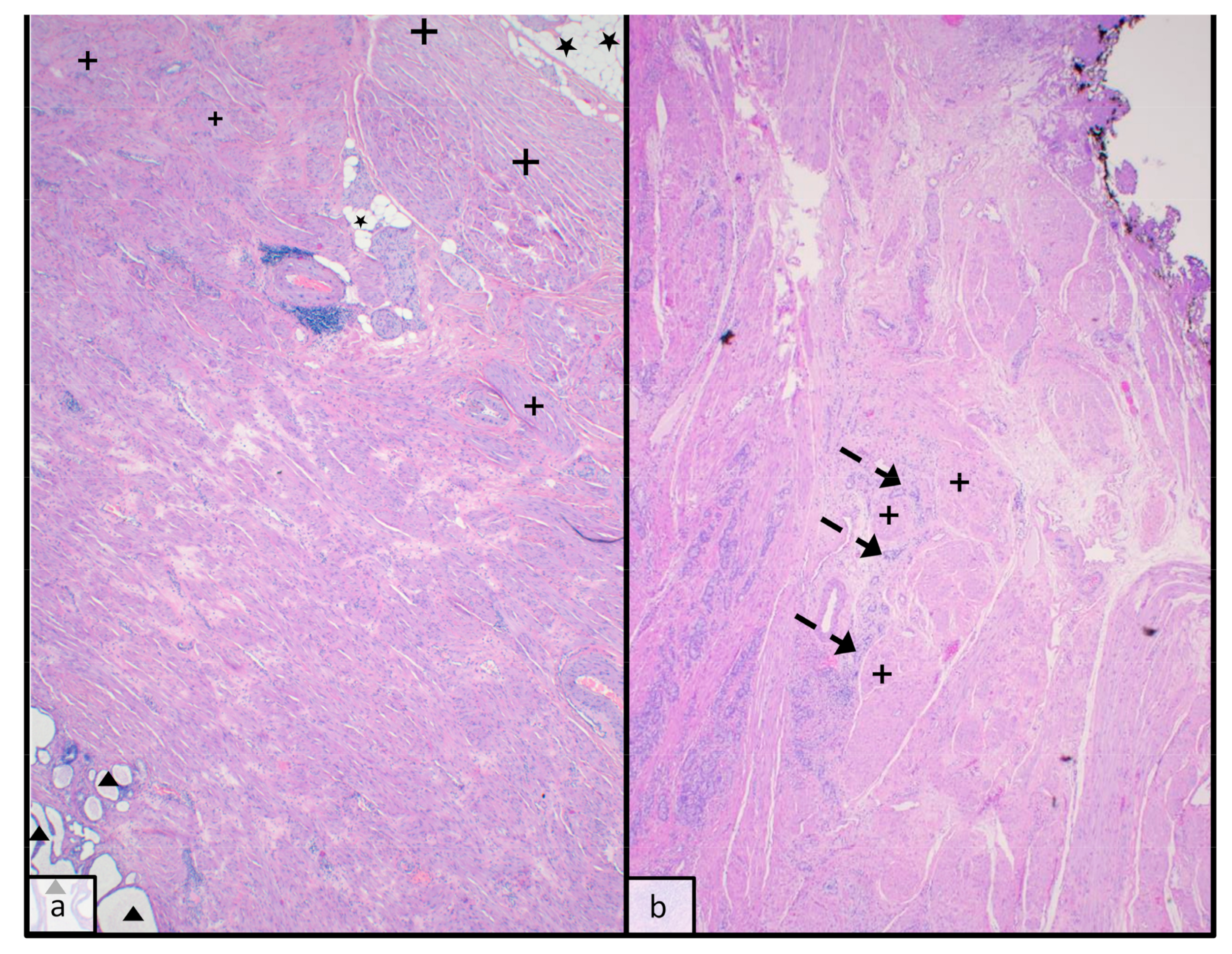
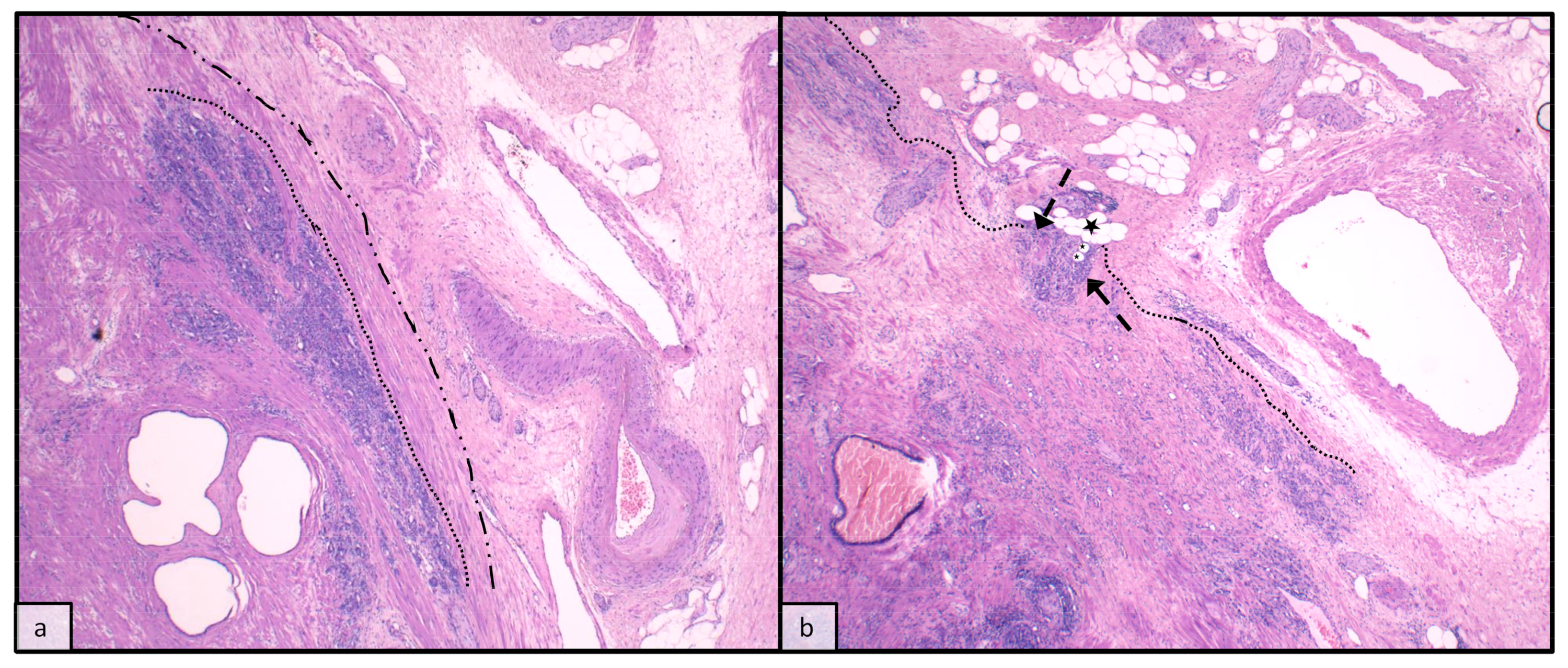
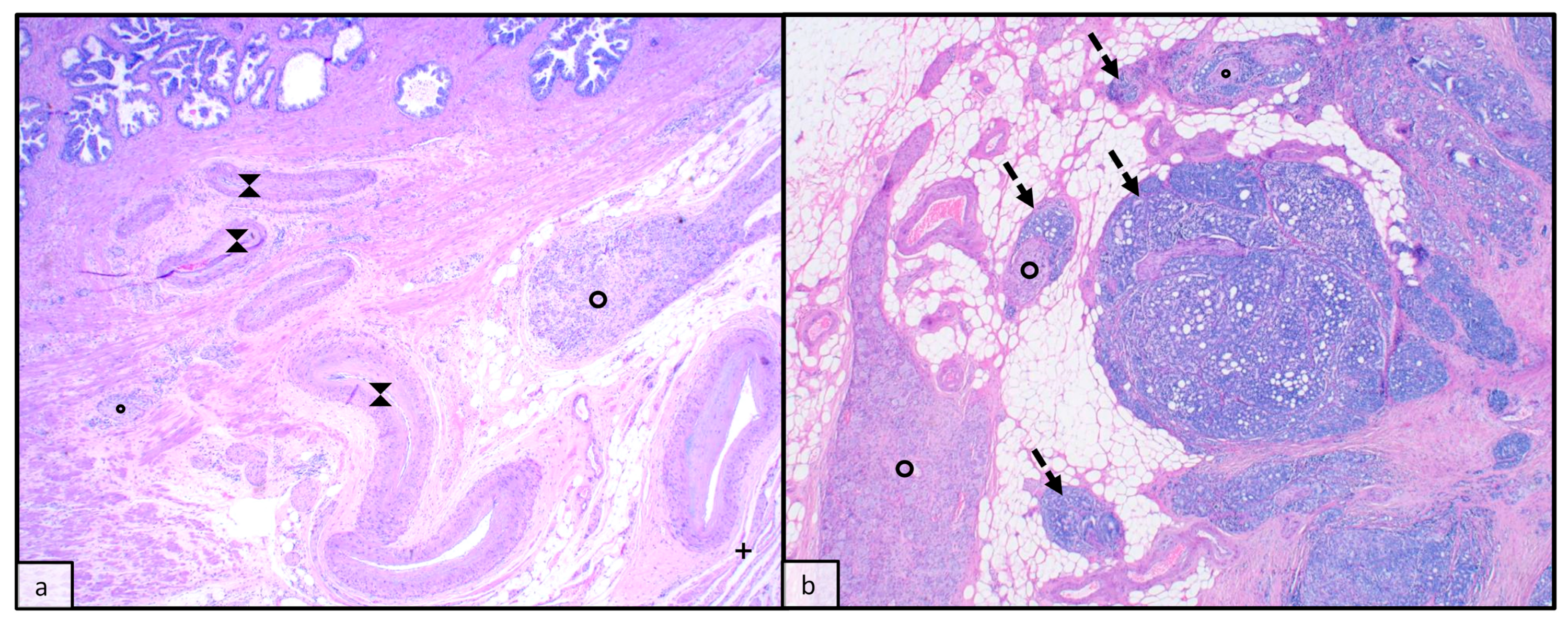
3. The Base and Anterior Surface of the Prostate
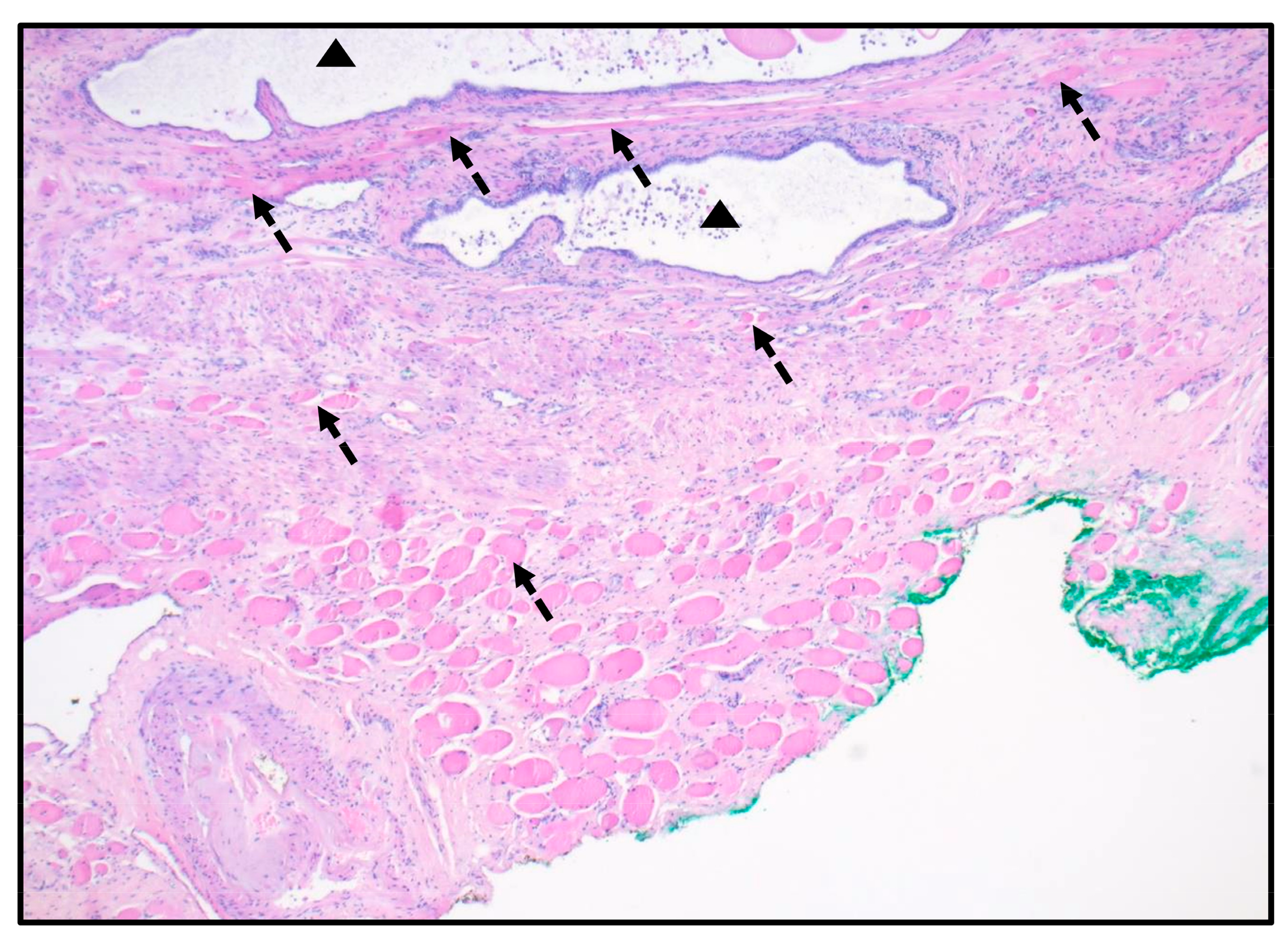
Cranially, at the base, the prostate is part of the neck of the bladder. Here, several anatomic structures, such as the detrusor vesicae, the male internal urethral sphincter and, of course, the prostate parenchyma, blend with each other [5,6,11]. Muscle fibers of the external longitudinal layer of the detrusor vesicae span over the prostate anteriorly and posteriorly [5,6,11]. Anteriorly, they form the detrusor apron and contribute to the pubovesical/puboprostatic ligaments, which stretch from the posterior pubic bone to the bladder and the neck of the bladder but are also attached to the ventral prostate [5]. These structures are ventrally covered by the visceral pelvic fascia and together they fuse in the midline with the prostate fibromuscular stroma [5,6]. The anterolateral zone is formed of adipose connective tissue filling the space between the prostate parenchyma and the visceral endopelvic fascia [5]. Determining extraprostatic tumor growth in this area may be simple, where malign glands lying in adipose tissue can be easily appreciated. However, it is more difficult in the midline, where the fibromuscular parenchyma blends with the fibrous tissue of the detrusor apron and visceral endopelvic fascia. In the absence of adipose tissue, anticipation of the normal parenchymal margin based on the adjacent contour might be a feasible approach to determine extraprostatic tumor growth [3,4]. At the bladder neck, the vesical outlet joins into the prostate [5]. There, the fibromuscular tissue blends with the detrusor muscle and the prostatic boundary may be difficult to discern (Figure 1a) [5]. However, invasion into smooth muscle bundles clearly representing the detrusor muscle of the bladder neck is staged similarly to extraprostatic extension (Figure 1b) [3]. It is important to note that this must not be seen as infiltration in an adjacent organ (bladder) [3].

4. The Apex

Caudally, at the apex, the prostate is delineated by the urethral sphincter, which consists of an internal longitudinal and an external striated muscle layer. The internal muscle layer around the urethra extends into the prostatic apex. The outer striated muscle also reaches the apex surface and may even insert into the parenchyma, resulting in histomorphologically benign glands intermingled with striated muscle (Figure 2). This makes it impossible to consider extraprostatic extension in the apex area. As outlined in the ISUP consensus report of Working Group 3 on “extraprostatic extension, lymphovascular invasion and locally advanced disease”, several approaches have been used, such as “ink on tumor” or extension across the anticipated contour of the normal gland [3]. However, ultimately, no reliable landmark at the apical region can be used [3]. Please note that this does not apply to the other prostatic margin sites.

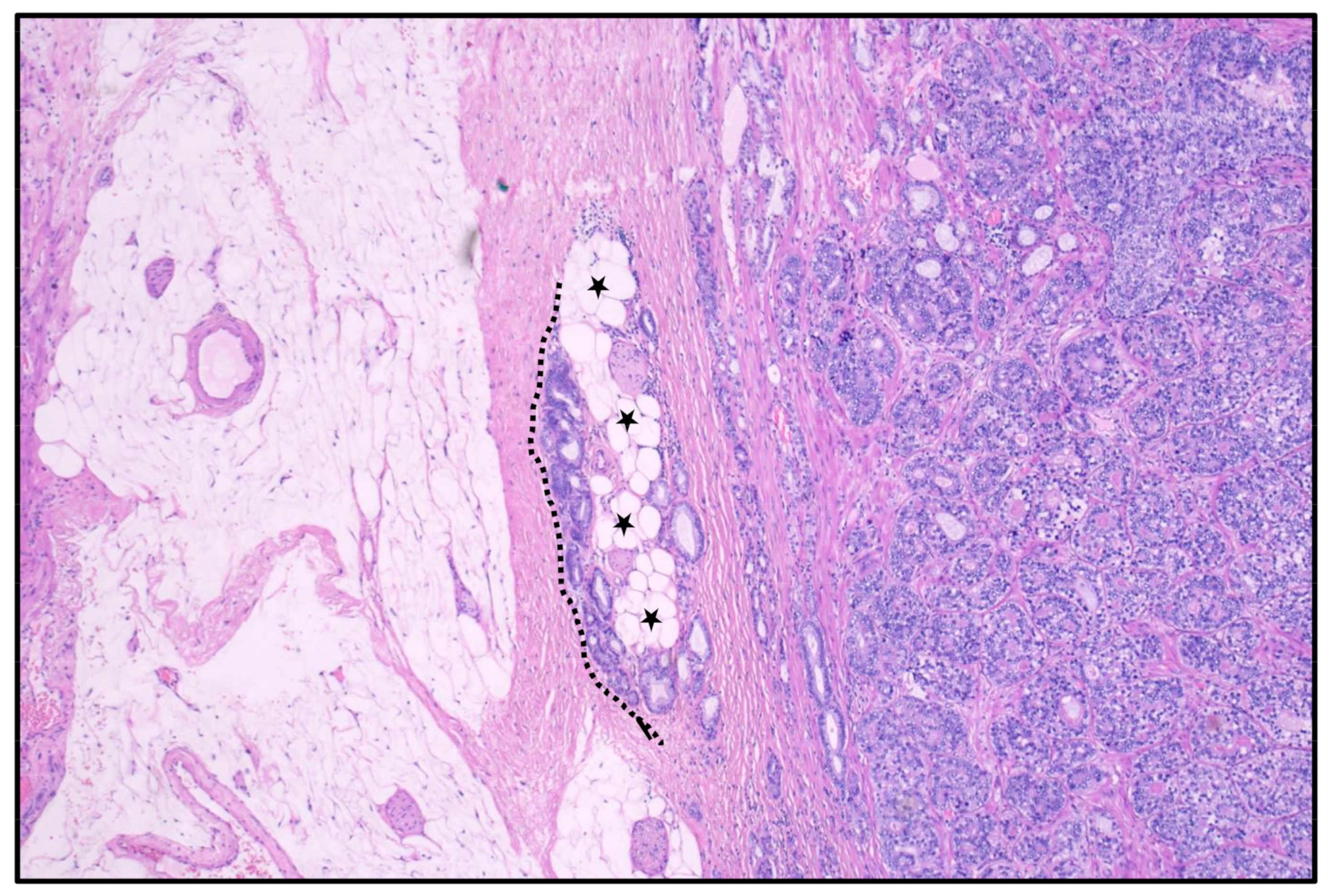
5. The Lateral and Dorsal Surface with Seminal Vesicles

Laterally, the prostate is surrounded by a variable amount of fibroadipose tissue containing vessels, nerves and fascial structures [5,6,11]. Starting from the anterior, the visceral endopelvic fascia continues after the fascial tendinous arch of pelvis as the levator ani fascia and marks the most lateral border of the periprostatic fascial structures [5,6,11]. Medially, within the connective tissue, and often multilayered, is the prostatic fascia [5,6,11]. Together, these fascial structures and the connective tissue are referred to as the periprostatic fascia [5,6,11]. This structure continues posteriorly to encounter the neurovascular bundle [5,6,11]. It is essential to note that the neurovascular bundle is not necessarily a well-circumscribed structure and may spread over the lateral periprostatic area [5,6,11]. There, at the turn to the dorsal surface, some of the facial structures join the posterior prostatic fascia, or Denonvilliers’ fascia [5,6,11]. This fascial structure is a continuous fibrous structure covering the dorsal prostate and extending cranially from immediately underneath the dip of the recto-vesical pouch downward to the prostate-urethral junction, where it blends with the central perineal tendon [5,6,11]. It also covers the seminal vesicles [5]. Similar to the ventral area, extraprostatic extension may be easily assessed in the presence of surrounding adipose tissue but it might be difficult in the more posterior areas where the fibrous tissue intermingles with the fibromuscular prostate parenchyma (Figure 3). Likewise, extension beyond the anticipated organ boundary extrapolated from the adjacent contour might be a feasible approach [3,4].

6. Extraprostatic Growth and Oncological Implications

Pathological evaluation of tumor extent in radical prostatectomies (RPE) forms the basis for staging and can be important for patient management [1]. We divide PCa after RPE into stage pT2, where the tumor is confined to the prostate gland, and pT3a, where the tumor displays extraprostatic extension (EPE), which is not always obvious (Figure 4). In the 1970s and 1980s, studies evaluated whether invasion or extension beyond the so-called “capsule” of the prostate plays a role in progression-free or overall survival [13,14]. More recent works have attempted to evaluate the significance of EPE, but often these studies do not differentiate between EPE alone, positive margins or invasion of the seminal vesicles, and the results on its significance are ambiguous [4,15,16,17,18,19]. McNeal et al. showed that EPE (formerly called capsule penetration) was correlated with the cancer volume, the location of positive surgical margins and the presence of nodal metastases or seminal vesicle (SV) invasion [20]. However, they could not demonstrate that it was related to prognosis independently of its correlation with the cancer volume [20]. Other authors who stratified patients based on the extent of EPE in different groups were repeatedly able to show an association with disease progression.
However, the measurement technique and the related stratification into different groups is another issue of discussion. The latest ISUP consensus paper dealing with this issue was released in 2010, and 94% of participants voted for the quantification of EPE [3]. However, they did not reach a consensus on the specific method to use [3]. In general, the option is either to use a subjective or quantitative approach. Two examples of a subjective approach are the methods described by Epstein and by Wheeler. Epstein et al. differentiated between focal, defined by “only a few neoplastic glands…exterior to the prostate”, and established capsular penetration, without further specification of “a few”, whereas Wheeler et al. defined focal as extraprostatic extension “of less than one high power field on no more than two separate sections” and any more than this as established [21,22]. Both of them were able to show an association between EPE and disease progression in their cohorts [21,22]. However, these results could not be confirmed by Sung et al. using the aforementioned measurement techniques. Instead, they suggested a quantitative measurement of the radial distance, defined as the distance that the tumor protrudes perpendicularly beyond the outer margin of the prostatic stroma [23,24]. With this approach, they could show EPE to be an independent predictor of prostate-specific antigen (PSA) recurrence in a multivariate analysis. Interestingly, this group also compared other quantitative methods such as circumferential length measurement and uni- vs. multifocality of EPE and could not find an association with PSA recurrence [23]. Similar results for radial distance measurement and circumferential length measurement were shown by Kir et al. [25]. Farchoukh et al. differentiated between focal and established EPE based on the measurement of less than 8 or equal to/more than 8 mm but did not further specify the measurement technique. Additionally, they formed a third group called “multifocal extraprostatic extension”, encompassing those cases with more than one focus less than 8 mm [26]. Established EPE was again associated with a higher risk of recurrence, but, interestingly, in contrast to the results of Sung et al., multifocality even showed a higher risk [26]. More recently, a study by Park et al. confirmed radial distance measurement as predictive of biochemical recurrence (BCR) and additionally suggested to include the number of foci to further improve the prediction [27]. However, there is no consensus yet, and one has to keep in mind that the method of evaluating EPE has to be applicable in the routine diagnostic workup of radical prostatectomies and should also be reproducible, a fact that, to our knowledge, has not yet been looked at.
In non-transition zone cancers, extraprostatic extension is most commonly observed posterolaterally, where nerves penetrate the outer parenchymal rim (Figure 5) [20]. In transition zone cancers, it is less common and located more anteriorly [20]. Apical positive margins are also relatively common in transition zone cancers [20]. From a clinical perspective, it is important to determine whether there is involvement of the anterior part of the prostate, as cancers of peripheral or transition zone origin may also be predominantly located in the anterior prostate gland [28]. These tumors are less likely to be detected by standard biopsy procedures and may be associated with a higher incidence of margin positivity and increased risk of biochemical recurrence [29]. Nowadays, with the help of multiparametric magnetic resonance imaging (mpMRI), these PCa are found more often [28]. A recent study showed that the detection of anterior PCa has doubled since the introduction of mpMRI. This study also confirmed the aggressiveness of anterior PCa with a significant rate of extraprostatic extension [28]. Some authors claim that modern imaging can accurately predict EPE, and, therefore, suggest the staging of PCa in radiology reports, which might help in planning and selecting the most appropriate surgical procedure [30,31]. The assessment of EPE in radiological MRI is based on different imaging features, but providing a detailed insight into the radiologists’ view on it and on the prostate margins is beyond the scope of this article. However, it is worth noting that in the radiological literature, many authors still use the term “capsule” as they can see the fibrotic limitation of the middle part of the prostate in imaging [30,32]. Nevertheless, this is an inappropriate term.

7. Conclusions and Future Directions

The prostate is enveloped with adipose tissue containing different fibrous sheets and the architecture of the different layers is complex. From our point of view, these structures should not be collectively referred to as capsules because (a) they do not meet the proper sense of the definition of a capsule, and (b) using the term might lead to the oversimplification of a complex issue and hinder attempts to identify potential prognostic implications of extraprostatic tumor growth. Knowledge of the anatomy is therefore important, especially in the context of PCa and tumor staging. The prostatic fibromuscular parenchyma is more or less sharply demarcated and often intermingles with the surrounding fibrous structures or, especially at the base and apex, with muscular bundles of the adjacent bladder neck or urethral sphincter. As outlined before, this is well known, and reference textbooks for surgical pathologists also describe the problems in discerning extraprostatic growth in a similar way [9,10]. Determination of extraprostatic extension can sometimes be difficult but must be included in the final report and should also be quantified. However, the best measurement technique and potential cutoffs for prognostic group stratification are still a matter of debate. Moreover, reproducibility and applicability are important aspects for a technique to become widely accepted and applied.

Author Contributions

J.K.: Writing and Original Draft Preparation, G.W.: Writing—Review and Editing, A.O.: Writing—Review and Editing, E.C.: Supervision, Writing and Original Draft Preparation. All authors have read and agreed to the published version of the manuscript.

Funding

This research received no external funding.

Institutional Review Board Statement

Not applicable.

Informed Consent Statement

Not applicable.

Data Availability Statement

Not applicable.

Conflicts of Interest

The authors declare no conflict of interest.

References

  1. Mottet, N.; van den Bergh, R.C.N.; Briers, E.; Van den Broeck, T.; Cumberbatch, M.G.; De Santis, M.; Fanti, S.; Fossati, N.; Gandaglia, G.; Gillessen, S.; et al. EAU-EANM-ESTRO-ESUR-SIOG Guidelines on Prostate Cancer—2020 Update. Part 1: Screening, Diagnosis, and Local Treatment with Curative Intent. Eur. Urol. 2021, 79, 243–262. [Google Scholar] [CrossRef] [PubMed]
  2. Ayala, A.G.; Ro, J.Y.; Babaian, R.; Troncoso, P.; Grignon, D.J. The prostatic capsule: Does it exist? Its importance in the staging and treatment of prostatic carcinoma. Am. J. Surg. Pathol. 1989, 13, 21–27. [Google Scholar] [CrossRef] [PubMed]
  3. Magi-Galluzzi, C.; Evans, A.J.; Delahunt, B.; I Epstein, J.; Griffiths, D.F.; van der Kwast, T.H.; Montironi, R.; Wheeler, T.M.; Srigley, J.R.; Egevad, L.L.; et al. International society of urological pathology (ISUP) consensus conference on handling and staging of radical prostatectomy specimens. working group 3: Extraprostatic extension, lymphovascular invasion and locally advanced disease. Mod. Pathol. 2011, 24, 26–38. [Google Scholar] [CrossRef] [PubMed] [Green Version]
  4. Epstein, J.I.; Amin, M.; Boccon-Gibod, L.; Egevad, L.; Humphrey, P.A.; Mikuz, G.; Newling, D.; Nilsson, S.; Sakr, W.; Srigley, J.R.; et al. Prognostic factors and reporting of prostate carcinoma in radical prostatectomy and pelvic lymphadenectomy specimens. Scand. J. Urol. Nephrol. 2005, 39, 34–63. [Google Scholar] [CrossRef] [PubMed]
  5. Walz, J.; Burnett, A.L.; Costello, A.J.; Eastham, J.A.; Graefen, M.; Guillonneau, B.; Menon, M.; Montorsi, F.; Myers, R.P.; Rocco, B.; et al. A Critical Analysis of the Current Knowledge of Surgical Anatomy Related to Optimization of Cancer Control and Preservation of Continence and Erection in Candidates for Radical Prostatectomy. Eur. Urol. 2010, 57, 179–192. [Google Scholar] [CrossRef] [PubMed]
  6. Hoeh, B.; Wenzel, M.; Hohenhorst, L.; Köllermann, J.; Graefen, M.; Haese, A.; Tilki, D.; Walz, J.; Kosiba, M.; Becker, A.; et al. Anatomical Fundamentals and Current Surgical Knowledge of Prostate Anatomy Related to Functional and Oncological Outcomes for Robotic-Assisted Radical Prostatectomy. Front. Surg. 2022, 8, 825183. [Google Scholar] [CrossRef]
  7. Srigley, J.R.; Amin, M.B.; Epstein, J.I.; Grignon, D.J.; Humphrey, P.A.; Renshaw, A.A.; Wheeler, T.M. Updated protocol for the examination of specimens from patients with carcinomas of the prostate gland. Arch. Pathol. Lab. Med. 2006, 130, 936–946. [Google Scholar] [CrossRef]
  8. Fanghänel, J.; Pera, F.; Anderhuber, F. Waldeyer Anatomie des Menschen; Walter de Gruyter GmbH&Co.KG: Berlin, Germany, 2009. [Google Scholar]
  9. McKenny, J.K.; Fine, S.W. Prostate. In Histology for Pathologists; Mills, S., Ed.; Lippincott Williams & Wilkins: Philadelphia, PA, USA, 2019; pp. 964–979. [Google Scholar]
  10. McKenny, J.K. Prostate and Seminal Vesicles. In Rosai and Ackerman’s Surgical Pathology; Elsevier Health Sciences: Amsterdam, The Netherlands, 2018; pp. 1097–1121. [Google Scholar]
  11. Walz, J.; Epstein, J.I.; Ganzer, R.; Graefen, M.; Guazzoni, G.; Kaouk, J.; Menon, M.; Mottrie, A.; Myers, R.P.; Patel, V.; et al. A Critical Analysis of the Current Knowledge of Surgical Anatomy of the Prostate Related to Optimisation of Cancer Control and Preservation of Continence and Erection in Candidates for Radical Prostatectomy: An Update. Eur. Urol. 2016, 70, 301–311. [Google Scholar] [CrossRef]
  12. FIPAT. Federative International Programme for Anatomical Terminology, 2nd ed. 2019. Available online: https://fipat.library.dal.ca/ta2/ (accessed on 4 July 2023).
  13. Byar, D.P.; Mostofi, F.K. Carcinoma of the prostate: Prognostic evaluation of certain pathologic features in 208 radical prostatectomies. Examined by the step-section technique. Cancer 1972, 30, 5–13. [Google Scholar] [CrossRef]
  14. Walsh, P.C.; Lepor, H. The role of radical prostatectomy in the management of prostatic cancer. Cancer 1987, 60, 526–537. [Google Scholar] [CrossRef]
  15. Tanaka, M.; Suzuki, N.; Nakatsu, H.; Murakami, S.; Matsuzaki, O.; Shimazaki, J. Significance of capsular attachment and invasion of cancer tissues in prostate cancer. Int. J. Urol. 2003, 10, 309–314. [Google Scholar] [CrossRef] [PubMed]
  16. Rehman, A.; El-Zaatari, Z.M.; Han, S.H.; Shen, S.S.; Ayala, A.G.; Miles, B.; Divatia, M.K.; Ketcham, M.S.; Chung, B.M.; Rogers, J.T.; et al. Seminal vesicle invasion combined with extraprostatic extension is associated with higher frequency of biochemical recurrence and lymph node metastasis than seminal vesicle invasion alone: Proposal for further pT3 prostate cancer subclassification. Ann. Diagn. Pathol. 2020, 49, 151611. [Google Scholar] [CrossRef] [PubMed]
  17. Billis, A.; Meirelles, L.L.; Freitas, L.L.L.; Magna, L.A.; Reis, L.O.; Ferreira, U. Influence of focal and diffuse extraprostatic extension and positive surgical margins on biochemical progression following radical prostatectomy. Int. Braz. J. Urol. 2012, 38, 175–184. [Google Scholar] [CrossRef] [Green Version]
  18. Jeong, B.C.; Chalfin, H.J.; Lee, S.B.; Feng, Z.; Epstein, J.I.; Trock, B.J.; Partin, A.W.; Humphreys, E.; Walsh, P.C.; Han, M. The relationship between the extent of extraprostatic extension and survival following radical prostatectomy. Eur. Urol. 2015, 67, 342–346. [Google Scholar] [CrossRef]
  19. Teramoto, Y.; Numbere, N.; Wang, Y.; Miyamoto, H. The Clinical Significance of pT3a Lesions as Well as Unilateral Versus Bilateral Invasion into the Seminal Vesicle in Men with pT3b Prostate Cancer. Arch. Pathol. Lab. Med. 2022. [Google Scholar] [CrossRef] [PubMed]
  20. McNeal, J.E.M.; Villers, A.A.M.; Redwine, E.A.B.; Freiha, F.S.M.; Stamey, T.A.M. Capsular penetration in prostate cancer: Significance for natural history and treatment. Am. J. Surg. Pathol. 1990, 14, 240–247. [Google Scholar] [CrossRef] [PubMed]
  21. Epstein, J.I.; Carmichael, M.J.; Pizovt, G.; Walsh, P.C. Influence of capsular penetration on progression following radical prostatectomy: A study of 196 cases with long-term followup. J. Urol. 1993, 150, 135–141. [Google Scholar] [CrossRef]
  22. Wheeler, T.M.; Dillioglugil, O.; Kattan, M.W.; Arakawa, A.; Soh, S.; Suyama, K.; Ohori, M.; Scardino, P.T. Clinical and pathological significance of the level and extent of capsular invasion in clinical stage T1-2 prostate cancer. Hum. Pathol. 1998, 29, 856–862. [Google Scholar] [CrossRef]
  23. Sung, M.T.; Lin, H.; Koch, M.O.; Davidson, D.D.; Cheng, L. Radial distance of extraprostatic extension measured by ocular micrometer is an independent predictor of prostate-specific antigen recurrence: A new proposal for the substaging of pT3a prostate cancer. Am. J. Surg. Pathol. 2007, 31, 311–318. [Google Scholar] [CrossRef]
  24. Bostwick, D.G.; Foster, C.S. Predictive factors in prostate cancer: Current concepts from the 1999 College of American Pathologists Conference on Solid Tumor Prognostic Factors and the 1999 World Health Organization Second International Consultation on Prostate Cancer. Semin. Urol. Oncol. 1999, 17, 222–272. [Google Scholar]
  25. Kir, G.; Arikan, E.A.; Seneldir, H.; Ankarali, H.; Oznergiz, S.; Olgun, Z.C.; Yildirim, A. Determining the cut-off values of tumor diameter, degree of extraprostatic extension, and extent of surgical margin positivity with regard to biochemical recurrence of prostate cancer after radical prostatectomy. Ann. Diagn. Pathol. 2020, 44, 151431. [Google Scholar] [CrossRef] [PubMed]
  26. Farchoukh, L.; LaFramboise, W.A.; Nelson, J.B.; Bastacky, S.; Parwani, A.V.; Dhir, R. Multifocal Extraprostatic Extension of Prostate Cancer: A Third Subtype with Worse Prognosis Than Focal Prostate Cancer. Am. J. Clin. Pathol. 2020, 153, 548–553. [Google Scholar] [CrossRef] [PubMed]
  27. Park, C.K.; Chung, Y.S.; Choi, Y.D.; Ham, W.S.; Jang, W.S.; Cho, N.H. Revisiting extraprostatic extension based on invasion depth and number for new algorithm for substaging of pT3a prostate cancer. Sci. Rep. 2021, 11, 13952. [Google Scholar] [CrossRef] [PubMed]
  28. Samaratunga, H.; Egevad, L.; Yaxley, J.W.; Johannsen, S.; Le Fevre, I.K.; Perry-Keene, J.L.; Gianduzzo, T.; Chabert, C.; Coughlin, G.; Parkinson, R.; et al. Clinicopathologic Significance of Anterior Prostate Cancer: Comparison with Posterior Prostate Cancer in the Era of Multiparametric Magnetic Resonance Imaging. Am. J. Surg. Pathol. 2023, 47, 701–708. [Google Scholar] [CrossRef]
  29. Miyake, H.; Sakai, I.; Ishimura, T.; Hara, I.; Eto, H. Significance of cancer detection in the anterior lateral horn on systematic prostate biopsy: The effect on pathological findings of radical prostatectomy specimens. BJU Int. 2004, 93, 57–59. [Google Scholar] [CrossRef] [Green Version]
  30. Granja, M.; Pedraza, C.; Flórez, D.; Romero, J.; Palau, M.; Aguirre, D. Predicting extracapsular involvement in prostate cancer through the tumor contact length and the apparent diffusion coefficient. Radiologia 2017, 59, 313–320. [Google Scholar] [CrossRef]
  31. Li, W.; Shang, W.; Lu, F.; Sun, Y.; Tian, J.; Wu, Y.; Dong, A. Diagnostic Performance of Extraprostatic Extension Grading System for Detection of Extraprostatic Extension in Prostate Cancer: A Diagnostic Systematic Review and Meta-Analysis. Front. Oncol. 2022, 11, 792120. [Google Scholar] [CrossRef]
  32. Choi, M.H.; Kim, D.H.; Lee, Y.J.; Rha, S.E.; Lee, J.Y. Imaging features of the PI-RADS for predicting extraprostatic extension of prostate cancer: Systematic review and meta-analysis. Insights Imaging 2023, 14, 77. [Google Scholar] [CrossRef]
Figure 1. (20×, H&E staining): (a) The fibromuscular stroma between peripheral acini (triangle) and detrusor muscle (cross) and adipose tissue (star) forms and indistinctive organ margin. (b) Tumour growth (arrows) into detrusor muscle fibres (cross) at the prostate base.
Anatomia 02 00023 g001
Figure 2. (40×, H&E staining): Prostate apex with striated muscle fibers (arrows) intermingling with fibromuscular stroma with dilated ducts (triangle).
Anatomia 02 00023 g002
Figure 3. (40×, H&E staining): posterolateral area with prostate fibromuscular stroma blending into the adjacent fibroadipose tissue. (a) showing tumour (dotted line) confined to the prostate with the anticipated organ border (dashed/dotted line), and (b) showing extraprostatic tumour extension (dotted line) with tumour (arrows) within adipose tissue (stars).
Anatomia 02 00023 g003
Figure 4. (40×, H&E staining): lateral prostate with parenchyma intermingling with fibrous connective tissue of periprostatic fascial structures but tumour extends (dotted line) beyond adipose tissue (stars), qualifying as extraprostatic growth.
Anatomia 02 00023 g004
Figure 5. (20×, H&E staining): the neurovascular bundle with nerval structures (circle) and vessels (hourglass shape) joins with the outer prostate pstromal rim without (a) and with tumour (arrows) extension beyond organ restriction (b).
Anatomia 02 00023 g005
Disclaimer/Publisher’s Note: The statements, opinions and data contained in all publications are solely those of the individual author(s) and contributor(s) and not of MDPI and/or the editor(s). MDPI and/or the editor(s) disclaim responsibility for any injury to people or property resulting from any ideas, methods, instructions or products referred to in the content.

