Anatomia 2024, 3(2), 93-109; https://doi.org/10.3390/anatomia3020008 - 10 Apr 2024
Abstract
►
Show Figures
Bone remodeling is essential for maintaining bone health. The imbalance between bone formation and bone resorption leads to bone diseases such as osteoporosis. Connexin43 (Cx43) is a gap junction molecule that plays an important role in bone homeostasis. The present study investigates the
[...] Read more.
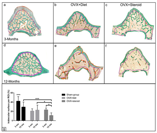
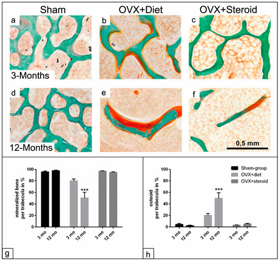
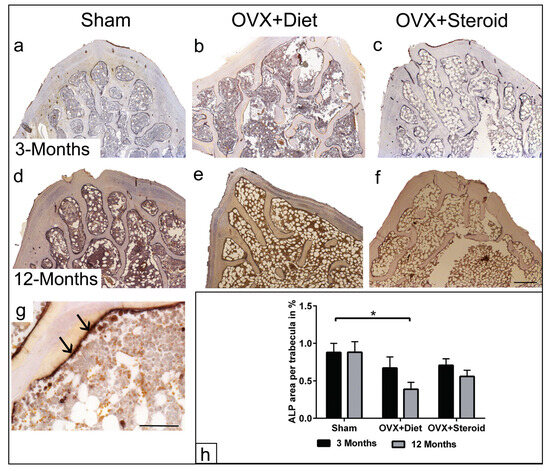
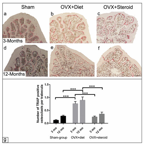
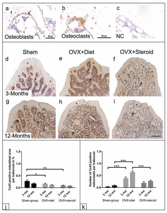
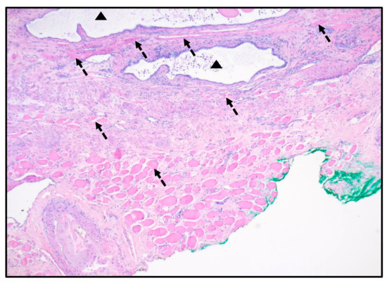
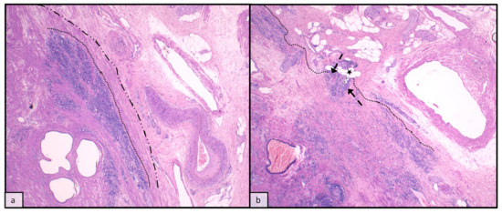
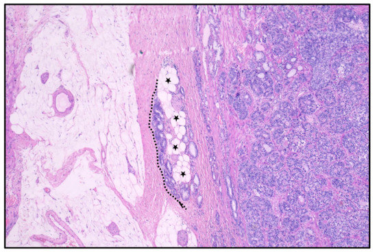
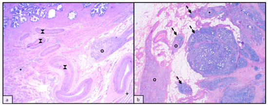
Bone remodeling is essential for maintaining bone health. The imbalance between bone formation and bone resorption leads to bone diseases such as osteoporosis. Connexin43 (Cx43) is a gap junction molecule that plays an important role in bone homeostasis. The present study investigates the morphological characteristics of bone trabeculae and the distribution of Cx43 in bone cells using osteoporotic rat models to explore the relationship between osteoporosis and bone remodeling. Female Sprague–Dawley rats were divided into three groups: sham, ovarectomy with food deprivation (OVX+diet), and ovarectomy with steroid administration (OVX+steroid) for 3 and 12 months to induce osteoporosis. The lumbar vertebrae were processed for histomorphometric and immunohistochemical evaluation of the trabeculae and the distribution of Cx43 in bone cells. The data showed a significant reduction in trabecular bone in both osteoporotic groups. After 12 months, the OVX+diet treatment resulted in reduced mineralization and an increase in unmineralized bone. The percentage of alkaline phosphatase-positive areas in the OVX+diet vertebrae was lower at 12 months compared to the sham group. A significant increase in tartrate-resistant acid phosphatase (TRAP) positive osteoclasts was observed in the OVX+diet group. Both osteoporotic groups showed a decrease in Cx43-positive osteoblasts areas. An increase in the number of osteoclasts positive for Cx43 was detected in the OVX+diet group. The changes in Cx43 distribution in bone cells, together with trabecular mineralization, suggest that Cx43 may play a role in the progression of osteoporosis and could be a valuable target to improve bone remodeling.
Full article
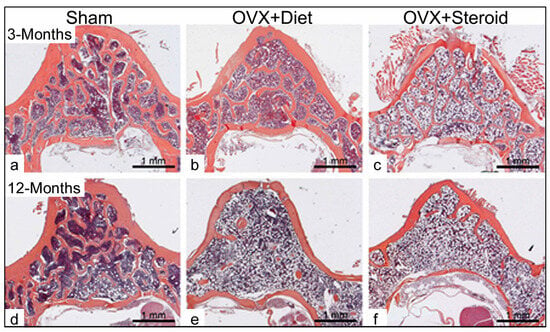
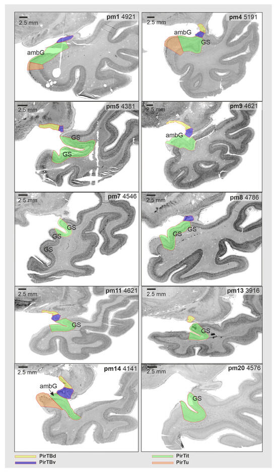
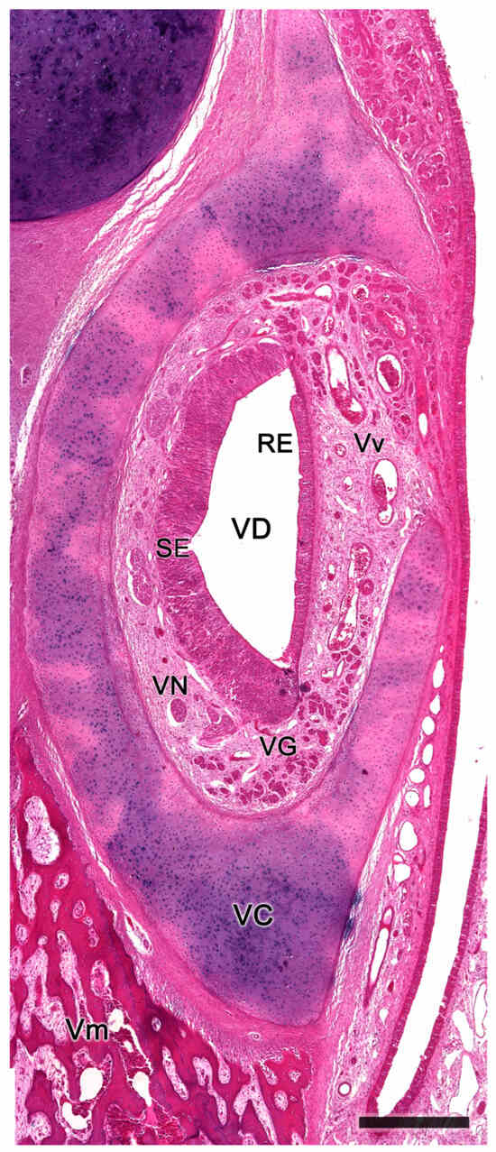
Figure 1


