Article

Morphological Relationships between the Cholinergic and Somatostatin-28(1-12) Systems in the Alpaca (Lama pacos) Brainstem

1
Regional Center for Biomedical Research (CRIB), Facultad de Medicina de Albacete, University of Castilla-La Mancha, 02006 Albacete, Spain
2
Laboratory of Neuroanatomy of the Peptidergic Systems, Institute of Neurosciences of Castilla y León (INCYL), University of Salamanca, 37007 Salamanca, Spain
3
Grupo GIR USAL: BMD (Bases Moleculares del Desarrollo), University of Salamanca, 37007 Salamanca, Spain
*
Author to whom correspondence should be addressed.
Anatomia 2022, 1(1), 54-67; https://doi.org/10.3390/anatomia1010006
Submission received: 5 May 2022 / Revised: 22 May 2022 / Accepted: 23 May 2022 / Published: 25 May 2022
(This article belongs to the Special Issue State-of-the-Art Anatomical Research in the Mediterranean Region 2022)

Abstract

:
In the alpaca brainstem, the distribution of the cholinergic system by the immunohistochemical detection of the enzyme choline acetyltransferase (ChAT) has been described, and its relationship with the distribution of somatostatin-28(1-12) is analyzed by double-immunostaining techniques. Overlapping distribution patterns for both substances were observed in many brainstem regions, suggesting that interactions between them may occur in the reticular formation, nucleus ambiguus or laterodorsal tegmental nucleus. Colocalization of the two substances in the same cell bodies was only observed in restricted areas, such as the nucleus of the solitary tract, reticular formation and nucleus ambiguus. In addition, in several regions, an apparent high innervation of the peptidergic fibers on cholinergic neurons has been observed. The results suggest that chemospecific interactions could be crucial for the control of specific cardiorespiratory and/or digestive functions in alpacas. These interactions may represent brain-adaptive mechanisms to particular environments and have a potential therapeutic use in respiratory disorders.

Graphical Abstract

1. Introduction

Cetaceans (dolphins, whales) and artiodactyls (even-toed ungulates, e.g., sheep, giraffe) belong to the order Cetartiodactyla. The family Camelidae is part of artiodactyls, and the alpaca (Lama pacos) is included in this family [1,2,3,4,5]. Alpacas are important animals for the economy of numerous South American countries due to the excellent quality of their wool, and in this sense, numerous studies focused on their maintenance and reproductive cycles have been performed [6,7]. The members of the Camelidae family have specific morphological characteristics, such as long necks and seven cervical vertebrae, and moreover alpacas can live at sea level and at 5000 m above sea level [6,7,8,9,10,11,12]. These characteristics suggest the existence of important and unique adaptation mechanisms, mainly related to cardiovascular and respiratory mechanisms, which are controlled by the central nervous system, specifically brainstem centers. Other members of the Certiodactyla order also exhibit brain specializations that help them to survive within their respective environments [1,2,3,4,5].
Since all these physiological functions are regulated by neuroactive substances, several studies have reported the distributions of different classical neurotransmitters and neuropeptides by means of immunohistochemical methods in the alpaca brain [6,7,8,9,10,11,12]. These studies complement previous works regarding the mapping of neuro-modulatory systems in the brain of Artiodactyla and confirm that the nuclear complement of neurotransmitters, such as acetylcholine or catecholamines, detected in the alpaca brainstem and diencephalon was similar to that found in other members of the same order, and this finding supports Manger’s hypothesis [13]. In addition, the distribution of some neuropeptides has been studied in the alpaca brainstem [6,7,8,9,12] and diencephalon [10,11]. Moreover, the morphological relationship between neuropeptides and neurotransmitters has been reported, and double-immunolabeling for tyrosine hydroxylase, which is the rate-limiting enzyme of the catecholaminergic synthesis, and somatostatin-28(1-12) (Som-28(1-12)) has been carried out in the diencephalon of the alpaca [11]. In the brainstem of this species, a double-labeling for choline acetyl transferase (ChAT, a marker for the cholinergic system) and calcitonin gene-related peptide (CGRP), as well as for CGRP and tyrosine hydroxylase [9,12], has been described. According to the distribution of double-labeled perikarya, the results observed in the brainstem in these studies suggest that CGRP might interact more with the catecholaminergic system than with the cholinergic system. However, although the physiological interactions between somatostatin and catecholamines have been reported in the literature [13], no double-labeled neurons for these two substances were detected in the alpaca diencephalon [11]. A possible explanation may be that the interaction between the two substances may occur with another somatostatin fragment different from the one studied in that work.
Somatostatin, in addition to inhibiting the growth hormone, acts as a neuromodulator in numerous physiological functions, blocking the release of noradrenaline and stimulating the release of serotonin and acetylcholine [14]. The precursor named pro-somatostatin is cleaved into somatostatin-28, somatostatin-12 (corresponding to the first 112 amino acids of somatostatin-28) and somatostatin-14 [15]. These fragments can elicit different responses in relation to the same mechanism, such as the cardiovascular regulation [15], probably due to the activation of different somatostatin receptor subtypes [16]. In addition, the interaction of somatostatin with other neurotransmitters such as acetylcholine is well-known [16,17,18,19,20,21], but the studies that described such interactions were more focused on memory processing.
Studies previously performed in the alpaca brainstem suggest a similar distribution pattern for CGRP and Som-28(1-12) [7,8]. The presence of CGRP and ChAT in the same perikarya was reported in some of the alpaca brainstem nuclei; thus, the goal of the present work was to know whether there is a neuroanatomical basis for possible interactions between Som-28(1-12) and ChAT in the alpaca brainstem. Moreover, due to the participation of these two substances in cardiovascular and respiratory functions, another aim of this study is to know whether these interactions can constitute a morphological basis for the control of cardiovascular and/or respiratory mechanisms in this species. The results obtained will help to understand the distinctive control mechanisms that exist in the alpaca as physiological adaptations to living in such different habitats in terms of altitude, which leads to changes in air quality and composition. Knowledge of these adaptive mechanisms and their morphological basis will contribute to a better understanding of the neuroanatomy and physiology of the alpaca. Comparison with the results obtained in other mammalian species will allow to assess whether these mechanisms may constitute therapeutic targets for the possible treatment of cardiovascular, respiratory and/or digestive disorders.

2. Materials and Methods

2.1. Animals

As reported previously in similar studies [8,12], six adult male alpacas (Lama pacos) (70–80 kg; 5–8 years) were used here. From birth to the perfusion day, animals were maintained at 0 m on the sea level and kept under standard conditions of temperature and light and with free access to water and food. The study was performed following the guidelines of the ethical and legal recommendations of the Spanish legislation [8,12].

2.2. Tissue Processing

The protocol has previously been published in the reports showing the distribution of immunoreactive structures containing Som-28(1-2) [8] and ChAT [12] in the alpaca brainstem. Animals were intravenously anaesthetized with ketamine (10 mg/kg) and xylazine (4 mg/kg), heparinized, and perfused (NaCl (0.9%): 3 L, paraformaldehyde (4%): 5 L in phosphate-buffered saline (PBS, pH 7.2, 0.15 M)) through the carotid artery [8]. Brainstems were dissected out, post-fixed in the latter fixative solution (overnight) and cryoprotected (using increasing sucrose baths). Using a cryostat, 50 µm frontal brainstem sections were obtained, kept at 4 °C in PBS and processed for the immunohistochemical detection of Som-28 (1-12) [8]. Then, these sections were used for double-immunohistochemistry with ChAT labeling.

2.3. Immunohistochemistry
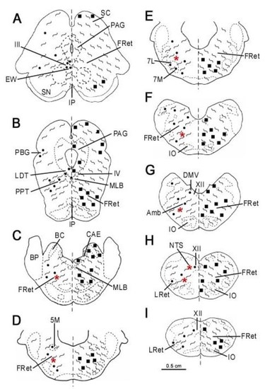
In the present work, the same frontal sections in which Som-28(1-12)-immunoreactivity was previously observed, after applying the diaminobenzidine (DAB) developing methodology [8], were used to detect ChAT according to a previously published protocol [22]. Som-28(1-12)-immunoreactive slides were immersed in PBS until the coverslip became detached, then sections were carefully removed from the surface of the slides. The traces of the mounting medium were removed with several rinses in PBS (6 × 10 min). To inactivate endogenous peroxidases, Som-28(1-12)-immunoreactive sections were treated with NH3 (20%), NaOH (1%) and H2O2 (30%). Sections were extensively rinsed in PBS (6 × 10 min) and pre-incubated in PBS containing normal horse serum (1%) and Triton X-100 (0.3%). This solution was also used for further dilution of streptavidin and antibodies. Sections were incubated with primary polyclonal antibody against choline acetyl transferase (ChAT, Millipore, ref. AB144 P, raised in goat; dilution 1/75; overnight, 4 °C), washed in PBS and incubated in biotinylated donkey anti-goat antibody (Jackson Laboratories; dilution 1/2000; 90 min, room temperature). After washing in PBS, sections were incubated in peroxidase-coupled streptavidin (Jackson Laboratories, dilution 1/2000; 90 min, room temperature). Finally, sections were washed in PBS and Tris-HCl buffer (pH 7.6, 0.05 M), and revealed with 4-chloro-1-napthol. This chromogen shows a blue precipitate, easily distinguishable from the DAB brown product used for the detection of Som-28(1-12). Sections were mounted on gelatin-coated slides and cover-slipped with glycerol/PBS. The specificity of the ChAT immunoreactivity was confirmed by the following histological controls [12]: (1) first incubation bath: omission of the antibody, (2) PBS instead of the secondary biotinylated antibody and (3) a non-appropriated secondary antibody instead of the secondary biotinylated antibody. No immunoreactivity was found. The specificity of the Som-28(1-12) antiserum was previously checked [8]: (1) the first antiserum was preabsorbed with the synthetic peptide, (2) omission of the antibody in the first incubation bath, (3) PBS instead of the secondary biotinylated antibody and (4) the Som-28(1-12) antiserum was preabsorbed with an excess of related peptides (somatostatin-28, somatostatin-14, methionine-enkephalin, substance P, angiotensin II, cholecystokinin and neuropeptide Y). These controls confirmed the specificity of the immunoreactivity for Som-28(1-12) [8]. In addition, the distributions of the cholinergic [12] and somatostatinergic [8] systems in the alpaca brainstem were verified by comparison to the previously published distribution of each substance.

2.4. Mapping

Frontal planes of the alpaca brainstem that were previously published were followed [6,7,8,9]. Atlases of non-camelid mammals and the brain atlas of Lama glama were also used [23,24]. For the nomenclature of the nuclei studied, previous published articles on the alpaca brainstem were used [6,7,8,9,12]. As the main references, the mapping of de Souza et al. [6,7,8] and Marcos and Coveñas [12] were used for both the location of the immunoreactivity and nomenclature. The distribution of cholinergic cell groups in other mammals was consulted [1,2,3,4,5,25,26,27,28,29,30,31,32,33,34,35,36,37,38,39,40]. To study the distribution of the immunoreactive neurons, the computerized digital mapping system Accustage MDPlot v5.2 (MD3-Digitizer, Accustage, Minnesota Datametrics, Saint Paul, MN, USA) was applied. A digital camera attached to a Nikon Eclipse 80i microscope was used to take photographs without any further manipulation of them. Image files have been prepared using Canvas 11 Build 1173 software (Deneba ADC Systems of America, Seattle, WA, USA).

3. Results

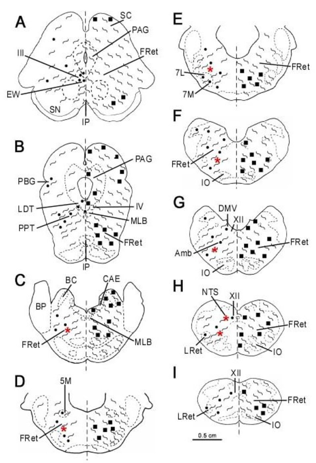
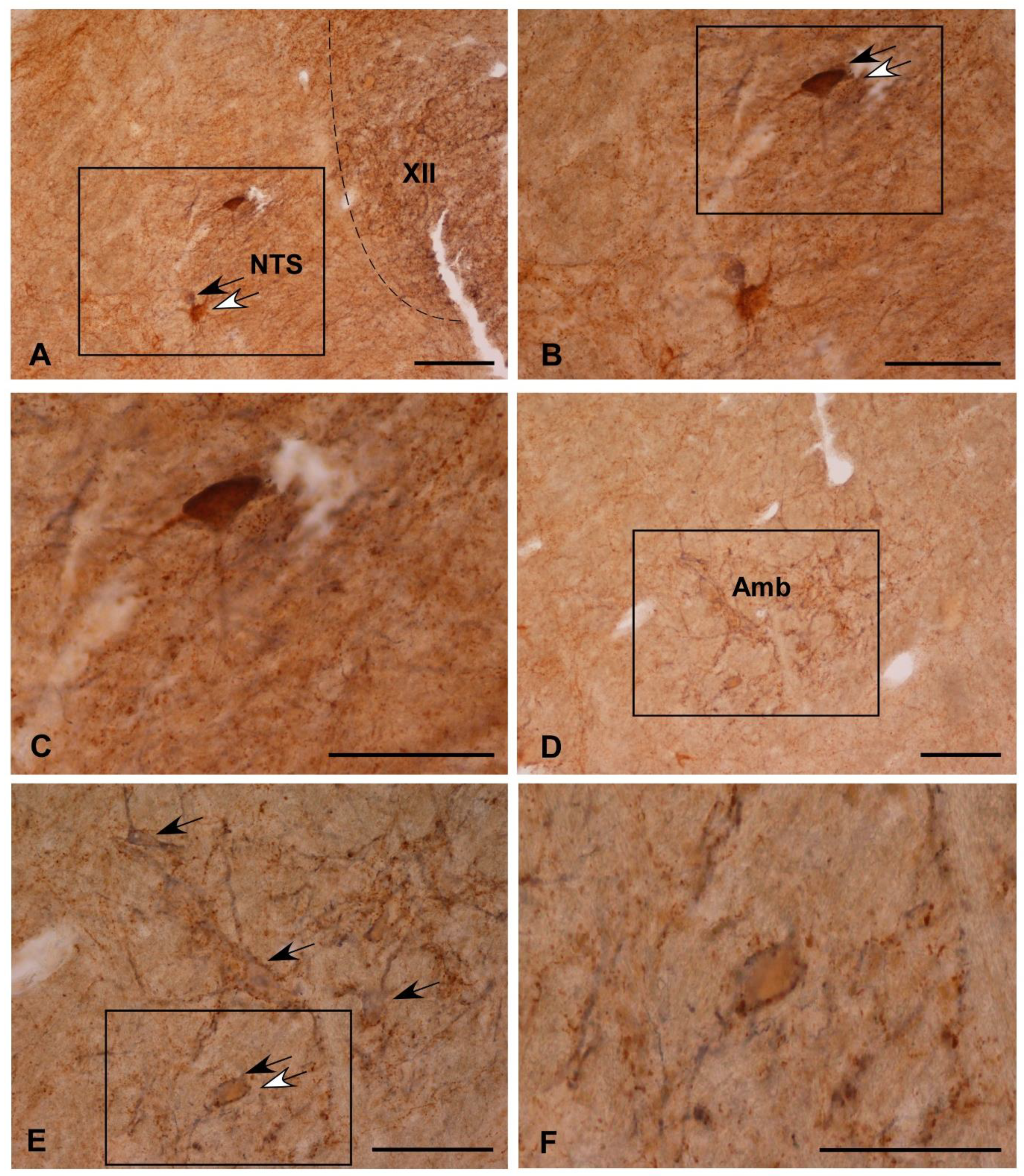
3.1. Single Immunolabeling for Som-28(1-12) and ChAT

The distribution of Som-28(1-12)- and ChAT-immunoreactive structures was studied according to the anatomical description performed by de Souza et al. [8] and Marcos and Coveñas [12]. Since the detailed mapping of the distribution of the immunoreactive structures containing Som-28(1-12) and ChAT in the alpaca brainstem has been performed previously in these two studies, only a brief description will be reported in the present work, more focused on the coexistence of ChAT and Som-28(1-12) in nerve cells. As in previous studies performed using a similar methodology, the brown precipitate (for Som-28(1-12)) and the blue staining (for ChAT) are easily distinguishable. Som-28(1-12)-immunoreactive profiles showed a typically peptidergic morphology, with Som-28(1-12)-positive cell bodies containing visible secretion granules and immunoreactive fibers of varicose appearance. In contrast, profiles containing ChAT showed no varicose labeling and a more homogeneous precipitate.

3.2. Colocalization of Som-28(1-12) and ChAT in Cell Bodies

Comparing the distribution patterns of ChAT- and Som-28(1-12)-immunoreactive structures in the alpaca brainstem (Table 1), perikarya containing ChAT or Som-28(1-12) were detected in the nucleus of the solitary tract (Figure 1A–C), nucleus ambiguus (Figure 1D–F), dorsal motor nucleus of the vagus, the entire reticular formation (Figure 2A,B), laterodorsal tegmental nucleus (Ch6 cholinergic cell group) (Figure 2C,D), pedunculopontine tegmental nucleus (Ch5 cholinergic cell group) (Figure 2C,D) and parabigeminal nucleus (Ch8 cholinergic cell group) (Figure 2E). However, coexistence of the two neuroactive substances into the same cell bodies was only observed in isolated neurons located in the nucleus of the solitary tract (Figure 1A–C), nucleus ambiguus (Figure 1D–F) and reticular formation (Figure 2A,B). In the remaining regions, neuronal populations containing ChAT or Som-28(1-12) displayed separate distributions.

3.3. Fibrilar Immunolabeling

Almost all the cholinergic regions of the alpaca brainstem contained numerous immunoreactive fibers for Som-28(1-12) (Table 1). This was especially noted in the nuclei of motor cranial nerves (Figure 2F and Figure 3A) and associated regions (e.g., Edinger–Westphal nucleus) (Figure 3B). In some of these nuclei, a rich innervation of ChAT-immunoreactive perikarya and dendrites by fibers containing Som-28(1-12) was observed, such is the case of the nucleus ambiguus and medial division of the facial nucleus (Figure 3C,D). The opposite pattern (peptidergic neurons innervated by ChAT-immunoreactive fibers) was more difficult to observe.

4. Discussion

The distribution of immunoreactive profiles for ChAT and Som-28(1-12) is widespread in the alpaca brainstem. According to their respective distributions, these neuroactive substances could participate in the regulation of nociceptive, motor, autonomic, visual, auditory, cardiovascular or respiratory mechanisms [8,12]. The distribution of cholinergic cell bodies suggests that acetylcholine might be implicated, among others, in the regulation of rapid eye movement (REM) sleep and wakefulness as well as in motor functions, since ChAT was observed in the nuclei of several motor cranial nerves and in the pedunculopontine and laterodorsal tegmental nuclei [12]. Moreover, neurons containing Som-28(1-12) detected in the brainstem could be the main source for the somatostatinergic innervation of the alpaca diencephalic and other brainstem nuclei [8,10,11].
Although immunoreactive cell bodies for ChAT or Som-28(1-12) were observed in several regions of the alpaca brainstem, the presence of the two substances in the same cell bodies was only detected in the nucleus ambiguus, reticular formation and nucleus of the solitary tract, suggesting that the interaction between the cholinergic and the somatostatinergic systems in the same neurons is restricted in the alpaca brainstem. The relation between the two systems in the mammalian brain is well-known, mainly in memory processes due to the important role played by acetylcholine in this function: somatostatin stimulates the release of acetylcholine from cholinergic terminals [18,19], and somatostatin administered intracerebrally improved memory failures in rodents showing brain cholinergic deficits [21]. In addition, a monosynaptic relationship between cholinergic forebrain neurons and somatostatin-containing axons has been described [41]. The results reported in the present study suggest that the intracellular interaction between acetylcholine and somatostatin could be mainly related with the regulation of cardiovascular, respiratory, gastrointestinal and gustatory systems [20,42]. This has been reported in other mammals [43] such as cats [20,42] and rodents [18,19,44,45]. A study performed in rats described the relationship between acetylcholine and several somatostatin molecules in the solitary tract complex (including the nucleus of the solitary tract and dorsal motor nucleus of the vagus) and reported that Som-28 and Som-14 could be considered as inhibitory neurotransmitters in the solitary tract complex, but neither Som-28(1-12) nor Som-28(1-10) hyperpolarized the same cells that showed Som-28- or Som-14-evoked hyperpolarization in this region [45]. This suggests that the different somatostatin fragments might exert different actions and that several somatostatin receptors could participate in these responses. It has been reported that cholinergic parasympathetic neurons in the dorsal motor nucleus of the vagus are immunoreactive for somatostatin receptor subtypes 2A, 2B, 4 and 5 [16]. The results obtained in the alpaca brainstem displayed neurons containing Som-28(1-12) and ChAT in the nucleus of the solitary tract but not in the dorsal motor nucleus of the vagus, although the two substances were detected in this later region in separated cellular populations. In cats, it has been suggested that the source of cholinergic elements for the nucleus of the solitary tract could arise from the dorsal motor nucleus of the vagus [42]. The methodology used in the present work does not allow to know whether the immunoreactive neurons detected in the alpaca brainstem are projection neurons or not. However, the results presented here suggest that the interactions between somatostatin and acetylcholine could be different in both regions, and that acetylcholine might be related to Som-28(1-12) in the nucleus of the solitary tract and/or to other somatostatin fragment(s) in the dorsal motor nucleus of the vagus. In other brain regions, different distribution patterns have been reported for distinct somatostatin fragments, at least in humans [43], and this could also be the case in alpaca.
The coexistence of somatostatin and acetylcholine in centers involved in autonomic regulation suggests that they could also be implicated in the regulation of the gastrointestinal tract. Esophageal afferents terminate in the nucleus of the solitary tract, which participates in deglution, eliciting the entire sequence of muscle activity. This nucleus projects to esophageal motoneurons located in the rostral portion of the nucleus ambiguus. It has been reported that this projection contains somatostatin, which can inhibit the neuronal firing in this pathway [19,46]. On the other hand, acetylcholine is present in motoneurons of the nucleus ambiguus that project to the esophagus and stimulates the contraction of the striated regions of the esophagus [46]. Somatostatin also participates in the generation of ambigual excitatory postsynaptic potential [19]. According to the results obtained in the brainstem of the alpaca, it can be suggested that acetylcholine and somatostatin might interact on neurons of the nucleus of the solitary tract and nucleus ambiguus, since both regions displayed double-labeled cells, and then could play a role in the central control of deglution and esophageal motility. In this sense, it has been reported that the somatostatinergic neurons of this circuit modulate viscerosensory signaling and provide a strong postsynaptic inhibition of this signal [44]. However, this regulatory function has been assigned to the somatostatinergic neurons containing GABA located in the nucleus of the solitary tract connected to neurons of the dorsal motor nucleus of the vagus that project to the antrum [44]. The direct stimulation of the latter nucleus increases phasic contractions and gastric tone, effects that are independent from changes in heart rate and blood pressure, which are also regulated by these regions of the brainstem [47,48]. The results obtained in the alpaca agree with these findings, since Som-28(1-12) has been detected in very few cholinergic neurons of the nucleus of the solitary tract and no colocalization has been observed in the dorsal motor nucleus of the vagus. Thus, although the interaction between both substances cannot be discarded, it seems more likely that the regulation of the gastrointestinal tract was mainly related to the somatostatin-GABAergic neurons and/or other somatostatin fragments different from Som-28(1-12). According to the literature, the results presented here emphasize the complex regulatory mechanisms carried out in the alpaca by brainstem structures, since the same neuroactive substances detected in the same regions might exert different regulatory roles on distinct physiological functions. Further studies are needed to confirm whether these complex regulatory mechanisms are common to several species or whether they are specific adaptations of the alpacas to their habitat, since these animals can live at sea level and at altitudes of more than 5000 m. Life in these environments, together with the special morphological characteristics of the alpaca, make it a good candidate to study the brain morphological characteristics underlying these adaptations.
The results obtained in the present paper describe an abundant somatostatinergic innervation in the alpaca brainstem. In two regions, the nucleus ambiguus and the medial division of the facial nucleus, these peptidergic fibers are especially numerous surrounding the perikarya of the cholinergic neurons, suggesting that these cell bodies may be strongly innervated by somatostatinergic terminals. Supporting this interaction, the colocalization of somatostatin receptors 2A in cholinergic neurons in the nucleus ambiguus of rats has been reported [16], as well as the innervation of the vagal motoneurons of the nucleus ambiguus by somatostatin in cats [46]. Below the facial nucleus, a region displaying numerous peptidergic and cholinergic fibers in the alpaca brainstem, a small group of neurons called the retrotrapezoid nucleus, are activated by increases in CO2 levels and regulate the breathing cycle [49]. These neurons are mainly glutamatergic and receive cholinergic inputs as well as somatostatinergic innervation from the pre-Bötzinger complex, and it has been reported that this could be a neuroanatomical substrate to interact with the chemosensory control of breathing [50,51]. This region is unknown in the alpaca brainstem, but the abundance of fibers containing Som-28(1-12) and ChAT as well as the possible somatostatinergic innervation on cholinergic cells detected in this area support the suggestion of a neuroanatomical basis for the chemosensory control of breathing, where somatostatin and acetylcholine might interact. Somatostatin has been involved in the chemosensory drive to breathe [49], and it should be clarified whether these results are again something common in several mammalian species or whether they are adaptive mechanisms of alpacas to their particular living conditions.
The opposite pattern (peptidergic neurons innervated by ChAT-immunoreactive fibers) was more difficult to observe in the alpaca brainstem. This was probably due to the appearance of the fibers containing the enzyme, that are usually harder to visualize near the Som-28(1-12)-immunoreactive cell bodies. The distinct appearance of cholinergic and peptidergic terminals has been reported in optical and electron microscopy [42], but the possibility of methodological aspects to this difficult visualization cannot be ruled out. It may be possible that the DAB precipitate prevents the correct visualization of cholinergic terminals when they are very thin, since the fibers containing ChAT of a larger diameter are easily visible.
The cholinergic projection from the laterodorsal tegmental nucleus links the forebrain limbic circuit with the limbic midbrain [17], and the presence of ChAT in the laterodorsal tegmental and pedunculopontine nuclei has been involved in the control of sleep, especially important in Artiodactyls to modulate the initiation of REM sleep, but not REM sleep maintenance [1]. It has been reported that putative sleep factors have hypnotic properties that could be related to changes in blood pressure, such as urotensin II [52], but this does not seem to be the case for Som-28(1-12) in the alpaca brainstem considering the results reported in the present study, since apparently the peptide was detected in a different population than acetylcholine. However, in the alpaca brainstem, the regulation of the initiation of REM sleep carried out by the cholinergic neurons of the laterodorsal tegmental nucleus seemed to be more related to CGRP, since double-labeled cholinergic neurons containing this peptide have been detected in this region [12]. Other regions such as the reticular formation and the nucleus ambiguus also displayed neurons double-labeled with CGRP and ChAT. It may be possible that some cholinergic neurons of the reticular formation and/or the nucleus ambiguus containing Som-28(1-12) are also immunoreactive for CGRP, and thus a possible influence of the two peptidergic systems (somatostatin and CGRP) on cholinergic cell bodies can be suggested associated with the regulation of cardiovascular, digestive and respiratory functions, and this could be related to other regions of the brainstem. In this regard, the pedunculopontine tegmental nucleus modulates breathing by releasing acetylcholine into the retrotrapezoid nucleus in rats [49]. As discussed previously, in the alpaca, these latter regions showed immunoreactive profiles for ChAT, CGRP and Som-28(1-12), suggesting the involvement of these neuroactive substances in the regulation of the breathing homeostasis. The deep knowledge of the morphological basis underlying these mechanisms may potentially have a therapeutic use for the treatment of respiratory control problems, especially those associated with disorders of breathing during sleep [49]. In this regard, the morphological study of the alpaca brainstem can be very useful for the comprehension of cardiorespiratory control mechanisms, given the anatomophysiological peculiarities of these animals.

5. Conclusions

Although the distributions of Som-28(1-12) and ChAT in the alpaca brainstem are similar, the colocalization of both substances in the same cell bodies is very scarce, suggesting a very limited interaction at the intracellular level. However, the abundant somatostatinergic innervation detected in some regions containing cholinergic cells points to a possible regulation of these neurons by the peptide, which may be related to respiratory control. Nevertheless, results obtained in previous studies suggest that the interaction between ChAT and CGRP would be more important than the interaction between ChAT and somatostatin in the alpaca brainstem, although ChAT, CGRP and somatostatin could be involved in the regulation of the sleep cycle. The detailed knowledge of these mechanisms may contribute to the development of therapies related to respiratory problems, especially those related to breathing disorders affecting some patients during sleep.

Author Contributions

Conceptualization, P.M. and R.C.; methodology, P.M. and R.C.; software, P.M. and R.C.; validation, P.M. and R.C.; formal analysis, P.M. and R.C.; investigation, P.M. and R.C.; resources, P.M. and R.C.; data curation, P.M. and R.C.; writing—original draft preparation, P.M and R.C.; writing—review and editing, P.M. and R.C.; visualization, P.M. and R.C.; supervision, P.M and R.C.; project administration, P.M. and R.C.; funding acquisition, P.M. and R.C. All authors have read and agreed to the published version of the manuscript.

Funding

This research received no external funding.

Institutional Review Board Statement

Experimental design, procedures and protocols were carried out under the guidelines of the legal and ethics recommendations of the Spanish legislation. Ethical review and approval were waived for this study due to the use of histological sections from a previous study.

Informed Consent Statement

Not applicable.

Data Availability Statement

Not applicable.

Conflicts of Interest

The authors declare no conflict of interest.

Abbreviations

IIInucleus of oculomotor nerve (III cranial nerve)
IVnucleus of the trochlear nerve (IV cranial nerve)
5Mmotor trigeminal nucleus
5SLlaminar spinal trigeminal nucleus
5SPspinal trigeminal nucleus
VIabducens nerve (VI cranial nerve)
7Lfacial nucleus, lateral division
7Mfacial nucleus, medial division
XIInucleus of the hypoglossal nerve (XII cranial nerve)
Ambnucleus ambiguus
BCbrachium conjunctivum
BCLmarginal nucleus of the brachium conjunctivum, lateral division
BCMmarginal nucleus of the brachium conjunctivum, medial division
CAElocus coeruleus
ChATcholine acetyl transferase
Cucuneate nucleus
CXexternal cuneate nucleus
DMVdorsal motor nucleus of the vagus nerve
DRNdorsal raphe nucleus
EWnucleus of Edinger–Westphal
FRetreticular formation
Grgracile nucleus
ICinferior colliculus
IOinferior olive
IPinterpeduncular nucleus
LDTlaterodorsal tegmental nucleus (Ch6 cholinergic cell group)
LRetlateral reticular nucleus
MLFmedial longitudinal fascicle
NRred nucleus
NTSnucleus of the solitary tract
Ppyramidal tract
PAGperiaqueductal gray
PBGparabigeminal nucleus (Ch8 cholinergic cell group)
Pedcerebral peduncle
PGpontine gray
PGLpontine gray, lateral division
PGMpontine gray, medial division
PHnucleus praepositus hypoglossi
PPTpedunculopontine tegmental nucleus (Ch5 cholinergic cell group)
Ssolitary tract
SCsuperior colliculus
SNCsubstantia nigra, pars compacta
SNRsubstantia nigra, pars reticulata
SOsuperior colliculus
Som-28(1-12)somatostatin-28 (1-12)
Tnucleus of the trapezoid body
TBtrapezoid body
TDCdorsal tegmental nucleus, central division
TDPdorsal tegmental nucleus, pericentral division
TRCtegmental reticular nucleus, central division
Vesvestibular nucleus

References

  1. Davimes, J.G.; Alagaili, A.N.; Bennett, N.C.; Mohammed, O.B.; Bhagwandin, A.; Manger, P.R.; Gravett, N. Neurochemical organization and morphology of the sleep related nuclei in the brain of the Arabian oryx, Oryx leucoryx. J. Chem. Neuroanat. 2017, 81, 53–70. [Google Scholar] [CrossRef] [PubMed]
  2. Dell, L.A.; Karlsson, K.A.; Patzke, N.; Spocter, M.A.; Siegel, J.M.; Manger, P.R. Organization of the sleep-related neural systems in the brain of the minke whale (Balaenoptera acutorostrata). J. Comp. Neurol. 2015, 524, 2018–2035. [Google Scholar] [CrossRef] [PubMed]
  3. Dell, L.A.; Patzke, N.; Spocter, M.A.; Bertelsen, M.F.; Siegel, J.M.; Manger, P.R. Organization of the sleep-related neural systems in the brain of the river hippopotamus (Hippopotamus amphibius): A most inusual Certiodactyl species. J. Comp. Neurol. 2015, 524, 2036–2058. [Google Scholar] [CrossRef] [PubMed]
  4. Dell, L.A.; Patzke, N.; Spocter, M.A.; Siegel, J.M.; Manger, P.R. Organization of the sleep-related neural systems in the brain of the harbour porpoise (Phocoena phocoena). J. Comp. Neurol. 2016, 524, 1999–2017. [Google Scholar] [CrossRef] [PubMed]
  5. Malungo, I.B.; Gravett, N.; Bhagwandin, A.; Davimes, J.G.; Manger, P.R. A preliminary description of the sleep-related neural systems in the brain of the blue wildebeest, Connochaetes taurinus. Anat. Rec. 2020, 303, 1977–1997. [Google Scholar] [CrossRef]
  6. De Souza, E.; Yi, P.; Aguilar, L.A.; Coveñas, R.; Lerma, L.; Andrade, R.; Mangas, A.; Narváez, J.A. Mapping of leucine-encephalin in the alpaca (Lama pacos) brainstem. In Focus on Neuropeptide Research; Coveñas, R., Mangas, A., Narváez, J.A., Eds.; Transworld Research Network: Trivandrum, India, 2007; pp. 103–113. [Google Scholar]
  7. De Souza, E.; Coveñas, R.; Yi, P.; Aguilar, L.A.; Lerma, L.; Andrade, R.; Mangas, A.; Díaz-Cabiale, Z.; Narváez, J.A. Mapping of CGRP in the alpaca (Lama pacos) brainstem. J. Chem. Neuroanat. 2008, 35, 346–355. [Google Scholar] [CrossRef]
  8. De Souza, E.; Sánchez, M.L.; Aguilar, L.A.; Díaz-Cabiale, Z.; Narváez, J.A.; Coveñas, R. Mapping of Somatostatin-28(1-12) in the alpaca (Lama pacos) brainstem. Microsc. Res. Tech. 2015, 78, 363–374. [Google Scholar] [CrossRef]
  9. Marcos, P.; Arroyo-Jiménez, M.M.; Lozano, G.; Aguilar, L.A.; Coveñas, R. Mapping of tyrosine hydroxylase in the alpaca (Lama pacos) brainstem and colocalization with CGRP. J. Chem. Neuroanat. 2011, 41, 63–72. [Google Scholar] [CrossRef]
  10. Coveñas, R.; Mangas, A.; Medina, L.E.; Sánchez, M.L.; Aguilar, L.A.; Díaz-Cabiale, Z.; Narváez, J.A. Mapping of somatostatin-28 (1-12) in the alpaca diencephalon. J. Chem. Neuroanat. 2011, 42, 89–98. [Google Scholar] [CrossRef]
  11. Marcos, P.; Arroyo-Jiménez, M.M.; Lozano, G.; González-Fuentes, J.; Lagartos-Donate, M.J.; Aguilar, L.A.; Coveñas, R. Mapping of tyrosine hydroxylase in the diencephalon of alpaca (Lama pacos) and co-distribution with somatostatin-28 (1-12). J. Chem. Neuroanat. 2013, 50–51, 66–74. [Google Scholar] [CrossRef]
  12. Marcos, P.; Coveñas, R. Immunohistochemical study of the brainstem cholinergic system in the alpaca (Lama pacos) and colocalization with CGRP. Eur. J. Histochem. 2021, 65, 3266. [Google Scholar] [CrossRef] [PubMed]
  13. Manger, P.R. Establishing order at the systems level in mammalian brain evolution. Brain Res. Bull. 2005, 66, 282–289. [Google Scholar] [CrossRef] [PubMed]
  14. Reichlin, S. Somatostatin. In Brain Peptides; Krieger, D.T., Browstein, M.J., Martin, J.B., Eds.; John Wiley and Sons: New York, NY, USA, 1983; pp. 711–752. [Google Scholar]
  15. Benoit, R.; Ling, N.; Bakhit, C.; Morrison, J.H.; Alford, B.; Guillemin, R. Somatostatin-28(1-12)-like immunoreactivity in the rat. Endocrinology 1982, 111, 2149–2151. [Google Scholar] [CrossRef] [PubMed]
  16. Spary, E.J.; Maqbool, A.; Batten, T.F.C. Expression and localization of somatostatin receptor subtypes sst1-sst5 in areas of the rat medulla oblongata involved in autonomic regulation. J. Chem. Neuroanat. 2008, 35, 49–66. [Google Scholar] [CrossRef] [PubMed]
  17. Sutin, E.L.; Jacobowitz, D.M. Immunocytochemical localization of peptides and other neurochemical in the rat laterodorsal tegmental nucleus and adjacent area. J. Comp. Neurol. 1988, 270, 243–270. [Google Scholar] [CrossRef]
  18. Wang, Y.T.; Neuman, R.S.; Bieger, D. Somatostatin inhibits nicotinic cholinoceptor mediated-excitation in rat ambigual motoneurons in vitro. Neurosci. Lett. 1991, 123, 236–239. [Google Scholar] [CrossRef]
  19. Wang, Y.T.; Zhang, M.; Neuman, R.S.; Bieger, D. Somatostatin regulates excitatory amino acid receptor-mediated fast excitatory postsynaptic potential components in vagal motoneurons. Neuroscience 1993, 53, 7–9. [Google Scholar] [CrossRef]
  20. Batten, T.F.C. Immunolocalization of putative neurotransmitters innervating autonomic regulating neurons of cat ventral medulla. Brain Res. Bull. 1995, 37, 487–506. [Google Scholar] [CrossRef]
  21. Matsuoka, N.; Yamazaki, M.; Yamaguchi, I. Changes in brain somatostatin in memory-deficient rats: Comparison with cholinergic markers. Neuroscience 1995, 66, 617–626. [Google Scholar] [CrossRef]
  22. Marcos, P.; Corio, M.; Dubourg, P.; Tramu, G. Reciprocal synaptic connections between neurotensin- and tyrosine hydroxylase-immunoreactive neurons in the mediobasal hypothalamus of the guinea pig. Brain. Res. 1996, 715, 63–70. [Google Scholar] [CrossRef]
  23. Jasper, A.L.; Ajmone-Marsan, C. A Stereotaxic Atlas of the Diencephalon of the Cat; National Research Council of Canada: Ottawa, ON, Canada, 1966. [Google Scholar]
  24. Paxinos, G.; Watson, C. The Rat Brain in Stereotaxic Coordinates; Academic Press: San Diego, CA, USA, 1998. [Google Scholar]
  25. Badlangana, N.L.; Bhagwandin, A.; Fuxe, K.; Manger, P.R. Observations on the giraffe central nervous system related to the corticospinal tract, motor cortex and spinal cord: What difference does a long neck make? Neuroscience 2007, 148, 522–534. [Google Scholar] [CrossRef] [PubMed]
  26. Bux, F.; Bhagwandin, A.; Fuxe, K.; Manger, P.R. Organization of cholinergic, putative catecholaminergic and serotonergic nuclei in the diencephalon, midbrain and pons of sub-adult male giraffes. J. Chem. Neuroanat. 2010, 39, 189–203. [Google Scholar] [CrossRef] [PubMed]
  27. Dell, L.A.; Gruger, J.L.; Bhagwandin, A.; Jillani, N.E.; Pettigrew, J.D.; Manger, P.R. Nuclear organization of cholinergic, putative catecholaminergic, and serotonergic systems in the brains of two megachiropteran species. J. Chem. Neuroanat. 2010, 40, 177–195. [Google Scholar] [CrossRef] [PubMed]
  28. Houser, C.R.; Crawford, G.D.; Barber, R.P.; Salvaterra, P.M.; Vaughn, J.E. Organization and morphological characteristics of cholinergic neurons: An immunocytochemical study with a monoclonal antibody to choline acetyltransferase. Brain Res. 1983, 266, 97–119. [Google Scholar] [CrossRef]
  29. Kimura, H.; Maeda, T. Aminergic and cholinergic systems in the dorsolateral pontine tegmentum. Brain Res. Bull. 1982, 9, 493–499. [Google Scholar] [CrossRef]
  30. Levey, A.I.; Wainer, B.H.; Mufson, E.J.; Mesulam, M.M. Co-localization of acetylcholinesterase and choline acetyltransferase in the rat cerebrum. Neuroscience 1983, 9, 9–22. [Google Scholar] [CrossRef]
  31. Manaye, K.F.; Zweig, R.; Wu, D.; Hersh, L.B.; de Lacalle, S.; Saper, C.B.; German, D.C. Quantification of cholinergic and select non-cholinergic mesopontine neuronal populations in the human brain. Neuroscience 1999, 89, 759–770. [Google Scholar] [CrossRef]
  32. Marcos, P.; Coveñas, R. Neuroanatomical relationship between the cholinergic and tachykininergic systems in the adult human brainstem: An immunohistochemical study. J. Chem. Neuroanat. 2019, 102, 101701. [Google Scholar] [CrossRef]
  33. Mesulam, M.M. Cholinergic pathways and the ascending reticular activating system of the human brain. Ann. N. Y. Acad. Sci. 1995, 757, 169–179. [Google Scholar] [CrossRef]
  34. Motts, S.D.; Slusarczyk, A.S.; Sowick, C.S.; Schofield, B.R. Distribution of cholinergic cells in guinea pig brainstem. Neuroscience 2008, 154, 186–196. [Google Scholar] [CrossRef] [Green Version]
  35. Oda, Y. Choline acetyltransferase: The structure, distribution and pathologic changes in the central nervous system. Pathol. Int. 1999, 49, 921–937. [Google Scholar] [CrossRef]
  36. Sakai, K.; Luppi, P.H.; Salvert, D.; Kimura, H.; Maeda, T.; Jouvet, M. Localization of cholinergic neurons in the cat lower brainstem. CR Acad. Sci. III 1986, 303, 317–324. [Google Scholar]
  37. Tago, H.; McGeer, P.L.; McGeer, E.G.; Akiyama, H.; Hersh, L.B. Distribution of choline acetyltransferase immunopositive structures in the rat brainstem. Brain Res. 1989, 495, 271–297. [Google Scholar] [CrossRef]
  38. Yasuhara, O.; Aimi, Y.; Matsuo, A.; Kimura, H. Distribution of a splice variant of choline acetyltransferase in the trigeminal ganglion and brainstem of the rat: Comparison with calcitonin gene-related peptide and substance P. J. Comp. Neurol. 2008, 509, 436–448. [Google Scholar] [CrossRef] [PubMed]
  39. Mesulam, M.M.; Geula, C.; Bothwell, M.A.; Hersh, L.B. Human reticular formation: Cholinergic neurons of the pedunculopontine and laterodorsal tegmental nuclei and some cytochemical comparisons to forebrain cholinergic neurons. J. Comp. Neurol. 1989, 283, 611–633. [Google Scholar] [CrossRef] [PubMed]
  40. Ichikawa, T.; Ajiki, K.; Matsuura, J.; Misawa, H. Localization of two cholinergic markers, choline acetyltransferase and vesicular acetylcholine transporter in the central nervous system of the rat: In situ hybridization histochemistry and immunohistochemistry. J. Chem. Neuroanat. 1997, 13, 23–39. [Google Scholar] [CrossRef]
  41. Zaborsky, L. Afferent connections of the forebrain cholinergic projection neurons, with special reference to monoaminergic and peptidergic fibers. Experientia 1989, 57, 12–32. [Google Scholar] [CrossRef]
  42. Maley, B.E. Immunohistochemical localization of neuropeptides and neurotransmitters in the nucleus solitarius. Chem. Senses 1996, 21, 367–376. [Google Scholar] [CrossRef] [Green Version]
  43. Cebada-Sánchez, S.; Insausti, R.; González-Fuentes, J.; Arroyo-Jiménez, M.M.; Rivas-Infante, E.; Lagartos, M.J.; Martínez-Ruiz, J.; Lozano, G.; Marcos, P. Distribution of peptidergic populations in the human dentate gyrus (Somatostatin [SOM-28, SOM-12] and neuropeptide Y [NPY]) during postnatal development. Cell Tissue Res. 2014, 358, 25–41. [Google Scholar] [CrossRef]
  44. Gillis, R.A.; Dezfuli, G.; Bellusci, L.; Vicini, S.; Sahibzada, N. Brainstem neuronal circuitries controlling gastric tonic and phasic contractions: A review. Cell. Mol. Neurobiol. 2022, 42, 333–360. [Google Scholar] [CrossRef]
  45. Jacquin, T.; Champagnat, J.; Madamba, S.; Denavit-Saubié, M.; Siggins, G.R. Somatostatin depresses excitability in neurons of the solitary tract complex through hyperpolarization and augmentation of IM, a non-inactivating voltage-dependent outward current blocked by muscarinic agonists. Proc. Natl. Acad. Sci. USA 1988, 85, 948–952. [Google Scholar] [CrossRef] [PubMed] [Green Version]
  46. Cunningham, E.T.; Sawchenko, P.E. Central neural control of esophageal motility: A review. Disphagia 1990, 5, 35–51. [Google Scholar] [CrossRef] [PubMed]
  47. Pagani, F.D.; Norman, W.P.; Gillis, R.A. Medullary parasympathetic projections innervate specific sites in the feline stomach. Gastroenterology 1988, 95, 277–288. [Google Scholar] [CrossRef]
  48. Norman, W.P.; Pagani, F.D.; Ormsbee, H.S., III; Kasbekar, D.K.; Gillis, R.A. Use of horseradish peroxidase to identify hindbrain sites that influence gastric motility in the cat. Gastroenterology 1985, 88, 701–705. [Google Scholar] [CrossRef]
  49. Moreira, T.S.; Sobrinho, C.R.; Falquetto, B.; Oliveira, L.M.; Lima, J.D.; Mulkey, D.K.; Takakura, A.C. The retrotrapezoid nucleus and the neuromodulation of breathing. J. Neurophysiol. 2021, 125, 699–719. [Google Scholar] [CrossRef]
  50. Tan, W.; Pagliardini, S.; Yang, P.; Janczewski, W.A.; Feldman, J.A. Projections of preBötzinger complex neurons in adult rats. J. Comp. Neurol. 2010, 518, 1862–1878. [Google Scholar] [CrossRef] [Green Version]
  51. Yang, C.F.; Feldman, J.L. Efferent projections of excitatory and inhibitory preBötzinger Complex neurons. J. Comp. Neurol. 2018, 526, 1389–1402. [Google Scholar] [CrossRef]
  52. de Lecea, L.; Bourgin, P. Neuropeptide interactions and REM sleep: A role for urotensin II? Peptides 2008, 29, 845–851. [Google Scholar] [CrossRef]
Figure 1. Pictures showing immunoreactivity for Som-28(1-12) and ChAT. (A) Image of the nucleus of the hypoglossal nerve (XII) and nucleus of the solitary tract (NTS). Abundant ChAT immunolabeling is present in XII, and in NTS, cell bodies containing ChAT (black arrow) and Som-28(1-12) (white arrow) were observed. (B) High-power image of the area squared in (A). A double-labeled neuron (white and black arrows) containing Som-28(1-12) and ChAT was detected. (C) Amplification of the double-immuno-stained neuron pointed out in (B). (D) Immunolabeling observed in the nucleus ambiguus. (E) High-power magnification of the region in the square in (D). Abundant immunoreactivity for ChAT was detected in this nucleus, in fibers and cell bodies (black arrows). Som-28(1-12) was detected in some neurons (white arrow), and in a high density of fibers. In some cases, these peptidergic fibers seemed to strongly innervate cell bodies containing ChAT. (F) Magnification of the double-labeled neuron in the square in (E), containing ChAT and Som-28(1-12). Scale bar = 100 μm in (A,D). Scale bar = 50 μm in (B,C,E,F).
Anatomia 01 00006 g001
Figure 2. Images of Som-28(1-12)- and ChAT-immunoreactive structures in the alpaca brainstem. (A) Neurons positive for Som-28(1-12) (white arrows) and for ChAT (black arrows) as well as a double-labeled cell body were found in the reticular formation (FRet). (B) High-power magnification of the region in the square in (A). (C) Strong ChAT immunoreactivity in the laterodorsal tegmental nucleus (LDT) and pedunculopontine tegmental nucleus (PPT). (D) Amplification of the region in the square in (C). Numerous cholinergic cell bodies (black arrows) and scarce neurons containing Som-28(1-12) (white arrows) were found in these two regions. No double-labeled structures were detected. (E) Immunoreactivity in the parabigeminal nucleus (PBG). Peptidergic neurons (white arrows) and cell bodies containing ChAT (black arrows) were observed in separated populations in this nucleus. (F) Strong cholinergic fibers and cell bodies (black arrows) detected in the hypoglossal nucleus (XII). Hypoglossal nerve (XIIn), also containing ChAT, can be observed coming out from the nucleus (dashed lines). Scale bar in (B): 50 μm. Scale bar in (A,CF): 100 μm.
Anatomia 01 00006 g002
Figure 3. Pictures displaying immunoreactive profiles for Som-28(1-12) and ChAT in the alpaca brainstem. (A) Strong cholinergic immunostaining in the trochlear nucleus (IV). (B) Cell bodies containing ChAT (black arrows) detected in the Edinger–Westphal nucleus. (C) Immunoreactive profiles observed in the lateral (7 L) and medial (7 M) parts of the facial nucleus. (D) High-power magnification of the region in the square in (C). Abundant cholinergic immunoreactivity was detected in fibers and cell bodies (black arrows) of 7 M, and in some cases (lower right part of the picture), a strong peptidergic innervation on ChAT-immunoreactive neurons was observed. Scale bar: 100 μm.
Anatomia 01 00006 g003
Table 1. Distributions of immunoreactive profiles for Som-28(1-12), ChAT and double-labeled neurons detected in the alpaca brainstem. CB: cell body; F: fibers. For nomenclature of the brainstem nuclei, see the list of abbreviations.
Som-28(1-12)ChATDouble-Labeling
NUCLEICBFCBF
III+++
IV+++
5M+++
5SL+
5SP+
VI++
7L+++
7M+++
XII+++
Amb+++++
BC+
BCL++
BCM++
CAE+
Cu+
CX+
DMV++++
DRM+
EW+++
FRet Mesencephalon+++++
FRet Medulla+++++
FRet Pons+++++
Gr+
IC++
IO+
IP+++
LDT++++
LRet++
MLF+
NR+
NTS+++++
P+
PAG+++
PBG++++
Ped+
PG+
PGL+
PGM+
PH++
PPT++++
s+
SC+++
SNC+++
SNR++
SO+
T++
TB+
TDC+
TDP++
TRC+
Ves++
Publisher’s Note: MDPI stays neutral with regard to jurisdictional claims in published maps and institutional affiliations.

